R A P I D C O M M U N I C A T I O N
Open Access
All-trans retinoic acid arrests cell cycle in
leukemic bone marrow stromal cells by
increasing intercellular communication
through connexin 43-mediated gap junction
Yao Liu
1,2, Qin Wen
1, Xue-lian Chen
2, Shi-jie Yang
1, Lei Gao
1, Li Gao
1, Cheng Zhang
1, Jia-li Li
1, Xi-xi Xiang
1,
Kai Wan
1, Xing-hua Chen
1, Xi Zhang
1*and Jiang-fan Zhong
2Abstract
Background:
Gap junctional intercellular communication (GJIC) is typically decreased in malignant tumors. Gap
junction is not presented between hematopoietic cells but occurred in bone marrow stromal cells (BMSCs). Connexin
43 (Cx43) is the major gap junction (GJ) protein; our previous study revealed that Cx43 expression and GJIC were
decreased in acute leukemic BMSCs. All-trans retinoic acid (ATRA) increases GJIC in a variety of cancer cells and has
been used to treat acute promyelocytic leukemia, but the effects of ATRA on leukemic BMSCs is unknown. In this
study, we evaluated the potential effects of ATRA on cell cycle, proliferation, and apoptosis of leukemic BMSCs. Effects
of ATRA on Cx43 expression and GJIC were also examined.
Methods:
Human BMSCs obtained from 25 patients with primary acute leukemia, and 10 normal healthy donors were
cultured. Effects of ATRA on cell cycle, cell proliferation, and apoptosis were examined with or without co-treatment with
amphotericin-B. Cx43 expression was examined at both the mRNA and protein expression levels. GJIC was examined by
using a dye transfer assay and measuring the rate of fluorescence recovery after photobleaching (FRAP).
Results:
ATRA arrested the cell cycle progression, inhibited cell growth, and increased apoptosis in leukemic BMSCs. Both
Cx43 expression and GJIC function were increased by ATRA treatment. Most of the observed effects mediated by ATRA
were abolished by amphotericin-B pretreatment.
Conclusions:
ATRA arrests cell cycle progression in leukemic BMSCs, likely due to upregulating Cx43 expression and
enhancing GJIC function.
Keywords:
All-trans retinoic acid, Bone marrow stromal cells, Connexin 43, Gap junctional intercellular communication,
Leukemia BMSCs
Background
Gap junctions (GJs) are specialized structures consisting
of highly conserved trans-membrane proteins connexins
and allow passage of small molecule (<1 kD) between
the cytoplasm of two adjacent cells [1, 2]. A total of 21
human connexin subtypes have been identified up to
date, with each one displaying unique tissue expression
pattern [3]. Decreased expression of connexins and thus
compromised gap junctional intercellular communication
(GJIC) is noted in a variety of malignant tumors and
con-tributes to their uncontrolled proliferation [1, 3
–
5].
In-creased connexin expression and restored GJIC using
molecular approach have been shown to inhibit the
growth of solid tumors [6
–
9], decrease invasive potential,
and jeopardize neovascularization [10, 11].
GJs do not exist between hematopoietic cells but are
present between bone marrow stromal cells (BMSCs)
and hematopoietic cells [12, 13]. Communication between
BMSCs and hematopoietic cells is an important part of
the microenvironment for establishment and growth of
* Correspondence:[email protected]
1
Department of Hematology, Xinqiao Hospital, the Third Military Medical University, Xinqiao Street, Chongqing 400037, China
Full list of author information is available at the end of the article
tumor cells [14
–
16]. For GJs at these sites, connexin
43 (Cx43) is the major subtype [12]. Functional GJIC
in BMSCs has direct impact on the proliferation and
differentiation of hematopoietic stem/progenitor cells
[17]. Our previous studies revealed decreased Cx43
ex-pression and GJIC function of acute leukemic BMSCs,
which could recover after adopting effective
chemo-therapy or transfection with Cx43 gene [18, 19]. These
findings suggested that Cx43 expression and GJIC
function in BMSCs are likely to be a dynamic process
and regulated by many factors in vivo and in vitro. This
hy-pothesis has been verified by some studies which
demon-strated that some hormones, cytokines, and drugs could
affect the expression of Cxs between coupling cells [20
–
23].
All-trans retinoic acid (ATRA) is a natural derivative
of vitamin A and could regulate various physiological
events, including cell cycle, embryonic development,
and cellular differentiation [24]. ATRA could inhibit
malignant transformation and produce anti-proliferative
ef-fect in several types of tumor cells, possibly through
enhan-cing GJIC function in tumor cells [25
–
27]. ATRA has been
used in the treatment of acute promyelocytic leukemia
(APL) [28] and other hematologic diseases [29, 30] based
on induction of differentiation and inhibition of growth.
However, whether ATRA affects GJIC in leukemic BMSCs
remains unknown.
In this study, we examined the potential effects of ATRA
on cell cycle progression of leukemic BMSCs from 25
pa-tients with acute leukemia. Increased Cx43 expression by
ATRA was verified with mRNA and Western blot.
En-hanced GJIC function was verified with fluorescent dye
dispersion as well as a bleaching assay. The experiments
revealed arrested cell cycle progression, inhibited growth,
and increased apoptosis upon ATRA exposure. These
findings suggest that GJIC function is implicated in the
therapeutic action of ATRA for leukemia.
Results
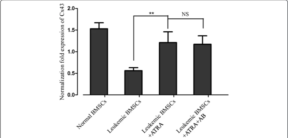
ATRA increases Cx43 mRNA
Quantitative reverse transcription polymerase chain
reac-tion (qRT-PCR) analysis showed that treatment with ATRA
(10 uM) increased Cx43 mRNA levels in leukemic BMSCs
by >50 % (
p
< 0.01) to a level closed to normal BMSCs
(
p
< 0.05, Fig. 1). Pretreatment with amphotericin-B did
not affect the ATRA action on Cx43 mRNA.
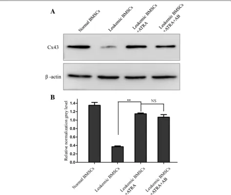
ATRA increases Cx43 protein expression
Western blot analysis showed that ATRA (10 uM)
increased Cx43 protein levels by approximately 70 %
(
p
< 0.01) to a level not statistically different from
nor-mal BMSCs (Fig. 2a, b). Moreover, treatment with
amphotericin-B did not affect Cx43 protein expression
levels mediated by ATRA.
ATRA enhances GJIC
Dye transfer assay was a rapid, sensitive method to detect
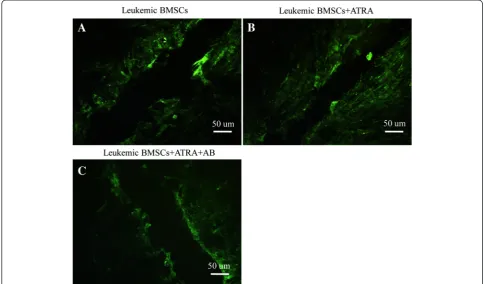
GJIC function between cells [31]. In this study, Lucifer
yel-low (LY) fluorescence in leukemic BMSCs dispersed to
Fig. 1Effects of ATRA and amphotericin-B pretreatment on Cx43 mRNA level in leukemic BMSCs. Cx43 mRNA (normalized againstβ-actin) was 1.53 ± 0.14 in healthy controls (N= 10), 0.56 ± 0.07 leukemic BMSCs (N= 10) treated with DMSO, 1.21 ± 0.36 in leukemic BMSCs (N= 25) exposed to ATRA (10 uM), and 1.17 ± 0.196 in leukemic BMSCs exposed to ATRA + amphotericin-B. Data are presented as mean ± SD.Double asterisks indicateP< 0.01
only 1
–
2 cell lines surrounding the wounded edge (Fig. 3a).
After ATRA treatment, LY fluorescence dispersed to 4
–
5
cell lines (Fig. 3b). Pretreatment with AB could block the
effect mediated by ATRA (Fig. 3c).
The
fluorescence
recovery
after
photobleaching
(FRAP) assay showed 20 % recovery of fluorescent signal
in leukemic BMSCs at 1 min after photobleaching. The
maximal recovery percentage was 36.25 % (±2.14); the
average recovery rate was 4.82 % (±0.78) min
−1(Fig. 4d
–
f).
ATRA treatment promoted the recovery of
fluores-cent signal after photobleaching (Fig. 4a
–
c): 35 % of
cells could be recovered in 1 min and 80.94 %
(±1.46) could be recovered within 30 min. The
aver-age recovery rate was increased to 15.03 % (±1.27)
min
−1(Fig. 4g, h). Pretreatment with amphotericin-B
blocked the effects of ATRA.
ATRA changes biologic characteristics of BMSCs through
GJIC
ATRA (10 uM for 48 h) inhibited the cell growth of
leukemic BMSCs by 40 % (
p
< 0.05; Fig. 5), and
pretreat-ment with amphotericin-B blocked the ATRA effects.
Flow cytometric analysis showed that ATRA treatment
led to decrease the percentage of the cells in S phase from
29.5 to 7.96 % and led to increase the percentage of cells in
the G0/G1 phase from 61.1 to 77.2 % (Additional file
1: Figure S1A
–
C). Such effects were abolished by
amphotericin-B pretreatment (Fig. 6).
ATRA treatment increased apoptosis rate from 5.16 ±
2.35 to 9.78 ± 4.88 % (
p
< 0.05) (Additional file 2: Figure
S2A
–
C). Such effects were attenuated by
amphotericin-B pretreatment (
p
< 0.05; Fig. 7).
Discussion
Abnormal expression of connexin genes in tumor cells is
the result of downregulation rather than genomic
dele-tion or mutadele-tion, and thus could be rescued by
pharma-cological treatment [32, 33]. Increasing the expression of
connexin genes could restore the function of gap
junc-tions [3, 34]. Recent studies showed that ATRA could
improve GJIC function in several types of tumor cells
[28, 35].
In the current study, we extended previous findings to
leukemic BMSCs. ATRA treatment increased the
expres-sion of Cx43 at both mRNA and protein levels and
resulted in increased intercellular communication. The
mechanisms of such action remain obscure, but based
on the secondary structure of Cx43 [36, 37], it is
reason-able to speculate that ATRA could bind to retinoic acid
nuclear receptors (i.e., RARs and RXRs) and activate the
PKC pathways, which in turn phosphorylate proteins
that bind to the phosphorylation site of the Cx43 mRNA.
The resulting instability due to AQUA repeated sequences
decrease the degradation of Cx43 mRNA. Alternatively,
ATRA could activate Cx43 gene transcription and
transla-tion more directly through binding to factors such as AP-1.
In a previous study from this laboratory [18], we
demonstrated that GJIC function is dysfunctional
be-tween leukemic BMSCs. Such a finding is consistent
with the reported decrease in GJIC in several solid
tu-mors [8, 38, 39]. In the current study, we showed that
GJIC between leukemic BMSCs is enhanced by ATRA
treatment. It is noteworthy to mention the discrepancy
of the reported action of ATRA on connexin gene
ex-pression in tumors in previous studies. For example,
Wageenblatt et al. [40] reported inhibited expression
of several connexin genes, including Cx43, by ATRA in
carcinoma of urinary bladder. Based on such a finding,
the authors argued that connexin gene upregulation
provides a
“
growth advantage
”
in certain type(s) of
tumor cells.
The results of the current study clearly showed that
ATRA could arrest cell cycle progression in leukemic
BMSCs. Consistently, cell proliferation was decreased
and apoptosis was enhanced. These effects could be
abol-ished by amphotericin-B pretreatment, and thus could
reasonably attributed to GJICs.
The connection between increased GJIC and BMSC
biological properties is now unknown. Potential
mecha-nisms could include purinergic signaling via extracellular
Fig. 3Effects of dye transfer between BMSCs by using laser confocal microscopy.aLeukemic BMSCs not exposed to ATRA;bleukemic BMSCs exposed to ATRA (10 M);cleukemic BMSCs treated with both ATRA and amphotericin-B. Scale bar = 50 um
adenosine triphosphate (ATP), change of Ca
2+internal
flow from extracellular through gap junction, and
down-regulation of monocyte chemotactic protein 1
expres-sion [41
–
44]. Nevertheless, the current study extended
previous findings of ATRA in solid tumors [11, 45] to
leukemia.
Conclusions
ATRA treatment could arrest cell cycle progression in
leukemic BMSCs, likely due to increasing Cx43
expres-sion and enhancing GJICs in human leukemic BMSCs.
These findings encourage future exploration of using
ATRA in the treatment of hematopoietic malignancies.
Materials
Sample source
Samples of acute leukemic bone marrow were obtained
from 25 patients with primary acute leukemia from the
Department of Hematology of Xinqiao Hospital between
April 2014 and February 2015. Patients included 14
males and 11 females, with a median age of 33. Among
the patients were 14 patients with ALL, 3 patients with
AML-M2, 3 patients with AML-M3, 3 patients with
AML-M5, and 2 patients with AML-M6. The diagnosis
was established on the basis of clinical data, tissue/cell
morphology, and immunophenotyping. Bone marrow
samples from ten normal healthy donors were used as
healthy controls. The research was approved by the Ethics
Committee of Xinqiao Hospital, and all subjects gave
writ-ten informed consent.
Cell culture and treatment
Human BMSCs were isolated as described previously
[46] and were plated in T75 flasks for continuous
pas-sage in Dulbecco
’
s modified Eagle
’
s medium (DMEM)
(Gibco, USA) supplemented with 12.5 % FBS (Gibco,
USA), 12.5 % HS (Gibco, USA), and 0.1 mM
hydrocorti-sone (Sigma, USA). Medium was changed twice weekly,
and cells were passaged into fresh culture flasks at a
ra-tio of 1:4 upon reaching confluence by using trypsin
(Sigma, USA). The cultures were incubated at 37 °C in a
humidified incubator with 5 % CO
2.
ATRA and amphotericin-B (both from Sigma) stock
solutions were prepared in dimethyl sulfoxide (DMSO;
Sigma, USA) and stored at
−
20 °C prior to use. The final
concentration of DMSO was <0.1 %. BMSCs at passage
3 (reach to 80 % confluence) were used for experiments.
qRT-PCR
Total RNA was extracted from BMSCs by using a
com-mercial kit from Stratagene (USA). cDNA synthesis and
amplification were carried out using 2
μg mRNA with
RevertAid
™
First Strand cDNA Synthesis Kit (Fermentas,
USA) and GeneAmp PCR System 2700 Thermal Cycler
(Applied Biosystems, USA). qRT-PCR conditions were as
follows: 95 °C for 15 min, then 40 cycles of 94 °C for 30 s,
60 °C for 30 s, and 72 °C for 30 s. Primers were as follows:
Cx43 (NM_000165), forward 5
′
-AACCTGGTTGTGAAA
ATGTC-3
′
; reverse 5
′
-GCAAGTGTAAACAGCACTCA-3
′
;
β-actin (NM_001101), forward 5
′
-CCTGTGGCATC
CACGAAACT-3
′
; reverse 5
′
-GAGCAATGATCCTGATC
TTC-3
′
. Results were normalized against
β-actin.
Western blot
Western blot analysis was performed using whole-cell
extract. Protein concentration of the samples was
deter-mined using a BCA method. Proteins (20
μg per lane)
were separated using 12 % SDS-PAGE gel, transferred to
nitrocellulose membrane, and blocked with 5 % skim
milk in PBS. The membrane was incubated with a rabbit
polyclonal anti-Cx43 antibody (Sigma, USA), and then
Fig. 5Effects of ATRA on the cell viability of BMSCs (MTT assay). Cell viability was evaluated by MTT assay in leukemic BMSCs, leukemic BMSCs treated with ATRA, and leukemic BMSCs treated with both ATRA and amphotericin-B. Data are presented as mean ± SD.Single asterisk indicates significant difference (P< 0.05)
Fig. 6Effects of ATRA on the cell cycle (FCM assay). Cell cycle was examined with FCM assay in leukemic BMSCs, leukemic BMSCs treated with ATRA, and leukemic BMSCs treated with both ATRA and amphotericin-B. Data are presented as mean ± SD.Single asterisk indicates significant difference (P< 0.05)
Fig. 7Effects of ATRA on the apoptosis of BMSCs (FCM assay). Cell apoptosis was examined with FCM assay in leukemic BMSCs, leukemic BMSCs treated with ATRA, and leukemic BMSCs treated with both ATRA and amphotericin-B. Data are presented as mean ± SD.Single asterisk indicates significant difference (P< 0.05)
with a secondary antibody linked to horseradish
peroxid-ase prior to ECA visualization (Amersham Biosciences
AB, Uppsala, Sweden). Blots were washed, reprobed with
an anti-GAPDH antibody (Sigma, USA), and then
devel-oped in an identical manner for assessment of GAPDH
to ensure even loading.
Dye transfer assay
Transfer assay was carried out as previously described
[47]. Briefly, cultured cells were rinsed thoroughly with
Hanks
’
balanced salt solution (HBSS) (with calcium and
magnesium) containing 1 % BSA. A 27-gauge needle was
used to create multiple scratches (minimum distance
between scratches 1 cm) in the cell monolayer in the
pres-ence of PBS containing 0.5 % Lucifer yellow (Gibco, USA).
After exactly 1 min, the culture was rinsed with HBSS and
then incubated for 1
–
10 min in the saved culture medium
to allow the loaded dye to transfer to adjoining cells. The
cells were then rinsed and fixed with 4 %
paraformalde-hyde and immediately analyzed under a laser confocal
scanning microscope (LCSM; Leica, German) at 485 nm.
GJIC was defined as the average distance of dye travel
(μm) at six different sites in each sample.
FRAP assay
FRAP assay was carried out in living cells as described
previously [48]. Briefly, cells cultured on glass cover slips
were loaded with C-FDA (10
μmol/L, 15 min; Sigma) at
37 °C and washed thoroughly with DMEM medium. A
cell with direct contact with 4
–
5 neighboring cells within a
cluster was selected under the LCSM. A pre-bleach image
of the whole field was taken using low laser intensity
(acousto-optic tunable filter (AOTF) = 10 %, zoom = ×2).
Then, laser intensity was increased by ×50 (AOTF = 50 %,
zoom = ×20), and the target BMSCs was photobleached for
1 s. Transfer of fluorescent dye from neighboring cells was
examined by scanning at 50 s intervals for a total period of
30 min. The rate of fluorescence recovery index was
calcu-lated as:
R
= (
I
t−
I
0) / (
I
−
I
0) × 100 %, where
I
0is the
inten-sity of the photo-bleached fluorescence and
I
is the
intensity of pre-bleached fluorescence.
MTT assay
Cell proliferation was examined using a methyl thiazolyl
tetrazolium (MTT) assay (Sigma, USA) at 610 nm. Data
were averaged from three independent experiments.
Flow cytometry
Flow cytometry (FCM) assay was carried out as described
previously [49]. BMSCs were washed, fixed in 70 %
etha-nol, and resuspended in 10 mL PBS. Cells were stained
with propidium iodide (5
μL 10 mg/mL) and DNAse-free
RNase (200
μg/mL) for 20 min prior to FACS analysis
using a FACSVantage flow cytometer (Becton Dickinson,
USA) and analyzed by CellQuest software. At least 1 × 10
4cells were analyzed for each sample.
Apoptosis
Apoptosis was determined by Annexin V-FITC (Gibco,
USA) and FCM analyses. After washing with PBS, 10
6BMSCs were resuspended in binding buffer containing
Annexin V-FITC (1 mg/mL). The mixture was incubated
for 10 min in the dark under room temperature and
then analyzed with FACSVantage flow cytometer and
CellQuest software.
Statistical analysis
Data are represented as mean with standard deviation
and analyzed with Student
’
s
t
test, except for GJIC
(Pearson
’
s chi-squared test). Statistical significance was
set at
p
< 0.05.
Additional files
Additional file 1: Figure S1.The original flow plots of cell cycle in leukemic BMSCs using FCM assay. (A) Leukemic BMSCs; (B) leukemic BMSCs exposed to ATRA; (C) leukemic BMSCs treated with both ATRA and amphotericin-B.
Additional file 2: Figure S2.The original flow plots of cell apoptosis in leukemic BMSCs using FCM assay. Leukemic BMSCs were treated DMSO, ATRA, and ATRA + AB, then cells were stained with Annexin V-FITC and propidium iodide (PI), followed by analysis on a flow cytometer. (A) Leukemic BMSCs; (B) leukemic BMSCs exposed to ATRA; (C) leukemic BMSCs treated with both ATRA and amphotericin-B.
Abbreviations
AOTF:acousto-optic tunable filter; ATP: adenosine triphosphate; ATRA: all-trans retinoic acid; BMSCs: bone marrow stromal cells; Cx43: connexin 43; DMSO: dimethyl sulfoxide; FCM: flow cytometry; FRAP: fluorescence recovery after photobleaching; GJIC: gap junctional intercellular communication; LCSM: laser confocal scanning microscope; LY: Lucifer yellow; MTT: methyl thiazolyl tetrazolium; qRT-PCR: quantitative reverse transcription polymerase chain reaction.
Competing interests
The authors declare that they have no competing interests.
Authors’contributions
YL, XZ, and QW carried out the cell culture and qRT-PCR, participated in the Western blot and dye transfer assay, and drafted the manuscript. XLC, SJY, LG, and CZ carried out the Western blot and FRAP assay. LG, JLL, XXX, KW, and XHC participated in the MTT assay and FCM and sample collection. XZ and JFZ participated in the design of the study and performed the statistical analysis. All authors read and approved the final manuscript.
Acknowledgements
This work was supported by grants from the National Natural Science Foundation of China (Nos. 81370594, 81070388, 81270569), Chongqing Key Discipline of Natural Science (No. 2009BA5056), and Youth Innovation Project of Military Medicine of Chinese People’s Liberation Army (No. 13QNP116).
Author details 1
Department of Hematology, Xinqiao Hospital, the Third Military Medical University, Xinqiao Street, Chongqing 400037, China.2Department of
Received: 1 July 2015 Accepted: 28 September 2015
References
1. Leithe E, Sirnes S, Omori Y, Rivedal E. Downregulation of gap junctions in cancer cells. Crit Rev Oncog. 2006;12:225–56.
2. Cotrina ML, Lin JH, Nedergaard M. Adhesive properties of connexin hemichannels. Glia. 2008;56:1791–8.
3. Oshima A. Structure and closure of connexin gap junction channels. FEBS Lett. 2014;588:1230–7.
4. Khan Z, Yaiw KC, Wilhelmi V, Lam H, Rahbar A, Stragliotto G, et al. Human cytomegalovirus immediate early proteins promote degradation of connexin 43 and disrupt gap junction communication: implications for a role in gliomagenesis. Carcinogenesis. 2014;35:145–54.
5. Anand RJ, Hackam DJ. The role of gap junctions in health and disease. Crit Care Med. 2005;33:S535–8.
6. Mehta PP, Perez-Stable C, Nadji M, Mian M, Asotra K, Roos BA. Suppression of human prostate cancer cell growth by forced expression of connexin genes. Dev Genet. 1999;24:91–110.
7. Ignatenko NA, Zhang H, Watts GS, Skovan BA, Stringer DE, Gemer EW. The chemopreventive agent alpha-difluoromethylornithine blocks Ki-ras-dependent tumor formation and specific gene expression in Caco-2 cells. MolCarcinog. 2004;39:221–33.
8. Tang B, Peng ZH, Yu PW, Yu G, Qian F, Zeng DZ, et al. Aberrant expression of Cx43 is associated with the peritoneal metastasis of gastric cancer and Cx43-mediated gap junction enhances gastric cancer cell diapedesis from peritoneal mesothelium. PLoS One. 2013;8:e74527.
9. Zhao K, Wang W, Guan C, Cai J, Wang P. Inhibition of gap junction channel attenuates the migration of breast cancer cells. MolBiol Rep. 2012;39:2607–13.
10. Qin H, Shao Q, Belliveau DJ, Laird DW. Aggregated DsRed-tagged Cx43 and over-expressed Cx43 are targeted to lysosomes in human breast cancer cells. Cell CommunAdhes. 2001;8:433–9.
11. Zhu D, Caveney S, Kidder GM, Naus CC. Transfection of C6 glioma cells with connexin 43 cDNA: analysis of expression, intercellular coupling, and cell proliferation. Proc Natl Acad Sci U S A. 1991;88:1883–7.
12. Yang J, Darley RL, Hallett M, Evans WH. Low connexin channel-dependent intercellular communication in human adult hematopoietic progenitor/stem cells: probing mechanisms of autologous stem cell therapy. Cell Commun Adhes. 2009;16:138–45.
13. Rosendaal M, Krenacs TT. Regulatory pathways in blood-forming tissue with particular reference to gap junctional communication. Pathol Oncol Res. 2006;6:243–9.
14. Sun Z, Wang S, Zhao R. The roles of mesenchymal stem cells in tumor inflammatory microenvironment. J Hematol Oncol. 2014;7:14.
15. Metcalf D. The molecular control of cell division, differentiation commitment and maturation in haemopoietic cells. Nature. 1989;339:27–30.
16. Ploemacher RE, Mayen AE, De Koning AE, Krenacs T, Rosendaal M.
Hematopoiesis: gap junction intercellular communication is likely to be involved in regulation of stroma-dependent proliferation of hemopoietic stem cells. Hematology. 2000;5:133–47.
17. Hurtado SP, Balduino A, Bodi EC, El-Cheikh MC, de Carvalho AC C, Borojevic R. Connexin expression and gap-junction-mediated cell interactions in an in vitro model of haemopoieticstroma. Cell Tissue Res. 2004;316:65–76. 18. Liu Y, Zhang X, Li ZJ, Chen XH. Up-regulation of Cx43 expression and GJIC
function in acute leukemia bone marrow stromal cells post-chemotherapy. Leuk Res. 2010;34:631–40.
19. Zhang X, Liu Y, Si YJ, Chen XH, Li ZJ, Gao L, et al. Effect of Cx43 gene-modified leukemic bone marrow stromal cells on the regulation of Jurkat cell line in vitro. Leuk Res. 2012;36:198–204.
20. Bodi E, Hurtado SP, Carvalho MA, Borojevic R, Carvalho AC. Gap junctions in hematopoietic stroma control proliferation and differentiation of blood cell precursors. An Acad Bras Cienc. 2004;76:743–56.
21. Zhang C, Li Y, Chen J, Gao Q, Zacharek A, Kapke A, et al. Bone marrow stromal cells upregulate expression of bone morphogenetic proteins 2 and 4, gap junction protein connexin-43 and synaptophysin after stroke in rats. Neuroscience. 2006;141:687–95.
22. Alves LA, Nihei OK, Fonseca PC, Carvalho AC, Savino W. Gap junction modulation by extracellular signaling molecules: the thymus model. Braz J Med Biol Res. 2000;33:457–65.
23. Tan XY, He JG. The remodeling of connexin in the hypertrophied right ventricular in pulmonary arterial hypertension and the effect of a dual ET receptor antagonist (bosentan). Pathol Res Pract. 2009;205:473–82. 24. Di Francesco AM, Ubezio P, Torella AR, Meco D, Pierri F, Barone G, et al.
Enhanced cell cycle perturbation and apoptosis mediate the synergistic effects of ST1926 and ATRA in neuroblastoma preclinical models. Invest New Drugs. 2012;30:1319–30.
25. Zhang KZ, Zhang QB, Zhang QB, Sun HC, Ao JY, Chai ZT, et al. Arsenic trioxide induces differentiation of CD133+ hepatocellular carcinoma cells and prolongs posthepatectomy survival by targeting GLI1 expression in a mouse model. J Hematol Oncol. 2014;7:28.
26. Chen W, Yan C, Hou J, Pu J, Ouyang J, Wen D. ATRA enhances bystander effect of suicide gene therapy in the treatment of prostate cancer. Urol Oncol. 2008;26:397–405.
27. Yang Y, Qin SK, Wu Q, Wang ZS, Zheng RS, Tong XH, et al. Connexin-dependent gap junction enhancement is involved in the synergistic effect of sorafenib and all-trans retinoic acid on HCC growth inhibition. Oncol Rep. 2014;31:540–50.
28. Shen ZX, Shi ZZ, Fang J, Gu BW, Li JM, Zhu YM, et al. All-trans retinoic acid/ As2O3 combination yields a high quality remission and survival in newly diagnosed acute promyelocytic leukemia. Proc Natl Acad Sci. 2004;101:5328–35. 29. Zhang L, Chen Q-S, Xu P-P, Qian Y, Wang A-H, Xiao D, et al. Catechins
induced acute promyelocytic leukemia cell apoptosis and triggered PML-RARαoncoprotein degradation. J Hematol Oncol. 2014;7:75. 30. Maeda Y, Yamaguchi T, Hijikata Y, Tanaka M, Hirase C, Takai S, et al. Clinical
efficacy of all-trans retinoic acid for treating adult T cell leukemia. J Cancer Res Clin Oncol. 2008;134:673–7.
31. el-Fouly MH, Trosko JE, Chang CC. Scrape-loading and dye transfer. A rapid and simple technique to study gap junctional intercellular communication. Exp Cell Res. 1987;168:422–30.
32. Bolhassani A, Khavari A, Bathaie SZ. Saffron and natural carotenoids: biochemical activities and anti-tumor effects. Biochim Biophys Acta. 2014;1845:20–30.
33. Matesic DF, Sidorova TS, Burns TJ, Bell AM, Tran PL, Ruch RJ, et al. p38 MAPK activation, JNK inhibition, neoplastic growth inhibition, and increased gap junction communication in human lung carcinoma and Ras-transformed cells by 4-phenyl-3-butenoic acid. J Cell Biochem. 2012;113:269–81. 34. Saez JC, Berthoud VM, Branes MC, Martinez AD, Beyer EC. Plasma
membrane channels formed by connexins: their regulation and functions. Physiol Rev. 2003;83:1359–400.
35. Zhang X, Ren Z, Zuo J, Su C, Wang R, Chang Y, et al. The effect of all-trans retinoic acid on gap junctional intercellular communication and connexin 43 gene expression in glioma cells. Chin Med Sci J. 2002;17:22–6. 36. Geimonen E, Jiang W, Ali M, Fishman GI, Garfield RE, Andersen J. Activation
of protein kinase C in human uterine smooth muscle induces connexin-43 gene transcription through an AP-1 site in the promoter sequence. J Biol Chem. 1996;271:23667–74.
37. Sullivan R, Ruangvoravat C, Joo D, Morgan J, Wang BL, Wang XK, et al. Structure, sequence and expression of the mouse Cx43 gene encoding connexin 43. Gene. 1993;130:191–9.
38. Gotow T, Shiozaki M, Higashi T, Yoshimura K, Shibata M, Kominami E, et al. Hepatic gap junctions in the hepatocarcinogen-resistant DRH rat. Histochem Cell Biol. 2008;130:583–94.
39. Lamiche C, Clarhaut J, Strale PO, Crespin S, Pedretti N, Bernard FX, et al. The gap junction protein Cx43 is involved in the bone-targeted metastatic behaviour of human prostate cancer cells. ClinExp Metastasis. 2012;29:111–22.
40. Hotz-Wagenblatt A, Shalloway D. Gap junctional communication and neoplastic transformation. Crit Rev Oncog. 1993;4:541–58.
41. Qian MX, Wen J, Zhu X, Jia XH, Yang XW, Du YZ, et al. Structurally differentiated Cis-elements that interact with PU.1 are functionally distinguishable in acute promyelocyticleukemia. J Hematol Oncol. 2013;6:25. 42. Makarenkova HP, Shestopalov VI. The role of pannexin hemichannels in
inflammation and regeneration. Front Physiol. 2014;25:1–8.
43. Xianrong Z, Yipeng Q. Role of intramolecular interaction in connexin 50: mediating the Ca2+-dependent binding of calmodulin to gap junction. Arc BiocBiop. 2005;2:111–7.
44. Akinleye A, Avvaru P, Furqan M, Song YP, Liu DL. Phosphatidylinositol 3-kinase(PI3K) inhibitors as cancer therapeutics. J HematolOncol. 2013;6:88. 45. Czyz J, Szpak K, Madeja Z. The role of connexins in prostate cancer
promotion and progression. Nat Rev Urol. 2012;5:274–82.
46. Kim H, Suh H, Jo SA, Kim HW, Lee JM, Kim EH, et al. In vivo bone formation by human marrow stromal cells in biodegradable scaffolds that release dexamethasone and ascorbate-2-phosphate. BiochemBiophys Res Commun. 2005;332:1053–60.
47. Boswell BA, Lein PJ, Musil LS. Cross-talk between fibroblast growth factor and bone morphogenetic proteins regulates gap junction-mediated intercellular communication in lens cells. MolBiol Cell. 2008;19:2631–41. 48. Wade MH, Trosko JE, Schindler M. A fluorescence photobleaching assay of
gap junction-mediated communication between human cells. Science. 1986;232:525–8.
49. Katarzyna AD, Wang Y, Li JM, Wayne AC, Cynthia RG, Huang CZ, et al. Host bone marrow-derived IL-12 enhances donor T cell engraftment in a mouse model of bone marrow transplantation. J Hematol Oncol. 2014;7:16.
Submit your next manuscript to BioMed Central
and take full advantage of:
• Convenient online submission
• Thorough peer review
• No space constraints or color figure charges
• Immediate publication on acceptance
• Inclusion in PubMed, CAS, Scopus and Google Scholar
• Research which is freely available for redistribution