Original Research Article
Antimicrobial Potential of Soil Isolates from
Uttarakhand Region against Pathogens and Drug
Resistant Strains
Kavita Sharma
Professor & Head, Dept. of Botany, Govt. A&C Girls College, Raipur, Chhattisgarh, India
ABSTRACT
Keywords: Soil isolates, antibiotic producers, antibiotic, pathogens and drug resistant strains, Uttarakhand region (Rishikesh and Dehradun).
INTRODUCTION
Soil organisms contribute a wide range of essential services to the sustainable functioning of all ecosystems by acting as the primary driving agents of nutrient cycling; regulating the dynamics of soil organic matter, soil carbon sequestration and greenhouse gas emission; modifying soil physical structure and water regime; enhancing the amount and efficiency of nutrient acquisition by vegetation; and enhancing plant health. [1] These services are not only essential to the functioning of natural ecosystems but also constitute an important resource for the sustainable management of agricultural systems. The bacterial genera Bacillus and Streptomyces along with the fungal genera Penicillium and Cephalosporium are commonly found in soil. [2-5] The genus Streptomyces are the most prolific antibiotic producers and, although bacteria are a unique subgroup of bacteria called the Actinomycetes. [6] Although soil has historically been used to find new antibiotic producers, at present many of the „old‟ antibiotics are now being manipulated in the lab and chemically modified to form new versions of older antibiotics. The actinomycetes produce about 70% of these, and the remaining 30% are products of filamentous fungi and non-actinomycete bacteria. Of the plethora of known bioactive compounds, approximately 160 are currently used in clinical practice. Streptomyces species produce about 55% of these. Most of the widely used antibacterial drugs have specific target sites in the physiological processes of microbial cells, which include (1) inhibition of cell wall synthesis, (2) inhibition of protein and nucleic acid synthesis, (3) inhibition of enzyme activity. [7] Since the introduction of antibiotics into clinical use in the mid 1940s, microorganisms have shown a remarkable ability to protect themselves by developing antibiotic resistance through different mechanisms. The major genetic
mechanism for antibiotic resistance is mainly through mutation or acquisition of new gene(s) through genetic exchange mechanisms, like conjugation, transduction, and transformation. Both chromosomal and plasmid-encoded genes are important in the development and dissemination of antibiotic resistance genes. Various antibiotic- resistance mechanisms are known in bacteria but the major mechanisms include destruction or modification of antibiotics (e.g. production of α-lactamases and amino-glycosides modifying enzymes), prevention of access to the target (e.g. alteration of permeability), and alteration of the target site. [8-10]
METHODOLOGY
The chemicals and reagents used were of Analytical Grade and were procured from Ranchem and CDH, India. The media used during the study was procured from Hi-Media Ltd. Mumbai, India.
2.1 Collection of soil samples
The soil samples were collected from different sites viz. Garden soil (Sample A), Near the Banks of Ganga River (Sample B) from Rishikesh region and soil sample from local gardens (Sample C) of Uttarakhand. Soil samples were collected using sterile screw-capped glass bottles and stored at 4ºC until analysis in the laboratory.
[11]
2.2 Isolation of bacterial isolates
The soil samples collected were serially diluted with N-saline. The maximally diluted samples were streaked on Nutrient Agar medium plates. The plates were further stored at 370C for 18-24 h in BOD incubator for the growth of the isolate.
[12]
(for Rishikesh isolates) and “D” (for Dehradun isolates). These pure cultures were further used for characterization and further studies.
2.3 Identification of bacterial isolates The isolates were further screened for gram staining [13] and biochemical tests viz. starch hydrolysis, casein hydrolysis, MRVP test, Urease test, Indole test, H2S production and Citrate test were performed for confirmation of the nature of bacterial isolates. Pure isolates were maintained at -700 C in LB broth supplemented with 15% (w ⁄v) glycerol.
2.4 Determination of in vitro antimicrobial activity of bacterial isolates against pathogenic strains and Methicillin resistant Staphylococcus aureus (MRSA)
The pure bacterial isolates obtained were inoculated in 250 ml Erlenmeyer flasks containing Nutrient broth. The flasks were incubated on a rotary shaker (200 rpm) at 30°C for two to three days. After incubation the culture was extracted by using equal volume of ethyl acetate. Ethyl acetate was evaporated and the residue was dissolved in 1:1 methanol: DMSO solvent. Further each of the culture was centrifuged to obtain the pellets. [14]
2.4.1 Culture Media
The media used for determination of antibacterial activity and growth of pathogenic bacterial cultures was Nutrient agar/broth while Sabouraud‟s dextrose agar/broth was used for determination of antifungal activity and growth of pathogenic fungal cultures.
2.4.2 Inoculum
The pathogenic bacteria cultures were inoculated separately into nutrient broth and incubated at 37 0C for 4 h. The suspension was checked to provide approximately 105 CFU/ml. Similar procedure was done for fungal strains by inoculating in Sabouraud‟s dextrose broth for 6 h.
2.4.3 Microorganisms used
Pure cultures of various pathogenic bacterial and fungal cultures viz. E. coli NCIM 2065, Lactobacillus plantarum
NCIM 2083, Micrococcus luteus ATCC 9341, Salmonella abony NCIM 2257, MRSA 8 (isolated from pus) and MRSA 35 (isolated from blood), pure fungal cultures viz. Candida albicans NCIM 3471 and Aspergillus niger NCIM 1196 were used for the study. The bacterial and fungal pathogens were procured from Roorkee Research & Analytical Labs Pvt. Ltd., Roorkee (Uttarakhand) and MRSA cultures were procured from Shooloni University, Solan (H.P), India.
2.4.4 Agar well diffusion method
The agar well diffusion method [15] was modified. Nutrient Agar medium (NAM) was used for bacterial cultures. The culture medium was inoculated with the bacterial strains separately suspended in nutrient broth. Sabouraud‟s dextrose agar (SDA) was used for fungal cultures. The culture medium was inoculated with the fungal strains separately suspended in Sabouraud‟s dextrose broth. A total of 8 mm diameter wells were punched into the agar and filled with bacterial supernatant, bacterial pellet (dissolved in N-saline), solvent blank (N-saline) and positive control (Erythromycin, 1 mg/ml) separately in the bacterial cultured plates separately. The bacterial plates were then incubated at 37 oC for 18 h. The same procedure was used for determining antifungal activity but in this case standard antibiotic (Fluconazole, concentration 1 mg/ml) was used as positive control. Further fungal culture plates were incubated at 37oC for 72 h. The antibacterial and antifungal activity was evaluated by measuring the diameter of zone of inhibition observed.
2.5 Separation of antimicrobial substance by TLC
The crude extracts of antimicrobial agent from the isolate were spotted on the preparative TLC plate as a single line or streak and the chromatogram was performed with the solvent system Chloroform: methanol (60:40). After drying the sheet was kept in closed iodine chamber to visualize the separated compound as clear spots. The spots were scrapped, mixed with ethyl acetate and centrifuged at 3000 rpm for 15 minutes. The supernatant was collected in a sterile vial and kept for evaporation. The partially purified bio-active compound obtained was pooled out from TLC plate for further analysis of the antimicrobial compound.
RESULTS & DISCUSSION
In the present study, the soil samples were collected from Rishikesh and Dehradun region of Uttarakhand. The study was localized to isolate and screen the antibiotic producers from soil.
3.1 Isolation and screening of antimicrobial isolates
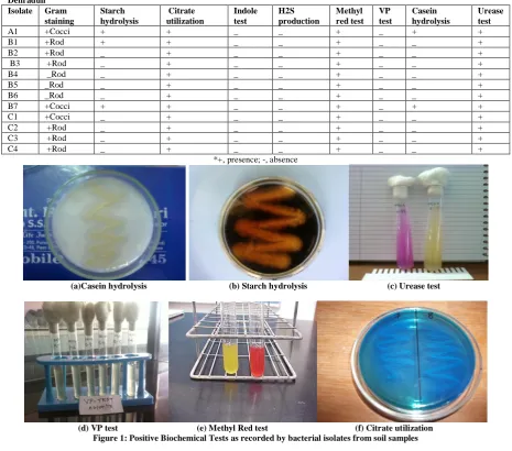
Total of twelve isolates were obtained from different soil samples i.e only one isolate was obtained from Garden soil (Sample A), seven isolates were obtained from Near the Banks of Ganga River (Sample B) and four isolates were obtained from soil sample from local gardens (Sample C) of Uttarakhand. The isolates were named as R/D on the basis of site of location (say Rishikesh and Dehradun) to avoid ambiguity on the culture plates and slants. The isolates were maintained on Nutrient agar plates and slants. The isolates were further screened for gram staining and different biochemical tests. The results showed that amongst twelve isolates obtained, 6 were gram positive bacilli, 3 were gram negative bacilli and 3 were gram positive cocci. The results of gram staining and biochemical tests are shown in Table 1. Positive biochemical tests shown by isolates are recorded in Figure 1.
3.2 Antimicrobial activity of bacteria isolates
antibacterial activity against Lactobacillus plantarum NCIM 2083. The pellet fractions, A1, B2 and B3 showed antifungal activity against Aspergillus niger NCIM 1196 while
Candida albicans NCIM 3471 was found to be sensitive against B1, B2, B5 and B6 pellet fractions. The results are shown in Table 3 and Figure 3 (a & b).
Table 1 (a): Biochemical Tests for confirmation of bacterial isolates recorded from soil samples of different sites of Rishikesh and Dehradun
Isolate Gram staining
Starch hydrolysis
Citrate utilization
Indole test
H2S production
Methyl red test
VP test
Casein hydrolysis
Urease test
A1 +Cocci + + _ _ + _ + +
B1 +Rod + + _ _ + _ _ +
B2 +Rod _ + _ _ + _ _ +
B3 +Rod _ + _ _ + _ _ +
B4 _Rod _ + _ _ + _ _ +
B5 _Rod _ + _ _ + _ _ +
B6 _Rod _ + _ _ + _ _ +
B7 +Cocci + + _ _ + _ + +
C1 +Cocci _ + _ _ + _ _ +
C2 +Rod _ + _ _ + _ _ +
C3 +Rod _ + _ _ + _ _ +
C4 +Rod _ + _ _ + _ _ +
*+, presence; -, absence
(a)Casein hydrolysis (b) Starch hydrolysis (c) Urease test
(d) VP test (e) Methyl Red test (f) Citrate utilization Figure 1: Positive Biochemical Tests as recorded by bacterial isolates from soil samples
Table 2: Antimicrobial activities of supernatant/ethyl acetate fractions of isolates obtained from soil Isolate supernatant Diameter of zone of inhibition (mm)
Pathogens studied
E.coli S. abony M. luteus L. plantarum MRSA 35 MRSA 8 niger C.albicans
A1 NA NA NA NA NA NA 05 03
B1 NA NA NA NA NA NA 04 02
B2 NA NA NA NA NA NA 03 03
B3 NA NA NA NA NA NA 02 04
B4 NA NA NA NA NA NA 03 03
B5 NA NA NA NA NA NA 04 07
B6 NA NA NA NA NA NA 03 NA
B7 NA NA NA NA 23 NA 02 NA
C1 NA NA NA NA NA NA 05 NA
C2 NA NA NA NA NA NA 07 NA
C3 NA NA NA NA 26 NA 04 NA
C4 NA NA NA NA NA NA 06 NA
Erythromycin (1 mg/ml)
18 12 15 15 10 12 NT NT
Fucanazole (1 mg/ml)
NT NT NT NT NT NT 12 15
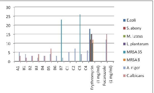
Figure 2 (a): Graphical representation of antimicrobial activity of supernatant/ethyl acetate fraction obtained from soil iso lates against pathogenic strains
3.3 TLC profile of potent antibiotic fraction
The supernatant/ethyl acetate fractions of the potent isolates, B2, B5 and C2 were analyzed by thin TLC plate using chloroform and methanol (60:40) as solvent
system and erythromycin as
control/standard antibiotic. Each extract
produced spots when the chromatogram was visualized under iodine vapor. The spot was near the solvent front with Rf value of 0.54 for B2, 0.56 for B5 and 0.57 for C2. Standard antibiotic, Erythromycin however formed a tailed spot with Rf of about 0.5. The results are shown in Table 4 and Figure 4.
Figure 3 (a): Graphical representation of antimicrobial activity of pellet fraction of isolates obtained from soil
Table 3: Antimicrobial activities of pellet fractions of isolates obtained from soil
Isolate supernatant Diameter of zone of inhibition (mm) Pathogens studied
E. coli S. abony M. luteus L. plantarum MRSA 35 MRSA 8 niger C. albicans
A1 NA NA NA 10 NA NA 07 06
B1 NA NA 10 NA NA NA 26 06
B2 NA NA NA NA NA NA 25 NA
B3 NA NA NA NA NA NA NA NA
B4 NA NA NA 09 NA NA NA 05
B5 NA NA NA 08 NA NA NA 07
B6 NA NA NA 10 NA NA NA NA
B7 NA NA 15 NA NA NA NA NA
C1 NA NA NA NA NA NA NA NA
C2 NA NA NA NA NA NA NA NA
C3 NA NA NA NA NA NA NA NA
C4 NA NA 13 15 NA NA NA NA
Erythromycin (1 mg/ml)
18 12 15 15 10 12 NT NT
Flucanazole (1 mg/ml)
NT NT NT NT NT NT 12 15
*NA, No Activity; NT, Not Tested
Table 4: TLC profile of potent antibiotic fraction
S.No. Ethyl acetate/supernatant fraction Rf values
1. B2 0.54
2. B5 0.56
3. C2 0.57
4. Erythromycin 0.5
Figure 4: TLC profile of potent antibiotic fraction
The results showed that antimicrobial substance produced by some isolates was membrane bound while some of the isolates produce non membranous bound antimicrobial substance. It was found that E. coli NCIM 2065, Lactobacillus plantarum NCIM 2083, Micrococcus luteus ATCC 9341, Salmonella abony NCIM 2257 and MRSA 8 (isolated from pus) were the most resistant pathogens which showed no activity against supernatant/ethyl acetate fraction of any of the isolates of soil samples collected from Rishikesh and Dehradun regions of Uttarakhand. Only B2 supernatant/ethyl acetate fraction showed significant antibacterial activity against
MRSA 35 (isolated from blood). Fractions, B1-B7 and C1-C4 showed antifungal activity against Candida albicans NCIM 3471 and Aspergillus niger NCIM 1196. The results indicate that E. coli NCIM 2065, Salmonella abony NCIM 2257, MRSA 8 (isolated from pus) and MRSA 35 (isolated from blood) were the most resistant pathogens which showed no activity against pellet fraction of any of the isolates. The pellet fractions B2, C2 and B6 showed antibacterial activity against Micrococcus luteus ATCC 9341. The pellet fractions, A1, B5, B6, C1 and C6 showed antibacterial activity against Lactobacillus plantarum NCIM 2083. The pellet fractions, A1, B2 and B3 showed antifungal activity against Aspergillus niger NCIM 1196 while Candida albicans NCIM 3471 was found to be sensitive against B1, B2, B5 and B6 pellet fractions. The results correlate the previous findings reported by large number of researchers showing the screening of antimicrobial isolates from soil samples.
[17-21]
CONCLUSION
antimicrobial activity suggests that water represents an ecological niche which harbours a largely uncharacterized microbial diversity and yet unexploited potential in the search for new secondary metabolites. The present study thus can be utilized further to explore the microbial biodiversity in sulphur water springs which are able to produce antibiotics that can be utilized in cure of skin infections etc. Further analysis of
compounds by spectrometric,
chromatographic techniques and strain improvement studies provides detailed information about the compounds produced by the isolates and the ability of the strains in producing newer compounds.
ACKNOWLEDGEMENT
The author acknowledges Dr. Abhishek Mathur, Sr. Scientist & Executive Director (R&D), NCS Group, Nagpur for contribution and support in her research work.
Conflict Of Interest
The author declares no conflict of interest.
REFERENCES
1. Classen ATS, Boyle I, Haskins KE, S. T. Overby, and S. C. Hart. Community-level physiological profiles of bacteria and fungi: plate type and incubation temperature influences on contrasting soils. Fems Microbiology Ecology. 2003; 44:319-328. 2. Cain CC, Lee DH, Waldo RH. Synergistic
antimicrobial activity of metabolites produced by a nonobligate bacterial predator. Antimicrobial Agents and Chemotherapy. 2008; 47:2113-2117. 3. Cain CC, Henry AT, Waldo RH, L. Casida,
and Falkinham JO. Identification and characteristics of a novel Burkholderia strain with broad-spectrum antimicrobial activity. Applied and Environmental Microbiology. 2009; 66:4139-4141.
4. Correa OS, Montecchia MS, Berti MF, Ferrari MCF, Pucheu NL. Bacillus amyloliquefaciens BNM122, a potential microbial biocontrol agent applied on soybean seeds, causes a minor impact on rhizosphere and soil microbial communities. Applied Soil Ecology. 2009; 41:185-194. 5. Casida LE. Bacterial Predators of
Micrococcus-Luteus in Soil. Applied and
Environmental Microbiology. 2009; 39: 1035-1041.
6. S. Chatterjee and LC Vinning. Nutrient utilization in Actinomycetes. Induction of Beta -glucosidases in Streptomyces
venezuelae. Canadian Journal of
Microbiology. 1981: 27: 639-645.
7. Kohnaski MA, Dwyer DJ, Collins JJ. How antibiotics kill bacteria: from targets to networks. Nat Rev Microbiology. 2010: 8(6): 423-435.
8. Rani P, Khullar N. Antimicrobial evaluation of some medicinal plants for their anti-enteric potential against multi drug resistant
Salmonella typhii. Phytotherapy Research.
2004; 18(8): 670-673.
9. Casey JT, Walsh PK, and O'Shea DG. Characterisation of adsorbent resins for the recovery of geldanamycin from fermentation broth. Separation and Purification Technology. 2007; 53:281-288. 10. Falkinham JO, Wall TE, Tanner JR, Tawaha K, Alali FQ, Li C, and Oberlies NH. Proliferation of Antibiotic Producing Bacteria and Concomitant Antibiotic Production as the Basis for the Antibiotic Activity of Jordan's Red Soils. Applied and Environmental Microbiology 2009; 75:2735-2741
11. Toroglu S, Dincer S. and Korkmaz H. Antibiotic resistance in Gram negative bacteria isolated from Aksu river in (Kahramanmaras), Turkey. 2005; 55, 229-233.
12. K. Khanna, D. R. Ecology and pollution of river Ganga. (pp. 1–241).NewDelhi: Ashish Pub. House, 1993.
13. Perez C, Anesini C. In vitro antimicrobial activity of Argentine folk medicinal plants against Salmonella typhii. Journal of Ethnopharmacology. 1993; 44: 41-46. 14. Balagurunathan, R., Palanivel, P.,
Ashokkumar, L., Prabakaran, R. (2012). Study on antimicrobial metabolite produced by soil bacteria isolated from less explored ecosystem. Life Sciences Leaflets. 12; 165-173.
15. Chandrashekhara S. Isolation and characterization of antibiotic production from soil isolates by Fermentation. Ph.D. Thesis. 2010., Vinayaka Missions University, Salem, Tamil Nadu pp. 16-220. 16. Oskay Mustafa. Isolation and purification of
Streptomyces sp. KGG32. International J. Agr. & Biol. 2011; 13 (3): 369-374.
17. Abo-Shadi Maha Abd Al-Rehman, Sidkey N.M., Al-Mutrafy A.M. Antimicrobial agent produced from some soils Rhizosphere in Al-Madinah Al-Munnawarah, KSA. J. American Sci. 2010; 6 (10): 915-925. 18. R. Thakur, MK Roy, NN Dutta and Rl
Bezbaruah. Coordinate production of Cephamycin C and clavulanic acid by
Streptomyces clavuligerus. Indian Journal of
Experimental Biology 1999; 37:1031-1033 19. Singh A.P., Singh R.B., Mishra S. Studies
on isolation and characterization of antibiotic producing microorganisms from
industrial waste soil sample. The Open Nutraceuticals Journal, 2012, 5, 169-173. 20. Compeau, G., B. J. Alachi, E. Platsouka,
and S. B. Levy. Survival of Rifampin-Resistant Mutants of Pseudomonas Fluorescens and Pseudomonas-Putida in Soil Systems. Applied and Environmental Microbiology. 2005: 54:2432-2438.
21. Cordell, G. A., N. R. Farnsworth, C. W. W. Beecher, D. D. Soejarto, A. D. Kinghorn, J. M. Pezzuto, M. E. Wall, M. C. Wani, D. M. Brown, M. J. Oneill, J. A. Lewis, R. M. Tait, and T. J. R. Harris. Novel Strategies for the Discovery of Plant-Derived Anticancer Agents. Acs Symposium Series. 2006: 534:191-204.
******