Available online on 15.09.2016 at http://jddtonline.info
Journal of Drug Delivery and Therapeutics
An International Peer Reviewed Journal Open access to Pharmaceutical and Medical research
© 2016, publisher and licensee JDDT, This is an Open Access article which permits unrestricted noncommercial use, provided the original work is properly cited
REVIEW ARTICLE
MICRONEEDLE TECHNOLOGY FOR TRANSDERMAL DRUG DELIVERY:
APPLICATIONS AND COMBINATION WITH OTHER ENHANCING TECHNIQUES
*Nayak Smita, Suryawanshi Sanidhya, Vaidhun Bhaskar
Department of Quality Assurance, Gahlot Institute of Pharmacy, Koparkhairane, Navi Mumbai-400709, Maharashtra, India
Received 19 June 2016; Review Completed 26 Aug 2016; Accepted 26 Aug 2016, Available online 15 Sep 2016
* Corresponding Author
Smita Nayak, Department of Quality Assurance,Gahlot Institute of Pharmacy
E-mail: [email protected], Tel: 022-27550816
ABSTRACT
Transdermal delivery system has been extensively employed over the years owing to its advantages over the conventional delivery systems. However, the stratum corneum hinders the penetration of substances through the skin. Microneedle technology is the latest advancement and has gained tremendous attention in transdermal delivery system, overcoming the obstacles of transdermal delivery system and providing advantages over the conventional delivery systems. Numerous studies have been carried out for exploring microneedles. This review article covers the types of microneedles, mechanism of action of microneedles, methodology of drug delivery via microneedles while mainly focusing on the applications of microneedles and their combination with other enhancing techniques in transdermal delivery of drug and other molecules.
Keywords: Transdermal delivery, Microneedles, Stratum corneum, Iontophoresis, Electroporation, Sonophoresis, Vibratory actuation, Immunologicals, Biopharmaceuticals, Phlebotomy, Diagnosis.
DOI:http://dx.doi.org/10.22270/jddt.v6i5.1285 URI: http://jddtonline.info/index.php/jddt/article/view/1285
1. INTRODUCTION
Administration of drugs can be achieved through various possible delivery routes available including the most common routes like the oral, parenteral, ophthalmic and transdermal route, as well as less explored routes such as nasal, pulmonary and buccal 1. Each of these routes has specific merits and demerits. Over the years transdermal drug delivery systems have shown a potential alternative for the oral and parenteral routes of drug delivery systems overcoming their demerits of drug degradation in the gastrointestinal tract, first-pass metabolism, poor absorption, local irritation and variability in absorption (due to factors like pH, motility, food, mucus layer, etc.), in case of oral drug delivery systems 2, 3 and pain associated with the injections, expertise required to deliver the drug, risk of infection and difficulty in obtaining sustained drug delivery in case of parenteral drug delivery systems4. . However transdermal technology has limitations due to the inability of a large majority of drugs to cross the skin at the desired therapeutic rates because of the presence of a relatively impermeable thick outer stratum corneum layer. This barrier posed by human skin limits transdermal
effectiveness in facilitating the passage of macromolecules, hydrophilic substances, and different carriers such as lipid vesicles and nanoparticles.
2. CONCEPT OF MICRONEEDLES
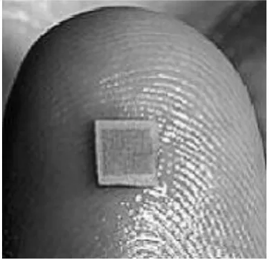
Microneedles are microstructured transdermal systems, consisting of an array of microstructured projections coated with a drug or vaccine that is applied to the skin to provide intradermal delivery of active agents, which otherwise would not cross the stratum corneum 6. Microneedles are somewhat like traditional needles, but are fabricated on the micro scale. They are smaller than hypodermic needle, generally one micron in diameter and ranges from 1-100 microns in length.
Figure 1: Experimental microneedle array consisting 400 microneedles.
Advantages of Microneedles 7-10:
rapid onset of action
painless administration of the active pharmaceutical ingredient
Large molecules can be administered first-pass metabolism is avoided
faster healing at injection site than with a hypodermic needle
patient compliance due to ease of administration decreased microbial penetration as compared with
a hypodermic needle, the microneedle bypasses the stratum corneum and punctures only the epidermis
specific skin area can be targeted for desired drug delivery
Drug can be administered at constant rate for a longer period
good reproducibility
good stability and enhanced drug efficacy may result in dose reduction
good tolerability without long-term oedema or erythema
Disadvantages of Microneedles 7:
skin irritation may result because of allergy or sensitive skin
local inflammation may result if the concentration of drug is high under the skin
careful use of the device may be needed to avoid particles ‘bouncing off’ the skin surface; if the device is not held vertically, the dose may escape or can penetrate the skin to differing degrees the thickness of the stratum corneum and other
skin layers varies between individuals and so penetration depth of particles could vary too external environment, like hydration of the skin,
could affect delivery
tip of the microneedle may break off and remain within the skin on removal of the patch
Compressed dermal tissue can block hollow microneedles
Selection of the material for constitution of the microneedle should be based on criteria such as gentle fabrication without damage to sensitive biomolecules, sufficient mechanical strength for insertion into skin and controlled or rapid drug release as per the requirement. Considering the requirement of microneedle to be long enough to penetrate the stratum corneum and at the same time should be short enough to be painless, they have been manufactured in a variety of shapes and sizes, as required for different applications using different materials as mentioned below 11. Most microneedle fabrication methods are based on the conventional microfabrication techniques of replicating microstructures using micromoulds, utilizing photolithographic processes, silicon etching, laser cutting, metal electroplating, metal electropolishing etc.
3. MECHANISM OF ACTION
Table 1: Material of construction for microneedles
Material of construction
Example Type of
microneedle produced
Merits Demerits
Metal Nickel-iron 11, stainless steel 12, titanium 13,
Hollow/solid/ coated
Good mechanical strength, cost
effective
Non-biodegradable, expensive, brittle
Silicon Silicon dioxide 14 Solid/hollow Greater mechanical
strength
Expensive, brittle
Glass --- Hollow High drug loading
capacity
Non-biodegradable Biodegradable
Polymers
Polylactic acid 15, Polyglycolic acid 15, Polylactide-co-glycolic acid (PLGA)
15, polycarbonate16,
Polyvinylpyrrolidone (PVP) 17
Solid Cost effective, good resistance,
biocompatible
Thermal instability
Non-biodegradable
polymers
Polyvinyl acetate (PVA) 18, Alginic acid 18, Gantrez AN-139, a copolymer
of methylvinylether and maleic anhydride (PMVE/MA) 18, Carbopol
971 P-NF 18, Polyetherimide 19
Solid Cost effective, good resistance
Non-biodegradable
Polysaccharide s
carboxymethylcellulose 20, Amylopectin 20, Maltose 21, Dextran 22, Galactose 22, Chondroitin Sulfate 22,
Thermoplastic starch 23
Solid, dissolving
Rapid drug delivery, biodegradable
Hygroscopic, caramelization
4. TYPES OF MICRONEEDLES
Classification for microneedles usually used in literature is based on the fabrication process: in-plane or out-of-plane microneedles.
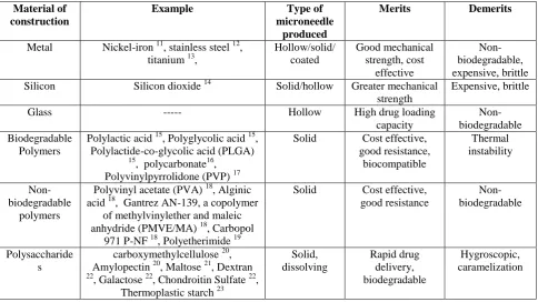
Another useful point of distinction is whether the microneedles are solid, hollow, coated or dissolving. The following sections will give an overview of these microneedles.
4.1 Solid microneedles
Solid micro needles are defined as the arrays of projections that are employed for creating holes in stratum corneum and are applied before the application of a drug and then removed afterwards. These can essentially create micron scale holes in the skin, through which drug molecules can easily enter 25. These needles are inserted into the skin for specified time period. The micro channels developed by the insertion of micro needles promote the drug transport in to the viable epidermis 26.
4.2 Coated microneedles
Coated microneedles are solid microneedles that act as vehicles to carry and deposit drug within the skin or other tissue. This includes coating microneedles with a drug in a formulation suitable for coating and subsequent dissolution. In this way, the desired dose of the drug is delivered into tissue quickly upon insertion of the microneedles. The drug dose that can be administered this way is limited to the amount that can be coated onto the tip and shaft of the microneedles, which is typically less than 1 mg for small microneedle arrays27
4.3. Dissolving microneedles
Unlike coated microneedles, dissolving microneedles completely dissolve in the skin and thus leave no bio hazardous waste behind after use. These microneedles typically constitute of water-soluble materials such as polymers and sugars that are safe and inert and will dissolve in the skin after insertion. While dissolving microneedles can be used as a skin pretreatment to increase permeability, drugs are often encapsulated inside the microneedle for release into the skin 28.
4.4 Hollow microneedles
Figure 2: Different types of microneedles: solid, coated, dissolving and hollow.
4.5 Hydrogel-forming microneedles
Hydrogel-forming microneedles are the latest development in microneedle technology. These are arrays of microneedles made up of a swelling material with a drug reservoir attached to the baseplate of the array 31-34. After insertion into the skin, the array absorbs interstitial fluid and swells to form continuous conduits between the dermal microcirculation and an attached patch-type drug reservoir leading to the diffusion of the drug into the skin 32, 33. Such microneedles act initially as a tool to penetrate the stratum corneum barrier. Upon swelling, they become a rate controlling membrane. These microneedle arrays are produced mainly using synthetic polymers such as an aqueous polymer gel that can be easily cross-linked by chemical or physical methods 35. These materials, once swollen, should maintain structural integrity and be reasonably robust during handling 33. Although hydrogel-forming microneedles are made from polymers, there are distinct differences between these microneedles and the regular dissolving polymer microneedles. Advantages of hydrogel-forming microneedles are that they can be fabricated in a wide range of patch sizes and geometries, can be easily sterilized, resist hole closure while in place and are removed completely intact from the skin 32. However more effort should be directed at widening the range of materials that can be used to fabricate these useful transdermal drug delivery systems. Hydrogel-forming MN arrays have been employed to deliver a wide range of different molecules from small molecules 32-33, 36-37 to high molecular weight compounds 32-33, 37. The use of hydrogel-forming microneedle arrays for minimally-invasive extraction and quantification of drug substances and glucose from skin in vitro and in vivo strongly illustrates the potential of hydrogel-forming microneedles in minimally-invasive patient monitoring and diagnosis 38.
5. METHODOLOGY FOR DRUG DELIVERY
A number of delivery strategies have been employed to use the microneedles for transdermal drug delivery.
These include
Poke and patch
Coat and poke
Poke and release
Poke and flow
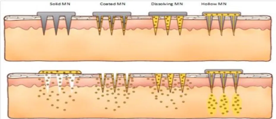
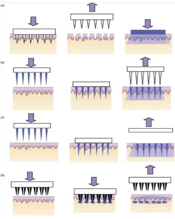
5.1 Poke and patch
It involves piercing an array of solid microneedles into the skin followed by application of the drug patch at the treated site. Transport of drug across skin can occur by diffusion or possibly by iontophoresis if an electric field is applied 39. This technique was also tried to extract the interstitial fluid to measure the glucose level by non-invasive method 40.
5.2 Coat and poke
In this approach needles are first coated with the drug and then inserted into the skin for drug release by dissolution. The entire drug to be delivered is coated on the needle itself 5. Dip and scrape approach is a variation of this approach, where microneedles are first dipped into a drug solution and then scraped across the skin surface to leave behind the drug within the micro abrasions created by the needles 39. A limited amount of drug could be coated over the microneedles (only about 1 mg) and extensive optimization was required for uniform coating in this ‘coat and poke’ approach.
5.3 Poke and release
be modulated as per the requirement using a variety of available polymers and polysaccharides 28.
5.4 Poke and flow
In this approach first the skin is pierced (external pressure) and then the drug is allowed to flow through hollow microneedles from the reservoir in the patch 41.
A large amount of drug can be administered by poke and flow method. Additionally, pressure-driven delivery adds the possibility to precisely steer the flow rate and to obtain a more controlled delivery.
All of the above-mentioned approaches can be employed to deliver drugs either systemically or at a restricted site (local action).
Figure 3 : Methodology for drug delivery by microneedles: (a) ‘poke and patch’ using solid microneedles, (b) ‘coat
and poke’ using coated solid microneedles, (c) ‘poke and release’ using polymeric microneedles, (d) ‘poke and flow’ using hollow microneedles.
6. APPLICATIONS OF MICRONEEDLES
Microneedles have been explored for different applications and are extended to many fields. Owing to benefit of piercing in a minimally invasive manner, apart from being an alternative to conventional hypodermic therapy, they have also been employed for ocular, systemic and intracellular delivery. Microneedles can be used to deliver high molecular weight compounds like proteins and peptides, immunobiologicals like vaccines, antibodies etc. 42,
bioactive agents or bio macromolecules like insulin, heparin, albumin, growth hormones 43. Microneedles have also gained prominent attention in the field of cosmetics and various cosmeceuticals have been used for the treatment of acne, pigmentation, scars and wrinkles as well as for skin toning 42.
6.1 Immunobiologicals
associated with insertion of needle into the skin, recent studies have focused on development of needle-free vaccination like liquid jet injectors, powder injectors, thermal ablation and microneedles for the administration of immunobiologicals via the subcutaneous, intramuscular or intradermal route for prevention of infectious diseases. Microneedles have an edge over the other methods due to lack of pain, self-administration and quick delivery of vaccine 42-43. Making use of the microneedles allows vaccines to cross the stratum corneum and stimulate a clinical response. In case of dissolving microneedles, controlled and complete penetration is an important parameter to be considered. These are capable of delivering a small dose, less than several milligrams, of peptides, proteins, vaccines, hormones and organic compounds. Numerous studies have been performed and reported on vaccine delivery using microneedles including human IgG 21, tetanus toxoid 23, DNA vaccine 39, influenza vaccine 44, hepatitis B vaccine 45, human papilloma virus vaccine 46, west nile virus vaccine 47, chikungunya virus vaccine 47, herpes simplex virus vaccine 48-49, Bacillus Calmette Guerin vaccine 50, ovalbumin 51, , live attenuated chimeric JE vaccine (ChimeriVax)-JE/flavivirus vaccine 52, diphtheria 53, malaria 53, combination and recombinant protein vaccines for anthrax, botulism, plague and staphylococcal toxic shock 54.
6.2 Bioactive macromolecules (Biopharmaceuticals)
Owing to the proteolytic degradation and hindered absorption, bioactive macromolecules such as insulin, heparin, and growth hormones are not administered orally. The majority of commercially available biopharmaceuticals are administered via the parenteral route and hence a suitable non-invasive route is desirable. Microneedle arrays have been found to enhance the transport across dermatomed human skin for both low and high molecular weight compounds and also that the length of the microneedles and the depth up to which the microneedles penetrated in the skin had no effect on the transport of either low or high molecular weight compounds55. For rapid release as well as controlled release of molecules, an approach of preparing dissolving microneedles constituting water-soluble polysaccharides has been done 20, 22. Studies performed and reported on administration of biopharmaceuticals using microneedles include delivery of low molecular weight heparin 22, insulin 56, L-Carnitine 57, calcein and bovine serum albumin 58, desmopressin 59, recombinant human growth hormone and desmopressin 60, albumin 61, calcein 62, erythropoietin 63, oligonucleotides 64, porphyrin precursor 5-aminolevulinic acid (ALA) 65, salmon calcitonin 66, daniplestim 67, leuprolide acetate 68, Para thyroid hormone 69, human growth hormone 70 etc.
6.3 Drugs
It is essential for a drug molecule to possess the necessary physicochemical properties to cross the skin barrier. Transport of a drug molecule through the skin and also the rate of transportation are governed by these physico-chemical properties like
hydrophilic-lipophilic balance, solubility, molecular weight, etc. The challenges posed in transdermal drug delivery can be overcome by use of microneedles. Use of microneedles enhances the bioavailability of drugs and can exclude the need for penetration enhancers, which may induce irritation. Microneedles may also reduce the side effects and complications associated with systemic administration. Drugs administered via microneedles include L-Ascorbic acid 19, riboflavin 27, galanthamine 71, aspirin 72, docetaxel 73, pilocarpine 74, methotrexate 75, prochlorperazine edisylate 76, lidocaine hydrochloride 77, ropinirole hydrochloride 78, ketoprofen 79, naltrexone with diclofenac sodium 80, phenylephrine 81, naltrexone 82, mannitol 83, glycerol 84 etc.
6.4 Phlebotomy
Phlebotomy refers to the withdrawal of blood samples for analysis of specific blood constituents for diagnosis of a disease. Blood samples are generally collected from capillaries by pricking the skin or from veins using evacuated collection tube, depending on the volume of blood required for analysis. These methods are associated with disadvantages such as excessive bleeding, infection, scarring, fainting or feeling light-headed. People may hesitate to give blood due to fear of needles and the moderate pain associated with the procedure. In such case, painless blood sampling using microneedles can be a very good alternative to hypodermic needles 85-86. Microneedles situated at a distance of 500–2000 mm in the dermis layer beneath the skin can be used to obtain precise body fluids as well as blood samples from the capillaries. Apart from making the procedure painless, microneedles also reduces the blood sample requirements (up to 200 nanolitres). However the most essential requirement is that the microneedle must penetrate to sufficient depth; hence care should be taken in the design, material selection and dimensions of the microneedle, to ensure penetration at low pressure without breakage. Painless hollow microneedle-based micro sampling can be used instead of traditional methods for glucose estimation in case of disease diabetes which requires frequent monitoring of blood for estimation of glucose concentration or disease severity 87. Microneedles can also be used for monitoring of therapeutic drug levels 88. A jagged-shaped, hollow in-plane silicon
microneedle resembling the proboscis of a mosquito has been prepared for collection of blood for testing 89. Arrays of 350m long, hollow, out-of-plane microneedles demonstrated in-situ analysis biological fluid extraction through capillary action 90.
6.5 Diagnosis
osteoarthritis in knees, hips and other joints 92. Work has been carried out on development of two sensing device based on hybrid microneedles array for diagnostic and therapeutic applications. Hybrid microneedles having a porous structure were prepared, which can include a variety of biological molecules, as bioprobes or drugs. The first device is an electrochemical sensor where microneedles contain enzymes in their matrix that interact with glucose. The redox reaction with glucose, mediated by ferrocene, creates a charge transfer resulting in a current proportional to the glucose concentration. The second device is a therapeutic tool with optically controlled release of drug. In this case the device includes a porous silicon membrane with a Bragg's mirror, whose reflection wavelength is related to the drug’s concentration in the microneedle 93. Microneedle sensors fabricated at the end of an endoscope using poly caprolactone (PCL) microneedles coated with poly (3, 4-ethylenedioxythiophene) (PEDOT), functionalized with hemin molecules on the surface were employed for both endomicroscopic imaging and biosensing of colon cancer by real-time electrical detection of nitric oxide 94. Microneedles have also been developed as sensors for hydrogen peroxide, lactate, dissolved oxygen and glutamate 95-97. Microneedles have been employed as bioelectrical interfaces, especially for neural recording and stimulation 98-106, as well as for electrocardiography (ECG) 107 and electroencephalography (EEG) measurements 108-109.
6.6 Cosmeceuticals
Cosmeceutical industry has shown great interest in microneedle technology, the majority of cosmetic products are lending themselves to microneedle technology for non-surgical and non-ablative treatment of skin conditions such as ageing (wrinkles, lax skin), scarring (acne, surgical), photo damage, hyperpigmentation (age/brown spots) and hair loss (alopecia). The process facilitates and stimulates skin’s natural repair without causing permanent epidermal damage. Microneedles can be used for cosmetic applications, mainly for treatment of skin blemishes and the delivery of active cosmetic ingredients 110-111. Marketed microneedles like Derma rollers® and stamps are available for treatment of skin problems as well as to improve looks. MTS Dermaroller® marketed by Clinical Resolution Laboratory is a cosmetic aid possessing needles that penetrate the skin up to a depth of 0.2–0.3 mm. It contains 200 very fine stainless-steel needles that pierce the epidermis, creating a micro-channel effect. Clinical studies from various countries have proven that therapeutic serum absorption is increased by as much as 1000 times when applied using the MTS Dermaroller® 112. A hyaluronic acid based dissolving microneedle patch developed for the intradermal delivery of ascorbic acid and retinyl retinoate displayed improved skin appearance in terms of roughness and wrinkle appearance when tested in human volunteers 113. An enhancement in the local delivery of eflornithine (used to reduce facial hirsutism) was observed both in vitro and in vivo when
the skin was exposed to microneedle pretreatment followed by local administration of an eflornithine cream 114. Microneedle technology can be used to treat different types of scars by dermabrasion 115-119. Dermabrasion involves piercing the skin multiple times with microneedles to induce collagen growth. A noticeable improvement was observed in the treatment of patients with dermal scars (striae distensae) using a disc microneedle therapy 116. However, similar to the treatment of acne, this treatment too is associated with limitations of skin bleeding and painful treatment. An aesthetic improvement in patients with acne scars was reported with a reduction in scar severity in all subjects when treated with Dermaroller® 117. Similar results were obtained for treatment of atrophic facial scars using Dermarollers®120. An alternative approach to the use of Dermarollers® for the treatment of acne scars reported is the fractional radio frequency microneedle system. This system was introduced recently as a new device for facial rejuvenation. It involves creating radiofrequency-induced thermal zones. This causes damage to the reticular dermis followed by the dermal thickening. This system was clinically evaluated and the results suggested that this novel technique is efficacious for the treatment of acne scars, as it reduced the severity of the scars in more than 80% of volunteers 121.
7. MICRONEEDLES IN COMBINATION WITH OTHER ENHANCING TECHNIQUES FOR IMPROVING TRANSDERMAL DRUG DELIVERY
In order to improve the skin permeability, different techniques have been developed and employed such as using permeation enhancers, either chemical or physical. These enhancers may exhibit different mechanisms of action however their sole aim is to modify the structure of stratum corneum which is considered as the main barrier to the permeation of substances. Even though the use of permeation enhancers increases the penetration of drugs through the skin, in many cases it proves to be insufficient in achieving the therapeutic levels. To overcome this, several experiments in which two or even more enhancing techniques are combined have been attempted to obtain a synergistic effect, thus achieving the required effect of enhanced drug delivery and better control of drug delivery across the skin. Following section gives an overview of the enhancing techniques.
7.1 Microneedles in combination with Iontophoresis
Iontophoretic transdermal transport mainly involves mechanisms of electromigration and electroosmosis. Electromigration is the movement of ionic species in response to the application of the electric field, and is the most important mechanism for the delivery of ionic drugs. Electroosmosis is the aqueous flow that occurs during the movement of charged species, carrying all the dissolved drugs across the skin. The technique of iontophoresis might be still limited by the molecular size of the drug candidate even though it offers an attractive alternative for the delivery of macromolecules 124.
7.2 Microneedles in combination with Sonophoresis
Sonophoresis involves application of ultrasound (frequency, 20 kHz to 10 MHz; intensity, up to 3 W/cm2) causing perturbation of lipid bilayer inducing change in the lipid arrangement of the stratum corneum, forming cavitation thus enhancing transportation of drugs across the skin. Drug permeation can be controlled by controlling the frequency of the ultrasound. An increase in the sound frequency from 20 kHz to 1 MHz causes 1000 fold increase in skin perturbation 125. A synergistic effect on the permeation of molecules through the skin can be achieved by combining sonophoresis with microneedles. Very few studies have been performed on employing this strategy and thus needs to be explored further.
7.3 Microneedles in combination with
Electroporation
Electroporation involves application of high voltage short-duration (milliseconds) current (generally 50– 1000 V/cm) causing a transitory, localized perturbation of lipid bilayer inducing structural rearrangements of the cell membrane due to the electric fields that are produced during current application. The aqueous pores formed as a result act as the aqueous pathways and provide a local driving force that facilitates the transport of molecules across the stratum corneum. A trans-membrane potential up to 1 kV for 10 ms to 500 ms was used for in-vitro electroporation of stratum corneum 125. Although this process is useful for delivering large hydrophilic drug species like small molecules, proteins, peptides and oligonucleotides, including biopharmaceuticals with molecular weights greater than 7 kilo Daltons, electroporation was also used for permeation enhancement of larger molecules having molecular weight up to several kilo Daltons 126. Combining electroporation with microneedles resulted in microneedles that behaved as microelectrodes for electroporation, which eradicated the need for electrodes. This combination provides a synergistic effect on the permeation of molecules through the skin.
7.4 Microneedles in combination with Vibratory actuation
Precise control of insertion force is required for the penetration of a microneedle into the skin and it should not exceed the fracture force of the microneedle. Structure rigidity and miniaturization of microneedles should be well considered and a satisfactory balance
between these two should be maintained. Vibration actuation causes tissue damage via fluid cavitation and thermal damage due to frictional interaction. When this mechanism of vibration actuation is combined with microneedles, a reduction in the microneedle insertion force is achieved as a result. A study employing this combination was performed wherein the combination of vibration actuation and microneedles helped in the preparation of microneedles using metals and polymers with low value of Young’s Modulus also the effect of vibratory actuation on microneedle insertion force was evaluated and greater than 70% reduction in the insertion force was observed 127.
7.5 Microneedles in combination with Vesicles
Several studies have been performed and reported on employing lipid vesicles for the transdermal drug delivery. However these systems are surrounded with controversy related to their delivery mechanism which is reported to be associated with the accumulation of the vesicle and the encapsulated drug in the stratum corneum because of their inability to penetrate the skin deep enough rendering them with little value as carriers for transdermal drug delivery. This paved the way for employing other vesicular systems which are able to penetrate into deeper skin layers, such as niosomes (vesicles composed of nonionic surfactants), ethosomes (vesicles with high alcohol content), transferosomes (vesicles composed of phospholipids as their main ingredient, with 10-25% surfactant and 3-10% ethanol), invasomes (vesicles formed by phospholipids, ethanol and terpenes as penetration enhancers) etc. Combining microneedles with these vesicular systems holds potential for enhanced transdermal drug delivery. Depending on the diameter and height of microneedles, liposomes can be located in different parts of the skin. Results may vary according to the needle geometries. Microneedles of short height render vesicles on the surface of the stratum corneum, while longer microneedles lead vesicles into deeper skin layers.
7.6 Microneedles in combination with
Microparticles and Nanoparticles
in a specific cellular compartment. Combining microneedles with these nanoparticulate systems may significantly enhance the transdermal drug delivery wherein microchannels created by the microneedles may prove to be large enough to deliver drug-loaded nanostructures into the skin. Delivery of microparticles into the skin sets a challenge. In such case combining microneedles with these solid particles could facilitate the delivery of the later into the skin. By making use of appropriate microneedle design and insertion methods, even relatively large microparticles can be delivered into the skin. Perhaps the greatest shortcoming of controlled-release microneedles is that limited dose can be administered. Since the drug is encapsulated within microneedles and microneedles are, of course, very small, the maximum total dose that can be administered is likely to be less than 1 mg 58.
7.7 Microneedles in combination with Micro pumps
Conjunction of microneedles with micro pumps provide precise delivery of drug as the pumps control the flow rate and pressure for delivery of concentrated
drug solution as required. This combination was employed in preparing an integrated system, with micro valves and micro-pumps, which was capable of controlling fluid withdrawal for medical analysis and delivering the drug in response to metabolites levels 129.
7.8 Pocketed and Grooved Microneedles
Modification of the microneedle surface can be employed for targeted drug delivery to a specific depth in the skin and for loading greater amount of drug onto the microneedle. This can be achieved by applying the protective coat or second drug coat on the same microneedles after filling the first part in the pockets. Such microneedles are known as pocketed microneedles. These can also be obtained by fabricating microneedles with one or more holes cut through the center 130. Also grooved microneedles that capable of loading greater amount of drug onto the microneedle can be employed for enhanced drug delivery 131.
Table 2: Summary of few results of the delivery of drugs to the skin via microneedles in combination with carriers and/or enhancers
S.N. Type of
microneedle
Enhancer Carrier Drug Model Result
1 Coated microneedles 2
--- 1-μm diameter barium sulfate particles and 10-μm diameter latex particles
--- Porcine cadaver skin, in vitro
Relatively large microparticles can be delivered into the skin for controlled drug release using appropriate microneedle design and insertion methods
2 Solid stainless microneedle arrays 12
Iontophoresis (0.2 mA/cm2)
Nanovesicles Insulin Pig skin, in vitro Rats, in vivo
Combination approach of microneedles and iontophoresis showed significant increase in the permeation of charged nanovesicles. Comparable levels to subcutaneous injection of insulin was achieved 3 Beveled-tip
and
tapered-cone biodegradable polymeric microneedles58
--- carboxymethylc ellulose or polyL:
-lactide micropar ticles
Calcein and bovine serum albumin
Human cadaver skin and test in saline, in vitro
Microneedles capable of encapsulating drug and providing controlled-release delivery in skin for hours to months. 4 Macroflux®
titanium or stainless Steel microprojection Array 64
Iontophoresis (100 μA/cm2)
--- Oligodeoxynu
cleotide ISIS 2302
(ODNs)
Hairless guinea pigs, in vivo
Delivering ODNs into and through the skin in therapeutically relevant amounts
5 Maltose microneedles66
Iontophoresis (0.2 mA/cm2 for 1 hr )
--- Salmon
Calcitonin (SCT)
Hairless rat skin, in vivo
flux. 6 Maltose
Microneedles67
Iontophoresis (0.5 mA/cm2)
--- Daniplestim Hairless rat skin, in vivo
The combination approach yielded much higher flux values of daniplestim compared to
iontophoresis alone 7 Maltose
microneedles68
Iontophoresis (0.1 mA/cm2 for 4 hrs)
--- Leuprolide
acetate
Hairless rats, in vivo
The combination approach yielded much faster and higher flux values of leuprolide acetate compared to iontophoresis and microneedles alone respectively 8 Microneedle
Array 73
--- Elastic liposomes
Docetaxel (DTX)
Rat skin, in vitro Porcine skin, in vitro
Microneedle treatment led to increased steady-state flux of DTX from all formulations and shorter lag time when applying elastic liposomes through microneedle 9 Maltose
Microneedles 75
Iontophoresis (0.4 mA/cm2 for 1 hr )
--- Methotrexate Hairless rat skin, in vitro and in vivo
Enhanced transdermal delivery of
methotrexate both in vitro and in vivo. 10 Dermaroller® 76 Iontophoresis
(0.4 mA/cm2 )
--- prochlorperazi
ne edisylate
dermatomed human skin, in vitro
Significantly enhanced the transdermal delivery of prochlorperazine edisylate
11 AdminPatch®78 Iontophoresis --- Ropinirole hydrochloride
Porcine ear skin, in vitro
Continuous iontophoresis in combination with microneedles showed enhanced reflux of Ropinirole hydrochloride 12 Dermarollers®83 --- Invasomes Mannitol Human
abdominal skin, in vitro
Enhanced penetration and permeation of hydrophilic model drugs due to skin perforation by Dermarollers® particularly when combined with invasosome formulation. 13 Microneedle
roller 85
Sonophoresis --- Glycerol Porcine
skin, in vitro
The combination method effectively enhanced transdermal delivery of glycerol by accelerating the diffusion rate through the skin barrier. 14 Silicon
microneedleele ctrode array126
Electroporati on
--- Fluorescein
isothiocyanate
(FITC)-dextran
Male hairless rats, in vivo
Enhanced permeation of FITC-dextran observed as a
combination approach 15 Hollow
microneedles127
Vibratory actuation
--- --- Excised
animal skin, in vitro
Greater than 70% reduction in
microneedle insertion force by using vibratory actuation 16 Pocketed solid
stainless steel microneedle130
--- --- Sulforhodamin e, sodium fluorescein,
luciferase plasmid DNA,
vit B2
Abdominal porcine cadaver skin, in vitro
Rapid release of the model drugs at a targeted depth in the skin.
17 Grooves-embedded microneedle arrays of poly-L-lactic acid 131
--- --- Ovalbumin Mice, in
vivo
Microneedles coated with ovalbumin render higher antibody response as the number and depth of grooves increase 18 Maltose
Microneedles132
Iontophoresis (0.5 mA/cm2)
--- Low mol. wt
heparin (LMWH)
Hairless rat skin, in vitro
Increased skin permeation of LMWH 19 Microneedle
Array 133
Iontophoresis (0.3 mA/cm2)
--- FITC-dextrans Hairless rat skin, in vitro
Enhanced transdermal delivery of dextrans 20 Silicon and
glass hollow microneedle array 134
Sonophoresis --- Calcein and bovine serum
albumin (BSA)
Pig skin, in vitro
In vitro results showed a significant improvement in the delivery rate even when large molecular weight compounds were used. 21 Stainless steel
& silicon solid microneedles135
Sonophoresis --- FITC-dextrans excised porcine skin, in vitro
Significantly increased skin permeation of FITC-dextrans.
22 Stainless steel solid
microneedles 136
Sonophoresis Cross-linked hydrogelof NaCMC and gel emulsion
Lidocaine Porcine skin, in vitro
Slight increase in the permeability of lidocaine due to the combination approach. 23 Dissolving
microneedles137
Electroporati on
---
p2CMVmIL-12
Mice, in vivo
in-situ cutaneous transfer of p2CMVmIL-12 to successfully treat B16F10 subcutaneous tumors
24 Silicon microneedle electrode arrays 138
Electroporati on
--- --- --- A uniform drug
release through hollow microneedle electrodes into tissue is expected to be achieved during electroporation. 25 Coated
microneedle array 139
Electroporati on
--- Plasmid DNA Mice, in
vivo
Robust antibody responses.
26 Solid
microneedles140
--- L595 vesicles (composed of sucrose-laurate ester &
octaoxyethylene -laurate ester) and sPC
hepatitis B surface antigen
(HBsAg)
Mice, in vivo
vesicles (composed of soybean– phosphatidylchol ne and Span-80)
27 Solid
microneedles141
--- anionic
surfactant-based vesicles
diphtheria toxoid
BALB/c mice, in vivo
Enhancement in the immunogenicity of topically applied diphtheria toxin due to microneedle pretreatment 28 Dermaroller®
142
--- liposomes --- Human skin,
iv vitro
Evaluation of potential of Dermaroller®, with different needle geometries, to deliver a lipophilic fluorescent compound
encapsulated in liposomes across the human skin
29 Nanopore turbo roller 143
--- nanoliposomes sepia melanin Human skin, iv vitro
Accelerates the delivery of melanin into hair structures allowing an even absorption, larger pigment deposits, and deeper penetration of the formulation into the hair.
30 Silicon microneedle arrays 144
--- Polystyrene nanospheres
--- Human skin, iv vitro
Potential for significant enhancement the intra/transdermal delivery of nanoparticle formulations.
31 Solid silicon microneedle arrays 145
--- poly(d,l-lactic-co-glycolic acid) (PLGA) nanoparticles
--- Human skin, iv vitro
Permeation of nanoparticles into the skin was enhanced by microneedles
32 Microneedle arrays 146
--- Microparticles Gene --- Microneedles
increase the
penetration depths of the particles. 33 Hollow
microneedle 147
--- nanoparticle and
microparticle suspensions
--- whole rabbit, pig, and human eyes ex vivo
Microneedles were shown for the first time to deliver nanoparticle and microparticle suspensions into the suprachoroidal space of rabbit, pig and human eyes. 34 Solid silicon
microneedle Arrays 148
--- Polystyrene latex nanospheres
--- Human cadaver epidermis, in vitro
A significant number of latex nanoparticles cross the skin.
35 Silicon microneedle arrays 149
--- Fluorescent latex nanospheres
--- Human skin, in vitro
charged fluorescent particles into the microchannels, & to the cells of the viable epidermis
36 Silicon-based Microneedles150
--- Fluorescent polystyrene nanospheres
Lipid: polycati on : pDNA
(LPD) nonviral gene
therapy vectors.
Heat-separated human epidermal sheets, in vitro
Microneedle membrane treatment resulted in enhanced diffusion of
fluorescent polystyrene nanospheres 37 Silicon
Microneedles151
--- Hydrogel / Fluorescent Nanoparticle Loaded
--- Human skin, in vitro
Microneedle treatment resulted in successful targeting of nanoparticles within the viable epidermis 38 Hollow
Microneedles152
--- Nanospheres of PLA with Nile red and latex fluorescein labeled microspheres
--- Human corpse sclera, in vitro
Successful delivery of soluble molecules and nanoparticle
Suspensions into the sclera.
39 Pocketed stainless steel microneedles153
--- --- Botulinum
toxin A (BT)
Excised human skin, in vitro
Microneedles provide less invasive
alternative that is suited to delivery of large proteins such as BT. Microneedles were not only able to accommodate therapeutic doses of a model protein but they were also able to deliver an inactivated form of the toxin into human skin.
40 Coated microneedles154
--- Poly(lactic-co-glycolic) acid (PLGA) nanoparticles
Doxorubicin porcine cadaver buccal tissues, in vitro
Coated microneedles provide uniform and effective delivery of drugs to localized oral cancers.
41 Silicon microneedles155
--- Hypoxia-sensitive hyaluronic acid-based vesicles
Insulin Mice, in vivo
42 Silicon microneedles156
--- Lipid
microparticles
Quercetin Pig skin, in vitro
Compared to intact skin, a marked increase in quercetin levels permeated into the stratum corneum and viable epidermis was achieved when skin was treated with the flavonoid-loaded LMs in combination with microneedle arrays
43 Solid silicon microneedles157
--- Poly-D, L-lactic acid (PDLLA) nanoparticles
Ketoprofen Porcine skin, in vitro
An enhanced flux of ketoprofen was observed in the skin treated with silicon microneedles over a prolonged period of time.
CONCLUSION
Various research studies carried out on microneedles are evident enough to state that microneedles are the efficient and potential transdermal delivery systems. They have been gaining a lot of importance due to their key advantages over the conventional delivery systems of being safe, convenient and painless. Microneedles have proved to be the solution for overcoming the barrier imposed by the stratum corneum, thus broadening the number of drugs and other molecules to be administered by this route. Extensive work has been carried out to create advances and to explore the wide applications of microneedles. Apart from playing the vital role in transportation of numerous drug molecules including poorly permeable ones, macromolecules and biopharmaceuticals, microneedles have also been applied in phlebotomy, diagnosis and cosmeceuticals. Numerous studies have been performed and reported on the synergistic effect of combining microneedles with other techniques for enhanced drug delivery such as sonophoresis,
electroporation, iontophoresis, vibratory actuation etc. and have turn out to be beneficial in efficient transportation of drug and other macromolecules across the skin. Conjugating microneedles with carriers such as vesicles, nanoparticles, microparticles etc. allows achieving a controlled delivery of drug molecules. Precise drug delivery has been possible by employing microneedles in combination of micro pumps while greater amount of drug can be delivered by loading into the pocketed and grooved microneedles. Thus it was concluded thatmicroneedles are more efficient as compared to the hypodermic needles and the other conventional delivery systems.
ACKNOWLEDGEMENT
The authors thank the management of Gahlot Institute of Pharmacy for providing support for carrying out the research work.
CONFLICT OF INTEREST: The authors declare
that there is no conflict of interest regarding the publication of this paper
REFERENCES
1. Langer R, Drug delivery. Drugs on target, Science, 2001, 293(5527), 58–59.
2. Gill HS, Prausnitz MR, Coated microneedles for transdermal delivery, Journal of Control Release, 2007, 117(2), 227–237. 3. Xie Y, Xu B, Gao Y, Controlled transdermal delivery of
model drug compounds by MEMS microneedle array, Nanomedicine, 2005, 1(2), 184– 190.
4. Avis KE Lieberman HA, Lachman L. Pharmaceutical Dosage Forms: Parenteral Modifications. 2nd ed. New York: Marcel Dekker Inc; 1992.
5. Bora P, Kumar L, Arvind K, Bansal AK, Microneedle technology for advanced drug delivery: Evolving vistas, Current Research & Information on Pharmaceutical Sciences (CRIPS), 2008, 9(1), 7-10.
6. Henry S, McAllister D, Allen MG, Prausnitz MR, Microfabricated microneedles, Journal of Pharmaceutical Sciences, 1998, 87(8), 922–925.
7. Bonilla MD, Molina TE, Microneedle: A valuable physical enhancer to increase transdermal drug delivery, Journal of Clinical Pharmacology, 2011, 51(7), 964-977.
8. Gupta J, Gill HS, Andrews SN,Prausnitz MR, Kinetics of skin resealing after insertion of microneedles in human subjects, Journal of Control Release, 2011, 154(2), 148–155. 9. Haq MI, Smith E, John DN, Kalavala M, Edwards C, Anstey
A, Morrissey A, Birchall JC, Clinical administration of microneedles: skin puncture, pain and sensation, Biomedical Microdevices, 2009, 11(1), 35–47.
10. Burton SA, , Ng CY, Simmers R, Moeckly C, Brandwein D, Gilbert T, Johnson N, Brown K, Alston T, Prochnow G, Siebenaler K, Hansen K, Rapid intradermal delivery of liquid formulations using a hollow microstructured array, Pharmaceutical Research, 2011, 28(1), 31–40.
12. Chen H, Zhu H, Zheng J, Mou D, Wan J, Zhang J, Shi T, Zhao Y, Xu H, Yang X, Iontophoresis-driven penetration of nanovesicles through microneedle-induced skin microchannels for enhancing transdermal delivery of insulin, Journal of Control Release, 2009, 139(1), 63-72.
13. Widera G, Johnson J, Kim L, Libiran L, Nyam K, Daddona PE, Cormier M, Effect of delivery parameters on immunization to ovalbumin following intracutaneous administration by a coated microneedle array patch system, Vaccine, 2006, 24(10), 1653–1664.
14. Ashraf MW Tayyaba S, Nisar A, Afzulpurkar N, Bodhale DW, Lomas T, Poyai A, Tuantranont A, Design, fabrication and analysis of silicon hollow microneedles for transdermal drug delivery system for treatment of hemodynamic dysfunctions, Cardiovascular Engineering, 2010, 10(3), 91–108.
15. Park JH, Allen MG, Prausnitz MR, Biodegradable polymer microneedles: fabrication, mechanics and transdermal drug delivery, Journal of Control Release 2005, 104(1), 51–66. 16. Jin CY, Han MH, Lee SS, Choi YH, Mass producible and
biocompatible microneedle patch and functional verification of its usefulness for transdermal drug delivery, Biomedical Microdevices, 2009, 11(6), 1195–1203.
17. Ji J, Tay FEH, Miao J, Iliescu C, Microfabricated microneedle with porous tip for drug delivery, Journal of Micromechanics and Microengineering, 2005, 16(5), 958– 964.
18. Donnelly RF, Majithiya R, Singh TR, Morrow DI, Garland MJ, Demir YK, Migalska K, Ryan E, Gillen D, Scott CJ, Woolfson AD, Design, optimization and characterisation of polymeric microneedle arrays prepared by a novel laser-based micromoulding technique, Pharmaceutical Research, 2011, 28(1), 41–57.
19. You SK, Noh YW, Park HH, Han M, Lee SS, Shin SC, Cho CW, Effect of applying modes of the polymer microneedle roller on the permeation of L-ascorbic acid in rats, Journal of Drug Targeting, 2010, 18(1), 15–20.
20. Lee JW, Park JH, Prausnitz MR, Dissolving microneedles for transdermal drug delivery, Biomaterials, 2008, 29(13), 2113– 2124.
21. Li G Badkar A, Nema S, Kolli CS, Banga AK, In vitro transdermal delivery of therapeutic antibodies using maltose microneedles. International Journal of Pharmaceutics, 2009, 368(1-2), 109–115.
22. Ito Y, Murakami A, Maeda T, Sugioka N, Takada K, Evaluation of selfdissolving needles containing low molecular weight heparin (LMWH) in rats. International Journal of Pharmaceutics, 2008, 349(1-2), 124-129.
23. Hirschberg HJ, Van de Wijdeven GG, Kelder AB, Van den Dobbelsteen GP, Kersten GF, Bioneedles as vaccine carriers, Vaccine, 2008, 26(19), 2389–2397.
24. Bagga M, Kumar R, Silpi C, Microneedle in transdermal drug delivery: An unique painless option, International Journal of Pharmaceutics, 2011, 2(4), 72-78
25. Kumar SL, Singh V, Nanoemulsification-a novel targeted drug delivery tool, Journal of Drug Delivery and Therapeutics’ 2012; 2(4):40-45
26. Morrow DIJ, McCarron PA, Woolfron AD, Donnelly RF, Innovative strategies for enhancing topical and transdermal drug delivery, Open Drug Delivery Journal, 2007, 1, 36-59. 27. Gill HS, Prausnitz MR, Coating formulations for
microneedles, Pharmaceutical Research, 2007, 24(7), 1369– 1380.
28. Kumar V, Kulkarni P, Raut R, Microneedle: Promising technique for transdermal drug delivery, International Journal of Pharma and Biosciences, 2011, 2(1), 684-708. 29. Vandervoort J, Ludig A, Microneedles for transdermal drug
delivery: a mini review, Frontiers in Bioscience, 2008, 1(13), 1711-1715.
30. Suner S, Fellows MR, Vargas-Irwin C, Nakata GK, Donoghue JP, Reliability of signals from a chronically implanted, silicon-based electrode array in non-human primate primary motor cortex, IEEE Transactions on Neural
Systems and Rehabilitation Engineering, 2005, 13(4), 524– 541.
31. Donnelly RF, Singh TR, Alkilani AZ, McCrudden MT, O’Neill S, O’Mahony C, Armstrong K, McLoone N, Kole P, Woolfson AD, Hydrogel-forming microneedle arrays exhibit antimicrobial properties: potential for enhanced patient safety, International Journal of Pharmaceutics, 2013, 451(1–2), 76–91.
32. Donnelly RF, Singh TRR, Garland MJ, Migalska K, Majithiya R, McCrudden CM, Kole PL, Mahmood TM, McCarthy HO, Woolfson AD, Hydrogel-forming microneedle arrays for enhanced transdermal drug delivery, Advanced Functional Materials, 2012, 22(23), 4879–4890. 33. Donnelly RF, McCrudden MTC, Zaid Alkilani A, Larrañeta
E, McAlister E, Courtenay AJ, Kearney MC, Singh TR, McCarthy HO, Kett VL, Caffarel-Salvador E, Al-Zahrani S, Woolfson AD, Hydrogel-forming microneedles prepared from “Super Swelling” polymers combined with lyophilised wafers for transdermal drug delivery, PLoS One, 2014, 9(10), e111547.
34. Larrañeta E, Lutton REM, Brady AJ, Vicente-Pérez EM, Woolfson AD, Thakur RRS, Donnelly RF, Microwave-assisted preparation of hydrogel-forming microneedle arrays for transdermal drug delivery applications, Macromolecular Materials and Engineering, 2015, 300(6), 586–595.
35. Kumar S, Proniosomal gel of flurbiprofen: formulation and evaluation, Journal of Drug Delivery and Therapeutics, 2012; 2(1):1-5
36. Donnelly RF, Morrow DI, McCrudden MT, Alkilani AZ, Vicente-Pérez EM, O’Mahony C, González-Vázquez P, McCarron PA, Woolfson AD, Hydrogel-forming and dissolving microneedles for enhanced delivery of photosensitizers and precursors, Photochemistry and Photobiology, 2014, 90(3), 641–647.
37. Tripathy S, Patel DK, Barob L, Naira SK, A review on phytosomes, their characterization, advancement & potential for transdermal application, Journal of Drug Delivery and Therapeutics, 2013; 3(3):147-152
38. Caffarel-Salvador E, Brady AJ , Eltayib E, Meng T, Alonso-Vicente A, Gonzalez-Vazquez P, Torrisi BM, Alonso-Vicente-Perez EM, Mooney K, Jones DS, Bell SEJ, McCoy CP, McCarthy H, McElnay JC, Donnelly RF, Hydrogel-forming microneedle arrays allow detection of drugs and glucose in vivo: potential for use in diagnosis and therapeutic drug monitoring, 2015, PLoS ONE 10(12): e0145644.
39. Prausnitz MR, Microneedles for transdermal drug delivery, Advanced Drug Delivery Reviews, 2004, 56(5), 581-587. 40. Wang PM, Cornwell M, Prausnitz MR, Minimally invasive
extraction of dermal interstitial fluid for glucose monitoring using microneedles, Diabetes Technol Ther, 2005, 7(1), 131– 141.
41. Milewski M, Brogden NK, Stinchcomb AL, Current aspects of formulation efforts and pore lifetime related to microneedle treatment of skin, Expert Opinion on Drug Delivery, 2010, 7(5), 617–629.
42. Jagannathan S, Dawood CS, Rajesh K, Ayyappan SR, A novel approach in delivering immunobiologicals : a glimpse, Advanced Biotech, 2009, 5, 22–31.
43. Carstens MG, Opportunities and challenges in vaccine delivery, European Journal of Pharmaceutical Sciences, 2009, 36(4-5), 605–608.
44. Kim YC, Quan FS, Compans RW, Kang SM, Prausnitz MR, Formulation and coating of microneedles with inactivated influenza virus to improve vaccine stability and immunogenicity, Journal of Control Release, 2010, 142(2), 187–195.
45. Hirschberg HJ, van de Wijdeven GG, Kraan H, Amorij JP, Kersten GF, Bioneedles as alternative delivery system for hepatitis B vaccine, Journal of Control Release, 2010, 147(2), 211–217.
47. Prow TW, Chen X, Prow NA, Fernando GJ, Tan CS, Raphael AP, Chang D, Ruutu MP, Jenkins DW, Pyke A, Crichton ML, Raphaelli K, Goh LY, Frazer IH, Roberts MS, Gardner J, Khromykh AA, Suhrbier A, Hall RA, Kendall MA, Nanopatch-targeted skin vaccination against West Nile virus and Chikungunya virus in mice, Small, 2010, 6(16), 1776–1784.
48. Chen XF, Kask AS, Crichton ML, McNeilly C, Yukiko S, Dong LC, Marshak JO, Jarrahian C, Fernando GJP, Chen DX, Koelle DM, Kendall MAF, Improved DNA vaccination by skin targeted delivery using dry-coated densely-packed microprojection arrays, Journal of Control Release, 2010 148(3), 327–333.
49. Kask AS, Chen X, Marshak JO, Dong L, Saracino M, Chen D, Jarrahian C, Kendall MA, Koelle DM, DNA vaccine delivery by densely-packed and short microprojection arrays to skin protects against vaginal HSV-2 challenge, Vaccine, 2010, 28(47), 7483–7491.
50. Hiraishi Y, Nandakumar S, Choi SO, Lee JW, Kim YC, Posey JE, Sable SB, Prausnitz MR, Bacillus Calmette-Guerin vaccination using a microneedle patch, Vaccine, 2011. 29(14), 2626–2636.
51. Matriano JA, Macroflux® microprojection array patch technology: a new and efficient approach for intracutaneous immunization, Pharmaceutical Research, 2002, 19, 63–70. 52. Dean C, Alarcon HJ, Waterston BA, Draper MK, Early R,
Guirakhoo F, Monath T, Mikszta PJA, Cutaneous delivery of a live, attenuated chimeric flavivirus vaccine against Japanese encephalitis (ChimeriVax)-JE) in non-human primates, Human vaccines, 2005, 1(3), 106–111.
53. Matsuo K, Hirobe S, Yokota Y , Ayabe Y, Seto M, Quan Ying-Shu, Kamiyama F, Tougan T, Horii T, Mukai Y ,Okada N, Nakagawa S, Transcutaneous immunization using a dissolving microneedle array protects against tetanus, diphtheria, malaria, and influenza, Journal of Control Release, 2012, 160(3), 495–501.
54. Morefield GL, Tammariello RF, Purcell BK, Worsham PL, Chapman J, Smith LA, Alarcon JB, Mikszta JA, Ulrich RG, An alternative approach to combination vaccines: intradermal administration of isolated components for control of anthrax, botulism, plague and staphylococcal toxic shock, Journal of Immune Based Therapies and Vaccines, 2008, 6(5), 1–11.
55. Verbaan FJ, Bal SM, van den Berg DJ, Groenink WH, Verpoorten H, Lüttge R, Bouwstra JA, Assembled microneedle arrays enhance the transport of compounds varying over a large range of molecular weight across human dermatomed skin, Journal of Control Release, 2007, 117(2), 238–245.
56. Martanto W, Davis SP, Holiday NR, Wang J, Gill HS, Prausnitz MR, Transdermal delivery of insulin using microneedles in vivo, Pharmaceutical Research, 2004, 21(6), 947–952.
57. Zhang S, Qin G, Wu Y, Gao Y, Qiu Y, Li F, Xu B, Enhanced bioavailability of L-carnitine after painless intradermal delivery vs. Oral administration in rats, Pharmaceutical Research, 2011, 28(1), 117–123.
58. Park JH, Allen MG, Prausnitz MR, Polymer microneedles for controlled-release drug delivery, Pharmaceutical Research, 2006, 23(5), 1008–1019.
59. Cormier M, Johnson B, Ameri M, Nyam K, Libiran L, Zhang DD, Daddona P, Transdermal delivery of desmopressin using a coated microneedle array patch system, Journal of Control Release, 2004, 97(3), 503–511.
60. Fukushima K, Ise A, Morita H, Hasegawa R, Ito Y, Sugioka N, Takada K, Two-layered dissolving microneedles for percutaneous delivery of peptide/protein drugs in rats, Pharmaceutical Research, 2011, 28(1), 7–21.
61. Häfeli UO, Mokhtari A, Liepmann D, Stoeber B, In vivo evaluation of a microneedle-based miniature syringe for intradermal drug delivery, Biomedical Microdevices, 2009, 11(5), 943–950.
62. Oh J, Park H, Do K, Han M, Hyun D, Kim CG, Kim CH, Lee S, Hwang S, Shin S, Cho C , Influence of the delivery
systems using a microneedle array on the permeation of a hydrophilic molecule, calcein, European Journal of Pharmaceutics and Biopharmaceutics, 2008, 69(3), 1040– 1045.
63. Ito Y, Yoshimitsu J, Shiroyama K, Sugioka N, Takada K, Self-dissolving microneedles for the percutaneous absorption of EPO in mice, Journal of Drug Targeting, 2006, 14(5), 255–261.
64. Lin W, Cornier M, Samiee A, Griffin A, Johnson B, Teng C, Hardee GE, Daddona P, Transdermal delivery of antisense oligonucleotides with microprojection patch (Macroflux) technology, Pharmaceutical Research, 2001, 18(2), 1789-1793.
65. Donnelly RF, Morrow DIJ, McCarron PA, Woolfson AD, Morrissey A, Juzenas P, Juzeniene A, Lani V, McCarthy HO, Moan J, Microneedle-mediated intradermal delivery of 5-aminolevulinic acid: Potential for enhanced topical photodynamic therapy, Journal of Control Release, 2008, 129(3), 154-162.
66. Vemulapalli V, Bai Y, Kalluri H, Herwadkar A, Kim H, Davis SP, Friden PM, Banga AK, In vivo iontophoretic delivery of salmon calcitonin across microporated skin, Journal of Pharmaceutical Sciences, 2012, 101(8), 2861-2869.
67. Katikaneni S, Badkar A, Nema S, Bang, AK, Molecular charge mediated transport of a 13 kD protein across microporated skin, International Journal of Pharmaceutics, 2009, 378(1-2), 93-100.
68. Sachdeva V, Zhou Y, Banga AK, In vivo Transdermal delivery of leuprolide using Microneedles and Iontophoresis, Current pharmaceutical Biotechnology, 2012, 14(2), 180-193.
69. Ameri M, Fan SC, Maa YF, Parathyroid hormone PTH(1-34) formulation that enables uniform coating on a novel transdermal microprojection delivery system, Pharmaceutical Research, 2010, 27(2), 303–313.
70. Cormier M, Daddona PE, Macroflux technology for transdermal delivery of therapeutic proteins and vaccines. In Rathbone MJ, Hadgraft J, Roberts MS editors, Modified-Release Drug Delivery Technology. New York: Marcel Dekker; 2003.
71. Li WZ, Huo MR, Zhou JP, Zhou YQ, Hao BH, Liu T, Zhang Y, Super-short solid microneedles for transdermal drug delivery application, International Journal of Pharmaceutics, 2010, 389(1-2), 122–129.
72. Park JH, Choi SO, Seo S, Choy YB, Prausnitz MR, A microneedle roller for transdermal drug delivery. European Journal of Pharmaceutics and Biopharmaceutics, 2010, 76, 282–289.
73. Qiu Y, Gao Y, Hu K, Li F, Enhancement of skin permeation of docetaxel: a novel approach combining microneedle and elastic liposomes, Journal of Control Release, 2008, 129(2), 144–150.
74. Jiang J Jiang J, Gill HS, Ghate D, McCarey BE, Patel SR, Edelhauser HF, Prausnitz MR, Coated microneedles for drug delivery to the eye, Investigative Ophthalmology and Visual Science, 2007, 48(9), 4038–4043.
75. Vemulapalli V, Yang Y, Friden PM, Banga AK, Synergistic effect of iontophoresis and soluble microneedles for transdermal delivery of methotrexate, Journal of Pharmacy and Pharmacology, 2008, 60(1), 27–33.
76. Kolli CS, Xiao J, Parsons DL, Jayachandra BR, Microneedle assisted Iontophoretic transdermal delivery of prochlorperazine edisylate, Drug development and Industrial pharmacy, 201, 38(5), 571-576.
77. Duan D, Moeckly C, Gysbers J, Novak C, Prochnow G, Siebender K, Albers L, Hansen K, Enhanced delivery of topically-applied formulations following skin pre treatment with a hand-applied, plastic microneedle array, Current Drug Delivery, 2013, 8(5), 557-565.
79. So JW, Park HH, Lee SS, Kim DC, Shin SC, Cho CW, Effect of microneedle on the pharmacokinetics of ketoprofen from its transdermal formulations, Drug Delivery, 2009, 16(1), 52–56.
80. Ghosh P, Pinninti RR, Hammell DC, Paudel KS, Stinchcomb AL, Development of a co drug approach for sustained drug delivery across microneedles-treatment skin, Journal of Pharmaceutical Sciences, 2013, 102(5), 1458-1467.
81. Back C, Han M, Min J, Prausnitz MR, Park JH Local transdermal delivery of phenylephrine to the anal sphincter muscle using microneedles Journal of Control Release, 2011, 154(2), 138-147.
82. Wermeling DP, Banks SL, Hudson DA, Gupta J, Prausnitz MR, Stinchcomb AL. Microneedles permit transdermal delivery of a skin-impermeant medication to humans, Proceedings of National Academy of Sciences, USA 2008, 105(6), 2058-2063.
83. Badran MM, Kuntsche J, Fahr A, Skin penetration enhancement by a microneedle device (Dermaroller®) in vitro: Dependency on needle size and applied formulation, European Journal of Pharmaceutics and Biopharmaceutics, 2009, 36(4-5), 511–523.
84. Yoon J, Park D, Son J, Seo J, Stuart J, Jung NB, A physical method to enhance transdermal delivery of a tissue optical clearing agent: Combination of microneedling and sonophoresis, Lasers in Surgery and Medicines, 2010, 42(5), 412-417.
85. Phlebotomy, The Internet Pathology Laboratory for Medical Education, Mercer University School of Medi- cine, USA. http://library.med.utah.edu/WebPath/TUTORIAL/PHLEB/P HLEB.html (accessed 11 July 2011).
86. Capillary sample, Mediline Plus, National Institute of Health.
http:// www.nlm.nih.gov/medlineplus/ency/
article/003427.htm (accessed 11 July 2011).
87. Smart WH, Subramanian K, The use of silicon microfabrication technology in painless blood glucose monitoring, Diabetes Technology and Therapeutics, 2000, 2(4), 549–559.
88. Yuzhakov VV, Tissue conforming microneedle array and patch for transdermal drug delivery and biological fluid collection, United States Patent, 2010, US7, 785, 301 B2. 89. Oka K, Aoyagi S, Arai Y, Isono Y, Hashiguchi G, Fujita H,
Fabrication of a micro needle for a trace blood test, Sensors and Actuators A: Physical, 2002, 97-98, 478–485.
90. Mukerjee EV, Collins SD, Isseroff RR, Smith RL, Microneedle array for transdermal biological fluid extraction and in situ analysis, Sensors and Actuators A: Physical, 2004, 114(2-3), 267–275.
91. Microneedle, quantum dot study opens door to new clinical cancer tools, North Carolina State University, USA, 2010. http://www.physorg.com/ news201950905.html (accessed 30 August 2010).
92. Magnetic nanoparticles and microneedle tip that collect biomarkers for safe early- stage arthritis diagnosis and monitoring, Office of technology licensing, University of Florida,
http://technologylicensing.research.ufl.edu/technologies/142 37_magnetic-nanoparticles-and-microneedle-tip-that-collect- biomarkers-for-safe-early-stage-arthritis-diagnosis-and-monitoring
93. Dardano P, Calio A, Politi J, Stefano L, Hybrid microneedles devices for diagnostic and therapeutic applications: Fabrication & preliminary results, Proceedings of SPIE, Bio- MEMS and Medical Microdevices, 9518, 2015, doi: 10.1117/12.2178919
94. Keum DH, Jung HS, Wang T, Shin MH, Kim Y, Kim K, Ahn G, Hahn SK, Microneedle Biosensor for Real-time Electrical detection of nitric oxide for insitu Cancer diagnosis During Endomicroscopy, Advanced Healthcare Materials, 2015, 4, 1153-1158.
95. Windmiller JR, Zhou ND, Chuang MC, Valdes-Ramirez G, Santhosh P, Miller PR, Narayan R, Wang J, Microneedle array-based carbon paste amperometric sensors and biosensors, Analyst, 2011, 136(9), 1846–1851.
96. Lee JH, Seo Y, Lim TS, Bishop PL, Papautsky I, MEMS needle-type sensor array for in situ measurements of dissolved oxygen and redox potential, Environmental Science and Technology, 2007, 41(22), 7857–7863. 97. Wassum KM, Tolosa VM, Wang J, Walker E, Monbouquette
HG, Maidment NT, Silicon wafer based platinum microelectrode array biosensor for near real-time measurement of glutamate in vivo, Sensors, 2008, 8(8), 5023–5036.
98. Bai Q, Wise KD, Anderson DJ, A high-yield microassembly structure for three-dimensional microelectrode arrays, IEEE Transactions on Biomedical Engineering, 2000, 47(3), 281– 289.
99. Chen CH, Yao DJ, Tseng SH, Lu SW, Chiao CC, Yeh SR, Micro-multi-probe electrode array to measure neural signals, Biosensors and Bioelectronics, 2009, 24(7), 1911–1917. 100.Hoogerwerf AC, Wise KD, A three-dimensional
microelectrode array for chronic neural recording, IEEE Transactions on Biomedical Engineering, 1994, 41(12), 1136–1146.
101.Normann RA, Maynard EM, Rousche PJ, Warren DJ, A neural interface for acortical vision prosthesis, Vision Research, 1999, 39(15), 2577–2587.
102.Rajaraman S, Bragg JA, Ross JD, Allen MG, Micromachined three-dimensional electrode arrays for transcutaneous nerve tracking, Journal of Micromechanics and Microengineering, 2011, 21(8), 085014.
103.Rajaraman S, Choi S-O, McClain MA, Ross JD, LaPlaca MC, Allen MG, Metal-Transfer- Micromolded Three-Dimensional Microelectrode Arrays for in-vitro Brain-Slice Recordings, Journal of Microelectromechanical Systems, 2011, 20(2), 396–409.
104.Rousche PJ, Pellinen DS, Pivin DP, Williams JC, Vetter RJ, Kipke DR, Flexible polyimide-based intracortical electrode arrays with bioactive capability, IEEE Trans Biomedical Engineering, 2001, 48(3), 361–371.
105.Vetter RJ, Williams JC, Hetke JF, Nunamaker EA, Kipke DR, Chronic neural recording using silicon-substrate microelectrode arrays implanted in cerebral cortex, IEEE Transactions on Biomedical Engineering, 2004, 51(6), 896– 904.
106.Wise KD, Sodagar AM, Yao Y, Gulari MN, Perlin GE, Najafi K, Microelectrodes, microelectronics, and implantable neural microsystems, Proceedings of the IEEE, 2008, 96(7), 1184–1202.
107.Yu LM, Tay FEH, Guo DG, Xu L, Yap KL, A microfabricated electrode with hollow microneedles for ECG measurement, Sensors and Actuators A: Physical, 2009, 151(1), 17–22.
108.Griss P, Tolvanen-Laakso HK, Merilainen P, Stemme G, Characterization of micromachined spiked biopotential electrodes, IEEE Transactions on Biomedical Engineering, 2002, 49(6), 597–604.
109.Griss P, Enoksson P, Tolvanen-Laakso H, Merilainen P, Ollmar S, and Stemme G, Micromachined electrodes for biopotential measurements, IEEE ASME Journal of Microelectromechanical Systems, 2001, 10(1), 6-10. 110.Doddaballapur S, Microneedling with Dermaroller, Journal
of Cutaneous and Aesthetic Surgery, 2009, 2(2), 110–111. 111.Park Y, Park J, Chu GS, Kim KS, Sung JH, Kim B,
Transdermal delivery of cosmetic ingredients using dissolving polymer microneedle arrays, Biotechnology and Bioprocess Engineering, 2015, 20(3), 543-549.
112.How does ‘Microneedle Therapy System’ work? Clinical Resolution Laboratory, Inc., USA. http://www. microneedle.com/main/whatismts. Html
113.Kim M, Yang H, Kim H, Jung H, Jung H, Novel cosmetic patches for wrinkle improvement: retinyl retinoate- and ascorbic acid-loaded dissolving microneedles, International Journal of Cosmetic Science, 2014, 36(3), 207-212.