.
The corresponding author can be contacted via e-mail:*[email protected]
Abstract— Several biodegradable polymers were reviewed in
regard of their properties to be used as bone substitute. Highlights were made on poly (glycolic acid) (PGA), the stereoisomers forms of poly (lactic acid) (PLA) and their copolymer poly (lactic-co-glycolide) (PLGA), poly(ε-caprolactone) (PCL) including natural biodegradable polymers; chitosan and collagen. Their composites, forms (scaffold and dense), drawbacks and potential usage were also discussed in detail.
Index Terms—Synthetic biodegradable polymers, Bone
substitute, Scaffold.
I. INTRODUCTION
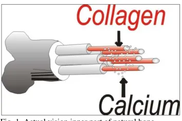
one is a complex living tissue which has an elegant structure at a range of different hierarchy scales. It is a composite comprise of an organic phase (base on collagen) by which calcium containing inorganic crystals are embedded (Fig. 1) [1]. Specifically, bone is a natural composite material, which by weight contains about 60% mineral, 30% matrix and 10% water [2]. The matrix bone is primarily consisting of collagen. This organic component of bone is predominantly responsible for the tensile strength. The mineral component of bone is of calcium phosphate, this mineral component gives rise to the compressive strength [3]. In the body, bone serves as number of functions, such as providing the cells found in the marrow, that differentiate into blood cells, and also acting as a calcium reservoir [4-6]. Nevertheless, its primary purpose is to provide mechanical support for soft tissues and serve as an anchor for the muscles that generate motion.
Fig. 1. Actual vision inner part of natural bone
There are two types of bone, compact or corical, and cancellous or trabecular (spongy) bone. The anisotropic structure of bone leads to mechanical properties that exhibit directionally. This directionality results from the fact that bone has evolved to be both tough and stiff, two competing properties which are optimized in bone but with an inherent loss in isotropy [7]. In fact, bone exhibits extraordinary mechanical properties, displaying both elastic and semi-brittle behavior [2, 8]. Compact bone has a compressive strength in the range of 131 - 224 MPa, and a Young’s modulus ranging from 17 - 20 GPa [2, 6], while compressive strength and Young’s modulus for trabecular bones are 5 – 10 MPa and 50 -100 MPa, respectively [2, 6].
Bone plays an important role in the movement, support and protection of vital organs. However, it is susceptible to fracture as a result of physiological resorption, dental losses, trauma injuries, bone pathology, infection, aging, population and bone disease [9-11]. The ways of bone repair improve year by year, parallel to the development of high technologies. Traditional biological methods of bone defects management includes: 1) Autogeneous bone graft which harvested from different side of the patient’s body, it is considered as the ideal bone graft substitute [12]. While the use of autograft material is the preferred technique, there are some limitations such as donor site morbidity, limited donor bone supply, anatomical and structural problems and elevated levels of resorption during healing [13] 2) Allograft bone which is obtain from
Biodegradable Polymers and their Bone
Applications: A Review
Saiful Izwan Abd Razak, Noor Fadzliana Ahmad Sharif and Wan Aizan Wan Abdul Rahman*,
Polymer Engineering Department, Faculty of Chemical Engineering, Universiti Teknologi Malaysia,
81310 Skudai, Johor, Malaysia
individuals of the same species as the receiver [9]. Allograft has the disadvantage of eliciting an immunological response due to the genetic differences and the risk of inducing transmissible disease [14, 15]. This necessities a thorough sterilization procedure that not only damages the graft’s biological and mechanical properties, but also expensive [16, 17].
Reconstruction of bone with fully synthetic artificial bone graft is another way to provide and create lost bone mineral. It was started past 30 -40 years, the terms of innovation of ceramic for bone repair and reconstruction become an interest in the major advance of medical applications [18]. Calcium phosphate is highly promising as a bone substitute in orthopaedics among the other ceramics [19-21]. This group of material exhibit high biocompatibility, bioactivity, self setting characteristic, low setting temperature, adequate stiffness and easy shaping in complicated geometries [22, 23]. However, several drawbacks do exist causing some problems that hinder clinical use of calcium phosphate. The problem is that calcium phosphate lack of macropores and low porosity making its resorbing rate rather slow and mechanically brittle, thus restricts its utilization in medical applications [24-26].
For the last two decades of the twentieth century, a paradigm shift occurred from the biostable materials to biodegradable materials (hydrolytically and enzymatically degradable) for the medical and related applications [27-29]. Current trend predicts that in the next couple of years, many of the permanent prosthetic devices used for temporary therapeutic applications will be replaced by biodegradable devices that could help the body to repair and regenerate the damage bone [30]. A biomaterial can be defined as a material intended to interface with biological system to evaluate, treat, augment or replace any organ or function of the body [31]. The essential prerequisite to qualify a material as a biomaterial is its biocompatibility, which is the ability of the material to perform with an appropriate host response in a specific application. The host response to an implant depends on factors ranging from the chemical, physical and biological of the materials to the shape and structure of the implant.
Biodegradable polymers are materials that fulfill the above requirement. The chemical, physical, mechanical polymers will vary with the time and degradation products produced have different level of bony compatibility. Notwithstanding, biodegradable polymers sometimes fall short of achieving satisfactory results in bone fixation [32]. These results should not necessarily be taken as a negative factor, since they are available to be reinforced with other materials like ceramics, metals, or clay in order to improve their mechanical properties and meet the promising requirements in bone substitute.
Often, from the medical point of view, biodegradable polymer with sufficient mechanical strength and optimized lifetime in the body, which could finally be replaced by bone, is most desirable. Biodegradable polymers lack in mechanical properties. It is desirable to reinforce it with other biocompatible substance to enhance its properties. The best
partner in reinforcing with ceramic is calcium phosphate, especially hydroxyapatite (HA) since their condition mimic natural bone composition [3]. Facing a complex biological and sensitive system as the human body, the requirement for bone repair using biodegradable polymers are manifold and challenging. Some of the important properties of biodegradable polymers can be summarized as follows [33]: 1) The material should not evoke or toxic response upon implantation in the body. 2) The material should have acceptable shelf life. 3) The degradation time of the material should match the healing or regeneration process. 4) The polymers should have appropriate mechanical properties for the indicated application and the variation in mechanical properties with degradation should be compatible with the healing process. 5) The degradation products should be nontoxic, and be able to metabolized and cleared from the body. 6) The polymers should have appropriate permeability and processability for the intended application.
Biodegradable polymers offer a number of advantages for developing bone at defect sites [34]. The key advantages include the ability to tailor mechanical properties and degradation kinetic. Biodegradable polymers can differ in their molecular weight (MW), polydispersity, crystallinity, structure
and thermal transition, allowing different absorption rates. By means of porosity, biodegradable polymers can be used to impart macroporosity to the cement as polymers degrade and expose macropores to bony ingrowth. The strengthening of the graft from the bone growth and the deposition of the new bone should offset the weakening of the graft due to polymer degradation [35].
In bone application, biodegradable polymer or its composite can be prepared in two form, scaffold and dense. Usually, scaffold is used to associate with cell seeded in purpose of cell growth to form bone. Scaffolds are able to promote cellular interactions and process uniformly interconnected pores with adequate structural integrity. The interaction between cell and substrate is related to the osteogenic cells attachment, adhesion and spreading and its quality will influence cell proliferation and differentiation [36]. Cell can be obtained from calvarine [37, 38], trabecular bone [39], human embryonic stem (hES) cells and bovine osteoblasts (bOB) [40].
TABLEI
COMPARISON OF THE IONIC CONCENTRATION IN BLOOD PLASMA AND SBF
(MM/L)[42]
Na+
K+
Mg2+
Ca2+
Cl-
HCO3 -
HPO4 2-
SO4 2-
Blood plasma
142.0 5.0 2.5 1.5 27.0 103.0 1.0 0.5
SBF 142.0 5.0 2.5 1.5 4.2 148.0 1.0 0.50
Though various studies have been done throughout the scientific community on biodegradable polymers, there is yet a systematic report on the application of biodegradable polymers on bone engineering. The purpose of this review is to observe various parameters (porosity, particle size, molecular weight, temperature, crystallinity, filler characteristic and etc) that possibly affect the mechanical properties and degradation rates of biodegradable polymers, whether in pure or mixing condition and also observation of bone growth after in vivo and in vitro implantation. Furthermore, biocompatibilities of biodegradable polymers in bone engineering were also investigated
II. SYNTHETIC BIODEGRADABLE POLYMERS
A. Polyesters / poly (α-hydroxy acids)
Naturally occurring hydroxyl acids, such as glycolic, lactic and ε-caproic acids have been utilized to synthesize an array of useful biodegradable polymers for a variety of medical product applications. Especially in the implantable devices (internal fixation devices for orthopedic repair) made from these polymers are becoming part of standard surgical protocol [43-45]. It was started 30 years ago where polyesters increased the popularization of polymers as material for sutures and bone fixation [46] and remain among the widely used synthetic degradable polymers. Although all polyesters are theoretically degradable through the method of esterification, it is a chemically reversible process, only aliphatic chains between ester bonds can degrade over the time frame required for most of the biomedical applications [47].
Poly (α-hydroxy acids) comprise one of the earliest and most extensively investigated class of biodegradable polymers. It can be developed from a variety of monomers via ring opening and condensation polymerization routes depending on the monomeric units. The ester bond of the poly (hydroxyl acids) is cleaved by hydrolysis, which results in a decrease in the polymer Mw (but not mass) of the implant [47]. The
degradation rate of polyester is determined by initial Mw,
exposed surface area, crystallinity, and (in case of the copolymers) ratio of the hydroxyl acid monomers [48].
Among the class of poly (α-hydroxy acid)s, the most extensively used polymers are poly (glycolic acid) (PGA), the stereoisomers forms of poly (lactic acid) (PLA) and their copolymer poly (lactic-co-glycolide) (PLGA) [49-52]. These polymers are candidate in bone tissue engineering since it does promote biocompatibility and biodegradability which would allow cell growth in human body [30]. The chemical
properties of these polymers allow hydrolytic degradation through de-esterification. Once they degraded, the monomeric components of each polymer are removed by natural pathway [53, 54]. The human body already contains highly regulated mechanism for completely removing monomeric components of lactic and glycolic acids. Due to these specialty polymers, such degradable material used in bone substitute have been approved by the US Food and Drug Administration (USFDA) [51]. With the exception of PGA, the polymers in this family are soluble in many common organic solvents and thus it can be processed by a variety of thermal and solvent-based methods [54, 55].
Due to several problems, it is advisable not to apply polyesters alone when dealing with bone substitution, since it can caused some drawbacks in the implementation in vitro and in vivo such as the degradation products reduce the local pH value, which in turn, may accelerate the polyesters’ degradation rates [56] and induce an inflammatory reaction, which are often ascribed to acidosis caused (chemically unavoidable) by the release of acidic degradation products (monomeric or oligomeric hydroxycarboxylic acids) [57-59]. Another disadvantage is that the mechanical properties of the highly porous scaffolds made from polyesters are relatively weak, which limits their use for bone-tissue engineering, especially the in vivo implant site [60].
B. Poly (lactic acid)
PLA has received the most attention due to its renewable resource [61], biocompatibility, biodegradation, excellent thermal and mechanical properties, and superior transparency of the processed materials [62]. It is a linear aliphatic thermoplastic polyester and is readily biodegradable through hydrolytic and enzymatic pathways [61, 63-65]. Since lactic acid is a chiral molecule, it exists in two stereoisomeric forms that give rise to four morphologically distinct polymers that is
D-PLA, L-PLA, D,L-PLA and meso-PLA. D-PLA and L-PLA are
the two stereoregular polymers, D,L-PLA is the racemic
polymer obtained from a mixture of D- and L- lactic acid, and
meso-PLA can be obtained from D,L-lactide. Generally, L-PLA
is more frequently employed than D-PLA, since the hydrolysis
of L-PLA yields L(+)-lactic acid, which is the naturally
occurring stereoisomer of lactic acid. On the other hand, the semicrystalline solids of L-PLA are preferred in the orthopedic
surgeries application where high mechanical strength and toughness are required [66-69].
In bone tissue engineering, the scaffolds require appropriate material properties, controlled porous architechture, proper porosity and appropriate mechanical properties [70]. Poly (L
hardly control the porous from 200-500 µm in size which also very important for bone regeneration and vascularization.
A thermally induced phase separation technique (TIPS) was used by Ruiyun and Peter, 1999, in order to create highly porous composite scaffolds for bone tissue engineering [60]. PLLA was mixed with hydroxyapatite (HA) using dioxane as solvent. Freeze drying procedure allows to produce hard and tough foam with a co-continuous structure of interconnected pores and HA/PLLA composite skeleton. The composite mixture was proved to be in good bone bonding [79, 80]. Their work showed that foam porosity could be reached as high as 95% while pore sizes ranging from several microns to few hundred microns were obtained. Mechanical properties of HA/PLLA scaffold was compared to those corresponding pure PLLA and it was found out that the compressive modulus of composite (11 MPa) was significantly higher than the pure PLLA (6.4 MPa).
Same technique and material was implemented by Nejati and co-worker [81], instead of using HA particles, they used nano and micro sized HA as filler in scaffolding. Both nano and micro composite scaffolds showed some molecular interactions and chemical linkages between HA particles and PLLA matrix, affecting the interfacial behavior and mechanical properties of the HA/PLLA composites. The porosity of scaffold was up to 85% and the average micropore diameter was in the range of 64-175 µm. The compressive strength of nano-composite scaffold was up to 14.9 MPa while pure PLLA and micro-HA/PLLA scaffolds were found to be 1.79 MPa and 13.68 MPa, respectively. This study demonstrated the positive effect nano-HA and micro-HA on improving the mechanical properties of the PLLA matrix, also indicating that the values are comparable to the high end strength of cancellous bone [82]. Furthermore, Mesenchymal stem cells (MSCs) for cell culture of scaffolds revealed that the cell affinity and biocompatibility of nano-HA/PLLA was found to be higher than the other two scaffolds.
Xiong and co-worker [78] fabricated PLLA scaffold using precise extrusion manufacturing (PEM). The measured value of scaffold porosity was only 60% but the pore size values
produced could not even reached by TIPS method, which was ranged from 200 µm to 500 µm. This happened due to the well prepared column inside the PEM with the additional of compressed air and nozzle. Effectiveness of the porosity formed increased the mechanical properties of the PLLA scaffolds up to 8.32 MPa.
From the observation of Table 2, the compression strength with the presence of filler was much higher than non-filler, indicating that most of the particles bonded to PLLA matrix were on the surfaces of the pores, and it showed good adhesion between them [79, 80]. Some molecular interaction and chemical bonding which were presented between HA and PLLA influenced the interfacial behavior and mechanical properties of the composites [83, 84]. It is obvious that among pure PLLA and composite scaffolds, nano-composite scaffolds showed the highest compressive strength because of some reasons: 1) Particle sizes may attribute to the large interfacial area between nano HA and PLLA matrix. 2) More uniform distribution of nano HA particles in polymer matrix. Xiong and his co-workers successfully used PEM in order to achieve high compression strength and modulus. However, it is still inconvenient since there are other alternative methods with reasonable cost [78]. A new artificial structural elastic, partially degradable bone graft for supporting weak bone in the proximal femur was presented by Lagoa and co-workers [34]. The implant materials consist of a functionally graded combination of titanium, PLA, HA and calcium carbonate. The implant has an outer elastic layer in contact with the bone, consisting of D,L-PLA, HA and calcium carbonate, inner layer
was made by manual dip-coating of the metal into solutions of PLLA with suspended calcium salts. The purpose of D,L-PLA
being placed at the outer layer is due to its fast degradation rate which gave an opportunity for bone on and ingrowth when it degrades. PLLA has slow degradation rate and provides a biocompatible interface between the living tissue and the bioinert metal while the bone consolidates. The artificial strut grafts strengthen its implant by inserting titanium which
TABLE2
COMPARISON MECHANICAL PROPERTIES OF SCAFFOLD COMPOSITES (PLA/FILLER)
Material characteristics Mechanical properties References Initial
PLLA Mw
(kDA)
Filler characterist
ics
Porosity (%) Average pore size
Methods Process condition Compression
Strength Modulus [60]
[81]
[78]
n.a.
140 (0.5g)
374
None
HA,
None
micro-HA
nano-HA
None
n.a.
95.00 (50-200
µm)
87.37 (64 µm)
86.56
85.06
60.30 (200-500
µm)
Thermally induced
phase separation
Thermally induced
phase separation
Precise extrusion
Stirred: 50oC, 2hrs
Solution: dioxane Immersed in liquid
nitrogen. Freeze Dry: -5 - -10oC, 4 days
Stirred: 4hrs* Solution: dioxane Freeze Dry: -200C,
72 hrs
160oC
0.24
0.4
1.8
4.8
8.7
8.32
6.4
11.0
2.2
14.0
14.9
TABLE3
COMPARISON MECHANICAL PROPERTIES OF DENSE COMPOSITES (PLA/FILLER)
Material characteristics Mechanical properties
References Initial PLA Mw
(kDA)
Filler characteristics
Methods Process
condition
Tension (MPa) Compression
(MPa)
Bending (MPa)
strength modulus strength modulus strength modulus
[91] 220 none Hot pressing 103oC 154.10 1300 123.5 4800 258.5 6500
202 HA, 50 wt%,
0.3-20 µm
Hot pressing 103oC 103 2400 115 6500 267.5 12,300
[92] 100 HA, 80 wt%,
0.5-0.7 mm
Hot pressing 176oC,
98.1 MPa
38 2000
nanoHA, 80 wt%, 100 nm
Hot pressing 176oC,
98.1 MPa
72 3500
430 HA, 80 wt%,
0.5-0.7 mm
Hot pressing 184oC,
98.1 MPa
50 2500
nanoHA, 80 wt%, 100 nm
Hot pressing 194oC,
98.1 MPa
139 10,000
[93] 160 none Hot pressing 180oC,
40 MPa
52 3500
HA fibers, 70 wt%
Hot pressing 180oC,
40 MPa
40 11,000
[94] 300 none Hot pressing 175oC,
10 MPa
60 1800
Grafted nanoHA,
4 wt%
Hot pressing 175oC,
10 MPa
63 2500
[85] 104 none Hot pressing 160oC,
300 MPa
56
nanoHA, 25 wt%, D=2.1 µm
Hot pressing 160oC,
300 MPa
88
nanoHA, 50 wt%, D=2.1 µm
Hot pressing 160oC,
300 MPa
100
[86] n.a none i) mixed
ii) pressing
i) 25oC, 24h ii) 200oC
1200
mMMT, 1.5 wt%
i) mixed ii) pressing
i) 25oC, 24 ii) 200oC
1400
mMMT, 3.0 wt%
i) mixed ii) press
i) 25oC, 24h ii) 200oC
2000
mMMT, 6.0 wt%
i) mixed ii) press
i) 25oC, 24h ii) 200oC
Besides using separation technique to produce scaffold composite at a range of porosity, there is a way to prepare the mixture of the same composite (nano-HA/PLLA, 0:100, 20:80 and 50:50) by using hot pressing, dense samples were investigated rather than porous scaffolds [85]. In addition, annealing treatment was performed with the hope to enhance the crystallinity of the polymers. However, annealing treatment does not influence the physical properties of the composites (particle sizes, porosity and mechanical properties, see Table 3) since the polymer has reached its ultimate crystallinity at the end of the cooling step, and yet, only remaining chloroform at the surface of the composites thoroughly eliminated after annealing treatment. Dispersion of HA nanoparticles into a PLLA matrix was a success whereby compressive strength of over 100 MPa have been achieved, this value expressed that the developed nano HA/PLLA properties was comparable to those of cortical bone. Nano-HA/PLLA composites are thus promising; to be develop as bioresorbable porous bone substitutes showing superior mechanical performance.
Tzong and Cheng [86] used solution mixing process of PLA reinforced with montmorillonite (MMT), the MMT modified by chitosan function was aimed to improve the chemical similarity between PLA and MMT. Polymer/clay composites have received significant attention in academic and industrial area in recent years due to the excellent enhancement in physical and/or chemical properties relative to the neat polymer matrix [87-89]. The results showed that the interaction between PLA and MMT was successful for the used in biomedical application and the detail is shown in Table 3.
Mechanical resistance was also evaluated in order to determine the best operating conditions of HA/PLA scaffolds to produce implants which offers optimized properties for use as bone substitutes [90]. A pore-forming agent (salt) was carried out in the forming of scaffolds which restricted to 70 wt%, whereby resistance as high as 2.2 MPa could distort the composites mechanical properties. It was shown that the addition of 30 % to 40 % of filler allows a satisfactory dispersion of the mineral within the matrix. A high percentage of filler is unfavorable for processing of porous scaffolds, as it prevents the matrix from playing its role as a binder.
Table 3 displays several examples of the composite according to the initial Mw, the nature of filler and the process
conditions. Presence of nano-particles as filler obviously improves the mechanical strength and stiffness of PLA composites. The maximum strength that can be reached was at 139 MPa [92]. Nano-particles have gained much recognition as bone dealer not only due to their composition and structural similarity with natural bone but also because of their unique functional properties such as larger surface area and superior mechanical strength than those of single or pure constituents [95]. The effect of micro-fiber demonstrated by Kasuga and co-workers [93] achieved a very high bending modulus but showed the vice versa effect on the bending compression. The compressive strength increased by increasing the quantity of
filler (%wt) and the initial Mw. Nano-particles obtain better
results in terms of mechanical strength and moduli. In addition, composite in the dense form more preferable compare to scaffold due to their capability to perform in high load bearing applications.
C. Poly (glycolic acid)
PGA has the simplest structure. It can be considered as one of the first biodegradable synthetic polymer investigated for biomedical implant. PGA is a highly crystalline polymer (45-50 % crystallinity) and therefore exhibits high tensile modulus with very low solubility in organic solvents. The glass transition temperature (Tg) of the polymer ranges from 35 to
40oC and the melting point is greater than 200oC. It shows excellent mechanical properties, also due to its high crystallinity. A self reinforced foam composed of PGA is stiffer than any other degradable polymeric system used clinically [96] and has been shown to exhibit a modulus approximately 12.5 GPa [97]. Due to its good initial mechanical properties, PGA has been investigated as bone internal fixation devices. However, PGA is also known to lose its strength in 1-2 months when hydrolyzed and losses mass within 6-12 months. Thus, this indicates that the degradation rate is very high, which in the end would limit the mechanical properties and biomedical applications for PGA. Therefore, as reported by Linhart and his co-workers [37], they used PGA reinforced with amorphous carbonate apatite to investigate any potential performance of PGA in bone substitute and found out that the composite could serve only as bone substitution materials for small or non-load bearing indications.
D. Poly (lactide-co-glycolide)
The combination of lactic and glycolic side formed PLGA amorphous polymer. Both L- and DL- have been used for
Fei and co-workers created a novel biodegradable PLGA for bone repair [104]. The moldable, degradable bone substitute with osteogenesis materials composed of PLGA loaded with bone morphogenetic protein-2 (BMP-2) microspheres incorporated with calcium phosphate cement (CPC) for bone growth. Lactic to glycolic ratio was 50:50. The scaffolds only appeared when the composites immersed in the phosphate buffer saline (PBS) in order to induce degradation towards BMP-2 which then formed PLGA/CPC scaffolds. The results showed that the CPC composites compressive strength decreased from 29.48 MPa to 8.26 MPa when PLGA was added during setting. This happened because the structure of interlocking CPC crystal needles, which produce the cement strength, was inhibited when microspheres were added into CPC [105]. Although the mechanical properties of the composites decreased after PLGA was added, better degradation and stable composite was produce and the results were equivalent to the cancellous bone.
Another PLGA pellets with a lactide/glycolide ratio of 85:15 with the pore sizes range of 250-450 µm were used by William and his co-workers in their investigation [106]. In this study, the process of mineralization in tooth and bone development was directly mimicked through the use of SBF as the mineral growth solution. In other words, PLGA was incubated in SBF at different times to demonstrate the growth of a continuous bonelike apatite layer within the pores of the polymer. In addition, the process was a single step and occurred in the room temperature. It resulted in a growth of a continuous layer on much of the inner pore surface, which significantly increased the compressive modulus up to 320 ± 60 kPa after 16 days of treatment, without a large decreased in total porosity. The mineral grown was a carbonated apatite mineral layer, similar to the mineral present in bone, and hence is expected to have a high degree of bioreactivity. Such a layer could be of great importance in the field of bone tissue engineering, providing the advantages of increased osteoconductivity and enhanced mechanical properties. Also, it is an efficient and relatively inexpensive mineral growth process.
Surface grafted of HA in the PLGA matrix deposition (g-HA/PLGA) showed excessive improvement on interface between the polymers and CPC rather than non-grafted HA/PLGA [107]. Three dimensional porous scaffolds of
grafted HA/PLGA was fabricated using solvent
casting/particulate leaching method to compare with conventional HA/PLGA scaffolds and to investigate its application in bone replacement [107]. In their report, in vivo mineralization and osteogenesis were investigated by replacement for repairing radius defects of rabbits. Table 4 analyzes the advantages of g-HA/PLGA and HA-PLGA.
Despite of decreased in calcium exposure when implantation, surface grafted-HA/PLGA showed some advantages and good requirements in bone fixation. Unfortunately, no mechanical properties have been reported for comparison, but it can be said that non-grafted composites
showed a better achievement after 20 months of post surgery. Other than that, both composites offered different advantages which make no differences in in vivo implantation where both showed similar ability to enhance in vivo mineralization and allowed rapid mineralization conjugation with neighboring bone better than neat PLGA.
Aligned nanofibrous scaffolds based on PLGA and nano-HA were synthesized by electrospinning for bone engineering [108]. At higher concentration (> 10wt %), a reduction in mechanical properties occurred because of the agglomeration of HA in the PLGA matrix fiber, whereby the presence of nano-HA resulted in fiber breaking (Table 5). In electrospinning, the major factors that have a direct effect on the fibers diameters (size particles) were solution concentration and electric field. At higher voltages, needle tip inside the machine produced unstable sources of fibers. The collection of fiber scaffolds on a high speed rotator resulted in a highly oriented fibers. The diameter of the scaffolds from this method can be affected by the choice of the solvent. It was suggested that the used of more polar and hydrophilic solvents can reduce the degree of aggloromeration of the HA powder and can thereby find a good dispersion of particles in the polymer matrix [109].
TABLE4
ADVANTAGES OF GRAFTED HA FILLER AND UN-GRAFTED HA FILLER IN PLGA MATRIX [107]
g-HA/PLGA HA/PLGA
More uniform distribution of granules on the pore walls surface. After 20 months post surgery, calcium content that attached to the
composite was 10 wt%, higher than grafted composite (8 wt %).
Improve the microstructure stability of the composite scaffolds Better osteogenesis after implantation
TABLE5
MECHANICAL PROPERTIES OF SCAFFOLD COMPOSITES (PLGA/FILLER)
Material characteristics Mechanical properties
Ref. Initial PLGA Mw
(kDA)
Filler characteristics
Lactic:glycolic ratio
Porosity (%) Average pore size
Methods Process condition Storage modulus
(MPa)
Compressive strength
(MPa)
Young modulus (MPa)
[40] 110 none 50:50 92.43
(134.22 µm)
Solvent casting/ particulate
leaching
PLGA: NaCl 1:10, Air dried 24h, vacuum dried 48h.
100-250 µm
1.20 8.33
110 Collagen, BSM 50:50 94.79
(121.84 µm)
0.83 5.56
[107] 196 none 80:20 83.6 Solvent casting/
particulate leaching
PLGA/HA:sucrose 1:6 100-450 µm
HA, 10 wt% 80:20 85.9
Grafted HA, 10
wt% 80:20
86.6
[106] 15 None
85:15
94.0 Solvent casting/
particulate leaching
PLGA:NaCl
250-450 µm
0.32
63 none
HA, 1 wt% HA, 5 wt% HA, 10 wt% HA, 20 wt%
85:15 85:15 85:15 85:15 85:15
72.0 61.0 62.0 64.0 72.0
electrospinning 13-15 kV, rotating
speed=6000 rpm, solvent=HFP
441.0 591.7 724.2 557.3 371.4
PLGA scaffolds reinforced with calcium phosphate like HA as filler showed better mechanical properties compared to pure PLGA, due to the HAs’ more similar characteristics to bone apatite, its’ ability to promote the attachment of cultured osteoblasts and to improve metabolic activity [110-112]. Addition of HA in polymer matrix had been approved to increase mineralization in matrices in vitro osteoblasts culture [113]. It provide more favorable surface for cell attachment and the ability of the material to support bone ingrowth [114]. In addition, calcium phosphate additives in PLGA matrix provides surface roughness which enhanced bone attachment and proliferation [115]. The advantage of PLGA is that the strength of the composite could be controlled by the amount of PLGA microspheres, which makes it possible for the surgeons to modify the material to obtain an adequate strength. The particles sizes also play a significant role in bone implantation, whereby particle sizes ranging from 50-300 µm have been found to be optimal for bone ingrowth [116]. However, previous studies noted that the size of interconnection in the scaffolds is the main limiting factor for bony ingrowth rather than the size of pores themselves [117].
Whether the method used are solvent casting, electrospinning or BMP loaded in PLGA matrix and degrade when immerse in the PBS solution, the mechanical properties produced (shown in Table 5) were all in the range of potential in forming cancellous bone, (compression strength, ≤ 10 MPa, modulus, 50-500 MPa). PLGA in the dense form was poorly studied and hardly found in the previous work, it could maybe due to the amorphous characteristics of PLGA whereby unknown melting temperature renders PLGA unsuitable to be prepared by melt blending or hot pressing.
E. Poly (ε-caprolactone)
Most research work has been directed at PLA, PGA and
PLGA copolymers. The success of these for pharmaceutical applications has further led to the evaluation of aliphatic polyesters such as poly(ε-caprolactone) (PCL). PCL is an aliphatic polyester that has been intensively investigated as a biomaterial. It appears as semicrystalline polyester and is
highly processable as it is soluble in a wide range of organic solvents. The uncommon things of PCL is its high thermal stability, with decomposition temperature (Td) of 350oC,
whereas others aliphatic polyesters are between 235oC and 255oC [118]. In bone engineering area, PCL can be categorized as promising biocompatible and biodegradable polymers since it is being used to enhance bone ingrowth and regeneration in the treatment of bone defects [119, 120], however, PCL is poorly studied due to the slow degradation [121].
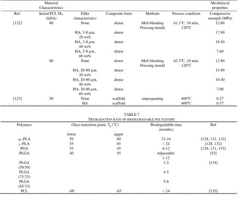
One of the work done by Chen and Sun [122] was by reinforcing PCL with HA by melt blending technique (Table 6). This method requires the exact melting temperature of the biodegradable polymers to ensure that the polymer fully melted and provide fillers with spaces for fine dispersion in the polymer matrix. Smaller HA particles (3-8 µm) distributed higher compressive strength than HA (20-80 µm), indicating that size of particle affect the improvement of the composites mechanical properties [122]. The smaller the particle diameter the greater the specific surface and interfacial areas within the matrix. Their results can simply be plotted as seen in Fig. 2. Fig. 2 indicates that the addition of HA increased the compression of composite towards bone fixation, but, until some level, the failure mechanism of the composites changes from plastic to brittle (easily rupture) and hence lowering the mechanical properties of PCL and other biodegradable polymers.
TABLE6
MECHANICAL PROPERTIES OF COMPOSITES (PCL/FILLER)
Material Characteristics
Mechanical properties Ref. Initial PCL Mw
(kDA)
Filler characteristics
Composite form Methods Process condition Compressive strength (MPa)
[122] 80 None dense Melt blending
Pressing mould
61.1oC, 10 min,
120oC
12.80
HA, 3-8 µm, 20 wt%
dense 17.90
HA, 3-8 µm, 40 wt%
dense 10.50
HA, 3-8 µm, 60 wt%
dense 7.60
80 None dense Melt blending
Pressing mould
62.3oC, 10 min, 120oC
12.80
HA, 20-80 µm, 20 wt%
dense 15.90
HA, 20-80 µm, 40 wt%
dense 10.40
HA, 20-80 µm, 60 wt%
dense 7.00
[123] 50 None scaffold impregnating 600oC 0.27
HA scaffold 600oC 0.57
TABLE7
DEGRADATION RATE OF BIODEGRADABLE POLYESTERS
Polymers Glass transition point, Tg (oC) Biodegradable time
(months)
Ref
lower upper
DL-PLA 50 60 12-16 [128, 131, 132]
L-PLA 55 65 > 24 [128, 132]
PGA 35 45 6-12 [128, 131, 133]
PLGA 40 55 Adjustable:
1-12
[52]
PLGA (50/50)
1-2 [134]
PLGA (75/25)
4-5
PLGA (85/15)
5-6
PCL -60 -65 > 24 [135]
F. Biodegradation of polyesters
Degradation could be characterized in many ways, like loss of Mw, loss of mass or loss of mechanical strength, chemical
composition, configurational structure, stress and strain, crystallinity, porosity, chain orientation, dispersion of chemically reactive compounds within the matrix [124, 125]. The polymers degrade by random chain scission [126]. Since only the monomer degradation products are soluble, a very large reduction in both Mw and mechanical strength typically
occurs before a decrease in the weight of the sample is observed. In the case of implantation in vivo, the body already contains highly regulated mechanism for completely removing monomeric components of lactic and glycolic acids. PGA is converted to metabolites or eliminated by other mechanisms, while PLA can be cleared through the tricarboxylic acid cycle. The crystallinity of L-PLA samples has been observed to
increase during degradation and particles of highly crystalline material can be found in vivo long after the bulk of the implant has disintegrated [56].
Biodegradable polyester degradation occurs by uptake of water followed by the hydrolysis of ester bonds. PLGA has a
wide range of degradation rates since the ratio of lactic over glycolic varied, the degradation kinetics being governed by both hydrophilic/hydrophobic balance and crystallinity. Composition of chain (lactic or glycolic units) determines the degradation rates of PLGA copolymers. Blends that contains higher amount of PGA have been shown to degrade faster [124]. PCL degradation proceeds with two stages: random hydrolytic ester cleavage and weight loss through the diffusion of oligometric species from the bulk. PCL with high Mw could
take several years to degrade in vivo [127-129]. In general, the sequence of polyesters degradation rates decreases in the following order [54]:
PGA > PDLLA > PLLA > PCL
The kinetic patterns of polyesters degradation are shown below [130]:
--COOR ···HOOC-- + H2O → 2COOH + ROH
Table 7 shows the degradation rate of some biopolymers. The degradation rate, physical and mechanical properties of PLA and PGA are adjustable by varying their Mw and
patients, 7.9 % developed a late noninfectious inflammatory response during healing process. It has been suggested that the consequences occurred due to the release of acidic degradation products (PGA and PLA) [136]. Böstman gave a solution to these problems; for orthopaedic applications, it requires the development of a polymer that is more hydrophobic than PLA and PGA and does not release acidic degradation products on hydrolysis [136].
New era with new technologies come out with better theories and materials. Linhart and his co-workers [37] announced that the used of calcium phosphate blended with polyesters could overcome the drawbacks. These composites showed good biocompatibility because of: 1. Compensation of acidic release from the polymer through the alkaline calcium phosphate. 2. Participation of carbonated apatite in the biological remodeling process. Less stability of apatite has higher solubility under conditions of acidic resorption [137, 138]. 3. Physicochemical and biological transformation of calcium phosphate into biological mineral as present in bone. 4. Good adhesion of cells on aliphatic polyesters [139]. 5. Better distribution of degradation products through the microporous structure, thereby avoiding accumulation of acidic or harmful products.
G. Benzyl ester of hyaluronic acid
Benzyl ester of hyaluronic acid, also known as HYAFF-11, has shown good degradation rate and the products of degradation are non-toxic and non carcinogenic [140]. It undergoes hydrolytic degradation via ester bonds in the absence of enzymatic activity with degradation times varying from 1-2 weeks to 2-3 months, depending on the degree of esterification. The de-esterified HYAFF-11 is more hydrated, soluble and resembles native hyarulonic acid [141, 142].
HYAFF-11 has been studied for the use in vascular grafts [143, 144] and for bone tissue engineering [145, 146]. Hyaluronic acid containing fibroblast growth factor is undergoing clinical trial as a synthetic bone graft to accelerate bone fracture healing [30]. Sanginario and co-workers [147] worked on HYAFF-11 reinforced with α-tri calcium phosphate in the quantity of (14/86 wt%, respectively) in the form of hydrogel. The highest compression strength was in the range of 17-12 MPa, compared to pure HYAFF-11 (3.0 MPa). This values are in the range of cancellous bone, thus, HYAFF-11 can be utilized bone fillers in orthopaedic and dental applications.
III. NATURALBIODEGRADABLE POLYMERS
A. Chitosan
Chitosan derived from chitin, is a natural biopolymer originating from crustacean and cell wall of fungi. It is known as linear polysaccharide, composed of glucosamine and N-acetyl glucosamine ratio being referred as the degree of deacetylation, which was found in marine environment. Depending on the source and the processing method, its Mw
may range from 300 to 1000 kDa. Chitosan is normally insoluble in water and aquoues solutions above pH 7, but, in diluted acid which pH less than 6, the protonated free amino group on glucosamine facilitate solubility of that molecule [159-162]. Chitosan has been found to be non-toxic after oral administration in humans and has FDA approval.
Enzymes, such as chitosanase, lysozyme and papain are known to degrade chitosan in vitro [163]. The in vivo degradation of chitosan is primarily due to lysozyme and takes place through the hydrolysis of the acetylated residues. The rate of degradation of chitosan inversely depends on the degree of acetylation and crystallinity of the polymer [164]. The highly deacetylated form exhibits the lowest degradation rates and may last several months in vivo. Apart from this, chemical modification of chitosan can significantly affect its solubility and degradation rate [164].
Chitosan is biocompatible, biodegradable, and can be molded into porous structures (allows osteoconduction) [165]. Several studies have focused on the use of chitosan /calcium phosphate composite for this purpose. Chitosan /nanocrystalline calcium phosphate scaffolds characterized by a relatively rough surface and approximately 20 times greater area/unit mass than chitosan scaffold indicate increased adsorption, improved cell attachment for bone regeneration [166]. In the development of chitosan/nano-HA scaffold conducted by Thein-Han and his co-workers [167], high Mw
chitosan scaffolds were examined by having superior mechanical properties compared to medium Mw chitosan
scaffolds (Table 8). In addition, the significant increased in compression modulus of the nano-HA/chitosan composite was attributed to the good dispersion of nano-HA and strong interaction between chitosan and nano-HA to formed a composite. In the nanocomposite scaffold, the organic chitosan network uniformly covered the HA nanoparticles, interactions may have occurred similar to those occurring components of bone. Possible interactions involve Ca2+ and PO43- charged
species of HA with NH3+ of chitosan. However, the
mechanical properties of the composites obtained were inadequate for bone regeneration.
membrane compared to that of using the pure chitosan one. Histomorphometric analysis performed 3 weeks after implantation revealed a fully closed defect in the hybrid membrane, whereas there was only 57% defect closure in the chitosan membrane.
Another scientific studies of modified chitosan structure, sulfated chitosans with varied sulfate group; the MW and sulfur
content were tailored to investigate the effects on the bioactivity of BMP-2 [172]. Low dose of synthetic sulfated chitosan (structure shown in Fig. 3) proved to stimulate osteoblast differentiation induced by BMP-2 in vitro and ectopic bone formation in vivo. Another foundation of chitosan modification is to allow water solubility in chitosan matrix, done by Gomez d’Ayala and co-workers [173]
(structure shown in Fig. 4). The transformation of chitosan into N-succinylchitosan incorporated with 50 wt% of hemihydrates calcium sulphate resulted in compression strength and Young’s Modulus of 0.80 MPa and 14.7 MPa, respectively.
Fig. 4. Succinylation reaction of chitosan [173].
Fig. 3. Chemical structures of chitosan modified with different sulfate groups. 2-N-Sulfated chitosan, 2SCS; 6-O-Sulfated chitosan, 6SCS; 2-N,6-O-Sulfated
TABLE8
MECHANICAL PROPERTIES OF SCAFFOLD COMPOSITES (CHITOSAN/FILLER)
Material characteristics Mechanical properties
Ref. Initial CS Mw
(kDA) (degree of deacetylation)
Filler characteristics
Composite form
Methods Process condition
Tensile strength (MPa)
Young modulus
(MPa)
Compressive modulus
(kPa)
[167] 250
(75%)
None Scaffold Thermally induce
phase separation
Solution used= acetic
acid, 1M Freeze dry =
-20oC, 24 h
4.5
nano-HA, 0.5 wt% (~50 nm)
scaffold 5.5
nano-HA, 1 wt% (~50 nm)
scaffold 6.8
nano-HA, 2 wt% (~50 nm)
8.6
400 (83%)
None Thermally
induce phase separation
Solution used= acetic
acid, 1M Freeze dry =
-20oC, 24 h
6.0
nano-HA, 1 wt% (~50 nm)
scaffold 9.4
[170] 100-170 (75-85%)
Collagen scaffold Solution
used= acetic acid, 0.5M Freeze dry =
-20oC, 2h
2
Collagen & HA
scaffold 10
[171] n.a.
(85%)
None membrane Sol gel Solution used= hydrochloric
acid, HCl, Methanol. Rotary pump and freeze dry
Thickness= 100-250 µm
0.9 15
Silica xerogel, 10 vol%
membrane Sol gel 0.52 16.11
Silica xerogel, 20 vol%
membrane 0.44 17.47
Silica xerogel, 30 vol%
membrane 0.96 55.53
Silica xerogel, 40 vol%
membrane 0.50 61.38
B. Collagen
Approximately 30% of the protein contain in the human body, collagen is the most abundant protein being the major component of skin and bone [30, 48]. Collagen is a rod type polymer nearly 300nm long with a Mw of 300.000. The
repeating sequence is responsible for the helical structure and the inherent and predictable mechanical strength of collagen [174]. Collagen undergoes enzymatic degradation within the body via enzymes. Due to the enzymatic degradability,
enzymatic degradation; 2) decrease the capacity of collagen to absorb water; 3) decrease its solubility, and 4) increase the tensile strength of collagen [175].
As a biomaterial, collagen is biocompatible, biodegradable and osteoconductive [176, 177]. The composition of human bone is a well known mineral/organic hybrid consisting of HA and organic mainly collagen constituents. Calcium phosphate /collagen composites are probably the most biomimetic system for osseous replacement or regenerative bony [178-182]. When associated with calcium phosphate particles or any other filler forming composite, collagen prevents the filler dispersion in implants, resulting in an easily molded biomaterial [183]. In order to investigate the vicinity between collagen and calcium phosphate in bone substitute biomaterial, composite constituted of collagen and calcium phosphate doped by europium were synthesized by a precipitation method [184]. Interaction checked by Förster resonance energy transfer (FRET) devices showed a good yield from the composites thus promising to be bone substitute.
Collagen coatings are known to enhance early cell adhesion and proliferation on calcium phosphate, increasing the in vivo osteointegration, osteoconduction and osteoregenerative capacity of these materials [39, 185]. For these reasons, composite materials containing calcium phosphate and collagen have been developed [186] using methods such as particle-gel mixing and powder compression [187-189].
One scientific study regarding the effect of collagen/citric acid on the setting reaction of brushite found out that collagen has the capability in speeding up the cement setting reaction and had compressive strength similar to that of spongeous bone (48.9 MPa) [190]. With the combination of citric acid, it helped to shorten the setting time, increase the injectability, reduce the viscosity and improve the mechanical strength of the composite [191]. Citric acid also enable the mixing of a high collagen content gel (3 wt%) with the cement powder to form brushite-collagen composite that was easy to work with. The effect of crosslinking on brushite-collagen composites was also studied by immersing the composite into 2% gluteraldehyde solution in wet condition, neither the addition of collagen nor glutaraldehyde crosslinked collagen seemed to increase the compressive strength of the composite after storage in wet condition [190].
IV. CONCLUSION
Biodegradable polymers properties do give definite impact on the bone healing, formation, regeneration or substitution for human body. Without the help of other fillers, biodegradable polymers application is limited to non-bearing bone. Solvent plays an important role in solubility of polymers. It is also important that the initial mechanical strength of the composite significantly higher compared with initially porous materials. A higher mechanical strength will assure defect stability in the early critical stage when few bones have yet grown onto the defects site. Mw, particle size, porosity, crystallinity,
methodology, polymers, fillers concentration, hydrophilic,
hydrophobic and even annealing process could tailor the mechanical properties and degradation rate of the composite. And yet, hot pressing is the best method in producing biostable, biodegradable and biocompatible composite because the parameters of polymers can be arrange to fulfill the mechanical integrity from non-load bearing to compact bone characteristic, while porous scaffold is only restricted to spongeous bone application. Further research should be embark upon the development of polymeric biomaterials for target applications.
ACKNOWLEDGMENT
The financial support provided by the Universiti Teknologi Malaysia through RUG grant is gratefully acknowledged.
REFERENCES
[1] F.C.M. Driessens, “The mineral in bone, dentin and tooth enamel,” Bull. Soc. Chim. Belg”, Bull. Soc. Chim. Belg, 89, 1980, 663-689.
[2] K.A. Athanasiou, C. Zhu, D.R. Lanctot, “Fundamentals of biomechanics in tissue engineering of bone”, Tissue Engineering, 6, 2000, 361-381.
[3] A.M. Rafal, “Polymer-calcium phosphate composites for use as an injectable bone substitute”, Materials Science and Engineering, 2001, 1-42.
[4] A. Guyton, “Textbook of Medical Physiology”, 1991, Saunders, Philadelphia.
[5] S. Weiner, T. Wolfie, H.D. Wagner, “Lamellar bone: structure-function relations”, Journal of Structural Biology, 126, 1999, 241-255.
[6] M.J. Yaszemski, R.G. Payne, W.C. Hayes, R. Langer, A.C. Mikos, “Evolution of bone transplantation: molecular, cellular, and tissue strategies to engineer human bone”, Biomaterials, 17, 1996, 175-185. [7] J.D. Currey, “Mechanical properties of vertebrate hard tissues,
Proceedings of the Institution of Mechanical Engineers, Part H”, 212, 1998, 399-411.
[8] P. Zioupos, “Recent developments in the study of solid biomaterials and bone: "fracture" and "pre-fracture toughness"”, Materials Science and
Engineering C, 6, 1998, 33-40.
[9] S.C.S.X.B. Cavalcanti, C.L. Pereira, R. Mazzonetto, “Histological and histomorphometric analyses of calcium phosphate cement in rabbit calvari”, Journal of Cranio-Maxillofacial Surgery, 36, 2008, 354-359. [10] A.B.M. Rabie, R.M.K. Wong, U. Hägg, “Composite autogenous bone
and demineralized bone matrices used to repair defects in the parietal bone of rabbits”, Br J Oral Maxillofac Surg, 38, 2000, 565-570. [11] P.J. Walsh, F.J., Buchanan, M. Dring, C. Maggs, S. Bell, G.M. Walker,
“ Low-pressure synthesis and characterisation of hydroxyapatite derived from mineralise red algae”, Chemical Engineering Journal, 137, 2008, 173-179.
[12] A.B.M. Rabie, S.H. Chay, A.M.K. Wong, “Healing of autogenous intramembranous bone in the presence and absence of homologous demineralized intramembranous bone”, Am. J. Orthod. Dentofacial
Orthop., 117, 2000, 288-289.
[13] J. Glowacki, J.B. Mulliken, “Demineralized bone implants”, Clin.
Plast. Surg., 12, 1985, 233-241.
[14] I. Binderman, N. Fin, (1990). “Bone substitutes-organic, inorganic, and polymeric: cell material interaction”, CRC handbook of bioactive biomaterials, Yamamuro, T., Hench, L.L., Wilson. J., 45-61, Boca Raton: CRC Press.
[15] B.E. Buck, T.I. Malinin, M.D. Brown, “Bone transplantation and human immunode"ciency virus. An estimate of risk of acquired immunode"ciency syndrome (AIDS)”, Clin. Orthop., 240, 1989, 129-136.
[16] S.C. Gamradt, J.R. Lieberman, Clin. Orthop. Relat. R., 417, 2003,183. [17] L. Vastel, A. Meunier, H. Siney, L. Sedel, J.P. Courpied, “Effect of
[18] S.M. Best, A.E. Porter, E.S. Thian, J. Huang, “Bioceramics: Past, present and for the future”, Journal of the European Ceramic Society, 28, 2008, 1319-1327.
[19] E.J. Blom, J. Klein-Nulend, J.G. Wolke, M.A., vanWaas, F.C. Driessens, E.H. Burger, “Transforming growth factor-beta1 incorporation in a calcium phosphate bone cement: material properties and release characteristics”, J. Biomed. Mater. Res., 59, 2002, 265-272. [20] R.H. Li, M.L. Bouxsein, C.A. Blake, D. D'Augusta, H. Kim, X.J. Li,
J.M. Wozney, H.J. Seeherman, “rhBMP-2 injected in a calcium phosphate paste (α-BSM) accelerates healing in the rabbit ulnar osteotomy model”, J. Orthop. Res, 21, 2003, 997-1004.
[21] P.Q. Ruhe, H.C. Kroese-Deutman, J.G.C. Wolke, P.H.M.Spauwen, J.A. Janse, “Bone inductive properties of rhBMP-2 loaded porous calcium phosphate cement implants in cranial defects in rabbits”, Biomaterials, 25, 2004, 2123-2132.
[22] Fernández, E. (2006). Bioactive bone cements, Wiley encyclopedia of biomedical engineering, Metin, A., 1-9, John Wiley & Sons, Inc. [23] X.P. Wang, J.D. Ye, Y.J. Wang, L. Chen, “Self-setting properties of a
b-dicalcium silicate-reinforced calcium phosphate cement”, J. Biomed.
Mater. Res. B, 82, 2007, 93-99.
[24] E.F. Burguera, H.H.K. Xu, S. Takagi, L.C. Chow, “High early strength calcium phosphate bone cement: effects of dicalcium phosphate dihydrate and absorbable fibers”, J. Biomed. Mater. Res. A, 75, 2005, 966–975.
[25] C.D. Friedman, P.D. Constantino, K. Jones, Arch. Otolaryngol Head
Neck Surg., 117, 1991, 385.
[26] M.L. Shindo, D.P. Costantino, C.D. Friedman, L.C. Chow, “Facial Skeletal Augmentation Using Hydroxyapatite Cement”, Arch.
Otolaryngol. Head Neck Surg., 119, 1993, 185-190.
[27] A.J. Domb, D.M. Wiseman, (1998). Handbook of Biodegradable Polymers, Boca Raton: CRC Press.
[28] E. Piskin, “Biodegradable polymers as biomaterial”, J. Biomat Sci
Polym Ed, 6, 1995, 775-795.
[29] S.W. Shalaby, K.J.L. Burg, (2003). Absorbable and biodegradable polymers (advances in polymeric materials), Boca Raton. CRC press. [30] L.S. Nair, C.T. Laurencin, “Biodegradable polymers as biomaterials”,
Prog. Polym. Sci, 32, 2007, 762-798.
[31] D.F. Williams, (1999). The Williams dictionary of biomaterials. Liverpool University Press.
[32] S. Vainionpaa, E. Laasonen, H. Pätlälä, M. Rusanen, P. Rokkannen, “Acute dislocation of the patella: Clinical, radiographic and operative findings in 64 consecutive cases”, Acta. Orthop. Scand., 57, 1986, 331-333.
[33] A.W. Lloyd, “Interfacial bioengineering to enhance surface biocompatibility”, Med .Device Technol., 13, 2002, 18-21.
[34] A.L.C. Lagoa, C. Wedemeyer, M. Knoch, F. Löer, M. Epple, “A strut graft substitute consisting of a metal core and a polymer surface”, J.
Mater. Sci.: Mater. Med., 19, 2008, 417–424.
[35] G.S.J. Carl, C. A. Khatri, S.A. Wight, F.W. Wang, “Preliminary report on the biocompatibility of a moldable, resorbable, composite bone graft consisting of calcium phosphate cement and poly(lactide-co-glycolide) microspheres”, Journal of Orthopaedic Research, 20, 2002, 473-482. [36] K. Anselme, “Osteoblast adhesion on biomaterials”, Biomaterials, 21,
2000, 667-681.
[37] W. Linhart, F. Peters, W. Lehmann, K. Schwarz, A. Schilling, M. Amling, J.M. Rueger, M. Epple, “Biologically and chemically optimized composites of carbonated apatite and polyglycolide as bone substitution materials”, Journal of Biomedical Materials Research, 54, 2001, 162-171.
[38] A.J. Salgado, M.E. Gomes, A. Chou, O.P. Coutinho, R.L. Reis, D.W. Hutmacher, “Preliminary study on the adhesion and proliferation of human osteoblasts on starch-based scaffolds”, Materials Science and
Engineering, 20, 2002, 27-33.
[39] C.V. Rodrigues, C.V.M. Rodrigues, P. Serricella, , Linhares, A.B.R., Guerdes, R.M., Borojevic, R., Rossi, M.A., Duarte, M.E.L. and Farina M. (2003). Characterization of a bovine collagen-hydroxyapatite composite scaffold for bone tissue engineering, Biomaterials, 24: 4987-4997.
[40] S.J. Lee, G.J. Lim, J. Lee, A. Atala, J.J. Yoo, “In vitro evaluation of a poly(lactide-co-glycolide)–collagen composite scaffold for bone regeneration”, Biomaterials, 27, 2006, 3466–3472.
[41] Y. Shikinami, M. Okuno, “Bioresorbable devices made of forged composites of hydroxyapatite (HA) particles and poly-L-lactide (PLLA): Part I. Basic characteristics”, Biomaterials, 20, 1999, 859-877. [42] M. Tanahashi, T. Yao, T. Kokubo, M. Minoda, T. Miyamoto, T.
Nakamura, T. Yamamuro, “Apatite coating on organic polymers by a biomimetic process”, J. American Ceramic Society, 77, 1994, 2805-2808.
[43] M.N. Helmus, J.A Hubbell, Materials selection, Cardiovasc. Pathol, 2, 1993, 53S-71S.
[44] J.A. Hubbell, “Biomaterials in tissue engineering, Biotechnology”, 13, 1995, 565-576.
[45] S.W. Shalaby, R.A. Johnson, (1994). “Synthetic absorbable polyester, Biomedical Polymers Designed-to-Degrade Systems”, Hanser Publishing.
[46] R.K. Kulkarni, E.G. Moore, A.F. Hegyeli, F. Leonard, “Biodegradable poly (lactic acid) polymers”, J. Biomed. Mater. Res., 5: 1971, 169-181. [47] M. Vert, S.M. Li, “Bioresorbability and biocompatibility of aliphatic
polyesters”, J. Mater. Sci. Mater. Med, 3, 1992, 432-446.
[48] J.M. Pachence, (2007). Biodegradable polymers, in Principles of Tissue Engineering, 3rd Edition, Academic Press: Burlington.
[49] J. Jagur-Grodzinski, “Biomedical application of functional polymers”,
Reactive Funct. Polym., 39, 1999, 99–138.
[50] J.R.L. Kohn (1996). Bioresorbable and bioerodible materials, Biomaterials science: an introduction to materials in medicine, Hoffman, B.D. and Schoen, A.S., 64-72, New York: Academic Press. [51] J.F. Mano, R.A. Sousa, L.F. Boesel, N.M. Neves, R.L. Reis, “Bioinert,
biodegradable and injectable polymeric matrix composites for hard tissue replacement: state of the art and recent developments”, Compos.
Sci. Technol., 64, 2004, 789-817.
[52] B.L. Seal, T.C. Otero, A. Panitch, “Polymeric biomaterials for tissue and organ regeneration”, Mater. Sci. Eng.: Rep., 34, 2001, 147-230. [53] W.T. Godbey, A. Atala, “In Vitro System Systems for Tissue
Engineering”, Annals of the New York Academy of Sciences, 961, 2002, 10-26.
[54] K. Rezwan, Q.Z. Chen, J.J.Blaker, A.R. Boccaccini, “Biodegradable and bioactive porous polymer/inorganic composite scaffolds for bone tissue engineering”, Biomaterials, 27, 2006, 3413-3431.
[55] L.G. Griffith, “Polymeric biomaterials”, Acta. Mater., 48, 2000, 263-277.
[56] M. Vert, J. Mauduit, S. Li, “Biodegradation of PLA/GA polymers:Increasing complexity”, Biomaterials, 15, 1994, 1209-1213. [57] C. Martin, H. Winet, J.Y. Bao, “Acidity near eroding
polylactide-polyglycolide in vitro and in vivo in rabbit tibial bone chambers”,
Biomaterials, 17, 1996, 2373–2380.
[58] C.M. Agrawal, K.A. Athanasiou, “Technique to control pH in vicinity of biodegrading PLA-PGA implant”, J. Biomed. Mater. Res., 38, 1997, 105-114.
[59] H. Winet, J.Y. Bao, “Comparative bone healing near eroding polylactide-polyglycolide implants of differing crystallinity in rabbit tibial bone chambers”, J. Biomater. Sci. Polym. Edn., 8, 1997, 517-532. [60] Z. Ruiyun, X.M. Peter, “Poly(alpha-hydroxyl acids)/hydroxyapatite
porous composites for bone-tissue engineering. I. Preparation and morphology”, Journal of Biomedical Materials Research, 44, 1999, 446-455.
[61] Y. Tsuji, Y.Ikada, “Blends of aliphatic polyesters. II. Hydrolysis of solution-cast blends from poly (L-lactide) and poly(E-caprolactone) in phosphate-buffered solution”, J. Appl. Polym. Sci., 67, 1998, 405-415. [62] H. Urayama, T. Kanamori, Y. Kimura, “Properties and biodegradability
of polymer”, Macromol. Mater. Eng., 287, 2002, 116-121.
[63] T. Iwata, Y. Doi, “Morphology and Enzymatic Degradation of Poly(l-lactic acid) Single Crystals”, Macromolecules, 31, 1998, 2461-2467. [64] D. Sawai, K.Takahashi, T. Imamura, K. Nakamura, T. Kanamoto, S.
Hyon, “Preparation of oriented β-form poly(L-lactic acid) by solid-state extrusion”, J. Polym. Sci. Polym. Phys., 40, 2002, 95-104.
[65] R.G. Sinclair, “The Case for Polylactic Acid as a Commodity Packaging Plastic”, J. Macromol. Sci. Pure. Appl. Chem., 33, 1996, 585-597. [66] P. Christel, F. Chabot, J.L. Leray, “Biodegradable composites for
internal fixation”, Biomaterials, 1982, 271-280.
![Fig. 2. Compression strength versus weight of HA in PCL [122].](https://thumb-us.123doks.com/thumbv2/123dok_us/1380495.1648641/10.612.348.529.408.534/fig-compression-strength-versus-weight-ha-pcl.webp)
![Fig. 4. Succinylation reaction of chitosan [173].](https://thumb-us.123doks.com/thumbv2/123dok_us/1380495.1648641/13.612.50.295.231.361/fig-succinylation-reaction-of-chitosan.webp)
