BRAIN
RESEARCH
ELSEVIER
Brain Research 713 (1996) 99-107R e s e a r c h r e p o r t
Differential and persistent expression patterns of CNS gene transfer by an
adeno-associated virus (AAV) vector
Thomas J. McCown a,b,c, *, Xiao Xiao b, Juan Li b, George R. Breese a,c,d, R. Jude Samulski b,d
a Brain and Development Research Center, University of North Carolina at Chapel Hill, Chapel Hill, NC 27599, USA h Gene Therapy Center, University of North Carolina at ChapelHill, ChapelI-Iill, NC 27599, USA ~ Department of Psychiatry, University of North Carolina at Chapel Hill, Chapel Hill, NC 27599, USA d Department of Pharmacology, University of North Carolina at Chapel Hill, Chapel Hill, NC 27599, USA
Accepted 21 November 1995
Abstract
Safe, long-term gene expression is a primary criteria for effective gene therapy in the brain, so studies were initiated to evaluate adeno-associated virus (AAV) vector transfer of a reporter gene into specific sites of the rat brain. In the 4 day old rat, site infusions of
AAV-CMV-lacZ ( l /./,1; 5 )< 104 particles) produced neuronal /3-galactosidase gene expression 3 weeks later in the hippocampus and inferior colliculus, but not in the cerebral cortex. Seven days after infusion of AAV-CMV-lacZ viral vectors (1 /xl) in the adult rat, /3-galactosidase gene expression was found in the olfactory tubercle, caudate, hippocampus, piriform cortex and inferior colliculus, primarily in multipolar neurons close to the infusion site. Three months after vector microinfusion, similar levels of gene expression remained in the olfactory tubercle and the inferior colliculus, with some reduction found in the caudate, but substantial reductions in /3-galactosidase gene expression occurred in the hippocampus and piriform cortex. In no case were obvious signs of toxicity noted. Therefore, AAV vectors can transfer foreign genes into the adult and neonatal CNS, but the pattern and longevity of gene expression depends upon the area of brain being studied.
Keywords: Gene therapy; Viral vector; Adeno-associated virus; Inferior colliculus; Hippocampus
1. I n t r o d u c t i o n
There are a number of degenerative diseases and ge- netic disorders where the resulting CNS pathology might be ameliorated, or even prevented, following the introduc- tion of specific genes into the CNS. In cases where treatment would be aimed towards a degenerative disease, such as Parkinsonism, the therapeutic gene(s) must be transduced within non-mitotic cells of the adult brain. For inborn genetic diseases, the gene(s) must be introduced into the developing CNS. In either situation, long term gene transfer and expression must be accompanied by a low neurotoxic liability.
Significant advances have been made towards these goals by a number of investigators who have demonstrated successful gene transfer into the adult CNS. Palella et al. [17] showed that a herpes simplex virus type-1 (HSV-1)
* Corresponding author, at: 223 Biological Sciences Research Center, Campus Box 7250, University of North Carolina, Chapel Hill, NC 27599, USA. Fax: (1) (919) 966-1844; E-mail: [email protected]
0006-8993/96/$15.00 © 1996 Elsevier Science B.V. All rights reserved SSDI 0 0 0 6 - 8 9 9 3 ( 9 5 ) 0 1 4 8 8 - 8
100 T.J. McCown et al. /Brain Research 713 (1996) 99-107
tially ameliorated the motor deficits caused by a prior, unilateral dopamine lesion. Even though the level of cor- rection remained the same from 1 to 3 months, the level of tyrosine hydroxylase expression diminished somewhat over time.
These studies in the caudate nucleus established the ability of AAV-mediated gene therapy to ameliorate a degenerative CNS process, but targets of CNS gene ther- apy include a variety of brain sites, as well as different stages of development. Given the heterogeneity of the CNS, gene expression and longevity, as well as neurotoxi- city, might vary across different brain areas. Therefore, the following studies characterized age-, time- and site-depen- dent gene expression patterns using the A A V vector deliv- ery system.
2. Materials and methods
2.1. Animals
All of the animals were pathogen-free, male or female Sprague-Dawley rats obtained from Charles Rivers Associ- ates (Raleigh, NC), or bred within the animal colony. The animals were maintained in a 12:12 h l i g h t / d a r k cycle and had free access to water and food. All care and procedures were in accordance with the Guide for the Care and Use of Laboratory Animals (DHHS Publication No. (NIH)85-23), and all procedures received prior approval by the I A C U C at the university.
2.2. AA V Vector preparation
The A A V vector used in these studies was constructed by inserting the E. coli /3-galactosidase gene (lacZ) into an A A V vector plasmid. Briefly, plasmid d13-94 neo [14] was partially digested with restriction enzyme PstI. The 2.2 kb fragment containing A A V terminal repeats and the neo gene was cloned into the PstI site of plasmid pGEM- 3Z (Promega), generating plasmid pdx-31. A plasmid con- taining the lacZ gene, driven by a C M V early promoter
(pCMV-lacZ, Clontech), was digested with Sall and filled in by Klenow fragment. The plasmid was subsequently digested with PstI. The 4.0 kb CMV-lacZ fragment was gel purified and ligated with the 3.4 kb NsiI-SnaBI frag- ment from pdx-31, generating A A V vector plasmid
pdx31-lacZ.
A A V - l a c Z viral particles were produced by cotransfect- ing the vector plasmid pdx31-lacZ with the helper plasmid A A V / A D into adenovirus-infected 293 cells [19]. Briefly, 25 /xg of plasmid D N A (6 /xg vector plus 19 /xg helper) was transfected by calcium phosphate precipitation into 293 cells at 80% confluency in D M E M plus 10% FBS. The medium was replaced after 8 to 12 h transfection with fresh D M E M plus 2% FBS. Adenovirus 5 was added to the cells at 1 m.o.i. (multiplicity of infection). After two
and a half days, the cells were harvested and f r e e z e / t h a w e d three times. Cell debris was removed by low-speed centrif- ugation. The supernatant containing A A V - l a c Z was gently extracted 2 to 3 times with equal volume of chloroform, and the residual chloroform was evaporated with nitrogen. To the supernatant, one-third volume of saturated ammo- nium sulfate solution was added to make 25% saturation. The sample was placed on ice for 10 min and centrifuged at 15,000 × g for 10 min. The supernatant was recovered and saturated ammonium sulfate solution was added to make 50% saturation. The sample was placed on ice for 10 min and centrifuged at 15,000 × g for 10 rain. The pellet was redissolved in cesium chloride-PBS solution (density 1.38 g / m l ) and centrifuged at 40,000 rpm for 48 h. This step separates the A A V from the adenovirus. The A A V band was collected, dialyzed against D M E M and heated at 56°C for 15 to 30 min. Thus, any adenovirus will be inactivated. The A A V - l a c Z virus titers were determined by infecting 293 cells at various dilutions. The cells were fixed and stained with X-gal, according to the methods of Hatton and Lin [5].
2.3. AA V Microinjection
T.J. McCown et al. / Brain Research 713 (1996) 99-107 101
2.4. fl-Galactosidase histochemistry and immunohisto- chemistry
On post-natal day 25, or for adults, 7 days or 3 months after A A V - l a c Z vector infusion, animals were anesthetized with 100 m g / k g pentobarbital, i.p. and perfused transcar- dially with ice-cold 100 mM sodium phosphate buffered saline (PBS) (pH 7.4), followed by 4% paraformaldehyde in 100 mM phosphate buffer (pH 7.4). After overnight fixation in the paraformaldehyde-phosphate buffer, vi- bratome sections (40 /xm thick) were taken and rinsed in PBS. For X-gal histochemistry, sections were mounted on slides and processed according to the methods of Hatton and Lin [5]. For the immunohistochemistry, tissue sections were incubated in 10% normal horse serum and 0.2% Triton X-100 in PBS for 30 min. Next, sections were incubated with a monoclonal antibody to /3-galactosidase (1:2000 dilution; Chemicon) in 3% normal rabbit serum, 0.2% Triton X-100 and PBS for 48 to 72 h at 4°C. In some instances, adjacent sections were incubated with a mono- clonal antibody to glial fibrillary acid protein (GFAP) (1:2000 dilution, Boehringer Mannheim). Tissue sections were then rinsed three times in PBS and processed through secondary biotinylated horse anti-mouse antibody and avidin-biotin complex using a Vectastain Mouse Elite ABC Kit (Vector Laboratories, Burlingame, CA). Visualization of FLI was achieved by nickel/cobalt enhancement of 3,3'-diaminobenzidine tetrahydrochloride [6].
3. Results
3.1. AAV-IacZ gene transfer and expression in the hip- pocampus
Fig. 1 illustrates the structure of the A A V - l a c Z vector used in the present experiments. Briefly, it can be seen that 96 percent of the A A V genome containing the replication and encapsidation genes has been removed and replaced with a lacZ-cytomegalovirus (CMV) promoter cassette. Thus, this viral vector does not have the ability to recom- bine with wild type virus. When 2 /zl of this A A V - l a c Z
vector (5 X 1 0 4 p a r t i c l e s / ~ l ) was infused into the hip- pocampus, 7 days later histochemical determination of /3-galactosidase activity revealed reaction product localized primarily to large multipolar neurons in the hilus and pyramidal cell layers (see Fig. 2). Some reaction product also was present in the neuropil, but reaction product was noticeably absent in the dentate granule cells. Fig. 2 also illustrates the restricted spread of the AAV vector trans- duction, and the lack of obvious neurotoxicity. Labeled cells were found 400-500 /.~m distal to the infusion site, in a rostral-caudal direction and up to 1.5 mm in a medial- lateral direction. However, neural damage was restricted to the tip of the injector and no more extensive than would be expected for a 2 /.1,1 infusion. There were no obvious signs
SV splicing
Fig. 1. Schematic diagram of the AAV-IacZ vector. ITR stands for the inverted terminal repeat of the AAV vector. CMV stands for the early promoter from human cytomegalovirus, lacZ is the fl-galactosidase gene from E. coli. SV splicing and SV polyA represent the SV40 splicing site and polyadenylation site, respectively.
of gliosis or cell damage within areas containing gene expression.
3.2. A A V-lacZ gene transfer and expression in the neonate
When 1 /zl of the A A V - l a c Z vector was microinjected into different brain sites of the 4-day-old rat, a variable pattern of expression was found at 25 days of age. In the hippocampus,/3-galactosidase immunoreactivity was found in the neuropil and large multipolar cells in close proxim- ity to the infusion site (see Fig. 3). In contrast, no neurons exhibited /3-galactosidase immunoreactivity after infusion into the piriform cortex of 4 neonates. When A A V - l a c Z
was infused into the inferior colliculus, many multipolar neurons exhibited /3-galactosidase immunoreactivity (see Fig. 3), as well a few glial cells. In one animal, the injector coursed through the inferior colliculus into the adjacent central gray. As seen in Fig. 3, the remnants of a labeled dendrite comprises the extent of /3-galactosidase immuno- reactivity along the injector path in the central gray, while substantial fl-galactosidase expression begins at the infe- rior collicular border and continues along the injector tract in the inferior colliculus. In all of the infusions, there were no obvious signs of damage beyond damage expected from the microinjection.
3.3. A A V-lacZ gene transfer and expression in different brain sites o f the adult at 1 week and 3 months
When 1 /xl volumes of A A V - l a c Z vectors were in- fused, differential site and temporal patterns of gene ex- pression were found. In the hippocampus, large multipolar neurons were labeled 1 week after A A V - l a c Z infusion, but 3 months after the same infusion, the number of/3-galacto- sidase positive cells was substantially reduced (see Fig. 4). Similarly, Fig. 4 shows that 1 week after the vector infusion into the piriform cortex, numerous large pyrami- dal cells contained /3-galactosidase immunoreactivity, but 3 months later, both the number and extent of /3-galacto- sidase positive cells was dramatically reduced. When
t---
T.J. McCown et al. / Brain Research 713 (1996) 99-107 103
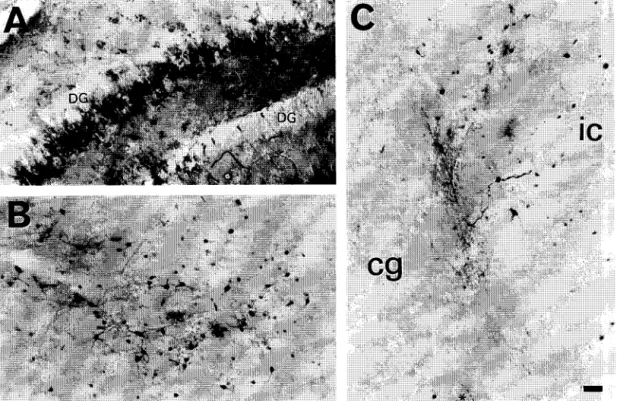
Fig. 3. The expression of fl-galactosidase 3 weeks after the infusion of 1 /xl of AAV-lacZ vectors (5 X 1 0 4 panicles//xl) into the hippocampus (A) or
inferior colliculus (B) of the 4-day-old rat. In both the hippocampus and the inferior colliculus, multipolar neurons comprise the majority of the cells labeled for/3-galactosidase immunoreactivity. In panel C, the injector coursed through the inferior colliculus and into the central gray. A dendritic remnant remains labeled near the tip of the injector ( * ), but little expression occurs until the border of the inferior colliculus, where substantial fl-galactosidase immunoreactivity is found. The large labeled cells demarcate the border between these adjacent brain structures. DG, dentate granule cell layer of the hippocampus; ic, inferior colliculus; cg, central gray. Bar = 50 /zm.
In direct contrast, t w o brain areas e x h i b i t e d similar a m o u n t s o f g e n e transfer and e x p r e s s i o n both at 1 w e e k and 3 m o n t h s . A s s e e n in Fig. 5, A A V - l a c Z v e c t o r infu- sion into the o l f a c t o r y t u b e r c l e resulted in a m o d e r a t e n u m b e r o f / 3 - g a l a c t o s i d a s e p o s i t i v e m u l t i p o l a r n e u r o n s at 1 w e e k . S i m i l a r n u m b e r s o f labeled, m u l t i p o l a r n e u r o n s w e r e f o u n d 3 m o n t h s after A A V - l a c Z v e c t o r infusion. W h e n A A V - l a c Z v e c t o r s w e r e i n f u s e d into the i n f e r i o r c o l l i c u - lus, a substantial n u m b e r o f /3-galactosidase p o s i t i v e neu- rons w e r e p r e s e n t b o t h at 1 w e e k and 3 m o n t h s (see Fig. 5). E v e n t h o u g h the i n f e r i o r c o l l i c u l u s exhibits a h i g h l e v e l o f l o n g - t e r m transduction, G F A P i m m u n o h i s t o c h e m i s t r y in a d j a c e n t s e c t i o n s did not r e v e a l o b v i o u s signs o f glial r e a c t i v i t y (see Fig. 6).
In all o f these brain areas, /3-galactosidase i m m u n o - r e a c t i v i t y w a s restricted to the i m m e d i a t e v i c i n i t y o f the infusion, o c c u r r i n g p r i m a r i l y in neurons. H o w e v e r , there w e r e a f e w i n s t a n c e s w h e r e f l - g a l a c t o s i d a s e i m m u n o -
r e a c t i v i t y w a s f o u n d in a clearly i d e n t i f i e d astrocyte. A l s o , w h e n 5 or 10 /zl o f the A A V - l a c Z v e c t o r s w e r e infused into the lateral ventricle, 7 d a y s later f l - g a l a c t o s i d a s e w a s f o u n d in e p e n d y m a l cells close to the site o f infusion, but not in a d j a c e n t brain structures.
4. Discussion
W h e n A A V - l a c Z - C M V p r o m o t e r v e c t o r s are m i c r o i n - fused into the brain, 15 to 40 /~m d i a m e t e r n e u r o n s r e p r e s e n t the m a j o r cell t y p e w h e r e transfer and e x p r e s s i o n o f f l - g a l a c t o s i d a s e occurs. H o w e v e r , d i f f e r e n t l e v e l s o f g e n e transfer and e x p r e s s i o n w e r e f o u n d b o t h across and w i t h i n d i f f e r e n t brain regions. A f t e r a 1 /xl infusion, the caudate n u c l e u s e x h i b i t e d the f e w e s t n u m b e r o f labeled cells, n e v e r m o r e than 10 cells in a g i v e n section. A greater n u m b e r o f cells w e r e f o u n d in the o l f a c t o r y tuber-
104 T.J. McCown et al. / B r a i n Research 713 (1996) 99-107
cle, between 10 and 20 cells in a given section. The greatest number of labeled cells were generally found in the piriform cortex, hippocampus and inferior colliculus. Usually, sections from these areas exhibited more than 20 labeled cells per section, while in the case of the inferior colliculus, some sections had greater than 50 /3-galacto- sidase positive cells in a given section (see Fig. 5). How-
ever, within given brain regions, not all neurons exhibited /3-galactosidase expression. For example, while many mul- tipolar neurons were /3-galactosidase positive in the hip- pocampus, j3-galactosidase activity was noticeably absent in the dentate granule neurons (see Fig. 2). Also, there were instances where other cells types exhibited gene transfer and expression. After ventricular administration,
' I
j
~j. ;i~ ~
T.J. McCown et al. / B r a i n Research 713 (1996) 99-107 105
CW
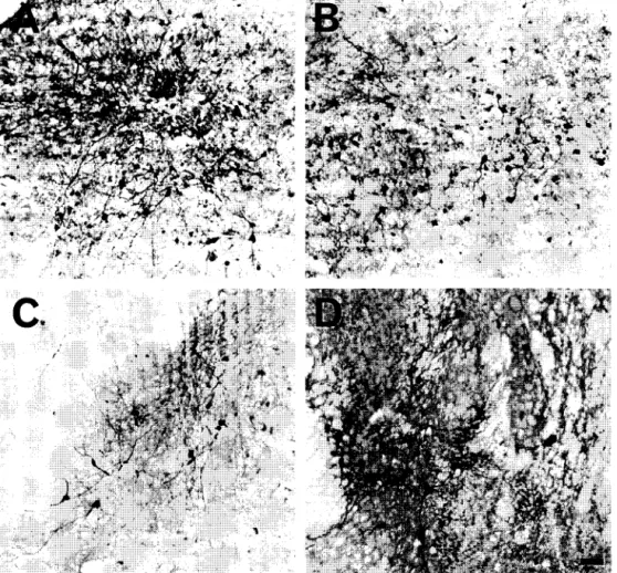
Fig. 5. The expression of /3-galactosidase in adult inferior colliculus and olfactory tubercle 7 days and 3 months after a 1 /.1.1 infusion of AAV-IacZ
(5 X 10 4 particles/#l). For the inferior colliculus, numerous neurons exhibited /3-galactosidase immunoreactivity both 7 days (A) and 3 months (B) after
AAV vector infusion. These findings were consistent for the 7-day (n = 5) and 3-month (n = 3) groups. Similarly, in the olfactory tubercle, a moderate number of neurons exhibited fl-galactosidase immunoreactivity both 7 days (C) and 3 months (D) after AAV vector infusion. Likewise, these findings were consistent for the 7-day (n = 4) and 3-month (n = 3) groups. Bar = 50 #m.
e p e n d y m a l c e l l s l i n i n g t h e v e n t r i c l e s w e r e p o s i t i v e f o r / 3 - g a l a c t o s i d a s e , a n d o c c a s i o n a l l y , g l i a d i d e x h i b i t e x p r e s - s i o n o f f l - g a l a c t o s i d a s e . In t h e c a s e o f a s t r o c y t e s , t h e r e l a t i v e l a c k o f l a b e l i n g d o e s n o t a p p e a r to b e a p r o b l e m w i t h d e t e c t i o n s e n s i t i v i t y , b e c a u s e in o n e c a s e , a l a b e l e d a s t r o c y t e w a s c l e a r l y i d e n t i f i e d in t h e c a u d a t e n u c l e u s . A l t h o u g h e x p r e s s i o n a p p e a r s to f a v o r l a r g e m u l t i p o l a r n e u r o n s , t h i s o b s e r v a t i o n d o e s n o t m e a n t h a t t h e A A V v e c t o r s a r e o n l y t a k e n u p i n t o m u l t i p o l a r n e u r o n s o r t h a t
g e n e t r a n s f e r is m o r e e f f i c i e n t in n e u r o n s . It o n l y m e a n s t h a t w i t h t h e A A V v e c t o r u n d e r c o n t r o l o f t h e C M V p r o m o t e r , e x p r e s s i o n o f t h e g e n e p r o d u c t o c c u r s p r e f e r - e n t i a l l y in m u l t i p o l a r n e u r o n s . G i v e n a o t h e r v e c t o r s o r p r o m o t e r s , t h e p a t t e r n o f g e n e t r a n s f e r c o u l d b e q u i t e d i f f e r e n t . F o r e x a m p l e , L e G a l L a S a l l e et al. [8] f o u n d / 3 - g a l a c t o s i d a s e e x p r e s s i o n in t h e d e n t a t e g r a n u l e c e l l s , b u t n o t t h e h i l u s o r p y r a m i d a l c e l l l a y e r , a f t e r h i p p o c a m p a l i n f u s i o n o f a n a d e n o v i r u s v e c t o r d r i v e n b y a R o u s s a r c o m a
106 T.J. McCown et al. /Brain Research 713 (1996) 99-107
A
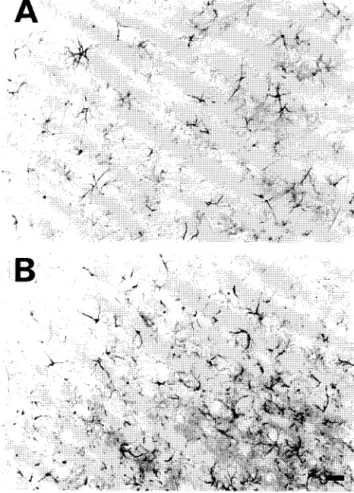
Fig. 6. GFAP immunoreactivity in sections from the inferior colliculus 3 months aflcr thc injection of 1 /zl AAV-lacZ (5 × 10⁴ particles/#1). It can be seen that the pattern of astrocyte GFAP immunoreactivity in the section contralateral to the AAV vector infusion (A) does not appreciably differ from the GFAP immunoreactivity on the infused side (B). The adjacent section to B exhibited /3-galactosidase immunoreactivity similar to that found in Fig. 5, panel B. Bar = 50 /xm.
virus long terminal repeat promoter. Further investigations will be necessary to delineate any cell specific differences in the A A V - v e c t o r uptake, gene transfer or promoter driven gene expression.
These studies also showed that A A V vectors can effec- tively transfer foreign genes into the neonatal brain, but the expression of /3-galactosidase proved to be site specific. Substantial /3-galactosidase expression was found in the h i p p o c a m p u s and inferior colliculus, but not in the piri- form cortex. One explanation for this difference could be the stage o f brain development. For example, the lack o f neural labeling in the cortex might be attributed to the fact that in 4-day-old rats, p y r a m i d a l cells are still migrating along the radial glia towards their final destination [15]. Alternatively, there could be site-specific differences in promoter regulation. Clearly, dramatic gene expression differences were found between two adjacent structures, the central gray and the inferior colliculus. Thus, in the neonate, the pattern of gene expression likely depends upon both differences in promoter regulation, as well as
the stage of development. Again, future studies will be necessary to define the actual contribution of these pro- cesses to gene transfer and expression in the neonatal brain.
In the adult rat, A A V vectors are capable of long-term transfer and expression of a foreign gene without overt signs of neurotoxicity, but the longevity of expression is brain site specific. In the piriform cortex and the hip- pocampus, the number of cells expressing /3-galactosidase at 3 months was dramatically lower than the number found 7 days post-infusion. In the caudate nucleus, the amount of expression at 7 days was not as high as in the cortex or hippocampus, but there still appeared to be some decline in the number of /3-galactosidase-positive cells at 3 months. However, it must be noted that even with such a decline in expression, Kaplitt et al. [7] found that A A V transfer and expression of tyrosine hydroxylase in the caudate was sufficient to partially reverse a lesion-induced motor deficit. Thus, for some disorders, partial long-term expression may be therapeutically sufficient. In marked contrast, both the olfactory tubercle and the inferior colliculus exhibited the same relative amount of /3-galactosidase expression at 7 days and 3 months. Furthermore, in the inferior colliculus, there were no obvious signs o f reactive astrocytes, so A A V - m e d i a t e d long term gene expression did not produce obvious signs of neurotoxicity.
These site-specific differences in long-term gene ex- pression could be attributed to variable expulsion of the vector or differential regulation of the C M V promoter. Given the difficulties with finding promoters capable of long term expression [4], and recent findings by Bloom et al. [3], it seems likely that inactivation of the promoter underlies our observed decline in gene product expression. For example, Bloom et al. [3] recently demonstrated long- term expression of /3-galactosidase in the hippocampus using an H S V vector and a latency associated transcript promoter from murine moloney leukemia virus. However, the 6-month level o f gene expression was substantially less than the levels of expression at 2 weeks, even though the amount of viral D N A did not change over this time period. In the present studies, differential suppression of the C M V promoter would explain why some brain areas exhibited a long term decline in /3-galactosidase expression, while other brain areas showed no decline in /3-galactosidase expression. Clearly, additional studies will be necessary to confirm this supposition.
T.J. McCown et al. / B r a i n Research 713 (1996) 9 9 - 1 0 7 107
in t h e i n f e r i o r c o l l i c u l u s . S i n c e n e o n a t a l a n d adult s e i z u r e g e n e s i s h a s b e e n w e l l c h a r a c t e r i z e d in t h e c o r t i c a l s u b d i v i - s i o n o f t h e i n f e r i o r c o l l i c u l u s [ 9 , 1 1 - 1 3 ] , this b r a i n area s h o u l d p r o v e to b e e x c e l l e n t m o d e l s y s t e m to e x p l o r e v a r i o u s s t r a t e g i e s o f g e n e t h e r a p y .
Acknowledgements
T h e s e s t u d i e s w e r e s u p p o r t e d in p a r t b y N I H D K 4 2 7 0 1 , a B a s i c R e s e a r c h G r a n t N o . 1 - F Y 9 4 - 0 8 9 1 f r o m the M a r c h o f D i m e s B i r t h D e f e c t s F o u n d a t i o n , N S 2 5 8 8 6 a n d H D 0311.
References
[1] Akli, S., Caillaud, C., Vigne, E., Stratford-Perricaudet, L.D., Poe- naru, L., Perricaudet, M., Kahn, A. and Peschanski, M.R., Transfer of a foreign gene into the brain using adenovirus vectors, Nature Genetics, 3 (1993) 224-228.
[2] Blacklow, N.R., Hoggan, M.D., Kapikian, A.Z., Austin, J.B. and Rowe, W.P., Epidemiology of adeno-associated virus infection in a nursery population, Am. J. Epidemiol., 8 (1968) 368-378. [3] Bloom, D.C., Maidment, N.T., Tan, A., Dissette, V.B., Feldman,
L.T. and Stevens, J.G., Long-term expression of a reporter gene from latent herpes simplex virus in the rat hippocampus, Mol. Brain Res., 31 (1995) 48-60.
[4] Glorioso, J.C., Goins, W.F. and Fink, D.J., Herpes simplex virus- based vectors, Semin. Virol., 3 (1992) 265-276.
[5] Hatton, J.D. and Lin, L., Demonstration of specific neuronal cell groups in rat brain /3-galactosidase enzyme histochemistry, J. Neu- rot. Methods, 45 (1992) 147-153.
[6] Hsu, S.M. and Soban, E., Color modification of diaminobenzidine (DAB) precipitation by metallic ions and its application for double immunohistochemistry, J. Histochem. Cytochem., 30 (1982) 1079- 1082.
[7] Kaplitt, M.G., Leone, P., Samulski, R.J., Xiao, X., Pfaff, D.W.,
O'Malley, K.L. and During, M.J., Long-term gene expression and phenotypic correction using adeno-associated virus vectors in the mammalian brain, Nature Genetics, 8 (1994) 148-154.
[8] Le Gal La Salle, G., Robert, J.J., Berrard, S., Ridoux, V., Stratford- Perricaudet, L.D., Perricaudet, M. and Mallet, J., An adenovirus vector for gene transfer into neurons and glia in the brain, Science, 259 (1993) 988-990.
[9] McCown, T.J., Greenwood, R.S., Frye, G.D. and Breese, G.R., Electrically elicited seizures from the inferior colliculus: a potential site for the genesis of epilepsy?, Exp. Neurol., 86 (1984) 527-542. [10] McCown, T.J., Givens, B.S. and Breese, G.R., Amino acid influ- ences on seizures elicited within the inferior colliculus, J. Pharma- col. Exp. Ther., 243 (1987) 603-608.
[11] McCown, T.J. and Breese, G.R., The role of the inferior collicular cortex in the neonatal rat: sensory-motor modulation, DeL. Brain Res., 59 (199) 1-5.
[12] McCown, T.J., Duncan, G.E and Breese, G.R., Neuroanatomical characterization of inferior collicular seizure genesis: 2-deoxyglu- cose and stimulation mapping, Brain Res., 567 (1991) 25-32. [13] McCown, T.J. and Breese, G.R., The developmental profile of
seizure genesis in the inferior collicular cortex of the rat: relevance to human neonatal seizures, Epilepsia, 33 (1992) 2-10.
[14] McLaughlin, S.K., Collis, P., Hermonat, P.L. and Muzyczka, N., Adeno-associated virus general transduction vectors: analysis of proviral structure, J. Virol., 62 (1988) 1963-1973.
[15] Miller, M.W., Development of projections and local circuit neurons in neocortex. In A. Peters and E.G. Jones (Eds.), Cerebral Cortex, Vol. 7, Plenum Press, New York, 1988, pp. 133-175.
[16] Pakzaban, P., Geller, A.1. and lsacson, O., Effect of exogenous nerve growth factor on neurotoxicity of neuronal gene delivery by a herpes simplex amplicon vector in the rat brain, Human Gene Ther., 5 (1994) 987-995.
[17] Paxinos, G. and Watson, C., The Rat Brain in Stereotaxic Coordi- nates, Academic Press, New York, 1986.
[18] Palella, T.D., Hidaka, Y., Silverman, L.J., Levine, M. Glorioso, J. and Kelley, W.N., Expression of human HPRT mRNA in brain of mice infected with recombinant herpes simplex virus-1 vector, Gene, 80 (1988) 137-144.