Methicillin Resistant Staphylococcus aureus (MRSA) has developed as a prevalent cause
of community acquired disease. S. aureus’s effectiveness is due to its exceptional resistance to
host immunity, including nitric oxide (NO•). Full NO• resistance requires activation of a gene
subset including hmp and ldh1. Hmp converts reactive nitric oxide into non-toxic nitrate. Ldh1
relieves redox imbalance incurred during anaerobic respiration required during NO•-stress.
Although preliminary work implicated SrrABfor controlling hmp expression, the regulation is
incomplete, implicating additional regulators. Ldh1 is stimulated by glucose, however the exact
regulatory mechanism is unknown; my research goal is to identify the precise regulatory
mechanisms for both genes. To find these regulators, I conducted a screen of mutants in every
non-essential regulatory S. aureus protein by transducing reporter plasmids for each gene into
each mutant. Growth curves measuring fluorescence upon NO•-stress to find atypical induction
of hmp or ldh1 indicated possible regulators, including SigB and HrcA.
INTRODUCTION
Staphylococcus aureus is a Gram positive, facultative anaerobe that is highly successful
human pathogen. As much as 30% of the population carries S. aureus on their skin
asymptomatically. Methicillin Resistant Staphylococcus aureus (MRSA) is an emerging threat
that, at one time only present in hospitals, has emerged in the community as a prevalent cause of
disease, with approximately 2% of the population carrying MRSA as part of their normal flora.
USA 300 is a community-acquired MRSA strain that is currently being studied. The virulence
immune response by inhibiting
opsonization, phagocytosis, and
neutrophil recruitment. In addition
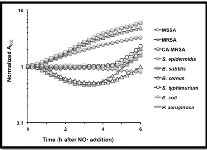
to these virulence factors, S.
aureus is also exceptionally
resistant to nitric oxide (NO•)
(Figure 1).
Figure 1. Staphylococcus aureus is uniquely resistant to NO• stress (Richardson lab, unpublished).
The immune system produces NO• to fight off pathogens infecting the body. NO• is a
radical that can react with the pathogen’s DNA, lipids, and proteins to kill the invading microbes.
S. aureus accomplishes NO• resistance by activating a subset of genes previously linked to
anaerobic metabolism. These genes include srrAB, hmp, and ldh1. Hmpis an NO• dioxygenase
that converts NO• into nitrate. In Bacillus subtilis, hmp expression has been linked to the SrrAB
counterpart, ResDE. SrrABis a two component system shown to be a global regulator of the
shift to anaerobic metabolism in S. aureus. In S. aureus, however, regulation of hmp by SrrAB is
incomplete, implicating additional unknown regulators (Richardson, et. al, 2006). Ldh1 is a
lactate dehydrogenase which restores redox balance (NADH/NAD+) in the cell. Ldh1 is
repressed by redox sensing regulator Rex, however it is also stimulated by glucose. The
mechanism of this glucose related regulation is unknown (Crooke et.al, 2013). This study
METHODS
NARSA Library
This study utilized a NARSA (Network on Antimicrobial Resistant of Staphylococcus
aureus) library of 105 mutants in every non-essential regulatory protein in S. aureus. The
NARSA library was created by the Center for Staphylococcal Research at the University of
Nebraska Medical Center through transposon mutagenesis of S. aureus strain USA300.
Lysate Preparation
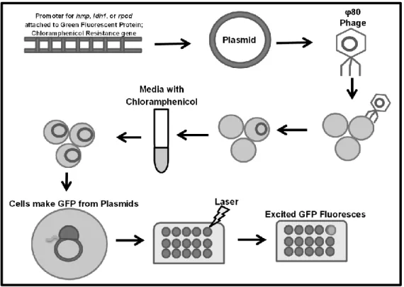
To prepare lysates for transduction of the reporter fusion in to the NARSA library, the
strains containing the reporter plasmids with prpod::gfp, phmp::gfp pldh1::gfp (premade) were
grown in an overnight culture in BHI and Chloramphenicol. The cultures were then diluted and
grown in CY broth for approximately 3 hours, after which, 1 ml of the culture, 1 ml of phage
buffer, and 100 µL of a phage stock made from lysate of φ80 RN4220 were combined and
allowed to shake at 30 degrees overnight. The lysate was then centrifuged, filter sterilized using
.45µM syringe filters, and stored at 4 degrees.
Transduction Protocol
Each regulator mutant in the NARSA library was transduced using the following
protocol. The strain was allowed to grow overnight in BHI. In 4 plastic falcons, 1 mL of the
overnight culture was added, in addition to 12 µL of 1M CaCl2. To each falcon, lysates from
prpod::GFP, phmp::GFP, or pldh1::GFP were added individually, or no lysate was added. The
tubes were allowed to incubate for 20 minutes. After incubation, 2 mL of 1% sodium citrate was
was discarded and resuspended in 2 mLs of TSB + 0.5% sodium citrate. The bacteria were then
allowed to rest in a 37 degree water bath for 1 hour. The tubes were then centrifuged again for 10
minutes at 4150 rpm. All but approximately 200 µL of the supernatant was removed; the culture
was then resuspended and plated on TSB plates containing 0.5% Sodium Citrate and
Chloramphenicol to grow overnight.
Miniprep, PCR, and DNA Gel Electrophoresis Protocol
Two colonies from the transduction plates were selected and grown overnight in BHI and
Chloramphenicol. Because of the possibility of spontaneous mutations causing chloramphenicol
resistance, each culture must be tested for the presence of the plasmid. This was completed using
a QIAprep Spin Miniprep kit. For each culture, 3 mL of culture was pelleted and resuspended in
250 µL of buffer P1 and 12 µL of 2.5 mg/ml lysostaphin in order to break the cell wall of the S.
aureus. The bacteria were allowed to incubate for 1 hour in a 37 degree water bath. Following,
250 µL of buffer P2 was added to each tube and mixed, proceeded by immediately adding 350
µL of buffer N3. Mixtures were then centrifuged for 10 minutes in a tabletop microcentrifuge.
The supernatant was then transferred to a QIA spin prep column and washed with 750 µL of
Buffer PE and centrifuged. The QIAprep column was then transferred to a clean microcentrifuge
tube and 30 µL of water was added to elute the DNA.
The DNA could then be directly run on a 1% agarose gel to detect the presence of the
plasmid. For further verification, PCR using the respective primers could be run to amplify the
Plasmid DNA pieces of the Rpod, Hmp, or Ldh1 gene promoters. This verification was only
contain the correct plasmid. Once the strain was verified as containing the correct plasmid, a
freezer stock could be made in BHI + 25% glycerol.
Growth Curve Protocol
In order to further identify possible regulatory candidates, growth curves were conducted.
The strain to be tested was grown overnight in BHI and Chloramphenicol. 500 µl of the culture
was washed twice and resuspended with PBS. Each washed culture was then diluted to an
OD660 of 0.01 in PN media with Glucose and Chloramphenicol. The diluted cultures were then
aliquoted into a 96 well plate and run on a Tecan Infinite M200 plate reader that measures both
OD and fluorescence of each well. At an OD660 of 0.15, 2mM of Nitric Oxide in the form of
DETANO was applied to the cells. The Tecan obtained readings for 24 hours.
RESULTS
The peak fluorescence of each mutant was averaged and normalized to Optical Density,
and to normalized expression of the Wildtype. Significant difference from the Wildtype (values
significantly >1 or <1) indicates possible regulators for hmp or ldh1.
Figure 3. A. (Fluorescencemutant /ODmutant) / (FluorescenceWT/ODWT) ratio of peak fluorescence for mutants containing phmp::GFP compared to the wildtype containing phmp::GFP.
B. (Fluorescencemutant /ODmutant) / (FluorescenceWT/ODWT) ratio of peak fluorescence for mutants containing pldh1::GFP compared to the wildtype containing pldh1::GFP. Δrex (Ratio 34.9) and ΔsrrA (ratio 6.3) not shown. Points in blue are 2 standard deviations from the average, points in red are 3 standard deviations from the average
0 0.2 0.4 0.6 0.8 1 1.2 1.4 1.6 1.8 2 Rati o
of Peak Fl
u
or
essenc
e
Mutants
Hmp Expression Change in NARSA Mutants
0 0.2 0.4 0.6 0.8 1 1.2 1.4 1.6 1.8 2 2.2 R atio o f Peak Fl u o re sen ce Mutants
Table 1. Mutants that displayed altered hmp expression levels derived from Figure 3A.
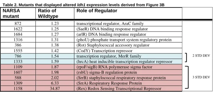
Table 2. Mutants that displayed altered ldh1 expression levels derived from Figure 3B
NARSA mutant
Ratio of Wildtype
Role of Regulator
872 1.23 transcriptional regulator, AraC family 1622 1.25 (SaeR) DNA binding response regulator 1684 1.27 (arlR) DNA binding response regulator
1316 1.31 (phoU) phosphate transport system regulatory protein 386 1.38 (Rot) Staphylococcal accessory regulator
1555 1.42 (CodY) Transcription repressor
786 1.54 transcription regulator, MerR family 2 STD DEV
1333 1.59 (hrcA) heat inducible transcription regulator repressor 1109 1.87 (rpoF/sigB) RNA polymerase sigma factor
1607 1.98 (rsbU) sigma-B regulation protein
588 2.02 (SrrB) staphylococcal respiratory response protein 3 STD DEV 1309 6.33 (SrrA) Respiratory Response Protein
1158 34.87 (Rex) Redox Sensing Transcriptional Repressor
Mutants of interest were selected by averaging the ratio of the wildtype for all 105
mutants and determining the standard deviation for the library. Mutants with 2 standard
deviations from the mean (P=.05) and 3 standard deviations from the mean (P=.01) were
determined to be significantly different from the Wildtype to justify further analysis. For ldh1,
both ΔsrrA and Δrex were excluded from the average and standard deviation as outliers.
NARSA mutant
Ratio of Wildtype
Role of Regulator
1532 1.277 (agrA) accessory gene regulator protein 755 1.34 (yfmC) transcription regulator, GntR family 367 1.367 (agrR) arginine repressor
454 1.37 (ArcA) transcriptional regulator, Crp/Fnr family 2 STD DEV 415 1.41 putative transcriptional regulator
1555 1.411 (codY) transcription repressor
DISCUSSION
Mutants that had corresponding expression changes observed in both Hmp and Ldh1
suggest that the mutated regulatory protein likely affects overall transcript levels. In order to
determine if the regulatory protein is a regulator for both genes, or simply affects overall
transcript levels, the expression of Rpod (constitutively active) must be compared. Mutants in
which all three genes show increased expression exclude these mutants from the list of
regulatory proteins to investigate further. One such mutant was observed- 1555 (CodY). ΔCodY
was elevated by 40% in both phmp::GFP and pldh1::GFP (Table 1 & 2). CodY is a known
transcript repressor that would be predicted to cause increased transcription levels when mutated.
Rpod expression must be analyzed in order to confirm the removal of CodY from the list of
possible regulators for either gene.
Change in transcription levels of phmp::GFP without a corresponding change in
transcription levels of pldh1::GFP and vice versa indicates a possible regulator for the gene. Of
the mutants identified by altered transcription levels for pldh1::GFP, ΔsigB and ΔrsbU are of
particular interest. SigB is an RNA polymerase sigma factor. During transcription, sigma factors
help recruit RNA polymerase to the site of transcription. S. aureus utilizes a normal
housekeeping sigma factor in addition to multiple alternative sigma factors with specificity to
different promoters. SigB is one such alternative sigma factor. The utilization of sigma factors
with different specificities allows the bacteria to alter transcription in response to a changing
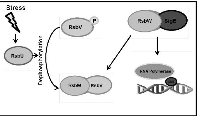
environment. The regulation of SigB in Bacillus subtilis is well characterized, and S. aureus is
suspected to have similar regulation with some key differences (Figure 4). In S. aureus, SigB and
related Rsb proteins (W, U, and V) are conserved, and in both S. aureus and B. subtilis, RsbU
appears to set a basal level of SigB activity which is subsequently increased by different cell
stresses. Increased RsbU did not appear, however, to increase the ratio of RsbV/RsbV-P,
indicating regulation that further differs from B. subtilis (Pane-Farre, 2009).
Figure 4. Regulation of SigB in Bacillus subtilis. In B. subtilis Sig B has been shown to be regulated by RsbW which forms a complex with SigB and prevents it from binding to promoter sequences. RsbV is an anti-sigma factor antagonist which competes with SigB to bind to RsbW when dephosphorylated. The release of SigB allows for binding to promoter sequences and subsequent transcription. RsbU acts as a phosphotase for RsbV during environmental stress, allowing it to form the complex with RsbW (Pane-Farre, 2009).
Similar GFP transcription in the ΔsigB and ΔrsbU backgrounds (table 2) is consistent
with the conserved role of RsbU as an activator of SigB. Both ΔsigB and ΔrsbU backgrounds
displayed a near two-fold increase in transcription levels. This increase is on par with that seen in
ΔsrrB, which is known to indirectly regulate ldh1 expression by increasing redox imbalance to
minimize Rex activity (Richardson lab, Unpublished). This evidence linking SigB to the nitric
oxide response of S. aureus makes it a prime candidate for further investigation.To elucidate the
role of SigB in the expression of ldh1, work will be done to evaluate expression of the other Rex
sequences specific to SigB by scanning for conserved binding sites.
Of the mutants identified by altered transcription levels for phmp::GFP and pldh1::GFP,
ΔhrcA is of particular interest. HrcA is known to be a protein related to heat shock resistance in
S. aureus, and has been shown to be significantly induced by NO• (Richardson, et. al, 2006).
However, HrcA is a repressor for a subset of genes that repair heat shock damage. Included in
this subset is dnaK. DnaK codes for protein refolding machinery that is critical for survival
during heat stress. DnaK has also been shown to have an important role in resistance to oxidative
stress which causes similar protein denaturing (Singh, 2007). The 60% increase in expression of
GFP in the ΔhrcA mutant therefore may be due to de-repression of hmp and ldh1, or it may be
due to increased stability of proteins due to the DnaK refolding machinery – resulting in
persistence of GFP. To test if this phenotype is a result of action of DnaK or due to transcription
regulation, the promoter regions of hmp and ldh1 will be checked for binding sequence
specificity to HrcA.
Future work
In order to verify the additional identified regulator candidates, Realtime PCR will be run
on promising candidates from Table 1 and 2 to analyze the genomic activity with regards to hmp
and ldh1. Because the screen only analyzed the activity of the reporter fusion on the plasmid,
actual genomic expression can vary widely, and a small drop in promoter::GFP expression can
correlate to no expression from the corresponding genomic promoter. Additionally, enzymatic
activity of Ldh1 can be measured as an additional method of verification.
It is possible that the regulators will not be found in the NARSA library. This could mean
translational modification of the regulator controls the expression of ldh1 or hmp. These would
not be found in our screen because we are not testing mutants in proteins that catalyze post
translational modifications. If the regulator is not found from the screen, these other possibilities
REFERENCES
Crooke AK, Fuller JR, Obrist MW, Tomkovich SE, Vitko NP, et al. (2013) “CcpA-Independent
Glucose Regulation of Lactate Dehydrogenase Staphylococcus aureus.” PLoS ONE 8(1):
e54293. doi:10.1371/journal.pone.0054293
Hecker, Michael, Alexander Reder, Stephan Fuchs, Martin Pagels, and Susanne Engelmann.
"Physiological Proteomics and Stress/starvation Responses in Bacillus Subtilis and
Staphylococcus Aureus." Research in Microbiology 160.4 (2009): 245-58. Web.
Pane-Farre, J., B. Jonas, S. W. Hardwick, K. Gronau, R. J. Lewis, M. Hecker, and S. Engelmann.
"Role of RsbU in Controlling SigB Activity in Staphylococcus Aureus following
Alkaline Stress." Journal of Bacteriology 191.8 (2009): 2561-573. Web.
Richardson A, Dunman P, Fang F. (2006) “The nitrosative stress response of Staphylococcus
aureus is required for resistance to innate immunity”. Mol Microbiol. 2006
Aug;61(4):927-39. Epub 2006 Jul 12.
Singh, V. K., S. Utaida, L. S. Jackson, R. K. Jayaswal, B. J. Wilkinson, and N. R. Chamberlain.
"Role for DnaK Locus in Tolerance of Multiple Stresses in Staphylococcus Aureus."