207
DPP4 INHIBITORS PROMOTE BIOLOGICAL FUNCTIONS OF HUMAN ENDOTHELIAL
PROGENITOR CELLS BY TARGETING THE SDF-1/CXCR4 SIGNALING PATHWAY
Feng Liu1, Guo-Dong Huang2,*, Jia-Zhen Tang1, Yu-Huan Peng3
1 Department of Endocrinology, The First Affiliated Hospital of Nanchang University, Nanchang 330006, China
2 Department of Integrated Traditional Chinese and Western Medicine, The First Affiliated Hospital of Nanchang University, Nanchang 330006, China
3Department of Pharmacy, The First Affiliated Hospital of Nanchang University, Nanchang 330006, China
*Corresponding author: [email protected]
Received: May 6, 2015; Revised: July 6, 2015; Accepted: July 9, 2015; Published online: December 28, 2015
Abstract:Dipeptidyl peptidase 4 (DPP4) inhibitors (oral hypoglycemic agents) have beneficial effects during the early stages of diabetes. In this study, we evaluated the role of DPP4 inhibitors on the biological functions of cultured human endothelial progenitor cells (EPCs). After treating EPCs with the DPP4 inhibitors sitagliptin and vildagliptin, we examined the mRNA expression of DPP4, vascular endothelial growth factor (VEGF), VEGF receptor 2 (VEGFR-2), endothelial nitric oxide synthase (eNOS), caspase-3, stromal cell-derived factor-1 (SDF-1), chemokine (C-X-C motif) receptor 4 (CXCR4) were measured by RT-PCR. The protein expression of SDF-1 and CXCR4 was determined by Western blot; cell prolifera-tion was tested by the MTT method, and DPP4 activity was determined by a DPP4 assay. Our results revealed that DPP4 expression and activity were inhibited following the treatment with various doses of DPP4 inhibitors. Cell proliferation and the expression of VEGF, VEGFR-2 and eNOS were upregulated, while cell apoptosis was inhibited by DPP4 inhibitors in a dose-dependent manner. DPP4 inhibitors activated the SDF-1/CXCR4 signaling pathway, shown by the elevated expres-sion of SDF-1/CXCR4. This further proved that after the SDF-1/CXCR4 signaling pathway was blocked by its inhibitor ADM3100, the effects of DPP4 inhibitors on the proliferation and apoptosis, and the expression of VEGF, VEGFR-2 and eNOS of EPCs were significantly reduced. These findings suggest that DPP4 inhibitors promote the biological functions of human EPCs by upregulating the SDF-1/CXCR4 signaling pathway.
Key words: DPP4 inhibitor; EPCs; biological function; SDF-1; CXCR4
INTRODUCTION
DPP4, a member of the prolyoligopeptidase family, is a transmembrane glycoprotein with serine exopepti-dase activity that cleaves X-proline dipeptides from the N-terminus of polypeptides such as chemokines, neuropeptides, and vasoactive peptides (Matteucci and Giampietro, 2009). DPP4 is known to be present in many tissues including bone marrow, kidney, lung, liver, spleen, pancreas, intestines and venular end of blood vessels (Augustyns et al., 1999; Lambeir et al., 2003), and it is also expressed on the surface of several cell types, including epithelial cells, embryonic stem cells, hematopoietic stem cells, hematopoietic
progeni-tor cells and memory T cells (Christopherson et al., 2004; Guo et al., 2005). DPP4 activity is associated with the development of obesity (Lamers et al., 2011), diabetes mellitus (Drucker et al., 2007), renal disease (Sato et al., 2014), cardiovascular disease (Jose et al., 2011; Zhong et al., 2013) and inflammation (Zhong et al., 2013). Therefore, it is very important to exam-ine potential agents that target DPP4 in order to treat these diseases.
barrier injury and oxidant stress in the obese Zucker rat (Nistala et al., 2014), and prevented obesity-in-duced renal injury in male mice (Nistala et al., 2014). Additionally, DPP4 inhibition combined with luminal nutrients was an effective therapy for the treatment of small intestinal ulcers in rats (Fujiwara et al., 2015). DPP4 inhibitors such as sitagliptin, vildagliptin, saxa-gliptin, alogliptin and linagliptin provide a compara-tively novel approach in diabetes treatment (Abad Pa-niagua et al., 2014; Kumar et al., 2014; Tomkin, 2014). Furthermore, several previous studies have suggested that DPP4 inhibitors directly or indirectly preserve renal function (Hocher et al., 2012; Sato et al., 2014), support cardiovascular functioning (Jose et al., 2011; Zhong et al., 2013), and prevent brain mitochondrial dysfunction and cognitive dysfunction caused by a high-fat diet (Pipatpiboon et al., 2013). The use of DPP4 inhibitors has opened new possibilities for the prevention and treatment of human diseases.
Numerous findings have demonstrated that en-dothelial dysfunction is a systemic process that is the first step in the pathogenesis of atherosclerosis and atherosclerotic plaque progression (Hattori et al., 2006). Endothelial progenitor cells (EPCs) can be used to repair tissues after myocardial infarction and contribute to therapeutic angiogenesis (Kawamoto et al., 2001). Many EPCs agonists, such as granulo-cyte-colony stimulating factor (G-CSF), VEGF and statins can mobilize EPCs in bone marrow (Rafii and Lyden, 2003). A well accepted hypothesis is that the chemokine SDF-1 and its main receptor CXCR4 are expressed in progenitor cells and play a pivotal role in EPC mobilization and homing (Peled et al., 1999; Zhou et al., 2014). However, little is known of the role of DPP4 inhibition in the biological functions of EPCs. Furthermore, whether there is a link between the SDF-1/CXCR4 axis and DPP4 inhibitors in EPCs remains unclear. In the present study we used an in vitro-cultured human EPCs as a model to evaluate the effects of DPP4 inhibitors on the biological functions of human EPCs and to investigate the mechanism of DPP4 inhibitors.
MATERIALS AND METHODS Cell culture
The human endothelial progenitor cells (EPCs), ob-tained from the China Center for Type Culture Collec-tion (Wuhan, China), were cultured in growth medi-um EBM-2 (Lonza Walkersville, USA, basal medimedi-um with 8 factors and 5% fetal bovine serum). Cells were propagated in a humidified environment at 37°C with 5% CO2 and 100% humidity.
DPP activity
After human EPCs were assigned to ten groups receiv-ing the DPP4 inhibitors sitagliptin (Carbosynth Lim-ited, Berkshire, UK) and vildagliptin (LGM Pharma, FL, USA) at doses of 0 (control), 0.01, 0.1, 1 or 10 μM for 48 h. DPP activity was determined by SensoLyte AMC DPP4 Assay Kit (AnaSpec, Inc., Fremont, CA) according to the manufacturer’s instructions. Briefly, a cellular lysate containing 10 μg/50 μL of protein was mixed with 50 μL of DPP substrate AMC. The mixture was then measured using a fluorescence plate reader with excitation at 354 nm and emission at 442 nm (Fluoro Skan Ascent, Thermo Fisher Scientific K.K., Kanagawa, Japan) after a 30-min incubation at room temperature. Cellular DPP activity was expressed as relative fluorescence units (RFU).
MTT methods
Real-time RT-PCR
Following treatment with 0, 0.01, 0.1, 1 and 10 μM of the DPP4 inhibitors sitagliptin or vildagliptin for 48 h, the mRNA expression of VEGF, VEGFR-2, eNOS, caspase-3, SDF-1 and CXCR4 in human EPCs was determined at the indicated times quantitative reverse transcription polymerase chain reaction (RT-PCR). Briefly, total cellular RNA was isolated from cells on 6-well plates using TRIzol reagent (Invitrogen, Carls-bad, CA, USA) according to the manufacturer’s in-structions. RNA quality was assessed by agarose gel electrophoresis, and complementary DNA (cDNA) was synthesized with a random hexamer (TaKaRa, Osaka, Japan). The real-time RT-PCR analysis was carried out using QuantiTect SYBR Green RT-PCR Kit (Qiagen, Valencia, CA) under an ABI Prism 7500 Sequence Detector (Applied Biosystems, Foster City, CA, USA) following the manufacturers’ instructions. The conditions were as follows: 1 cycle of denaturation at 95°C for 30 s, followed by 42 cycles at 95°C for 5 s, 58°C for 40 s, 1 cycle at 95°C for 15 s, and 60°C for 30 s. Specific primer sequences were synthesized in BIOSUNE Biological Technology Corp (Shanghai, China), and sequences of the primers are shown in Table 1. Expression of target genes was normalized to the expression of β-actin.
Table 1. Primers used in this study
Gene Serial number Primers
β-actin X00351.1 Sense: 5’-GCCGATCCACACGGAGTACT-3’Anti-sense: 5’-CTGGCACCCAGCACAATG-3’
DPP4 NM_001935.3 Sense:5’-AAATGGGATTTGTGGACA-GCAAG-3’Anti-sense:5’-CCGATCCCAGGACCATTGAG-3’
Caspase-3 NM_032991.2 Sense:5’-TATTCTTAAAGTATTTTCGTTAC-3’Antisense: 5’-TCGTTATATAGACAACTCGATA-3’
CXCR4 AY242129.1 Sense: 5’-CCTCGCCTTCTTCCACTGTT-3’Anti-sense: 5’-CTGGGCAGAGCTTTTGAACTTG-3’
SDF-1 NM_199168.3 Sense: 5’-GTGTCACTGGCGACACGTAG-3’Antisense: 5’-TCCCATCCCACAGAGAGAAG-3’
eNOS BC069465.1 Sense:5’-CACCAAACGTCGGGACCCGG-3’Antisense: 5’-TATGTCCTGAGTCCTACCCG-3’
VEGF M32977.1 Sense: 5’-TACCGTCTTCCTCCTCGAT-3’Antisense: 5’-CACTGTTCGGCTCCGCCA-3’
VEGFR2 EU826563.1 Sense: 5’-CGGAGCCAGTAAATCGATCAAG-3’Antisense: 5’-CCTCGAATTCTTACGTAGGAACG-3’
Caspase-3 activity assay
The caspase-3 colorimetric activity assay was per-formed according to the manufacturer’s instructions. In brief, the reaction buffer and the specific enzyme DEVD-pNA were added to each sample and incubated for 1-2 h at 37°C. The developed colorimetric reac-tion was measured at 405 nm in a 96-well microplate reader (Biorad Model) and values plotted as arbitrary units.
Western blot assay
Western blot analysis was performed to measure the expression of CXCR4 and SDF-1. Briefly, proteins were separated by 10% sodium dodecyl sulfate-polyacryl-amide gel electrophoresis (SDS-PAGE) and transferred electrophoretically to PVDF membranes. Then the membranes were blocked in 5% non-fat milk for 2 h at room temperature and incubated at 4°C overnight with antibodies SDF-1 and CXCR4 (1:1000 dilution; R&D Systems, Minneapolis, MN, USA). After overnight in-cubation, the membranes were washed and immunob-lotted with HRP-conjugated anti-rabbit IgG antibody (diluted 1:1000; Amersham Biosciences, Piscataway, NJ, USA) at 37°C for 1 h. The membranes were then developed using enhanced chemiluminescence (ECL) (Amersham, Buckinghamshire, UK) and exposed to X-ray film. Band density was quantitated using Image J software and normalized to the β-actin levels.
RESULTS
Effects of DPP4 inhibitors on the expression and activity of DPP4 in human EPCs
dose-dependent (Fig. 1A). Next, we measured the cellular DPP activity in human EPCs. The DPP activity assay revealed that, compared to the control, the distribution of DPP4 enzyme activity showed no difference in the 0.01-μM sitagliptin or vildagliptin group, while it was considerably decreased by sitagliptin or vildagliptin at concentrations of 0.1, 1 and 10 μM (P<0.05) (Fig. 1B). This suggested that DPP inhibitors could successfully inhibit the expression and enzyme activity of DPP4 in human EPCs, and that the effects of the higher dose of DPP4 inhibitors were more significant (P<0.05).
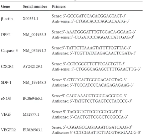
Effects of DPP4 inhibitors on the proliferation of human EPCs
To determine the influence of DPP4 inhibitors on the growth of human EPCs, cell proliferation was detected by MTT colorimetry. The data showed that, compared to the control without the stimulation of DPP4 in-hibitors, DPP4 inhibitors sitagliptin or vildagliptin could promote the proliferation of human EPCs in a dose-dependent manner. Briefly, sitagliptin at con-centrations of 0.01 and 0.1 μM or vildagliptin at a concentration of 0.01 μM increased the proliferation of human EPCs, however, the effects were not signifi-cant (P>0.05). Sitagliptin at concentrations of 1 and 10 μM or vildagliptin at concentrations of 0.1, 1 and 10 μM caused statistically significant cell growth stimula-tion (P<0.05). Furthermore, the effect of vildagliptin
Fig. 1. The effects of DPP4 inhibitors on the expression and activity of DPP4 in human EPCs. Human EPCs were treated with 0, 0.01, 0.1, 1 and 10 μM of DPP4 inhibitors sitagliptin or vildagliptin for 48 h, after which mRNA expression (A) and activity (B) of DPP4 were measured. Data are expressed as mean±SD of three independent experiments performed in triplicate. *P<0.05, **P<0.01, or P>0.05 vs control.
Fig. 2. The effect of DPP4 inhibitors on the proliferation of human EPCs. Human EPCs were treated with 0, 0.01, 0.1, 1 and 10 μM of DPP4 inhibitors sitagliptin or vildagliptin for 48 h, after which the proliferation of human EPCs was measured by the MTT method. Data are expressed as mean±SD of three independent experiments performed in triplicate. *P<0.05, **P<0.01, or P>0.05 vs control.
on cell proliferation was more significant than that of sitagliptin (Fig. 2).
Effects of DPP4 inhibitors on human EPC apoptosis
Fig. 4. Effect of DPP4 inhibitors on pro-angiogenic factors VEGF, VEGFR-2 and eNOS expression in human EPCs. After human EPCs were treated with 0, 0.01, 0.1, 1 and 10 μM of DPP4 inhibitors sitagliptin (A) or vildagliptin (B) for 48 h, mRNA expression of pro-angiogenic factors VEGF, VEGFR-2 and eNOS in human EPCs was determined by RT-PCR. Data are expressed as the mean±SD of three independent experiments performed in triplicate. *P<0.05., **P<0.01., or P>0.05 vs control.
in Fig. 4, this suggested that, compared to the control, 0.01 μM of sitagliptin or vildagliptin did not change the mRNA expression of VEGF, VEGFR2 and eNOS significantly (P>0.05), while sitagliptin (Fig. 4A) and vildagliptin (Fig. 4B) at concentrations of 0.1, 1 and 10 μM promoted mRNA expression of VEGF, VEGFR2 and eNOS in a dose-dependent manner (P<0.05), indicating that DPP4 inhibitors could enhance the angiogenesis of human EPCs.
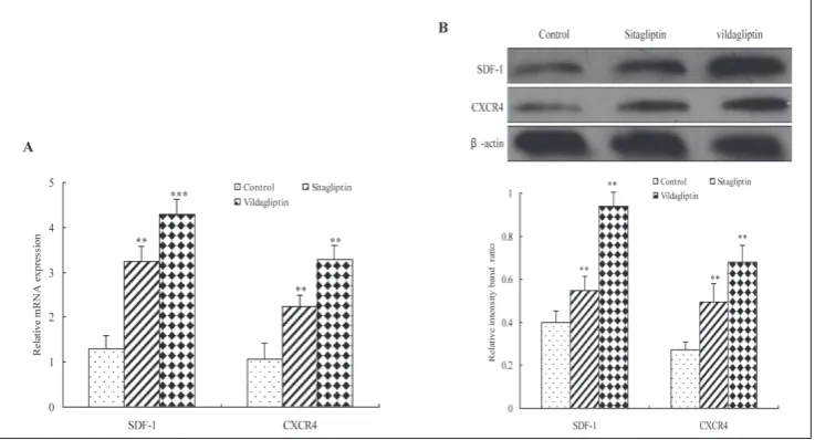
Effects of DPP4 inhibitors on the activation of the SDF-1/CXCR4 signaling pathway in human EPCs
To understand the molecular mechanism involved in the regulatory functions of DPP4 inhibitors, the alterations in the expression of SDF-1 and CXCR4 in
Fig. 3. The effect of DPP4 inhibitors on the apoptosis of human EPCs. Human EPCs were treated with 0, 0.01, 0.1, 1 and 10 μM of DPP4 inhibitors sitagliptin or vildagliptin for 48 h after which mRNA expression (A) and the activity (B) of caspase-3 in human EPCs were determined by RT-PCR and a caspase-3 kit. Data are expressed as the mean±SD of three independent experiments performed in triplicate. *P<0.05, **P<0.01, or P>0.05 vs control.
control and the 0.01-μM sitagliptin- or vildagliptin-treated groups (P>0.05). Moreover, compared with the control group, mRNA expression and caspase-3 activ-ity were significantly lower in the 0.1-, 1- and 10-μM sitagliptin- or vildagliptin-treated groups (P<0.05), and the effects were dose-dependent. These results indicated that DPP4 inhibitors could decrease the apoptosis of human EPCs.
Effects of DPP4 inhibitors on pro-angiogenic factors VEGF, VEGFR-2 and eNOS expression in human EPCs
human EPCs treated with 10 μM sitagliptin or vilda-gliptin for 48 h were assessed using real-time RT-PCR and Western blot analysis. We observed a marked increase in SDF-1 and CXCR4 mRNA expression (P<0.01) (Fig. 5A) and protein expression (P<0.05) (Fig. 5B) following the treatment of sitagliptin or vildagliptin. This indicated that DPP4 inhibitors could activate the SDF-1/CXCR4 signaling pathway in hu-man EPCs.
Role of the SDF-1/CXCR4 signaling pathway on the proliferation and apoptosis of DPP4 inhibitor-treated human EPCs
To evaluate the role of the SDF-1/CXCR4 signaling pathway in DPP4 inhibitor-triggered biological func-tions in human EPCs, the proliferation and apoptosis of EPCs and the expression of pro-angiogenic factors including VEGF, VEGFR2 and eNOS were investi-gated following the blocking of the SDF-1/CXCR4 sig-naling pathway activation by AMD3100. SDF-1 and CXCR4 protein expression was significantly down-regulated by AMD3100 (P<0.05), revealing the block-ing efficiency of the AMD3100 inhibitor (Fig. 6A).
Furthermore, the DPP4 inhibitors sitagliptin and vildagliptin promoted the proliferation and mRNA expression of VEGF, VEGFR2 and eNOS while inhib-iting the apoptosis of human EPCs (P<0.05). However, there was no significant difference in cell proliferation (Fig. 6B), apoptosis (Fig. 6C, D) of mRNA expres-sion of VEGF, VEGFR2 and eNOS (Fig. 6E) (P>0.05) between the control group and either the AMD3100 group or the group treated with the DPP4 inhibitor and AMD3100. Taken together, these results strongly suggest that the activation of the SDF-1/CXCR4 sig-naling pathway was required for the DPP4 inhibitor-triggered promotion of proliferation and decrease in apoptosis in human EPCs.
DISCUSSION
DPP4, with a multiplicity of functions and targets, can mediate the degradation of many chemokines and neuropeptides, play a critical role in providing costim-ulatory signals to T cells via adenosine deaminase and regulate inflammatory responses in innate immune cells such as monocytes and dendritic cells (Zhong et al., 2013). Numerous in vitro and in vivo studies have
suggested that DPP4 activity is correlated with obesity, diabetes, heart failure and renal disease (Jose et al., 2011; Zhong et al., 2013; Sato et al., 2014). A recent
in vitro study reported that DPP4 also possesses anti-thrombotic properties and may behave as an immobi-lized anticoagulant on endothelial cells (Hocher et al., 2012). DPP4 inhibitors such as sitagliptin, vildagliptin, saxagliptin, alogliptin and linagliptin have been used to cure diabetes, obesity, cardiovascular disease and kidney disease (Jose et al., 2011; Lamers et al., 2011; Zhong et al., 2013; Sato et al., 2014). In the present study, we evaluated the effects of DPP4 inhibitors on the biological functions of human EPCs and further investigated the mechanism of DPP4 inhibitors.
DPP4 expression and activity were found on the endothelium of intramyocardial blood vessels and en-dothelial cells (Hocher et al., 2012). Hyperglycemia is able to increase DPP4 activity and mRNA expression in a significant manner in microvascular endothelial
cells (Pala et al., 2012). DPP4 inhibitors are currently used as glucose-lowering agents in type 2 diabetes, due to their effects on insulin and glucagon secretion. Sev-eral prospective clinical trials in humans have proven that the DPP4 inhibitor dapagliflozin alone or in com-bination with metformin, would be a cost-effective al-ternative in the treatment of type-2 diabetes mellitus (Abad Paniagua et al., 2014; Kumar et al., 2014). The expression level of DPP4 was significantly decreased by the addition of sitagliptin in human embryonic kidney 293 (HEK293) cells, human renal cancer cells (Sato et al., 2014), microvascular endothelial cells, and human umbilical vein endothelial cells (Pala et al., 2012). This study also indicated that the DPP4 inhibitors sitagliptin and vildagliptin could successfully inhibit the expres-sion and enzyme activity of DPP4 in human EPCs.
1) and gastric inhibitory polypeptide (GIP), which caused an increase in glucose-dependent stimulation of insulin secretion, resulting in a lower blood glucose levels (Schmiedl et al., 2014). DPP4 inhibitors such as sitagliptin, vildagliptin, and saxagliptin worked by inducing a significant reduction in glycosylated he-moglobin levels (Doupis and Veves, 2008), slowing incretin metabolism, increasing endogenous GLP-1 concentrations and improving postprandial glycemic control in type 2 diabetes (Tomkin, 2014). Addition-ally, the DPP4 inhibitor vildagliptin preserved β-cell mass through an amelioration of endoplasmic reticu-lum stress in C/EBPB transgenic mice (Shimizu et al., 2012), and prevented neuronal insulin resistance by restoring insulin-induced long-term depression and neuronal IRS-1 phosphorylation, IR phosphorylation and Akt/PKB-Ser phosphorylation (Pipatpiboon et al., 2013). The DPP4 inhibitor linagliptin delayed the onset of diabetes and preserved β-cell mass in non-obese diabetic mice (Jelsing et al., 2012). In contrast, a study demonstrated that protein expressions of eNOS, CXCR4, SDF-1α and VEGF were remarkably higher in wild-type rats than in DPP4-deficient rats. Furthermore, vasorelaxation and nitric oxide produc-tion of the normal femoral artery were significantly reduced in DPP4-deficient than in wild-type Fischer rats, suggesting a positive role of DPP4 in maintain-ing vascular function and tissue perfusion in this experimental setting (Sun et al., 2013). The in vitro
studies demonstrated that DPP4-inhibitor treatment facilitated an increase in hormones glucagon-like pep-tide-2 (GLP-2) receptor levels, intestinal growth and intestinal epithelial cell proliferation (Sueyoshi et al., 2014). Besides, DPP4-inhibition treatment enhanced engraftment of mouse bone marrow hematopoietic stem cells (Broxmeyer et al., 2013), and treatment with GLP-1 was able to increase the proliferation of the vas-culoprotective EPCs as shown through an action on VEGF (Ku et al., 2011). Moreover, we found that the DPP4 inhibitors sitagliptin and vildagliptin promoted the proliferation of human EPCs and the expression of eNOS, CXCR4, SDF-1α, VEGFR-2 and VEGF in hu-man EPCs, while they inhibited the apoptosis of this kind of cells. Why there is a difference between in vivo
and in vitro studies still needs further investigation.
Acknowledgments: This work was supported by Jiangxi Provin-cial Natural Science Foundation (No.20114BAB215004). Conflict of interest disclosure: The authors declare that they have no conflict of interest.
REFERENCES
Abad Paniagua, E. J., Casado Escribano, P., Fernández Rodriguez, J. M., Morales Escobar, F. J., Betegón Nicolás, L., Sánchez-Covisa, J. and M. Brosa (2014). Cost-effectiveness analysis of dapagliflozin compared to DPP4 inhibitors and other oral antidiabetic drugs in the treatment of type-2 diabetes mellitus in Spain. Aten. Primaria. In press.
Augustyns, K., Bal, G., Thonus, G., Belyaev, A., Zhang, X. M., Bollaert, W., Lambeir, A. M., Durinx, C., Goossens, F. and
A. Haemers (1999). The unique properties of dipeptidyl-peptidase IV (DPP IV/CD26) and the therapeutic potential of DPP IV inhibitors. Current Med. Chem. 6, 311-327.
Broxmeyer, H. E. and L. M. Pelus (2014). Inhibition of DPP4/ CD26 and dmPGE₂ treatment enhances engraftment of mouse bone marrow hematopoietic stem cells. Blood. Cells. Mol. Dis. 53, 34-38.
Christopherson, K. W., Hangoc, G., Mantel, C. R., and H. E. Broxmeyer (2004). Modulation of hematopoietic stem cell homing and engraftment by CD26. Science. 305, 1000-1003.
Doupis, J. and A. Veves (2008). DPP4 inhibitors: a new approach in diabetes treatment. Adv. Ther. 25, 627-643.
Drucker, D. J. (2007). Dipeptidyl peptidase-4 inhibition and the treatment of type 2 diabetes: preclinical biology and mech-anisms of action. Diabetes Care. 30, 1335-1343.
Fadini, G. P., Boscaro, E., Albiero, M., Menegazzo, L., Frison, V., de Kreutzenberg, S., Agostini, C., Tiengo, A. and A. Avogaro
(2010). The oral dipeptidyl peptidase-4 inhibitor sitagliptin increases circulating endothelial progenitor cells in patients with type 2 diabetes: possible role of stromal-derived fac-tor-1alpha. Diabetes Care. 33, 1607-1609.
Fujiwara, K., Inoue, T., Yorifuji, N., Iguchi, M., Sakanaka, T., Nara-bayashi, K., Kakimoto, K., Nouda, S., Okada, T., Ishida, K., Abe, Y., Masuda, D., Takeuchi, T., Fukunishi, S., Umegaki, E., Akiba, Y., Kaunitz, J. D. and K. Higuchi (2015). Com-bined treatment with dipeptidyl peptidase 4 (DPP4) inhibi-tor sitagliptin and elemental diets reduced indomethacin-induced intestinal injury in rats via the increase of mucosal glucagon-like peptide-2 concentration. J. Clin. Biochem. Nutr. 56, 155-162.
Glorie, L. L., Verhulst, A., Matheeussen, V., Baerts, L., Magielse, J., Hermans, N., D’Haese, P. C., De Meester, I. and A. De Beuf
(2012). DPP4 inhibition improves functional outcome after renal ischemia-reperfusion injury. Am. J. Physiol. Renal. Physiol. 303, F681-688.
Guo, Y., Hangoc, G., Bian, H., Pelus, L. M. and H. E. Broxmeyer
(2005). SDF-1/CXCL12 enhances survival and chemotaxis of murine embryonic stem cells and production of primi-tive and definiprimi-tive hematopoietic progenitor cells. Stem. Cells. 23, 1324-1332.
Haider, H. K., Jiang, S., Idris, N. M. and M. Ashraf (2008). IGF-1-overexpressing mesenchymal stem cells accelerate bone marrow stem cell mobilization via paracrine activation of SDF-1alpha/CXCR4 signaling to promote myocardial repair. Circ. Res. 103, 1300-1308.
Herrera, C., Morimoto, C., Blanco, J., Mallol, J., Arenzana, F., Lluis, C. and R. Franco (2001). Comodulation of CXCR4 and CD26 in human lymphocytes. J. Biol. Chem. 276, 19532-19539.
Hocher, B., Reichetzeder, C. and M. L. Alter (2012). Renal and cardiac effects of DPP4 inhibitors--from preclinical devel-opment to clinical research. Kidney Blood Press. Res. 36, 65-84.
Huber, B. C., Brunner, S., Segeth, A., Nathan, P., Fischer, R., Zaruba, M. M., Vallaster, M., Theiss, H. D., David, R., Ger-bitz, A. and W. M. Franz (2011). Parathyroid hormone is a DPP-IV inhibitor and increases SDF-1-driven homing of CXCR4+ stem cells into the ischaemic heart. Cardiovasc. Res. 90, 529-537.
Imanishi, T., Tsujioka, H. and T. Akasaka (2008). Endothelial pro-genitor cells dysfunction and senescence: contribution to oxidative stress. Curr. Cardiol. Rev. 4, 275-286.
Jelsing, J., Vrang, N., van Witteloostuijn, S. B., Mark, M. and T. Klein (2012). The DPP4 inhibitor linagliptin delays the onset of diabetes and preserves β-cell mass in non-obese diabetic mice. J. Endocrinol. 214, 381-387.
Jose, T. and S. E. Inzucchi (2012). Cardiovascular effects of the DPP-4 inhibitors. Diabetes Vas. Dis. Res. 9, 109-116.
Kawamoto, A., Gwon, H. C., Iwaguro, H., Yamaguchi, J. I., Uchida, S. Masuda, H., Silver, M., Ma, H., Kearney, M., Isner, J. M. and T. Asahara (2001). Therapeutic Potential of Ex Vivo Expanded Endothelial Progenitor Cells for Myocardial Ischemia. Circulation. 103, 634-637.
Ku, H. C., Chen, W. P. and M. J. Su (2011) DPP4 deficiency preserves cardiac function via GLP-1 signaling in rats subjected to myocardial ischemia/reperfusion. Naunyn Schmiedebergs Arch. Pharmacol. 384, 197-207.
Kumar, K. V. and A. K. Gupta (2014). Clinical audit of patients using DPP4 inhibitors in longstanding type 2 diabetes. Dia-betes Metab. Syndr. In press.
Lambeir, A. M., Durinx, C., Scharpé, S. and I. De Meester (2003). Dipeptidyl-peptidase IV from bench to bedside: an update on structural properties, functions, and clinical aspects of the enzyme DPP IV. Crit. Rev. Clin. Lab. Sci. 40, 209-294.
Matteucci, E. and O. Giampietro (2009). Dipeptidyl peptidase-4 (CD26): knowing the function before inhibiting the enzyme. Curr. Med. Chem. 16, 2943-2951.
Nistala, R., Habibi, J., Aroor, A., Sowers, J. R., Hayden, M. R., Meuth, A., Knight, W.,Hancock, T., Klein, T., DeMarco, V. G. and A. Whaley-Connell (2014). DPP4 inhibition atten-uates filtration barrier injury and oxidant stress in the Zucker obese rat. Obesity (Silver Spring). 22, 2172-2179.
Nistala, R., Habibi, J., Lastra, G., Manrique, C., Aroor, A. R., Hay-den, M. R., Garro, M., Meuth, A., Johnson, M., Whaley-Connell, A. and J. R. Sowers (2014). Prevention of obesity-induced renal injury in male mice by DPP4 inhibition.
Endocrinology. 155, 2266-2276.
Pala, L., Pezzatini, A., Dicembrini, I., Ciani, S., Gelmini, S., Van-nelli, B. G., Cresci, B., Mannucci, E. and C. M. Rotella
(2012). Different modulation of dipeptidyl peptidase-4 activity between microvascular and macrovascular human endothelial cells. Acta Diabetologica. 49, S59-S63.
Peled, A., Grabovsky, V., Habler, L., Sandbank, J., Arenzana-Seisde-dos, F., Petit, I., Ben-Hur, H., Lapidot, T. and R. Alon (1999). The chemokine SDF-1 stimulates integrin-mediated arrest of CD34(+) cells on vascular endothelium under shear flow. J. Clin. Invest. 104, 1199-1211.
Peled, A., Petit, I., Kollet, O., Magid, M., Ponomaryov, T., Magid, M., Ponomaryov, T., Byk, T., Nagler, A., Ben-Hur, H., Many, A., Shultz, L., Lider, O., Alon, R., Zipori, D. and T. Lapidot
(1999). Dependence of human stem cell engraftment and repopulation of NOD/SCID mice on CXCR4. Science. 283, 845-848.
Pipatpiboon, N., Pintana, H., Pratchayasakul, W., Chattipakorn, N. and S. C. Chattipakorn (2013). DPP4-inhibitor improves neuronal insulin receptor function, brain mitochondrial function and cognitive function in rats with insulin resist-ance induced by high-fat diet consumption. Eur. J. Neuro-sci. 37, 839-849.
Rafii, S. and D. Lyden (2003). Therapeutic stem and progenitor cell transplantation for organ vascularization and regenera-tion. Nat. Med. 9, 702-712.
Rizzo, M., Rizvi, A. A., Spinas, G. A., Rini, G. B. and K. Berneis
(2009). Glucose lowering and anti-atherogenic effects of incretin-based therapies: GLP-1 analogues and DPP-4-inhibitors. Expert Opinion Invest. Drugs. 18, 1495-1503.
Sato, Y., Kamada, T. and A. Yamauchi (2014). The role of dipep-tidyl peptidase 4 (DPP4) in the preservation of renal func-tion: DPP4 involvement in hemoglobin expression. J. Endo-crinol. 223, 133-142.
Schmiedl, A., Grützner, D., Hoffmann, T., von Hörsten, S. and M. Stephan (2014). DPP4 inhibitors increase differentially the expression of surfactant proteins in Fischer 344 rats. Acta Physiol (Oxf).212, 248-261.
Segers, V. F. M., Revin, V., Wu, W., Qiu, H., Yan, Z., Lee, R. T. and A. Sandrasagra (2011). Protease-resistant stromal cell-derived factor-1 for the treatment of experimental periph-eral artery disease. Circulation. 123, 1306-1315.
Shimizu, S., Hosooka, T., Matsuda, T., Asahara, S., Koyanagi-Kimura, M., Kanno, A., Bartolome, A., Etoh, H., Fuchita, M., Teruyama, K., Takahashi, H., Inoue, H., Mieda, Y., Hashimoto, N., Seino, S. and Y. Kido (2012). DPP4 inhibitor vildagliptin preserves β-cell mass through amelioration of endoplasmic reticulum stress in C/EBPB transgenic mice.
J. Mol. Endocrinol. 49, 125-135.
Sueyoshi, R., Woods Ignatoski, K. M., Okawada, M., Hartmann, B., Holst, J. and D. H. Teitelbaum (2014). Stimulation of intesti-nal growth and function with DPP4 inhibition in a mouse short bowel syndrome model. Am. J. Physiol. Gastrointest. Liver. Physiol. 307, G410-419.
Sun, C. K., Leu, S., Sheu, J. J., Tsai, T. H., Sung, H. C., Chen, Y. L., Chung, S. Y., Ko, S. F., Chang, H. W. and H. K. Yip (2013). Paradoxical impairment of angiogenesis, endothelial func-tion and circulating number of endothelial progenitor cells in DPP4-deficient rat after critical limb ischemia. Stem. Cell. Res. Ther. 4, 31.
Tomkin, G. H. (2014). Treatment of type 2 diabetes, lifestyle, GLP1 agonists and DPP4 inhibitors. World. J. Diabetes. 5, 636-650.
Wang, C. H., Verma, S., Hsieh, I. C., Chen, Y. J., Kuo, L. T., Yang, N. I., Wang, S. Y., Wu, M. Y., Hsu, C. M., Cheng, C. W. and
W. J. Cherng (2006). Enalapril increases ischemia-induced endothelial progenitor cell mobilization through manipula-tion of the CD26 system. J. Mol. Cell. Cardiol. 41, 34-43.
Zhong, J., Rao, X., Deiuliis, J., Braunstein, Z., Narula, V., Hazey, J., Mikami, D., Needleman, B., Satoskar, A. R. and S. Rajagopa-lan (2013). A potential role for dendritic cell/macrophage-expressing DPP4 in obesity-induced visceral inflammation.
Diabetes. 62, 149-157.
Zhong, J., Rao, X. and S. Rajagopalan (2013). An emerging role of dipeptidyl peptidase 4 (DPP4) beyond glucose control: potential implications in cardiovascular disease. Atheroscle-rosis. 226, 305-314.
Zhou, X., Porter, A. L., Robinson, D. K., Shim, M. S. and Y. Guo
(2014). Nano-enabled drug delivery: a research profile.