551
N-CoR MODULATES OSTEOGENIC DIFFERENTIATION OF RAT MESENCHYMAL STEM
CELLS THROUGH THE PI3K/AKT-CELL SIGNALING PATHWAY
Yi Qin1,2, Guoping Cao3, Jichao Ye1, Peng Wang1, Liangbin Gao1, Suwei Wang2 and Huiyong Shen1,*
1Department of Orthopedics, Sun Yat-Sen Memorial Hospital, Sun Yat-Sen University, Guangzhou 510120, China 2Department of Orthopedics, Zhuhai People’s Hospital, Zhuhai 519000, China
3Guangzhou University of Traditional Chinese Medicine, Guangzhou 510405, China *Corresponding author: [email protected]
Received: September 1, 2015; Revised: October 16, 2015; Accepted: October 21, 2015; Published online: April 27, 2016
Abstract: The nuclear receptor corepressor (N-CoR) is involved in the regulation of diverse transcription factors. We pre-viously found that N-CoR could regulate adipogenic differentiation of rat mesenchymal stem cells (MSCs), but whether it modulated osteogenic differentiation of this type of cells was uncertain. Therefore, in the present study, we investigated the effect and mechanism of N-CoR on osteogenic differentiation of rat MSCs. The results showed that MSCs cultured in osteogenic medium successfully differentiated into osteogenic cells. Overexpression of N-CoR decreased cell proliferation, alkaline phosphatase (ALP) activity, calcium accumulation, mRNA expression of genes including bone sialoprotein (BSP), osteocalcin (OCN), osteopontin (OPN), Osterix and Runx2, and protein expression of phosphor-Akt (pAkt). Conversely, knocking down cellular N-CoR by small interfering RNA (siRNA) promoted pAkt activity and cell differentiation. Over-expression or knockdown of N-CoR had no significant influences on the protein Over-expression of pyruvate dehydrogenase kinase isozyme 1 (PDK1), phosphatidylinositol 3-kinase (PI3K) and total Akt, indicating that N-CoR regulated the changes in the PI3K/Akt signaling pathway by targeting pAkt. To further prove the function of the PI3K/Akt signaling in N-CoR-regulated osteogenic differentiation, we used the PI3K inhibitor (LY294002) to block the activation of the PI3K/Akt pathway and found that overexpression of N-CoR showed no effects on ALP activity, calcium level and mRNA expression of BSP, osteocalcin OCN, OPN, Osterix and Runx2 in rat MSCs following the inhibition of the PI3K/Akt pathway. These findings suggest that N-CoR regulates osteogenic differentiation of rat MSCs through suppression of the PI3K/Akt signaling pathway.
Key words: N-CoR; MSCs; osteogenic differentiation; PI3K; Akt
INTRODUCTION
Mesenchymal stem cells (MSCs) isolated from bone marrow with properties of low immunologic rejec-tion and self-renewal potential [1,2] can differentiate into a variety of cell types, including fibroblasts, adi-pocytes, osteoblasts, chondrocytes and myoblasts [3]. Osteogenesis of MSCs is a complex biological process that involves the differentiation of mesenchymal cells into preosteoblastic cells and mature osteoblasts, and finally results in the synthesis of bone matrix proteins and calcium deposition. Furthermore, osteoblastic differentiation from MSCs plays a crucial role in bone formation [4]. In order to treat osteoporosis and other
bone diseases, it is important to understand the roles of the factors that regulate differentiation of MSCs.
suppressed by cellular N-CoR siRNA knockdown [8], indicating a novel regulatory role of N-CoR.
PI3K/Akt signaling pathways exist in a large number of mammalian cells and exert multiple pro-survival functions, including cell proliferation, dif-ferentiation, survival, migration, invasion and me-tabolism. Studies proved that aberrant regulation of PI3K/Akt signaling led to diseases such as cancer and autoimmunity [9,10]. In the PI3K/Akt signaling pathway, the lipid kinase-PI3K generates phosphati-dylinositol-3,4,5-phosphate (PIP3), which further ac-tivates PDK1 and Akt [11]. After being fully activated by PDK1, Akt becomes powerful and translocates into the cell cytosol and nucleus, where it causes the ac-tivation of a variety of other important signals [12]. Previous results revealed N-CoR as a novel regulator of PI3K signaling, which modulates thyroid tumor progression [8]. A more recent study suggested that N-CoR negatively regulated adipogenic differentia-tion of rat MSCs [13]. However, whether N-CoR and its activated signaling pathways were involved in the osteogenic differentiation of rat MSCs remained un-certain. Therefore, in the present study, we made a mechanistic study of the proliferation and osteogenic differentiation of rat MSCs induced by N-CoR, which might provide a base knowledge for our understand-ing the correlation between N-CoR and its meditated PI3K/AKT-cell signaling pathway in the regulation of osteogenic differentiation of rat MSCs.
MATERIALS AND METHODS
Ethics statement
The in vivo experiment was carried out according to the recommendations in the Guide for the Care and Use of Laboratory Animals of the National Institutes of Health. The protocol was approved by the Shanghai Laboratory Animal Center (Permit Number: 15-632). All animals received humane care in compliance with the Guide for the Care and Use of Experimental Ani-mals (Animal Care Committee, 2002).
Isolation, culture and osteogenic differentiation induction of rat MSCs
Sixty 6-week-old Wistar rats (200±10 g) were purchased from the Shanghai Laboratory Animal Center, Chinese Academy of Sciences. Bone marrow cells were isolated from the bilateral femora and tibias of rats and incubat-ed in mincubat-edium containing an α-minimal essential mincubat-edi- medi-um (α-MEM, Invitrogen, CA, USA) supplemented with 10% fetal bovine serum (FBS) and 1% penicillin-strep-tomycin at 37°C in 5% CO2. When cells had grown to 90% confluency, they were cultivated as the first genera-tion. All cells used for this experiment were MSCs of the third generation. Rat MSCs of passage 3 were cultured in the osteogenic medium, which was α-MEM supple-mented with 10% FBS, 100 nM dexamethasone, 5 mM β-glycerophosphate (Sigma-Aldrich, St. Louis., MO, USA) and 50 μg/mL L-ascorbic acid (Sigma-Aldrich, St. Louis., MO, USA), as previously described [14]. Rat MSCs and MSC-derived cells in culture were identified through visualizing the morphology under an inverted microscope (Olympus, Tokyo, Japan). After culture in osteogenic medium for 1, 7, 14, 21 and 28 days, ALP activity and calcium accumulation were tested and the expression levels of BSP, OCN, OPN, Osterix and Runx2 gene expression were analyzed by real-time PCR.
XTT assay
The endogenous effects of N-CoR on cell viability were evaluated using the 2,3-Bis-(2-methoxy-4-ni-tro-5-sulfophenyl)-2H-tetrazolium-5-carboxanilide (XTT) Cell Viability Assay Kit (Sigma, CA, USA), as described [15]. Briefly, isolated and identified rat MSCs were seeded in 96-well plates at a density of 1.0×10⁶/mL in osteogenic medium. When cells
Real time RT-PCR
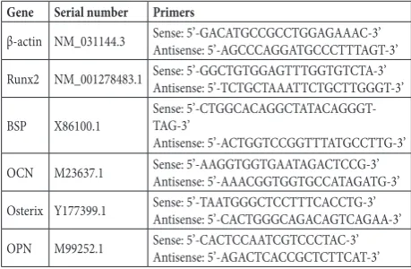
Expressions of BSP, OCN, OPN, Osterix and Runx2 in MSC-derived cells were measured by RNA preparation and quantitative reverse transcription polymerase chain reaction (RT-PCR). Total cellular RNA isolation (Invit-rogen, Carlsbad, CA, USA) and real-time RT-PCR anal-ysis (Qiagen, Valencia, CA) were performed according to the manufacturers’ instructions. The reaction run was 1 cycle of 56°C for 2 min and 94°C for 15 min, followed by 40 cycles of 94°C for 10 s, 58°C for 30 s and 72°C for 45 s. We used β-actin expression as an internal control. Specific primer sequences were synthesized in the BIO-SUNE Biological Technology Corp (Shanghai, China), and sequences of the primers are shown in Table 1. Western blot analysis
The Western blot method was used to analyze pro-tein expression of N-CoR, PI3K, PDK1, total Akt and pAkt. Briefly, proteins were separated by 10% sodium dodecyl sulfate-polyacrylamide gel electrophoresis (SDS-PAGE) and transferred to polyvinylidene fluo-ride (PVDF) membranes. Then the membranes were blocked in 5% non-fat milk for 2 h at room tempera-ture and incubated at 4°C overnight with polyclonal anti-N-CoR (1:1000 diluted; Sigma, CA, USA), PI3K (1:1000 diluted; Cell Signaling Technology, MA, USA), PDK1 (1:1000 diluted; Cell Signaling Technol-ogy, MA, USA), total Akt (1:1000 diluted; Cell Signal-ing Technology, MA, USA) and pAkt (1:1000 diluted; Cell Signaling Technology, MA, USA). After overnight
incubation, the membranes were immunoblotted with HRP-conjugated anti-rabbit IgG antibody (diluted 1:1000; Abcam, Cambridge, UK) at 37°C for 40 min, developed with enhanced chemiluminescence (ECL) substrate (Pierce, Wisconsin, USA) and exposed to X-ray film. β-actin (diluted 1/1000, Santa Cruz Bio-technology, CA, USA) was used to ensure adequate sample loading for all Western blots. Band density was quantitated using Image J software.
Small interference RNA (siRNA) assay
N-CoR-specific siRNA was as follows: GATCCAG-GAAGAGTGTTCCTGATTTTCAAGA (sense) GAAATCAGGAACACTCTTCCTTTTTTTGGAAA (anti-sense). For transient transfections, cells cultured in osteogenic medium were grown to 60% confluence and then transfected with 6 μg of N-CoR-specific siRNA construct or the negative control siRNA using HiPerFect (Qiagen, Valencia, CA, USA), according to the manufacturer’s protocols.
ALP activity
Measurement of cellular ALP activity was performed as previously described [16]. On days the experiments were performed, the medium was removed, and the cell layers were rinsed with PBS three times and lysed by lysis buffer containing 0.1% Triton X-100 (Sigma, CA, USA). Twenty μL of cell lysate was mixed with 100 μL of Tris-glycine buffer (PH 10.3) (BioRad, Her-cules, CA, USA), 2 mM MgCl2 (Sigma, CA, USA) and 100 Μl p-nitrophenyl phosphate (Sigma, CA, USA). The reaction mixture was incubated at 37°C for 30 min and the reaction was stop by adding 50 μL of NaOH. Absorbance was read at 405 nm using a mi-croplate reader (Olympus, Tokyo, Japan).
Calcium assay
Calcium deposition was measured in 0.5 N HCl ex-tracts according to the manufacturer’s instructions contained in Diagnostic Kit 587 (Sigma, CA, USA). Total calcium was expressed as μg/mg cellular protein. Cell numbers were determined using the nuclear dye, crystal violet [17].
Table 1. Primers used in this study
Gene Serial number Primers
β-actin NM_031144.3 Sense: 5’-GACATGCCGCCTGGAGAAAC-3’Antisense: 5’-AGCCCAGGATGCCCTTTAGT-3’ Runx2 NM_001278483.1 Sense: 5’-GGCTGTGGAGTTTGGTGTCTA-3’Antisense: 5’-TCTGCTAAATTCTGCTTGGGT-3’
Statistical analysis
Statistical analysis was carried out with one-way analy-sis of variance (ANOVA) using SPSS17.0 software. Val-ues are expressed as means±standard deviation (SD). The mean values and standard deviations were calcu-lated from three independent experiments. Differences were considered statistically significant at P<0.05.
RESULTS
Induction of rat MSC osteogenic differentiation
It was found that, as shown in Fig. 1A, MSCs were derived from rat bone marrow, and MSCs of the third generation (a in Fig. 1A) were successfully induced to differentiate into osteogenic cells (b and c in Fig. 1A) by adding osteogenic medium. Under this condition, the calcium deposition level, ALP activity and marker genes related to rat MSC osteogenic differentiation were measured at 1, 7, 14, 21 and 28 days. Calcium deposition was detected at 7 days, and increased mark-edly at 21 days (Fig. 1B) (P<0.05). ALP activity was increased at 7 days, while decreasing the following days (Fig. 1C) (P<0.05). Moreover, real-time RT-PCR analy-ses showed that mRNA expression of Runx2 (Fig. 1D) and Osterix (Fig. 1E) was found at 7 days and reached a peak at 14 days (P<0.05), and BSP (Fig. 1F), OCN (Fig. 1G) and OPN (Fig. 1H) expression was found at 21 days of osteogenic differentiation induction (P<0.05). Therefore, under the osteogenic induction condition, the onset of osteogenic differentiation of rat MSCs be-gan 7 days post induction initiation.
Effects of N-CoR on MSC-derived cell proliferation
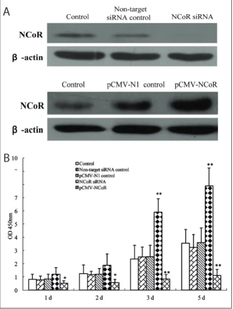
To further determine the individual roles of N-CoR in osteogenic differentiation of rat MSCs, we established osteogenic differentiation cells constitutively express-ing siRNA targetexpress-ing N-CoR and pCMV-N-CoR at 7 days when rat MSCs were induced to differentiate. After 2 days, the efficiency of knockdown and over-expression of N-CoR in osteogenically differentiated rat MSCs was tested by Western blot. It was seen that,
following N-CoR inhibition or overexpression, the protein expression of N-CoR was significantly de-creased as compared with cells that were transfected with non-targeting siRNA (Fig. 2A), and increased heavily in pCMV-N-CoR-treated cells in compari-son with the control (Fig. 2B). On days 1, 2, 3 and 5 after transfection, cell viability was detected by the XTT method. It was suggested that, compared to the control, XTT activity of cells in N-CoR siRNA was significantly promoted at the examined times, whilst it
was inhibited in the N-CoR overexpressing cells (Fig. 2C) (P<0.05).
Effects of N-CoR on osteogenic differentiation of rat MSCs
Following 21 days of osteogenic differentiation in-duction, cells were transfected with N-CoR siRNA, non-targeting N-CoR siRNA, pCMV-N-CoR and pCMV-N1. About 3 days later, calcium deposition, ALP activity and osteogenic differentiation related genes were measured by different methods. As shown
in Fig. 3, it demonstrated that, compared to the con-trol, overexpression of N-CoR in cells significantly abrogated calcium deposition (Fig. 3A), ALP activity (Fig. 3B), and Runx2, Osterix, BSP, OCN and OPN expression (Fig. 3C), whereas N-CoR siRNA induced higher activity of these indexes (P<0.05).
Effects of N-CoR on the activation of the PI3K/ Akt signaling pathway
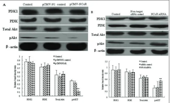
The effects of N-CoR on the activation of the PI3K/ Akt signaling pathway were determined by treatment
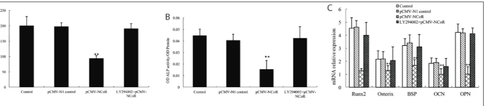
Fig. 3. Effects of siRNA knockdown or overexpression of cellular N-CoR on osteogenic differentiation of rat MSCs. On day 21, calcium deposition (A), ALP activity (B) and osteogenic differentiation-related marker genes Runx2, Osterix, BSP, OCN and OPN (C) signifi-cantly decreased in overexpressing N-CoR-treated cells, and increased in siRNA knockdown N-CoR-treated cells as compared with the control. Data are expressed as means±SD of three independent experiments in six replicates.
of cells with pCMV-N-CoR and N-CoR siRNA. The results showed that overexpression of N-CoR de-creased the expression of pAkt (Fig. 4A) (P<0.05), whereas N-CoR siRNA produced a promotion of pAkt as determined by Western blot (Fig. 4B) (P<0.05). To-tal Akt, PDK1 and PI3K in both pCMV-N-CoR cells and N-CoR siRNA cells showed no changes in com-parison with that in the control (P>0.05). Taken to-gether, these findings indicated that N-CoR influenced the activation of the PI3K/Akt signaling pathway. The PI3K/Akt pathway signaling is involved in N-CoR-dependent functions
To elucidate whether PI3K/Akt signaling is neces-sary for N-CoR-dependent osteoblast differentiation, LY294002, a selective inhibitor of PI3K, at a dose of 50 mM, was used to prevent the activation of PI3K/Akt signaling. pCMV-N-CoR or N-CoR siRNA was stably transfected in cells, and osteogenic differentiation was quantified after 2 days of stimulation. The results sug-gested that, compared to the control, overexpression of N-CoR heavily prohibited calcium deposition (Fig. 5A), ALP activity (Fig. 5B), and Runx2, Osterix, BSP, OCN and OPN mRNA expression (Fig. 5C) induced by the osteogenic medium (P<0.05). However, when PI3K/Akt signaling was blocked by LY294002, N-CoR overexpression showed no influences on calcium deposition, ALP activity and osteogenic differentia-tion-related gene expression (P>0.05). These findings indicated that PI3K/Akt signaling was involved in N-CoR-dependent rat MSCs osteoblast differentiation.
DISCUSSION
Mesenchymal stem cells (MSCs) isolated from bone marrow and other adult tissues are proved to be multipotent progenitor cells, and they can be differ-entiated into multiple cell lineages, including fat, bone, muscles, ligament, cartilage and neurons [18,19]. Re-cent studies have reported that several factors, such as bone morphogenetic proteins (BMPs) [20], osteogenic growth peptide (OGP) [21] and 1, 25-dihydroxyvita-min D3 [22] play an important role in the differen-tiation of MSCs. N-CoR was also found to interact with the ligand-binding domains of PPARδ as well as PPARγ and PPARα, indicating that N-CoR was in-volved in cell differentiation. Therefore, in the present study, the role and mechanism of N-CoR on osteo-genic differentiation of rat MSCs were investigated.
We found that rat MSCs began to differentiate after cells were cultured in osteogenic medium for 7 days. The XTT assay was used to determine cell vi-ability in osteogenically differentiated rat MSCs. It was indicated that N-CoR was able to negatively regulate cell proliferation. To further elucidate the function and mechanism of N-CoR, six marker genes related to osteoblast differentiation were investigated. ALP secreted by osteoblasts is an early marker frequently used to demonstrate osteoblast differentiation [23,24]. Runx2, which belongs to the Runx family, contributes to osteoblastic differentiation of multipotent mesen-chymal cells [25], and induces ALP activity and bone matrix protein expression in osteoblastic cells [26]. Furthermore, Runx2 regulates its target genes such as
BSP, OCN and OPN by binding the promoter region. OPN and Osterix act as direct downstream targets of Runx2 [27]. Osterix is also required for differentiation of preosteoblasts into mature osteoblasts [28]. OCN regulates osteoblast maturation and matrix minerali-zation at the late stage of bone formation [29]. This study suggested that N-CoR overexpression down-regulated ALP activity and Runx2, Osterix, BSP, OCN and OPN expression, while N-CoR knockdown ex-erted the contrary functions, indicating N-CoR nega-tively regulated osteogenic differentiation of rat MSCs. The PI3K/Akt signaling has been implicated in differentiation of cells such as skeletal muscle cells, adipocytes, osteoblasts, chondrocytes and myoblasts [30-32]. Furthermore, Akt1/Akt2 is the key element in osteoclast differentiation [33], and is involved in bone development [34]. We assessed three members of the PI3K/Akt signaling pathway in order to confirm pathway activation. PDK1, a novel serine/threonine kinase, can activate a group of protein kinases in the AGC kinase subfamily in response to different stimu-lation [35]. Numerous studies suggest that PDK1 has pivotal roles in mediating cell proliferation, migration and energy metabolism [36,37]. We found that overex-pression of N-CoR could inhibit protein exoverex-pression of pAkt, while knockdown of N-CoR could promote the expression of this protein, indicating the regulation of N-CoR in the PI3/Akt signaling pathway. We fur-ther hypothesized that this inactivation of the PI3K/ Akt pathway could be an important factor in N-CoR-exerted proliferation and differentiation of rat MSCs. To further verify our hypothesis, we used LY294002, the specific inhibitor of PI3K to block the activation of the PI3K/Akt pathway. We observed that N-CoR overexpression was capable of inhibiting osteogenic differentiation of rat MSCs, and that this effect was not significant following blockage of the PI3K/Akt pathway by LY294002, indicating that N-CoR could inhibit osteogenic differentiation of rat MSCs through suppression of the PI3K/Akt signaling pathway.
CONCLUSIONS
This study underscores the importance of N-CoR in regulating the differentiative responses of rat MSCs cultured in osteogenic medium. N-CoR could sup-press osteogenic differentiation of rat MSCs through targeting the PI3K/Akt signaling pathway. These ob-servations might provide a potential mechanism for modulating PI3K/Akt inactivation and the commit-ment of rat MSCs to osteogenic differentiation.
Authors’ contributions: Yi Qin and Guoping Cao designed this study, carried out Western blot analysis and drafted the manu-script; Yi Qin and Guoping Cao contributed equally to this work; Jichao Ye and Peng Wang performed RT-PCR and participated in the interpretation of data; Liangbin Gao and Suwei Wang main-tained the cell culture and performed data analysis; as the corre-sponding author of this study, Huiyong Shen participated in the design of the study and revised the manuscript. All authors read and approved this version to be published.
Conflict of interest disclosure: The authors declare that they have no competing interests.
REFERENCE
1. Ding DC, Shyu WC, Lin SZ. Mesenchymal stem cells. Cell Transplant. 2011;20:5-14.
2. Ma L, Feng XY, Cui BL, Law F, Jiang XW, Yang LY, Xie QD, Huang TH. Human umbilical cord Wharton’s Jelly-derived mesenchymal stem cells differentiation into nerve-like cells. Chin Med J (Engl). 2005;118:1987-93.
3. Caplan AI. Mesenchymal stem cells. J Ortho Res. 1991;9:641-50.
4. Aubin J, Triffitt JT. Mesenchymal stem cell and osteoblast Dif-ferentiation. In: Bilezikian J, Raisz LG, Rodan GA, editors. Principles of bone biology. San Diego: Academic Press; 2002. p. 59-82.
5. Hermanson O, Jepsen K, Rosenfeld MG. N-CoR controls differentiation of neural stem cells into astrocytes. Nature. 2002;419:934-9.
6. Sardi SP, Murtie J, Koirala S, Patten BA, Corfas G. Presenilin-dependent ErbB4 nuclear signaling regulates the timing of astrogenesis in the developing brain. Cell. 2006;127:185-97. 7. Horlein AJ, Naar AM, Heinzel T, Torchia J, Gloss B, Kurokawa
R. Ligand-independent repression by the thyroid hormone receptor mediated by a nuclear receptor co-repressor. Nature. 1995;377:397-404.
9. Jiang BH, Liu LZ. PI3K/PTEN signaling in angiogenesis and tumorigenesis. Adv Cancer Res. 2009;102:19-65.
10. Oak JS, Fruman DA. Role of phosphoinositide 3-kinase sig-naling in autoimmunity. Autoimmunity. 2007;40:433-41. 11. Akanbi KA, Brodie AE, Suryawan A, Hu CY. Effect of age on
the differentiation of porcine adipose stromal-vascular cells in culture. J Anim Sci. 1994;72(11):2828-35.
12. Kim EH, Suresh M. Role of PI3K/Akt signaling in memory CD8 T cell differentiation. Front Immunol. 2013;4:20. 13. Gao HW, Liu Lan, Xing DG, Liu ZH, Ren P, Li ZQ, Shan GQ,
Gong MZ. NCoR negatively regulates adipogenic differentia-tion of mesenchymal stem cells. In Vitro Cell Dev Biol Anim. 2015;51(7):749-58
14. Li B, Dong Z, Gao L, He X, Liao L, Hu C, Wang Q, Jin Y. Lipopolysaccharide differentially affects the osteogenic dif-ferentiation of periodontal ligament stem cells and bone marrow mesenchymal stem cells through Toll-like receptor 4 mediated nuclear factor κB pathway. Stem Cell Res Ther. 2014;5(3):67.
15. Huyck L, Ampe C, Van Troys M. The XTT cell proliferation assay applied to cell layers embedded in three-dimensional matrix. Assay Drug Dev Technol. 2012;10(4):382-92. 16. Son KM, Park HC, Kim NR, Lee IS, Yang HC. Enhancement
of the ALP activity of C3H10T1/2 cells by the combination of an oxysterol and apatite. Biomed Mater. 2010;5(4):044107. 17. Westergren-Thorsson G, Onnervik PO, Fransson LA, Malm-ström A. Proliferation of cultured fibroblasts is inhibited by L-iduronate-containing glycosaminoglycans. J Cell Physiol. 1991;147(3):523-30.
18. Pittenger MF, Mackay AM, Beck SC, Jaiswal RK, Douglas R, Mosca JM, Moorman MA, Simonetti DW, Craig S, Marshak DR. Multilineage potential of adult human mesenchymal stem cells. Science. 1999;284:143-7.
19. Wang YK, Chen CS. Cell Adhesion and mechanical stimula-tion in the regulastimula-tion of mesenchymal stem cell differentia-tion. Cell Mol Med. 2013;17(7):823-32.
20. Chen D, Ji X, Harris MA, Feng JQ, Karsenty G, Celeste AJ, Rosen V, Mundy GR, Harris SE. Differential roles for bone morphogenetic protein (BMP) receptor type IB and IA in differentiation and specification of mesenchymal precur-sor cells to osteoblast and adipocyte lineages. J Cell Biol. 1998;142:295-305.
21. Tontonoz P, Hu E, Spiegelman B. Regulation of adipocyte gene expression and differentiation by peroxisome prolif-erator activated receptor gamma. Curr Opin Genet Dev. 1995;5:571-6.
22. Kveiborg M, Flyvbjerg A, Eriksen EF, Kassem M. 1,25-Dihy-droxyvitamin D3 stimulates the production of insulin-like growth factor-binding proteins-2, -3 and -4 in human bone marrow stromal cells. Eur J Endocrinol. 2001;144(5):549-57. 23. Allen MJ. Biochemical markers of bone metabolism in ani-mals: uses and limitations. Vet Clin Pathol. 2003;32:101-13.
24. Sila-Asna M, Bunyaratvej A, Maeda S, Kitaguchi H, Bunyarat-avej N. Osteoblast differentiation and bone formation gene expression in strontiunm-inducing bone marrow mesenchy-mal stem cell. Kobe J Med Sci. 2007;53:25-35.
25. Komori T. Runx2, a multifunctional transcription factor in skeletal development. J Cell Biochem. 2002;87:1-8.
26. Harada H, Tagashira S, Fujiwara M, Ogawa S, Katsumata T, Yamaguchi A. Cbfa1 isoforms exert functional differences in osteoblast differentiation. J Biol Chem. 1999;274:6972-8. 27. Westendorf JJ, Zaidi SK, Cascino JE, Kahler R, van Wijnen AJ.
Runx2 (Cbfa 1, AML-3) interacts with histone deacetylase 6 and represses the p21 (CIP1/WAF1) promoter. Mol Cell Biol. 2002;22(22):7982-92.
28. Zhang C. Transcriptional regulation of bone formation by the osteoblast specific transcription factor Osx. J Orthop Surg Res. 2010;5:37.
29. Peng S, Zhou G, Luk KD, Cheung KM, Li Z. Strontium pro-motes osteogenic differentiation of mesenchymal stem cells through the Ras/MAPK signaling pathway. Cell Physiol Bio-chem. 2008;23:165-74.
30. Ghosh-Choudhury N, Abboud SL, Nishimura R, Celeste A, Mahimainathan L, Choudhury GG. Requirement of BMP-2-induced phosphatidylinositol 3-kinase and Akt serine/threo-nine kinase in osteoblast differentiation and Smad-dependent BMP-2 gene transcription. J Biol Chem. 2002;277:33361-8. 31. Kaliman P, Vinals F, Testar X, Palacin M, Zorzano A.
Phos-phatidylinositol 3-kinase inhibitors block differentiation of skeletal muscle cells. J Biol Chem. 1996;271:19146-51. 32. Sakaue H, Ogawa W, Matsumoto M, Kuroda S, Takata M,
Sugimoto T, Spiegelman BM, Kasuga M. Posttranscriptional control of adipocyte differentiation through activation of phosphoinositide 3-kinase. J Biol Chem. 1998;273:28945-52. 33. Sugatani T, Hruska KA. Akt1/Akt2 and mammalian target
of rapamycin/Bim play critical roles in osteoclast differentia-tion and survival, respectively, whereas Akt is dispensable for cell survival in isolated osteoclast precursors. J Biol Chem. 2005;280(5):3583-9.
34. Peng XD, Xu PZ, Chen ML, Hahn-Windgassen A, Skeen J, Jacobs J. Dwarfism, impaired skin development, skeletal muscle atrophy, delayed bone development, and impeded adipogenesis in mice lacking Akt1 and Akt2. Genes Dev. 2003;17:1352-65.
35. Alessi DR, Deak M, Casamayor A, Caudwell FB, Morrice N, Norman DG. 3-Phosphoinositide-dependent protein kinase-1 (PDK1): structural and functional homology with the Dro-sophila DSTPK61 kinase. Curr Biol. 1997;7(10):776-89. 36. Mora A, Komander D, van Aalten DM, Alessi DR. PDK1, the
master regulator of AGC kinase signal transduction. Semin Cell Dev Biol. 2004;15(2):161-70.