An Updated Report by the American Society of
Anesthesiologists Task Force on Perioperative Visual Loss
P
RACTICE Advisories are systematically developed re-ports that are intended to assist decision-making in ar-eas of patient care. Advisories provide a synthesis and analysis of expert opinion, clinical feasibility data, open forum com-mentary, and consensus surveys. Practice Advisories devel-oped by the American Society of Anesthesiologists (ASA) are not intended as standards, guidelines, or absolute require-ments, and their use cannot guarantee any specific outcome. They may be adopted, modified, or rejected according to clinical needs and constraints and are not intended to replace local institutional policies.Practice Advisories are not supported by scientific litera-ture to the same degree as standards or guidelines because of the lack of sufficient numbers of adequately controlled stud-ies. Practice Advisories are subject to periodic update or
re-vision as warranted by the evolution of medical knowledge, technology, and practice.
This document updates the “Practice Advisory for Peri-operative Visual Loss Associated with Spine Surgery: A Re-port by the American Society of Anesthesiologists Task Force on Perioperative Blindness,” adopted by the ASA in 2005 and published in 2006.*
Methodology
A. Definition of Perioperative Visual loss
Visual loss after spine surgery is an uncommon occur-rence.1–3Ophthalmic complications have been reported to occur in less than 0.2% of spine surgeries.4 – 8For this Advisory,perioperative visual lossrefers to permanent im-pairment or total loss of sight associated with a spine procedure during which general anesthesia is adminis-tered. The perioperative period includes the time period from the immediate preoperative assessment through dis-charge from the acute healthcare facility. Conditions ad-dressed in this Advisory include posterior ischemic optic neuropathy (ION), anterior ION, and central retinal ar-tery occlusion (CRAO). “High-risk patients” are defined as those who undergo spine procedures while positioned
Updated by the Committee on Standards and Practice Parame-ters, Jeffrey L. Apfelbaum, M.D. (Committee Chair), Chicago, Illi-nois; Steven Roth, M.D. (Task Force Chair), Chicago, IlliIlli-nois; Rich-ard T. Connis, Ph.D., Woodinville, Washington; Karen B. Domino, M.D., Seattle, Washington; Lorri A. Lee, M.D., Seattle, Washington; David G. Nickinovich, Ph.D., Bellevue, Washington; and Mark A. Warner, M.D., Rochester, Minnesota. The original document was developed by the American Society of Anesthesiologists Task Force on Perioperative Visual Loss: Mark A. Warner, M.D. (Chair), Roch-ester, Minnesota; James F. Arens, M.D., Houston, Texas; Richard T. Connis, Ph.D., Woodinville, Washington; Karen B. Domino, M.D., Seattle, Washington; Lorri A. Lee, M.D., Seattle, Washington; Neil Miller, M.D., Baltimore, Maryland; Sohail Mirza, M.D., Seattle, Wash-ington; Nancy Newman, M.D., Atlanta, Georgia; David G. Nickinov-ich, Ph.D., Bellevue, Washington; Steven Roth, M.D., Chicago, Illi-nois; Peter Savino, M.D., Philadelphia, Pennsylvania; and Philip Weinstein, M.D., San Francisco, California.
Received from the American Society of Anesthesiologists, Park Ridge, Illinois. Submitted for publication October 20, 2011. Ac-cepted for publication October 20, 2011. Supported by the Ameri-can Society of Anesthesiologists and developed under the direction of the Committee on Standards and Practice Parameters, Jeffrey L. Apfelbaum, M.D. (Chair). Approved by the ASA House of Delegates on October 19, 2011. The original Advisory was endorsed by the North American Neuro-Ophthalmology Society and supported by the North American Spine Society. A complete bibliography used to develop this updated Advisory, arranged alphabetically by author, is available as Supplemental Digital Content 1, http://links.lww.com/ALN/A785.
Address correspondence to the American Society of Anesthesi-ologists: 520 North Northwest Highway, Park Ridge, Illinois 60068-2573. This Practice Advisory, as well as all published ASA Practice Parameters, may be obtained at no cost through the Journal Web site, www.anesthesiology.org.
* American Society of Anesthesiologists Task Force on Periop-erative Visual Loss: Practice Advisory for PeriopPeriop-erative Visual Loss Associated with Spine Surgery: A Report by the American Society of Anesthesiologists Task Force on Perioperative Blindness. ANESTHESIOLOGY 2006; 104:1319 –28.
Copyright © 2012, the American Society of Anesthesiologists, Inc. Lippincott Williams & Wilkins.Anesthesiology 2012; 116:274 – 85
• What other guideline statements are available on this topic? XThis Practice Advisory updates the “Practice Advisory for
Perioperative Visual Loss Associated with Spine Surgery,” adopted by the ASA in 2005 and published in 2006.*
• Why was this Advisory developed?
XIn October 2010, the Committee on Standards and Practice Parameters elected to collect new evidence to determine whether recommendations in the existing Practice Advisory were supported by current evidence.
• How does this Advisory differ from existing guidelines? XNew evidence presented includes an updated evaluation of
scientific literature. The new findings did not necessitate a change in recommendations.
• Why does this Advisory differ from existing guidelines? XThe ASA advisory differs from the existing guidelines
be-cause it provides new evidence obtained from recent sci-entific literature.
Supplemental digital content is available for this article. Direct URL citations appear in the printed text and are available in both the HTML and PDF versions of this article. Links to the digital files are provided in the HTML text of this article on the Journal’s Web site (www.anesthesiology.org).
prone and who have prolonged procedures, experience substantial blood loss, or both.
B. Purpose of the Advisory
The purpose of this Advisory is to enhance awareness and reduce the frequency of perioperative visual loss.
C. Focus
This Advisory focuses on the perioperative management of patients who are undergoing spine procedures while they are positioned prone and receiving general anesthesia. This Ad-visory does not address the perioperative management of patients who receive regional anesthesia or sedation. This Advisory also does not include other causes of visual loss, such as cortical blindness. It does not include nonspine sur-gical procedures (e.g., cardiac surgery, radical neck dissec-tion). In addition, this Advisory does not apply to young children because of the rarity of visual loss in children younger than 12 years of age undergoing spine surgery. D. Application
This Advisory is intended for use by anesthesiologists, spine surgeons, and all other individuals who deliver or who are responsible for anesthesia or perioperative care. These indi-viduals may include orthopedic surgeons, neurosurgeons, ophthalmologists, neuro-ophthalmologists, neurologists, nurse anesthetists, perioperative nurses, and anesthesiology assistants. The Advisory may also serve as a resource for other physicians, nurses, and healthcare professionals who manage anesthetized patients.
E. Task Force Members and Consultants
The original Advisory was developed by an ASA-appointed task force of 10 members, consisting of four anesthesiologists from various geographic areas of the United States, three neuro-ophthalmologists (one neurologist, two ophthalmol-ogists), an orthopedic spine surgeon, a neurosurgeon, and two methodologists from the ASA Committee on Standards and Practice Parameters. Three physicians served as official liaisons from national organizations. They included a neuro-ophthalmologist (North American Neuro-Ophthalmology Society [NANOS]), an orthopedic surgeon (American Acad-emy of Orthopaedic Surgeons), and a neurosurgeon (Amer-ican Association of Neurological Surgeons).
The Task Force developed the original Advisory by means of a six-step process. First, it reached consensus on the criteria for evidence of effective perioperative interventions for the prevention of visual loss. Second, original published articles from peer-reviewed journals relevant to perioperative visual
loss were evaluated. Third, consultants who had expertise or interest in perioperative visual loss and who practiced or worked in various settings (e.g., academic and private prac-tice) were asked to: (1) participate in opinion surveys on the effectiveness of various perioperative management strategies, and (2) review and comment on a draft of the Advisory developed by the Task Force. Fourth, additional opinions were solicited from active members of the Society for Neu-roscience in Anesthesiology and Critical Care (SNACC), NANOS, and the North American Spine Society (NASS). Fifth, the Task Force held an open forum at a national anes-thesia meeting to solicit input on the key concepts of this Advisory.† Sixth, all available information was used to build consensus within the Task Force on the Advisory. A sum-mary of recommendations may be found in appendix 1.
The draft document was made available for review on the ASA website, and input was invited viae-mail announce-ment to all ASA members. All submitted comannounce-ments were considered by the Task Force in preparing the final draft.
In 2009, the ASA Committee on Standards and Practice Parameters requested that scientific evidence for this Advisory be updated. The update consists of an evaluation of literature published after completion of the original Advisory.
F. Availability and Strength of Evidence
Preparation of this update used the same methodological process as was used in the original Advisory to obtain new scientific evidence. Opinion-based evidence obtained from the original Advisory is reported in this update. The protocol for reporting each source of evidence is described below. G. Scientific Evidence
Study findings from published scientific literature were ag-gregated and are reported in summary form by evidence cat-egory, as described below. All literature (e.g., randomized controlled trials, observational studies, case reports) relevant to each topic was considered when evaluating the findings. However, for reporting purposes in this document, only the highest level of evidence (i.e., level 1, 2, or 3 identified below) within each category (i.e., A, B, or C) is included in the summary.
Category A: Supportive Literature. Randomized controlled trials report statistically significant (P⬍0.01) differences be-tween clinical interventions for a specified clinical outcome.
Level 1: The literature contains multiple randomized con-trolled trials, and the aggregated findings are supported by meta-analysis.‡
Level 2: The literature contains multiple randomized con-trolled trials, but there is an insufficient number of studies to conduct a viable meta-analysis.
Level 3: The literature contains a single randomized con-trolled trial.
Category B: Suggestive Literature.Information from obser-vational studies permits inference of beneficial or harmful rela-tionships among clinical interventions and clinical outcomes.
† Society for Ambulatory Anesthesia, 20th Annual Meeting, May 13, 2005, Scottsdale, Arizona.
‡ Practice Advisories lack the support of a sufficient number of ade-quately controlled studies required to conduct an appropriate meta-anal-ysis. Therefore, Categories A1 and C1 evidence are not reported in this document.
Level 1: The literature contains observational compari-sons (e.g., cohort, case-control research designs) of clinical interventions or conditions and indicates statistically signif-icant differences between clinical interventions for a specified clinical outcome.
Level 2: The literature contains noncomparative observa-tional studies with associative (e.g., relative risk, correlation) or descriptive statistics.
Level 3: The literature contains case reports.
Category C: Equivocal Literature. The literature cannot determine whether there are beneficial or harmful relation-ships among clinical interventions and clinical outcomes.
Level 1: Meta-analysis did not find significant differences among groups or conditions.
Level 2: The number of studies is insufficient to conduct meta-analysis, and (1) randomized controlled trials have not found significant differences among groups or conditions or (2) randomized controlled trials report inconsistent findings. Level 3: Observational studies report inconsistent findings or donotpermit inference of beneficial or harmful relationships.
Category D: Insufficient Evidence from Literature.Thelack of scientific evidence in the literature is described by the fol-lowing terms.
Inadequate: The available literature cannot be used to assess relationships among clinical interventions and clinical out-comes. The literature either does not meet the criteria for con-tent as defined in the “Focus” of the Advisory or does not permit a clear interpretation of findings due to methodological con-cerns (e.g., confounding in study design or implementation).
Silent: No identified studies address the specified rela-tionships among interventions and outcomes.
H. Opinion-based Evidence
The original Advisory contained formal survey information collected from expert consultants and samples of active members of the SNACC, NANOS, and the NASS. Addi-tional information was obtained from open forum presenta-tions and other invited and public sources. All opinion-based evidence relevant to each topic (e.g., survey data, open-forum testimony, Internet-based comments, letters, editorials) was considered in the development of the original Advisory. However, only the findings obtained from formal surveys are reported.
Survey responses from Task Force-appointed expert con-sultants and specialty society members obtained during de-velopment of the original Advisory are summarized in the text and reported in appendix 2, tables 1– 4.
Responses were solicited from four response categories: agree, equivocal, disagree, and no opinion. Survey informa-tion is summarized in the text based on modal responses (e.g.,
a modal response of “agree” will be listed in the text as an agreement).
Advisories
I. Preoperative Patient Evaluation and Preparation There are no clinical trials addressing the impact of perform-ing a focused preoperative evaluation for perioperative visual loss.§However, two observational studies report that preop-erative anemia, vascular risk factors (e.g., hypertension, dia-betes, peripheral vascular disease, coronary artery disease), obesity, and tobacco use may be associated with periopera-tive visual loss (Category B2 evidence).5,9
Case reports suggest that perioperative visual loss may occur after prolonged procedures10 –17or substantial intra-operative blood loss18 –25(Category B3 evidence). In addition, observational studies report visual loss among patients un-dergoing prolonged procedures during which substantial blood loss occurred (Category B2 evidence).5,26,27
The consultants and specialty society members disagree that an ophthalmic or neuro-ophthalmic evaluation is effec-tive in identifying patients at risk for perioperaeffec-tive visual loss. The consultants and specialty society members agree that vascular risk factors increase the risk of perioperative visual loss. In addition, they agree that (1) the preoperative pres-ence of anemia, (2) prolonged procedures, (3) substantial blood loss, and (4) prolonged procedures combined with substantial blood loss all increase the risk of perioperative visual loss. The consultants and specialty society members consider procedures to be prolonged when they exceed an average of 6.5 h (range, 2–12 h) duration. They consider blood loss to be substantial when the loss reaches an average of 44.7% (range, 10 –200%) of estimated blood volume. Advisory for Preoperative Patient Evaluation and Prepa-ration.Although the consultants and specialty society mem-bers agree that there are identifiable preoperative risk factors, at this time the Task Force does not believe that there are identifiable preoperative patient characteristics that predis-pose patients to perioperative ION. In addition, the Task Force believes that there is no evidence that an ophthalmic or neuro-ophthalmic evaluation would be useful in identifying patients at risk for perioperative visual loss. The Task Force believes that the risk of perioperative ION may be increased in patients who undergo prolonged procedures, have sub-stantial blood loss, or both.储Consider informing patients in whom prolonged procedures, substantial blood loss, or both are anticipated that there is a small, unpredictable risk of perioperative visual loss. Because the frequency of visual loss after spine surgery of short duration is very low, the decision to inform patients who arenotanticipated to be “high risk” for visual loss should be determined on a case-by-case basis. II. Intraoperative Management
Intraoperative management consists of (1) blood pressure management, (2) management of intraoperative fluids, (3) §Refer to appendix 2 for details of the literature review and data
analyses.
储For the purposes of this Advisory, the Task Force considers such patients (hereafter referred to as “high-risk patients”) to have a higher risk for perioperative visual loss than patients who do not undergo prolonged procedures, have substantial blood loss, or both.
management of anemia, (4) use of vasopressors, (5) patient positioning, and (6) staging of surgical procedures.
Blood Pressure Management.Blood pressure management of high-risk patients depends on multiple patient characteristics, such as the preoperative presence of chronic hypertension, car-diac dysfunction, and renal and vascular disease. In addition, there are many intraoperative factors, such as fluid manage-ment, rate of blood loss, hypotension, and administration of vasopressors, that affect blood pressure management. Case re-ports indicate perioperative visual loss occurring after proce-dures in which intraoperative hypotension was maintained for patients without hypertension10,19,20,28 –30or for patients with
well-controlled chronic hypertension30,31(Category B3 evidence).
The Consultants and specialty society members disagree with the survey statement “Deliberate hypotension tech-niques may be used in high-risk patients” (i.e., for high-risk patients without preoperative chronic hypertension or for high-risk patients with well-controlled preoperative chronic hypertension). However, NASS members are split equally in their opinions between agree and disagree for patients with-outpreoperative chronic hypertension. Consultants and spe-cialty society members who agree that deliberate hypoten-sion may be used in patients withoutpreoperative chronic hypertension indicate that blood pressure should be main-tained on average within 24% (range, 0 – 40%) of estimated baseline mean arterial pressure or with a minimum systolic blood pressure of 84 mmHg (range, 50 –120 mmHg). Advisory for Blood Pressure Management.Systemic blood pressure should be monitored continually in high-risk pa-tients. The Task Force believes that the use of deliberate hy-potensive techniques during spine surgery has not been shown to be associated with the development of perioperative visual loss. Therefore, the use of deliberate hypotension for these pa-tients should be determined on a case-by-case basis.
Management of Intraoperative Fluids.The literature is in-sufficient to assess the relationship between the monitoring of intravascular volume and the occurrence of visual loss among spine surgery patients (Category D evidence). Although the use of large volumes of crystalloids may be associated with increased intraoperative ocular pressure, periorbital edema, and double-vision in patients undergoing cardiopulmonary bypass,32 the
literature is insufficient to address these issues in spine surgery patients (Category D evidence).
The consultants, SNACC, NANOS, and NASS members agree that intravascular volume should be monitored contin-ually in high-risk patients. The consultants, SNACC mem-bers, and NANOS members agree that the balance between colloid and crystalloid fluid resuscitation and replacement has an impact on the potential for perioperative vision loss; the NASS members report no opinion. The Consultants and SNACC members are equivocal regarding the preference of colloids over crystalloids for fluid resuscitation and replace-ment to reduce the potential for perioperative vision loss; the
NANOS and NASS members report no opinion. The Con-sultants, SNACC members, and NASS members agree that central venous pressure monitoring should be used in high-risk patients; the NANOS members report no opinion. Advisory for Management of Intraoperative Fluids.Central venous pressure monitoring should be considered in high-risk patients. Colloids should be used along with crystalloids to maintain intravascular volume in patients who have sub-stantial blood loss.
Management of Anemia. The literature is insufficient to evaluate the efficacy of intraoperative management of anemia during spine surgery (Category D evidence). One retrospective comparison of patients who experienced perioperative visual loss after spine surgery with a matched control group found no difference in lowest recorded hematocrit values between groups27(Category B2 evidence).
The consultants and specialty society members agree that hemoglobin or hematocrit values should be monitored peri-odically to detect anemia in high-risk patients. Those who agree indicate that intraoperative hemoglobin or hematocrit should be maintained at a minimum average of 9.4 g/dl (range, 6 –13 g/dl) or 28% (range, 18 –37%), respectively. Advisory for Management of Anemia.Hemoglobin or he-matocrit values should be monitored periodically during sur-gery in high-risk patients who experience substantial blood loss. The Task Force believes that there is no documented lower limit of hemoglobin concentration that has been asso-ciated with the development of perioperative visual loss. Therefore, the Task Force believes a transfusion threshold that would eliminate the risk of perioperative visual loss re-lated to anemia cannot be established at this time.
Use of Vasopressors.The literature is insufficient to evalu-ate the prolonged use of high-dose ␣-adrenergic agonists during spine surgery (Category D evidence). The SNACC members agree that prolonged use of high-dose␣-adrenergic agonists may reduce perfusion of the optic nerve in high-risk patients; the consultants are equivocal, and the NANOS and NASS members report no opinion.
Advisory for Use of Vasopressors.The Task Force consen-sus is that there is insufficient evidence to provide guidance for the use of␣-adrenergic agonists in high-risk patients dur-ing spine surgery. Therefore, the decision to use␣-adrenergic agonists should be made on a case-by-case basis.
Patient Positioning.Case reports suggest that patient posi-tioning resulting in direct pressure to eyes (e.g., from the use of a headrest, sheet roll, or other device) may precede the onset of CRAO or retinal ischemia in spine surgery patients (Category B3 evidence).#13–16,25,33– 42 One observational study indicates that, in 19% of ION cases listed in the ASA Visual Loss Registry, patient head position was maintained with Mayfield pins with the eyes free of pressure9(Category
B2 evidence).
The consultants and specialty society members agree that direct pressure on the eye should be avoided to reduce the risk of CRAO and other ocular damage. The consultants and
# Although observational literature on the pathophysiology of retinal vascular occlusion in humans is lacking, animal studies are available.
SNACC members agree that the patient’s head should be positioned level with or higher than the heart in high-risk patients; NANOS member opinion is split equally between agree and equivocal, and NASS member opinion is split equally among agree, equivocal, and no opinion. The con-sultants, SNACC members, and NASS members agree that the patient’s head should be placed in a neutral forward po-sition in high-risk patients; the NANOS members report no opinion. The consultants, SNACC, and NANOS members agree that the type of head positioning device is not associ-ated with perioperative ION; the NASS members disagree. The consultants and all specialty society members agree that the use of a horseshoe headrest may increase the risk of ocular compression and perioperative CRAO. They all agree that the eyes of prone-positioned patients should be assessed reg-ularly and documented. In addition, they all agree that peri-operative facial edema is common in high-risk patients. Advisory for Patient Positioning. The Task Force believes that there is no pathophysiologic mechanism by which facial edema can cause perioperative ION. There is no evidence that ocular compression causes isolated perioperative ante-rior ION or posteante-rior ION. However, direct pressure on the eye should be avoided to prevent CRAO. The high-risk pa-tient should be positioned so that the head is level with or higher than the heart when possible. The high-risk patient’s head should be maintained in a neutral forward position (e.g., without significant neck flexion, extension, lateral flex-ion, or rotation) when possible.
Staging of Surgical Procedures.The majority of spine sur-gery patients who experience perioperative ION undergo pro-longed procedures with substantial blood loss while they are positioned prone. Although the literature is insufficient to ex-amine the impact of surgical staging on reducing the frequency of perioperative visual loss in spine surgery patients (Category D evidence), an observational study indicates that, in 94% of ION cases listed in the ASA Visual Loss Registry, anesthetic duration exceeded 6 h9(Category B2 evidence). A related retrospective
study reported an association between duration of anesthesia and frequency of eye injury after a mix of nonocular surgeries43 (Category B2 evidence).
The consultants and specialty society members agree that consideration should be given to staging procedures that are anticipated to be lengthy. Members of the spe-cialty societies agree with the staging of procedures that are anticipated to have substantial blood loss; consultant opinion is split equally between agree and equivocal. All groups agree with the staging of procedures that are antic-ipated to be lengthy and have substantial blood loss. The consultants and specialty society members consider pro-cedures to be prolonged when they exceed an average 6.5 h (range, 2–12 h) in duration. They consider blood loss to be substantial when the loss reaches an average of 44.7% (range, 10 –200%) of estimated blood volume.
Advisory for Staging of Surgical Procedures.Although the use of staged spine surgery procedures in high-risk
pa-tients may entail additional costs and patient risks (e.g., infection, thromboembolism, or neurologic injury), it also may decrease these risks and the risk of perioperative visual loss in some patients. Therefore, consideration should be given to the use of staged spine procedures in high-risk patients.
IV. Postoperative Management
The literature is insufficient to evaluate the use of magnetic resonance imaging to assess the extent of visual loss after spine surgery in patients with posterior ION (Category D evidence). A case report of a spine surgery patient with bilat-eral POIN indicated that visual recovery occurred after the de-liberate maintenance of increased hematocrit and blood pres-sure (Category B3 evidence).8One case report found no visual
improvement after a 1-week course of high-dose steroids to treat a patient with ION subsequent to lumbar spinal fusion (Cate-gory C3 evidence).30The literature is insufficient to evaluate the
use of antiplatelet agents or intraocular pressure-lowering agents in the treatment of ION (Category D evidence).
The consultants and specialty society members agree that magnetic resonance imaging may be useful to detect causes of visual loss other than ION and CRAO (e.g., cortical blind-ness, pituitary apoplexy). All groups agree that a high-risk patient’s vision should be assessed when the patient becomes alert. The consultants and specialty society members agree that, in high-risk patients for whom ION is suspected, he-moglobin or hematocrit values should be adjusted upward, blood pressure should be increased, and oxygen should be administered.
The consultants and SNACC members are equivocal, NANOS member opinion is split equally between agree and equivocal, and NASS members report no opinion regarding the statement that there is no role for steroids, antiplatelet agents, or intraocular pressure-lowering agents in the treat-ment of perioperative ION. All groups agree that there is no proven treatment for perioperative ION.
Advisory for Postoperative Management.The consensus of the Task Force is that a high-risk patient’s vision should be assessed when the patient becomes alert (e.g., in the recovery room, intensive care unit, or nursing floor). If there is concern regarding potential visual loss, an urgent ophthalmologic con-sultation should be obtained to determine its cause. Additional management may include optimizing hemoglobin or hemato-crit values, hemodynamic status, and arterial oxygenation. To rule out intracranial causes of visual loss, consider magnetic res-onance imaging. The Task Force believes that there is no role for antiplatelet agents, steroids, or intraocular pressure-lowering agents in the treatment of perioperative ION.
Appendix 1: Summary of Advisory Statements
I. Preoperative Patient Evaluation and Preparation • Although the consultants and specialty society members agree
Task Force does not believe that there are identifiable preoperative patient characteristics that predispose patients to perioperative ION. • Further, the Task Force believes that there is no evidence that an ophthalmic or neuro-ophthalmic evaluation would be useful in identifying patients at risk for perioperative visual loss. • The Task Force believes that the risk of perioperative ION may
be increased in patients who undergo prolonged procedures, have substantial blood loss, or both.**
• Consider informing patients in whom prolonged procedures, substantial blood loss, or both are anticipated that there is a small, unpredictable risk of perioperative visual loss.
• Because the frequency of visual loss after spine surgery of short duration is very low, the decision to inform patients who arenot anticipated to be “high risk” for visual loss should be determined on a case-by-case basis.
II. Intraoperative Management Blood Pressure Management
• Systemic blood pressure should be monitored continually in high-risk patients.
• The Task Force believes that the use of deliberate hypotensive techniques during spine surgery has not been shown to be associated with the development of perioperative visual loss. 䡩 Therefore, the use of deliberate hypotension for these
pa-tients should be determined on a case-by-case basis. Management of Intraoperative Fluids
• Central venous pressure monitoring should be considered in high-risk patients.
• Colloids should be used along with crystalloids to maintain in-travascular volume in patients who have substantial blood loss. Management of Anemia
• Hemoglobin or hematocrit values should be monitored periodi-cally during surgery in high-risk patients who experience substan-tial blood loss.
• The Task Force believes that there is no documented lower limit of hemoglobin concentration that has been associated with the development of perioperative visual loss.
䡩 Therefore, the Task Force believes a transfusion threshold that would eliminate the risk of perioperative visual loss related to anemia cannot be established at this time.
Use of Vasopressors
• The Task Force consensus is that there is insufficient evidence to provide guidance for the use of␣-adrenergic agonists in high-risk patients during spine surgery.
䡩 Therefore, the decision to use␣-adrenergic agonists should be made on a case-by-case basis.
Patient Positioning
• The Task Force believes that there is no pathophysiologic mech-anism by which facial edema can cause perioperative ION. • There is no evidence that ocular compression causes isolated
peri-operative anterior ION or posterior ION.
䡩 However, direct pressure on the eye should be avoided to prevent CRAO.
• The high-risk patient should be positioned so that the head is level with or higher than the heart when possible.
• The high-risk patient’s head should be maintained in a neutral forward position (e.g., without significant neck flexion, exten-sion, lateral flexion, or rotation) when possible.
III. Staging of Surgical Procedures
• Although the use of staged spine surgery procedures in high-risk patients may entail additional costs and patient risks (e.g., infec-tion, thromboembolism, or neurologic injury), it also may de-crease these risks and the risk of perioperative visual loss in some patients.
䡩 Therefore, consideration should be given to the use of staged spine procedures in high-risk patients.
IV. Postoperative Management
• The consensus of the Task Force is that a high-risk patient’s vision should be assessed when the patient becomes alert (e.g., in the recovery room, intensive care unit, or nursing floor). • If there is concern regarding potential visual loss, an urgent
oph-thalmologic consultation should be obtained to determine its cause.
• Additional management may include optimizing hemoglobin or he-matocrit values, hemodynamic status, and arterial oxygenation. • To rule out intracranial causes of visual loss, consider magnetic
resonance imaging.
• The Task Force believes that there is no role for antiplatelet agents, steroids, or intraocular pressure-lowering agents in the treatment of perioperative ION.
Appendix 2: Methods and Analyses A. State of the Literature
For this updated Advisory, a review of studies used in the develop-ment of the original Advisory was combined with a review of studies published subsequent to approval of the original Advisory. The updated literature review was based on evidence linkages, consisting of directional statements about relationships between specific peri-operative management activities associated with a spine procedure during which general anesthesia is administered and permanent impairment or total loss of sight. The evidence linkage interven-tions are listed below.††
Preoperative Patient Evaluation and Preparation Ophthalmic or neuro-ophthalmic evaluation Vascular risk factors
Preoperative anemia Prolonged procedures Substantial blood loss
Prolonged procedures combined with substantial blood loss Intraoperative Management
Blood Pressure Management
Deliberate hypotension techniques inhigh-riskpatients with-outpreoperative chronic hypertension
Deliberate hypotension techniques inhigh-riskpatientswith well-controlled preoperative chronic hypertension Management of Intraoperative Fluids
Continual intravascular volume monitoring forhigh-riskpatients Central venous pressure monitoring forhigh-riskpatients ** For the purposes of this Advisory, the Task Force considers
such patients to have a higher risk for perioperative visual loss than patients who do not undergo prolonged procedures, have substan-tial blood loss, or both.
†† Unless otherwise specified, outcomes for the listed interven-tions refer to the occurrence of perioperative visual loss.
Colloid and crystalloid balance for fluid resuscitation Colloidsversuscrystalloids for fluid resuscitation and replacement Management of Anemia
Periodic monitoring of hemoglobin or hematocrit values Vasopressors
Prolonged use of high-dose␣-adrenergic agonists inhigh-riskpatients Patient Positioning
Avoidance of direct pressure on the eye
Positioning of head level with or higher than the heart inhigh-risk patients
Placing head in a neutral forward position inhigh-riskpatients Type of head positioning device
Use of a horseshoe headrest
Regular assessment and documentation of the eyes of prone-positioned patients
Occurrence of perioperative facial edema inhigh-riskpatients Surgical Procedures
Staging of procedures anticipated to be lengthy
Staging of procedures anticipated to have substantial blood loss Staging of procedures anticipated to be lengthy with substantial
blood loss
Postoperative Management
Assessing a high-risk patient’s vision when the patient becomes alert Magnetic resonance imaging
Adjusting hemoglobin or hematocrit values upward in patients for whom ION is suspected
Increasing blood pressure in patients for whom ION is suspected Administering arterial oxygenation in patients for whom ION is
suspected
Administering antiplatelet agents, steroids, or intraocular pres-sure-lowering agents
For purposes of literature review, potentially relevant clinical studies were identifiedviaelectronic and manual searches of the literature. The updated electronic search covered a 10-yr period from 2002 through 2011. The manual search covered a 15-yr period of time from 1997 through 2011. More than 100 new citations that addressed topics related to the evidence linkages were identified. These articles were reviewed and combined with pre-2006 articles used in the original Advisory, resulting in a total of 51 articles that contained direct linkage-related evidence.
No evidence linkage contained sufficient literature with well-defined experimental designs and statistical information to con-duct an analysis of aggregated studies (i.e., meta-analysis). A complete bibliography used to develop this updated Advisory, organized by section, is available as Supplemental Digital Con-tent 2, http://links.lww.com/ALN/A786.
A study or report that appears in the published literature can be included as evidence in the development of an advisory if it meets four essential criteria. Failure to meet one or more of these criteria means that a study had features that did not make it suitable for analytic purposes. The four essential criteria are as follows: (1) the study must be related to one of the specified linkage statements; (2) the study must report a clinical finding or set of findings that can be tallied or quantified. This criterion
eliminates reports that contain only opinion; (3) the study must report a clinical finding or set of findings that can be identified as the product of an original investigation or report. This crite-rion eliminates the repetitive reporting and counting of the same results, as may occur in review articles or follow-up studies that summarize previous findings, and (4) the study must use sound research methods and analytical approaches that provide a clear test or indication of the relationship between the intervention and outcome of interest. Because none of the studies in this updated Advisory met all four criteria, the published literature could not be used as a source of quantitative support.
Although evidence linkages are designed to assess causality, the reviewed studies did not provide a clear indication of causality. There-fore, the published literature could not be used as a source of quantita-tive support. However, many published studies were evaluated that provided the Task Force with important noncausal evidence. For ex-ample, descriptive literature (i.e., reports of frequency or incidence) is often useful in providing an indication of the scope of a problem, and case reports may be useful in identifying perioperative events that may be precursors to permanent visual impairment or total loss of sight. In conclusion, the current literature has not been helpful in determining the efficacy of specific perioperative management activities (i.e., associ-ated with a spine procedure during which general anesthesia is admin-istered) in reducing permanent impairment or total loss of sight. Until controlled studies are conducted, evidence from noncausal sources will need to be used, such as consensus-driven data and the opinion of practitioners and experts. It is recommended that future research on perioperative visual loss focus on the identification of patients at higher risk of perioperative visual loss in the context of prospective research designs when feasible.
In the original Advisory, interobserver agreement among Task Force members and two methodologists was established by interra-ter reliability testing. Agreement levels using a kappa () statistic for two-rater agreement pairs were as follows: (1) type of study design, ⫽0.64 – 0.78; (2) type of analysis,⫽0.74 – 0.87; (3) evidence linkage assignment,⫽0.69 – 0.94; and (4) literature inclusion for database,⫽0.77–1.00. Three-rater chance-corrected agreement values were: (1) study design, Sav⫽0.69, Var (Sav)⫽0.022; (2) type of analysis, Sav⫽0.82, Var (Sav)⫽0.017; (3) linkage assign-ment, Sav⫽0.79, Var (Sav)⫽0.007; and (4) literature database inclusion, Sav⫽0.86 Var (Sav)⫽0.030. These values represent moderate-to-high levels of agreement. For the updated Advisory, the same two methodologists involved in the original Advisory con-ducted the literature review.
B. Consensus-based Evidence
For the original Advisory, consensus was obtained from multiple sources, including (1) survey opinion from consultants who were selected based on their knowledge or expertise regarding periop-erative visual impairment or total loss of sight associated with a spine procedure during which general anesthesia is adminis-tered; (2) survey opinions from selected samples of active mem-bers of the SNACC, NANOS, and NASS; (3) testimony from attendees of a publicly held open forum at a national anesthesia meeting,‡‡ (4) Internet commentary, and (5) Task Force opin-ion and interpretatopin-ion. The consultant survey rate of return was 60% (N ⫽18 of 30). Modal survey responses for consultants and specialty group members are presented in the text of the Advisory, and complete listings of survey responses are reported in tables 1– 4.
‡‡ Society for Ambulatory Anesthesia 20th Annual Meeting, Scottsdale, Arizona, May 13, 2005.
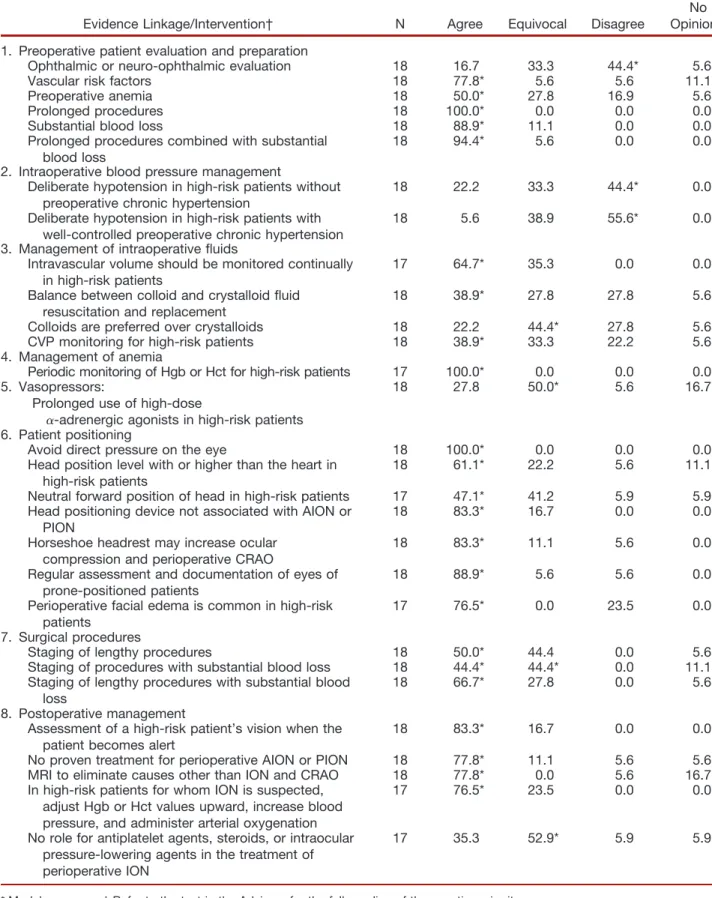
Table 1. Consultant Survey: Percentage Responses*
Evidence Linkage/Intervention† N Agree Equivocal Disagree
No Opinion 1. Preoperative patient evaluation and preparation
Ophthalmic or neuro-ophthalmic evaluation 18 16.7 33.3 44.4* 5.6
Vascular risk factors 18 77.8* 5.6 5.6 11.1
Preoperative anemia 18 50.0* 27.8 16.9 5.6
Prolonged procedures 18 100.0* 0.0 0.0 0.0
Substantial blood loss 18 88.9* 11.1 0.0 0.0
Prolonged procedures combined with substantial blood loss
18 94.4* 5.6 0.0 0.0
2. Intraoperative blood pressure management
Deliberate hypotension in high-risk patients without preoperative chronic hypertension
18 22.2 33.3 44.4* 0.0
Deliberate hypotension in high-risk patients with well-controlled preoperative chronic hypertension
18 5.6 38.9 55.6* 0.0
3. Management of intraoperative fluids
Intravascular volume should be monitored continually in high-risk patients
17 64.7* 35.3 0.0 0.0
Balance between colloid and crystalloid fluid resuscitation and replacement
18 38.9* 27.8 27.8 5.6
Colloids are preferred over crystalloids 18 22.2 44.4* 27.8 5.6
CVP monitoring for high-risk patients 18 38.9* 33.3 22.2 5.6
4. Management of anemia
Periodic monitoring of Hgb or Hct for high-risk patients 17 100.0* 0.0 0.0 0.0
5. Vasopressors:
Prolonged use of high-dose
␣-adrenergic agonists in high-risk patients
18 27.8 50.0* 5.6 16.7
6. Patient positioning
Avoid direct pressure on the eye 18 100.0* 0.0 0.0 0.0
Head position level with or higher than the heart in high-risk patients
18 61.1* 22.2 5.6 11.1
Neutral forward position of head in high-risk patients 17 47.1* 41.2 5.9 5.9
Head positioning device not associated with AION or PION
18 83.3* 16.7 0.0 0.0
Horseshoe headrest may increase ocular compression and perioperative CRAO
18 83.3* 11.1 5.6 0.0
Regular assessment and documentation of eyes of prone-positioned patients
18 88.9* 5.6 5.6 0.0
Perioperative facial edema is common in high-risk patients
17 76.5* 0.0 23.5 0.0
7. Surgical procedures
Staging of lengthy procedures 18 50.0* 44.4 0.0 5.6
Staging of procedures with substantial blood loss 18 44.4* 44.4* 0.0 11.1
Staging of lengthy procedures with substantial blood loss
18 66.7* 27.8 0.0 5.6
8. Postoperative management
Assessment of a high-risk patient’s vision when the patient becomes alert
18 83.3* 16.7 0.0 0.0
No proven treatment for perioperative AION or PION 18 77.8* 11.1 5.6 5.6
MRI to eliminate causes other than ION and CRAO 18 77.8* 0.0 5.6 16.7
In high-risk patients for whom ION is suspected, adjust Hgb or Hct values upward, increase blood pressure, and administer arterial oxygenation
17 76.5* 23.5 0.0 0.0
No role for antiplatelet agents, steroids, or intraocular pressure-lowering agents in the treatment of perioperative ION
17 35.3 52.9* 5.9 5.9
* Modal response. † Refer to the text in the Advisory for the full wording of the questionnaire items.
AION⫽anterior ischemic optic neuropathy; CRAO⫽central retinal artery occlusion; CVP⫽central venous pressure; Hct⫽hematocrit; Hgb⫽hemoglobin; ION⫽ischemic optic neuropathy; MRI⫽magnetic resonance imaging; N⫽number of consultants who responded to each item; PION⫽posterior ischemic optic neuropathy.
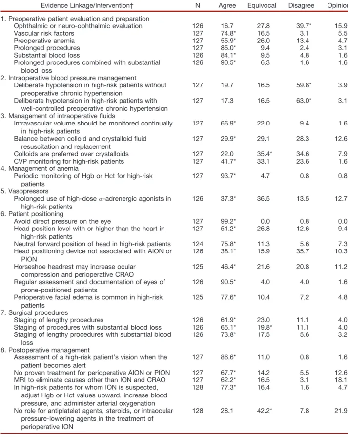
Table 2. Society for Neuroscience in Anesthesiology and Critical Care Member Survey: Percentage Responses*
Evidence Linkage/Intervention† N Agree Equivocal Disagree Opinion
1. Preoperative patient evaluation and preparation
Ophthalmic or neuro-ophthalmic evaluation 126 16.7 27.8 39.7* 15.9
Vascular risk factors 127 74.8* 16.5 3.1 5.5
Preoperative anemia 127 55.9* 26.0 13.4 4.7
Prolonged procedures 127 85.0* 9.4 2.4 3.1
Substantial blood loss 126 84.1* 9.5 4.8 1.6
Prolonged procedures combined with substantial blood loss
126 90.5* 6.3 1.6 1.6
2. Intraoperative blood pressure management
Deliberate hypotension in high-risk patients without preoperative chronic hypertension
127 19.7 16.5 59.8* 3.9
Deliberate hypotension in high-risk patients with well-controlled preoperative chronic hypertension
127 17.3 16.5 63.0* 3.1
3. Management of intraoperative fluids
Intravascular volume should be monitored continually in high-risk patients
127 66.9* 22.0 9.4 1.6
Balance between colloid and crystalloid fluid resuscitation and replacement
127 29.9* 29.1 28.3 12.6
Colloids are preferred over crystalloids 127 22.0 35.4* 34.6 7.9
CVP monitoring for high-risk patients 127 41.7* 33.1 23.6 1.6
4. Management of anemia
Periodic monitoring of Hgb or Hct for high-risk patients
127 93.7* 4.7 0.8 0.8
5. Vasopressors
Prolonged use of high-dose␣-adrenergic agonists in high-risk patients
126 37.3* 36.5 13.5 12.7
6. Patient positioning
Avoid direct pressure on the eye 127 99.2* 0.0 0.8 0.0
Head position level with or higher than the heart in high-risk patients
127 51.2* 26.8 12.6 9.4
Neutral forward position of head in high-risk patients 124 75.8* 11.3 5.6 7.3
Head positioning device not associated with AION or PION
126 38.1* 15.9 35.7 10.3
Horseshoe headrest may increase ocular compression and perioperative CRAO
125 46.4* 21.6 20.8 11.2
Regular assessment and documentation of eyes of prone-positioned patients
126 90.5* 4.0 4.0 1.6
Perioperative facial edema is common in high-risk patients
125 77.6* 10.4 7.2 4.8
7. Surgical procedures
Staging of lengthy procedures 126 61.9* 23.0 11.1 4.0
Staging of procedures with substantial blood loss 126 65.1* 19.8* 11.1 4.0
Staging of lengthy procedures with substantial blood loss
126 73.8* 17.5 5.6 3.2
8. Postoperative management
Assessment of a high-risk patient’s vision when the patient becomes alert
127 86.6* 11.0 0.8 1.6
No proven treatment for perioperative AION or PION 127 67.7* 14.2 5.5 12.6
MRI to eliminate causes other than ION and CRAO 127 62.2* 16.5 3.1 18.1
In high-risk patients for whom ION is suspected, adjust Hgb or Hct values upward, increase blood pressure, and administer arterial oxygenation
128 77.3* 16.4 1.6 4.7
No role for antiplatelet agents, steroids, or intraocular pressure-lowering agents in the treatment of perioperative ION
128 28.1 42.2* 7.8 21.9
* Modal response. † Refer to the text in the Advisory for the full wording of the questionnaire items.
AION⫽anterior ischemic optic neuropathy; CRAO⫽central retinal artery occlusion; CVP⫽central venous pressure; Hct⫽hematocrit; Hgb⫽hemoglobin; ION⫽ischemic optic neuropathy; MRI⫽magnetic resonance imaging; N⫽number of consultants who responded to each item; PION⫽posterior ischemic optic neuropathy.
Table 3. North American Neuro-Ophthalmology Society Member Survey: Percentage Responses*
Evidence Linkage/Intervention† N Agree Equivocal Disagree Opinion
1. Preoperative patient evaluation and preparation
Ophthalmic or neuro-ophthalmic evaluation 32 15.6 40.6 43.8* 0.0
Vascular risk factors 30 83.3* 10.0 6.7 0.0
Preoperative anemia 32 75.0* 12.5 12.5 0.0
Prolonged procedures 32 81.3* 15.6 3.1 0.0
Substantial blood loss 32 96.9* 3.1 0.0 0.0
Prolonged procedures combined with substantial blood loss
32 93.8* 6.3 0.0 0.0
2. Intraoperative blood pressure management
Deliberate hypotension in high-risk patients without preoperative chronic hypertension
31 12.9 19.4 41.9* 25.8
Deliberate hypotension in high-risk patients with well-controlled preoperative chronic hypertension
31 12.9 12.9 48.4* 25.8
3. Management of intraoperative fluids
Intravascular volume should be monitored continually in high-risk patients
30 80.0* 3.3 0.0 16.7
Balance between colloid and crystalloid fluid resuscitation and replacement
30 43.3* 13.3 3.3 40.0
Colloids are preferred over crystalloids 29 31.0 17.2 0.0 51.7*
CVP monitoring for high-risk patients 29 30.1 17.2 0.0 51.7*
4. Management of anemia
Periodic monitoring of Hgb or Hct for high-risk patients
31 83.9* 9.7 0.0 6.5
5. Vasopressors
Prolonged use of high-dose␣-adrenergic agonists in high-risk patients
31 38.7 12.9 3.2 45.2*
6. Patient positioning
Avoid direct pressure on the eye 31 96.8* 3.2 0.0 0.0
Head position level with or higher than the heart in high-risk patients
31 32.3* 32.3* 6.5 29.0
Neutral forward position of head in high-risk patients 30 30.0 26.7 3.3 40.0*
Head positioning device not associated with AION or PION
31 32.3* 22.6 29.0 16.1
Horseshoe headrest may increase ocular compression and perioperative CRAO
31 64.5* 9.7 12.9 12.9
Regular assessment and documentation of eyes of prone-positioned patients
31 83.9* 6.5 3.2 6.5
Perioperative facial edema is common in high-risk patients
31 67.7* 6.5 9.7 16.1
7. Surgical procedures
Staging of lengthy procedures 32 50.0* 21.9 6.3 21.9
Staging of procedures with substantial blood loss 31 71.0* 12.9* 6.5 9.7
Staging of lengthy procedures with substantial blood loss
31 71.0* 9.7 6.5 12.9
8. Postoperative management
Assessment of a high-risk patient’s vision when the patient becomes alert
32 81.3* 9.4 3.1 6.3
No proven treatment for perioperative AION or PION 32 75.0* 15.6 6.3 3.1
MRI to eliminate causes other than ION and CRAO 32 81.3* 12.5 3.1 3.1
In high-risk patients for whom ION is suspected, adjust Hgb or Hct values upward, increase blood pressure, and administer arterial oxygenation
32 90.6* 6.3 3.1 0.0
No role for antiplatelet agents, steroids, or intraocular pressure-lowering agents in the treatment of perioperative ION
31 35.5* 35.5* 22.6 6.5
* Modal response. † Refer to the text in the Advisory for the full wording of the questionnaire items.
AION⫽anterior ischemic optic neuropathy; CRAO⫽central retinal artery occlusion; CVP⫽central venous pressure; Hct⫽hematocrit; Hgb⫽hemoglobin; ION⫽ischemic optic neuropathy; MRI⫽magnetic resonance imaging; N⫽number of consultants who responded to each item; PION⫽posterior ischemic optic neuropathy.
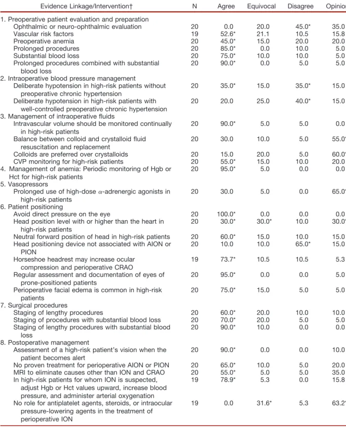
Table 4. North American Spine Society Member Survey: Percentage Responses*
Evidence Linkage/Intervention† N Agree Equivocal Disagree Opinion
1. Preoperative patient evaluation and preparation
Ophthalmic or neuro-ophthalmic evaluation 20 0.0 20.0 45.0* 35.0
Vascular risk factors 19 52.6* 21.1 10.5 15.8
Preoperative anemia 20 45.0* 15.0 20.0 20.0
Prolonged procedures 20 85.0* 0.0 10.0 5.0
Substantial blood loss 20 75.0* 10.0 10.0 5.0
Prolonged procedures combined with substantial blood loss
20 90.0* 0.0 5.0 5.0
2. Intraoperative blood pressure management
Deliberate hypotension in high-risk patients without preoperative chronic hypertension
20 35.0* 15.0 35.0* 15.0
Deliberate hypotension in high-risk patients with well-controlled preoperative chronic hypertension
20 20.0 25.0 40.0* 15.0
3. Management of intraoperative fluids
Intravascular volume should be monitored continually in high-risk patients
20 90.0* 5.0 5.0 0.0
Balance between colloid and crystalloid fluid resuscitation and replacement
20 30.0 10.0 5.0 55.0*
Colloids are preferred over crystalloids 20 15.0 20.0 5.0 60.0*
CVP monitoring for high-risk patients 20 55.0* 15.0 10.0 20.0
4. Management of anemia: Periodic monitoring of Hgb or Hct for high-risk patients
20 95.0* 5.0 0.0 0.0
5. Vasopressors
Prolonged use of high-dose␣-adrenergic agonists in high-risk patients
20 30.0 5.0 0.0 65.0*
6. Patient positioning
Avoid direct pressure on the eye 20 100.0* 0.0 0.0 0.0
Head position level with or higher than the heart in high-risk patients
20 30.0* 30.0* 10.0 30.0*
Neutral forward position of head in high-risk patients 20 60.0* 15.0 10.0 15.0
Head positioning device not associated with AION or PION
20 10.0 10.0 65.0* 15.0
Horseshoe headrest may increase ocular compression and perioperative CRAO
19 73.7* 10.5 10.5 5.3
Regular assessment and documentation of eyes of prone-positioned patients
20 95.0* 0.0 0.0 5.0
Perioperative facial edema is common in high-risk patients
20 75.0* 15.0 5.0 5.0
7. Surgical procedures
Staging of lengthy procedures 20 60.0* 20.0 10.0 10.0
Staging of procedures with substantial blood loss 20 70.0* 20.0 5.0 5.0
Staging of lengthy procedures with substantial blood loss
20 90.0* 10.0 0.0 0.0
8. Postoperative management
Assessment of a high-risk patient’s vision when the patient becomes alert
20 90.0* 0.0 0.0 10.0
No proven treatment for perioperative AION or PION 20 65.0* 10.0 5.0 20.0
MRI to eliminate causes other than ION and CRAO 20 55.0* 5.0 5.0 35.0
In high-risk patients for whom ION is suspected, adjust Hgb or Hct values upward, increase blood pressure, and administer arterial oxygenation
19 78.9* 5.3 0.0 15.8
No role for antiplatelet agents, steroids, or intraocular pressure-lowering agents in the treatment of perioperative ION
19 0.0 31.6* 5.3 63.2*
* Modal response. † Refer to the text in the Advisory for the full wording of the questionnaire items.
AION⫽anterior ischemic optic neuropathy; CRAO⫽central retinal artery occlusion; CVP⫽central venous pressure; Hct⫽hematocrit; Hgb⫽hemoglobin; ION⫽ischemic optic neuropathy; MRI⫽magnetic resonance imaging; N⫽number of consultants who responded to each item; PION⫽posterior ischemic optic neuropathy.
References
1. Buono LM, Foroozan R: Perioperative posterior ischemic optic neuropathy: Review of the literature. Surv Ophthalmol 2005; 50:15–26
2. Ho VT, Newman NJ, Song S, Ksiazek S, Roth S: Ischemic optic neuropathy following spine surgery. J Neurosurg An-esthesiol 2005; 17:38 – 44
3. Roth S: Postoperative blindness, Anesthesia, 7th edition.. Edited by Miller RD. Philadelphia, Churchill Livingstone, 2010, pp 2821– 41
4. Chang SH, Miller NR: The incidence of vision loss due to perioperative ischemic optic neuropathy associated with spine surgery: The Johns Hopkins Hospital Experience. Spine 2005; 30:1299 –302
5. Patil CG, Lad EM, Lad SP, Ho C, Boakye M: Visual loss after spine surgery: A population-based study. Spine 2008; 33:1491– 6 6. Roth S, Barach P: Postoperative visual loss: Still no answers—
yet. ANESTHESIOLOGY2001; 95:575–7
7. Shen Y, Drum M, Roth S: The prevalence of perioperative visual loss in the United States: A 10-year study from 1996 to 2005 of spinal, orthopedic, cardiac, and general surgery. Anesth Analg 2009; 109:1534 – 45
8. Stevens WR, Glazer PA, Kelley SD, Lietman TM, Bradford DS: Ophthalmic complications after spinal surgery. Spine 1997; 22:1319 –24
9. Lee LA, Roth S, Posner KL, Cheney FW, Caplan RA, Newman NJ, Domino KB: The American Society of Anesthesiologists Postoperative Visual Loss Registry: Analysis of 93 spine sur-gery cases with postoperative visual loss. ANESTHESIOLOGY
2006; 105:652–9
10. Chalam KV, Shah VA: Severe bilateral posterior ischemic optic neuropathy as a complication of spinal surgery. Eye 2005; 19:367– 8
11. Heitz JW, Audu PB: Asymmetric postoperative visual loss after spine surgery in the lateral decubitus position. Br J Anaesth 2008; 101:380 –2
12. Kothari MT, Maiti A: Ophthalmic artery occlusion: A cause of unilateral visual loss following spine surgery. Indian J Oph-thalmol 2007; 55:401–2
13. Kumar N, Jivan S, Topping N, Morrell AJ: Blindness and rectus muscle damage following spinal surgery. Am J Oph-thalmol 2004; 138:889 –91
14. Leibovitch I, Casson R, Laforest C, Selva D: Ischemic orbital compartment syndrome as a complication of spinal surgery in the prone position. Ophthalmology 2006; 113:105– 8 15. Locastro A, Novak KD, Biglan AW: Central retinal artery
occlusion in a child after general anesthesia. Am J Ophthal-mol 1991; 112:91–2
16. Mertens E, Smets RM, Sys J, Michielsen J, Verstreken J, Tassignon MJ: Central retinal artery occlusion after back surgery: A case report. Bull Soc Belge Ophtalmol 1995; 255:127–31
17. West J, Askin G, Clarke M, Vernon SA: Loss of vision in one eye following scoliosis surgery. Br J Ophthalmol 1990; 74:243– 4 18. Bermejo-Alvarez MA, Carpintero M, García-Carro G, Acebal
G, Fervienza P, Cosío F: [Ischemic optic neuropathy after lumbar spine surgery]. Rev Esp Anestesiol Reanim 2007; 54:621–5
19. Bradish CF, Flowers M: Central retinal artery occlusion in associ-ation with osteogenesis imperfecta. Spine 1987; 12:193– 4 20. Brown RH, Schauble JF, Miller NR: Anemia and hypotension as
contributors to perioperative loss of vision. ANESTHESIOLOGY1994; 80:222– 6
21. Dunker S, Hsu HY, Sebag J, Sadun AA: Perioperative risk factors for posterior ischemic optic neuropathy. J Am Coll Surg 2002; 194:705–10
22. Hoff JM, Varhaug P, Midelfart A, Lund-Johansen M: Acute visual loss after spinal surgery. Acta Ophthalmol 2010; 88: 490 –2
23. Huber JF, Grob D: Bilateral cortical blindness after lumbar spine surgery. A case report. Spine 1998; 23:1807–9 24. Lee AG: Ischemic optic neuropathy following lumbar spine
surgery. Case report. J Neurosurg 1995; 83:348 –9
25. Wolfe SW, Lospinuso MF, Burke SW: Unilateral blindness as a complication of patient positioning for spinal surgery. A case report. Spine 1992; 17:600 –5
26. Cheng MA, Sigurdson W, Tempelhoff R, Lauryssen C: Visual loss after spine surgery: A survey. Neurosurgery 2000; 46: 625–30
27. Myers MA, Hamilton SR, Bogosian AJ, Smith CH, Wagner TA: Visual loss as a complication of spine surgery. A review of 37 cases. Spine 1997; 22:1325–9
28. Katz DM, Trobe JD, Cornblath WT, Kline LB: Ischemic optic neuropathy after lumbar spine surgery. Arch Ophthalmol 1994; 112:925–31
29. Montero JA, Ruiz-Moreno JM, Galindo A, Fernandez-Mun˜oz M: Release hallucinations and visual loss as first manifesta-tions of postoperative unilateral blindness. Eur J Ophthalmol 2007; 17:844 – 6
30. Roth S, Nunez R, Schreider BD: Unexplained visual loss after lumbar spinal fusion. J Neurosurg Anesthesiol 1997; 9:346 – 8 31. Stang-Veldhouse KN, Yeu E, Rothenberg DM, Mizen TR: Unusual presentation of perioperative ischemic optic neu-ropathy following major spine surgery. J Clin Anesth 2010; 22:52–5
32. Abbott MA, McLaren AD, Algie T: Intra-ocular pressure dur-ing cardiopulmonary bypass: A comparison of crystalloid and colloid priming solutions. Anaesthesia 1994; 49:343– 6 33. Bekar A, Tu¨reyen K, Aksoy K: Unilateral blindness due to
patient positioning during cervical syringomyelia surgery: Unilateral blindness after prone position. J Neurosurg Anes-thesiol 1996; 8:227–9
34. Grossman W, Ward WT: Central retinal artery occlusion after scoliosis surgery with a horseshoe headrest. Case report and literature review. Spine 1993; 18:1226 – 8
35. Hollenhorst RW, Svien HJ, Benoit CF: Unilateral blindness occurring during anesthesia for neurosurgical operations. AMA Arch Ophthalmol 1954; 52:819 –30
36. Hoski JJ, Eismont FJ, Green BA: Blindness as a complication of intraoperative positioning. A case report. J Bone Joint Surg 1993; 75:1231–2
37. Jampol LM, Goldbaum M, Rosenberg M, Bahr R: Ischemia of ciliary arterial circulation from ocular compression. Arch Ophthalmol 1975; 93:1311–7
38. Manfredini M, Ferrante R, Gildone A, Massari L: Unilateral blindness as a complication of intraoperative positioning for cervical spinal surgery. J Spinal Disord 2000; 13:271–2 39. Nakra D, Bala I, Pratap M: Unilateral postoperative visual loss
due to central retinal artery occlusion following cervical spine surgery in prone position. Paediatr Anaesth 2007; 17:805– 8
40. Roth S, Tung A, Ksiazek S: Visual loss in a prone-positioned spine surgery patient with the head on a foam headrest and goggles covering the eyes: An old complication with a new mechanism. Anesth Analg 2007; 104:1185–7
41. Sys J, Michielsen J, Mertens E, Verstreken J, Tassignon MJ: Central retinal artery occlusion after spinal surgery. Eur Spine J 1996; 5:74 –5
42. Yu YH, Chen WJ, Chen LH, Chen WC: Ischemic orbital compartment syndrome after posterior spinal surgery. Spine 2008; 33:E569 –72
43. Roth S, Thisted RA, Erickson JP, Black S, Schreider BD: Eye injuries after nonocular surgery: A study of 60,965 anesthet-ics from 1988 to 1992. ANESTHESIOLOGY1996; 85:1020 –7