ALTERATIONS TO THE EPIGENOME REPRESENT CRITICAL MECHANISMS OF CHEMICALLY INDUCED CARCINOGENESIS
Grace A. Chappell
A dissertation submitted to the faculty at the University of North Carolina at Chapel Hill in partial fulfillment of the requirements for the degree of Doctor of Philosophy in the Department of
Environmental Sciences and Engineering in the Gillings School of Global Public Health.
Chapel Hill 2015
Approved by:
Wanda Bodnar
Rebecca Fry
Terrence Furey
Igor Pogribny
ABSTRACT
Grace A. Chappell: Alterations to the epigenome represent critical mechanisms of chemically-induced carcinogenesis
(Under the direction of Ivan Rusyn)
Environmental and occupational exposures to both natural and anthropogenic substances
are known to play a causative role in carcinogenesis. While it is well accepted that carcinogenesis
occurs by both genotoxic and non-genotoxic mechanisms, genotoxicity has been more thoroughly
studied. Accumulating evidence suggests that epigenetic alterations also play an important role
in chemically-induced carcinogenesis, and that these changes to the epigenome may be as
important as genotoxicity. To gain a better understanding of the epigenetic alterations that occur
as a consequence of exposure to a genotoxic chemical, we evaluated genetic and epigenetic
alterations in two mouse models of genotoxicity. First, to determine the relative contribution of
these molecular changes in liver tumorigenesis, we evaluated epigenetic modifications and the
mutational profile of genes commonly altered in liver tumors in mice treated with a combination of
a genotoxic and a pro-fibrogenic agent. Marked epigenetic changes in liver tumors and
surrounding fibrotic liver tissue were associated with tumor incidence, while mutations in known
cancer-related genes were not observed. Second, to better characterize the diverse molecular
mechanisms of 1,3-butadiene (BD)-induced tumorigenicity, we evaluated DNA adducts and
epigenetic alterations caused by exposure to BD in a set of two genetically divergent mouse
strains (C57BL/6J and CAST/EiJ) and across both target (lung and liver) and non-target (kidney)
tissues of BD-induced tumorigenesis. Although BD-DNA adducts were observed in all three
epigenetic alterations were clearly tissue- and strain-specific. These results suggest that
epigenetic alterations represent a disease-relevant class of specific, and potentially
mechanistically important, molecular features that are involved in chemically-induced
carcinogenesis. Our findings emphasize the necessity of inclusion of epigenetic marks in
ACKNOWLEDGEMENTS
First and foremost I thank my advisor, Dr. Ivan Rusyn, for giving me with the opportunity
to join his research group. From the high anxiety of my first lab meeting presentations to
winning awards at conferences, this has truly been a learning experience in more dimensions
than I ever expected. Thank you for training my critical eye, for teaching me how to effectively
present my research, and for always encouraging me to better my best.
I also thank all of my lab-mates, past and present, for their feedback, help with
experiments, and general camaraderie throughout my tenure as a member of the Rusyn Lab
group, both at UNC and at TAMU. I am grateful of the members of the Furey Lab for welcoming
me into their group for a semester and not only graciously helping me with analytical methods
that were new to me, but also for treating me as ‘one of their own.’
Each member of my committee has offered me support throughout my pursuit of a PhD
in a unique way, whether it be technical, analytical, motivational, or some combination of the
three. I am incredibly appreciative of your interest in my research and your involvement in my
scientific journey; thank you.
I would like to acknowledge all of the coauthors of the published work presented herein
for their essential contributions to the studies: Frederick Beland, Wanda Bodnar, Mark
Hoenerhoff, Hue-Hua Hong, Tetyana Kobets, Oksana Kosyk, Kristy Kutanzi, Bridget O’Brien,
Igor Pogribny, Dewakar Sangaraju, Kenneth Sexton, Natalia Tretyakova, Volodymyr Tryndyak,
and Takeki Uehara.
I thank my parents for their unwavering (I think) faith in me no matter what I choose to
TABLE OF CONTENTS
LIST OF TABLES ... ix
LIST OF FIGURES ... xi
LIST OF ABBREVIATIONS ... xii
CHAPTER 1: GENERAL INTRODUCTION ... 1
1.1 Description of the need for better characterization of the epigenetic alterations induced by exposure to genotoxic chemicals ... 2
1.2 Epigenetic effects of genotoxic human carcinogens: a systematic review ... 3
1.2.1 Methods ... 3
1.2.2 Evidence of genotoxic mechanisms of carcinogenesis for chemicals and associated occupations included in the review ... 8
1.2.3 Epigenetic alterations induced by chemicals and associated occupations included in the review ... 16
1.2.3.1 DNA Methylation ... 16
1.2.3.2 Histone modifications ... 25
1.2.3.3 Non-coding RNA ... 29
1.2.3.4 Summary ... 36
1.3 Filling in data gaps: studies of epigenetic alterations in complex disease phenotypes in mice exposed to genotoxic chemicals ... 39
References ... 42
CHAPTER 2: GENETIC AND EPIGENETIC CHANGES IN FIBROSIS-ASSOCIATED HEPATOCARCINOGENESIS IN MICE ... 59
2.1 Introduction ... 59
2.3 Results ... 63
2.4 Discussion ... 74
References ... 80
CHAPTER 3: EPIGENETIC EVENTS DETERMINE TISSUE-SPECIFIC TOXICITY OF INHALATIONAL EXPOSURE TO THE GENOTOXIC CHEMICAL 1,3-BUTADIENE IN MALE C57BL/6J MICE ... 84
3.1 Introduction ... 84
3.2 Materials and Methods ... 86
3.3 Results ... 90
3.4 Discussion ... 98
References ... 105
CHAPTER 4: EXPOSURE TO THE GENOTOXIC CHEMICAL 1,3-BUTADIENE CONFER TISSUE- AND STRAIN-SPECIFIC ALTERATIONS IN MRNA AND MICRORNA EXPRESSION ... 109
4.1 Introduction ... 109
4.2 Materials and Methods ... 112
4.3 Results ... 117
4.4 Discussion ... 130
References ... 138
CHAPTER 5: SUMMARY, LIMITATIONS, FUTURE DIRECTIONS, AND CONCLUSIONS ... 142
5.1 Summary of findings ... 142
5.2 General discussion ... 144
5.3 Limitations ... 145
5.4 Future directions ... 147
5.5 Conclusions ... 150
References ... 151
APPENDIX 2.1 LIVER TUMOR HISTOLOGY OF DEN+CCl4-TREATED MICE ... 153 APPENDIX 2.2 H-RAS GENE EXPRESSION ... 154 APPENDIX 2.3 EXPRESSION OF CANCTER STEM CELL MARKERS IN
FIBROSIS-ASSOCIATED HCC ... 155
APPENDIX 3.1 QPCR PRIMERS USED FOR GENE EXPRESSION ANALYSES
DISCUSSED IN CHAPTER 3 ... 156
APPENDIX 4.1 NUMBER OF SEQUENCES READ AND ALIGNED FOR TOTAL
RNA SEQUENCING DISCUSSED IN CHAPTER 4 ... 157
APPENDIX 4.2 NUMBER OF SEQUENCES READ AND ALIGNED FOR SMALL
RNA SEQUENCING DISCUSSED IN CHAPTER 4 ... 159
APPENDIX 4.3 DIFFERENTIALLY EXPRESSED (Q<0.1) MIRNAS IN BD-EXPOSED
LIST OF TABLES
TABLE 1.1 Chemicals and associated occupations included in the systematic
literature review ... 5
TABLE 1.2 Evidence of genotoxicity of the chemicals and associated occupations
included in the systematic literature review ... 15
TABLE 1.3 Number of publications concerning epigenetics for each chemical
included in the systematic literature review ... 37
TABLE 4.1 Compilation of the top enriched genesets for each strain and exposure
group ... 122
TABLE 4.2 Top-most significantly differentially expressed miRNAs ... 127
TABLE 4.3 miRNA families identified to be master regulators of mRNA expression
in C57BL/6J lung ... 128
TABLE 4.4 Functional annotation clustering of differentially expressed mRNA
LIST OF FIGURES
FIGURE 1.1 Literature tree from HAWC assessment ... 8
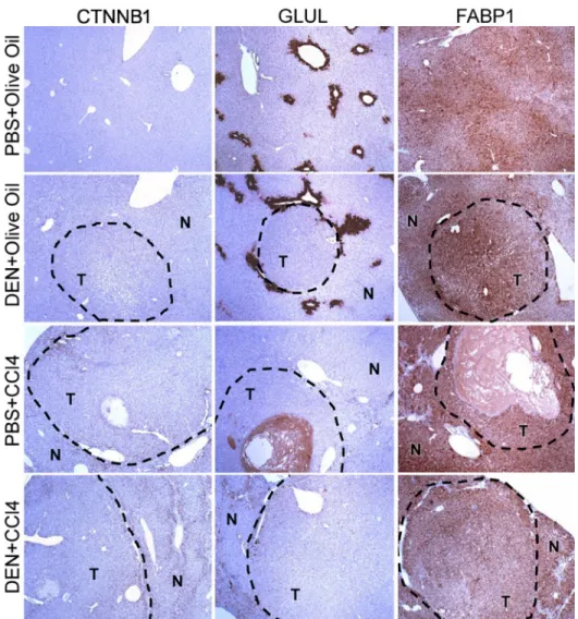
FIGURE 2.1 Immunostaining of CTNNB1, GLUL, and FABP1 in formalin-fixed paraffin-embedded sections of liver tumor and adjacent non-tumor liver tissue ... 66
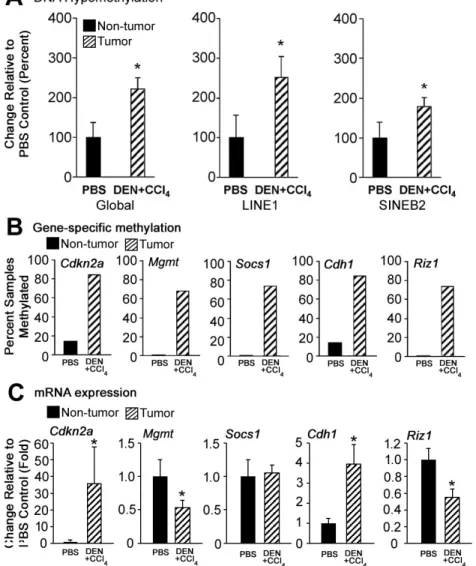
FIGURE 2.2 DNA methylation and gene expression changes in liver tumors ... 67
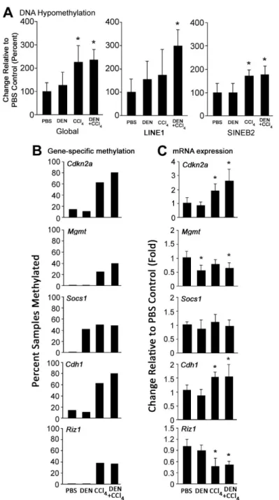
FIGURE 2.3 DNA methylation and gene expression changes in non-tumor liver tissues during fibrosis-associated hepatocarcinogenesis in mice ... 69
FIGURE 2.4 H3K9, H3K279, and H4K20 trimethylation in liver tissues during fibrosis-associated liver carcinogenesis in mice ... 71
FIGURE 2.5 Expression of chromatin-modifying genes in the liver during fibrosis- associated hepatocarcinogenesis in mice ... 72
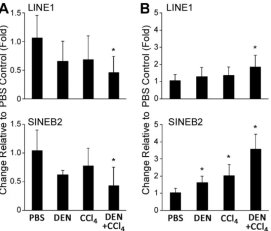
FIGURE 2.6 Level of H3K9me3 at LINE1 and SINE B2 repetitive sequences and expression of LINE1 and SINE B2 in the liver during fibrosis-associated liver carcinogenesis in mice ... 73
FIGURE 3.1 THB-Gua-BD adducts and bis-N7G-BD crosslinks in tissues from mice exposed to 425ppm of BD ... 91
FIGURE 3.2 Effects of BD exposure on the extent of DNA methylation in mouse tissues ... 93
FIGURE 3.3 Effects of BD exposure on the extent of 5-hydroxymethylcytosine in mouse tissues ... 94
FIGURE 3.4 Effects of BD exposure on histone modifications ... 95
FIGURE 3.5 Effects of BD exposure on the expression of DNA methylation and DNA demethylation genes DNA methylation and gene expression changes in liver tumors... 97
FIGURE 3.6 Effects of BD exposure on the expression of histone modifying genes ... 98
FIGURE 3.7 Summary of genotoxic and epigenetic changes in the tissues of BD- exposed mice ... 103
FIGURE 4.1 Mechanisms of gene silencing by miRNAs ... 110
FIGURE 4.2Concentration of 1,3-butadiene inside the inhalation chamber ... 118
FIGURE 4.3 Level of N-7-(2,3,4-trihydroxybut-1-yl)-guanine adducts ... 118
FIGURE 4.5BD-induced differentially expressed miRNAs in C57BL/6J and CAST/EiJ
mice ... 126
FIGURE 4.6 Differentially expressed genes in CAST/EiJ mice relative to C57BL/6J
LIST OF ABBREVIATIONS
AFB1 Aflatoxin B1
ANOVA Analysis of variance
ATAC Assay for transposase-accessible chromatin
BaP Benzo[a]pyrene
BD 1,3-butadiene
bp Base pair
BPDE (±)-anti-7β-8α-dihydroxy-9α,10α-epoxy-7,8,9,10-tetrahydro-benzo[a]pyrene
cDNA Complementary DNA
CCl4 Carbon tetrachloride
CT Cycle threshold
DEN N-nitrosodiethylamine
GSAASeqSP Gene Set Association Analysis for RNA-seq (sample permutation)
HAWC Health Assessment Workspace Collaborative
HCC hepatocellular carcinoma
IARC International Agency for Research on Cancer
LINE Long interspersed nuclear element
miRNA microRNA
mmu Mus musculus
MOCA 4,4'-methylene-bis(2-chloroaniline)
mRNA Messenger RNA
nt Nucleotide
PAH Polycyclic aromatic hydrocarbon
RISC RNA induced silencing complex
RNA-seq RNA sequencing
RT-qPCR Reverse transcriptase quantitative polymerase chain reaction
LINE Long interspersed nuclear element
SINE Short interspersed nuclear element
UTR Untranslated region
CHAPTER 1: GENERAL INTRODUCTION
Environmental and occupational exposures to both natural and anthropogenic substances
are known to play a causative role in carcinogenesis (Pogribny and Rusyn, 2013; Langie et al.,
2015). While it is well accepted that carcinogenesis may occur by both genotoxic and
non-genotoxic mechanisms, non-genotoxicity has been more thoroughly studied. Accumulating evidence
suggests that epigenetic alterations also play an important role in chemically-induced
carcinogenesis, and that epigenetic mechanisms may be as important as genetic mechanisms in
cancer development and progression (Ushijima and Asada, 2010). Epigenetic alterations
represent non-genotoxic mechanisms of carcinogenesis that may occur independently or
concomitantly with genotoxic aberrations. Further, the epigenomic landscape may directly
influence the genotoxic potential of a chemical; for example, several studies have indicated
preferential binding of reactive chemicals to regions of DNA that harbor specific histone
modification marks and/or DNA methylation patterns (Denissenko et al., 1997; Chen et al., 1998;
Yoon et al., 2001; Hu et al., 2003; Tretyakova et al., 2008; O'Hagan et al., 2011).
There are several major types of epigenetic and epigenomic alterations: DNA methylation,
alterations to histones/chromatin structure, nucleosome positioning, and expression of
non-coding RNAs. A wealth of data exists that demonstrates that alterations in DNA methylation,
histone modifications, and expression of non-coding RNAs occur as a consequence of exposure
to environmental chemicals, and such changes to the genome have been associated with various
1.1. There is a need for better characterization of the epigenetic alterations induced by exposure to genotoxic chemicals
To enable the efficacious incorporation of epigenetic endpoints in chemical hazard
assessments, further characterization of the role of epigenetic alterations induced by
environmental exposure is necessary (Koturbash et al., 2011a). In 2012, Richard Stein reviewed
the current knowledge of epigenetics and environmental exposures, emphasizing that evaluating
genotoxic factors is insufficient for the characterization of chemical hazards, and ultimately stating
that epigenetic features do not receive adequate attention relative to genetic endpoints (Stein,
2012). Concurrently, the appropriateness of current safety assessment paradigms that utilize a
no observed adverse effect level (NOAEL) for epigenetic mechanisms was also considered (Alyea
et al., 2012). Using several case studies on 1,3-butadiene, arsenic, and diethylstilbesterol, the
authors found that epigenetic alterations largely fell within the dose-response curve for apical
effects, but also highlighted several important gaps in the data, including dissimilar study designs
and limited epigenetic dose-response data for only a small subset of known epigenetic marks.
The authors concluded that current safety assessment models would be protective of various
mechanisms that lead to adverse effects typically observed in guideline animal studies, because
reference doses for a risk assessment are set based on an overall NOAEL that is driven by observed apical effects. However, additional studies are needed to characterize the relationship
between epigenetic alterations and toxicity phenotypes, and the epigenetic-specific
dose-response.
Exponentially growing evidence indicates that epigenetic events may be indispensable
key mechanisms by which chemical carcinogens induce alterations in target cells. It has been
demonstrated that these alterations occur early during exposure and may have significance as
biomarkers of chemical exposure. However, it is not necessary that an observation of the
epigenetic effect in response to chemical exposure, usually within hours or few days after
transitory adaptive or non-specific cellular responses to chemical treatment. Overall, although
there has been an increase in reporting of, and interest in, epigenetic mechanisms in the fields of
environmental science and toxicology, the application of epigenetic endpoints in regulatory
science is lacking.
The US Environmental Protection Agency has held multiple workshops and State of the
Science evaluations (http://cfpub.epa.gov/ncea/cfm/recordisplay.cfm?deid=308271) regarding
epigenetics and environmental chemicals, but no standard exists for how these endpoints are to
be incorporated into chemical assessments. The International Agency for the Research of Cancer
(IARC) recently named “Epigenetic Alterations” as one of 10 key characteristics of human
carcinogens (Smith et al, in press). However, it is recognized that most carcinogens were
evaluated by IARC before new data on their epigenetic effects became available (Herceg et al.,
2013).
Research in the field of epigenetic responses to chemicals is increasing, and as additional
information is made available regarding epigenetic mechanisms of cancer (and other toxicity
phenotypes), such information should be utilized by regulators and governing bodies in the
assessment of known or potentially hazardous chemicals. As the relationship between epigenetic
alterations and toxicity phenotypes is further clarified, improved congruent guidelines for the
application of epigenetic data in safety assessments can be accomplished.
1.2. Epigenetic effects of genotoxic human carcinogens: a systematic review 1.2.1. Methods
To address the need for comprehensive information regarding epigenetic alterations and
chemically-induced carcinogenesis, we conducted a systematic review of published studies
concerning epigenetic alterations caused by known human carcinogens that have strong
evidence of a genotoxic mechanism of carcinogenesis. We used the Health Assessment
literature for the assessment of chemicals or groups of chemicals (https://hawcproject.org). The
review focused on human carcinogens as classified by IARC. IARC evaluations of carcinogenic
risks to humans assign potential carcinogenic chemicals into one of five categories based on the
body of evidence from epidemiological studies, animal cancer bioassays, and mechanistic and
other relevant data (Tomatis, 1976). In 2012, re-evaluation of data on known human carcinogens
was undertaken and published as a compendium of six monographs comprising volume 100. To
date, there are 117 agents classified as known human carcinogens; these include chemical
agents and related occupations, personal habits and indoor combustions, radiation, arsenic,
metals, fibers and dusts, biological agents, and pharmaceuticals. For the purpose of this
systematic review, we focused on environmental and occupational hazards; specifically, the
agents and occupations listed in the IARC monograph volume 100F. Of the 31 chemicals and
associated occupations included in this volume, 27 were included in this review because they
were (1) classified as a known human carcinogen, and (2) the IARC Monographs working group
concluded that there was sufficient evidence for a genotoxic mechanism of carcinogenesis. Of
the chemicals and associated occupations included in the search, we identified publications
Table 1.1. Chemicals and associated occupations included in the systematic literature review.
Chemical or associated occupation volume # Original
Evidence of genotoxicity:
animal
Evidence of genotoxicity:
human Epigenetics
1,3-butadiene 97 yes yes yes
2-naphthylamine 99 yes yes no
4,4'-methylenebis(2-chlorobenzenamine)
(MOCA) 99 yes yes yes
4-aminobiphenyl 99 yes yes yes
aflatoxins (naturally occurring mixtures) 82 yes yes yes
aluminium production 92 no moderate* weak-to- no
benzene supp. 7 yes yes yes
benzidine 99 yes yes yes
benzo[a]pyrene 92 yes yes yes
bis(chloromethyl)ether and chlormethyl
methyl ether supp. 7 no moderate-to-strong no
chimney soot 92 no moderate* no
coal gasification, occupational exposures 92 yes† no no
coal-tar distillation, occupational 92 yes# yes no
coal-tar pitch 92 yes# moderate no
coke production, occupational exposures 92 yes& yes* yes
ethylene oxide 97 yes yes no
formaldehyde 88 yes yes yes
iron and steel founding supp. 7 no yes* no
isopropyl alcohol manufacture supp. 7 no plausible no
mineral oils supp. 7 no weak no
ortho-toluidine 99 yes moderate no
painter (occupational exposure) 98 no yes* yes
rubber manufacturing supp. 7 no yes* no
shale oils supp. 7 yes no no
strong-inorganic-acid mists containing
sulfuric acid 54 no plausible no
sulfur mustard supp. 7 yes yes yes
vinyl chloride 97 yes yes yes
*genotoxicity is attributable to the presence of known genotoxic chemicals in the exposure scenario †animal mechanistic data based on treatment with coal-tars or manufactured gas plant residues #animal mechanistic data based on treatment with coal-tars
Using HAWC, we queried all available literature in the PubMed database using search
terms for both epigenetic alterations and the chemicals listed in Table 1.1. The full search terms
are detailed in Appendix 1.1.
The following exclusion criteria were applied:
Not chemical of interest. The exclusion criteria “not chemical of interest” was assigned to cases in which no chemical in the list of Group 1 carcinogens included in the 100F IARC monographs
(or their recognized reactive metabolites) were used in the study, or in cases in which the chemical
was used for purposes other than exposure assessment. For example, many studies used
formaldehyde for cross-linking of proteins or benzidine for staining of Hb, but the chemicals were
not used for the purpose of studying cancer-relevant consequences of exposure. Such studies
were excluded from the assessment. Similarly, studies of endogenous sources of formaldehyde
were also excluded.
No epigenetics. Several studies made mention of potential epigenetic mechanisms, or epigenetic alterations reported for a chemical that was not actually studied. Studies that investigated direct
binding of a chemical (or metabolite) to histones, and no other epigenetic mark, were also
excluded.
Reviews. Reviews were not considered.
Unable to access full text. Articles that were not publicly available or accessible through either the Texas A&M Univsersity or University of North Carolina online portal were excluded. Articles
that were not available in English were also excluded.
γ-H2AX. This category was created to bin all studies that measured phosphorylation of histone 2AX (γ-H2AX) as a consequence of exposure to one of the chemicals of interest in any system. γ-H2AX is used as a marker for DNA double strand breaks (Rogakou et al., 1998), and studies
that included this endpoint for that purpose alone were not included in our review if there was no
collected from exposures to benzo[a]pyrene, sulfur mustard, and benzene (or the
metabolites/analogs of these chemicals).
Other. A total of 47 studies were excluded for reasons other than those detailed above, and were included in this category. For example, some studies included an analysis of one of our chemicals
of interest and of epigenetic alterations, but not of epigenetic events due to a chemical we queried.
Methods papers also were assigned to this exclusion category. A sub-category was created for
studies that reported specific binding of benzo[a]pyrene (BaP) to DNA at methylated vs.
unmethylated cytosines. While these studies are pertinent to this review, and are briefly discussed
herein (section B.3.a), they were not included with the rest of the studies because the DNA
methylation was either assumed or definitely independent of exposure to BaP.
A total of 919 references were returned as of the search date 10/27/2015. Of these, 148
met the inclusion criteria and 771 were excluded (Figure 1). The publicly available assessment
Figure 1.
Literature tree of the 919 studies returned by the search after assignment to appropriate categories. The human studies of benzene are expanded as an example.
1.2.2. Evidence of genotoxic mechanisms of carcinogenesis for chemicals and associated occupations included in the review
Common routes of exposure, associated cancers, and previously reported evidence of
genotoxicity of the 12 chemicals included in this review are briefly described below. Genotoxic
attributes are is summarized in Table 1.2.
1,3-butadiene
1,3-butadiene is a gas monomer used in the production of synthetic rubber. Exposure
typically occurs in occupational settings in the production of 1,3-butadiene itself, as well as in
widely detected in ambient air, albeit at much lower levels than in occupational settings, from
sources such as vehicle exhaust, cigarette smoke, and wood fires. An excess of hematopoietic
cancers has been reported among workers occupationally exposed to 1,3-butadiene, and lung
and bladder cancers have also been associated with 1,3-butadiene exposure. 1,3-butadiene is a
multi-site carcinogen in experimental animals. The carcinogenicity of 1,3-butadiene is contingent
upon the metabolism of 1,3-butadiene to reactive epoxides, which can bind with DNA and proteins
(Huff et al., 1985; Jackson et al., 2000). bd-DNA adducts have been observed in
occupationally-exposed humans and experimental animals, and have been associated with mutations in
cancer-related genes (Ton et al., 2007; Abdel-Rahman et al., 2001; Sills et al., 2001; Lee et al., 2002;
Abdel-Rahman et al., 2003).
4-aminobiphenyl
4-aminobiphenyl is an aromatic amine used as a dye intermediate and as a rubber
antioxidant, and human exposure predominantly occurs in occupational settings. Industrial
production of 4-aminobiphenyl was ceased in 1955, and current exposures are due to
contamination or metabolic release from benzidine. 4-aminobiphenyl is also a byproduct of
tobacco combustion, and has been detected in fumes from cooking oils. Bladder carcinoma is the
primary cancer associated with exposure to 4-aminobiphenyl, observed in human chemical plant
workers and in experimental animal models. Multiple metabolic pathways activate aromatic
amines, including 4-aminobiphenyl, to DNA-reactive intermediates, which are known to result in
mutations. 4-aminobiphenyl-DNA adducts have been detected in human bladder, lung, and breast
tissue of exposed humans (Reimann and Erdogan, 1976; Beland et al., 1983; Lin et al., 1994),
and mutations in the HPRT locus and in the H-ras gene have been detected in human and mouse tissues, respectively, after exposure to 4-aminobiphenyl (IARC, 2012).
4,4’-methylenebis(2-chlorobenzenamine)
4,4’-methylenebis(2-chlorobenzenamine), also referred to as MOCA, is a curing agent
occupational settings, with non-occupational exposures in areas contaminated with MOCA or
consumption of foods that were grown in contaminated soil. Limited human data is suggestive of
an association between MOCA exposure and bladder cancer, and MOCA caused lung, liver, and
bladder cancer in experimental animals (Stula et al., 1978). The assignment of MOCA to IARC
“Group 1 carcinogens” was largely based on the strong evidence of genotoxic mechanism of
action, involving metabolism of the aromatic amine to DNA-reactive intermediates, which lead to
DNA adducts, mutations, sister chromatid exchange, and increased micronuclei (Silk et al., 1989;
Kaderlik et al., 1993; Murray and Edwards, 1999).
Aflatoxins
Aflatoxins are naturally occurring potent hepatocarcinogens produced by Aspergillus flavus and Aspergillus parasiticus, with Aflatoxin B1 (AFB1) considered to be the most toxic type. Human exposure to AFB1 primarily occurs by consumption of contaminated food sources, most
commonly stored grains, but occupational exposure also occurs during processing and handling
of contaminated grains (inhalation and dermal). The main mode of carcinogenicity is attributed to
the metabolic activation of AFB1 to a genotoxic epoxide, with a high prevalence of point mutations
in the p53 gene (Tam et al., 1999; Gomaa et al., 2008). AFB1 exposure causes sister chromatid
exchange, micronuclei, chromosomal alterations, and DNA and protein adducts (IARC, 2012).
Benzene
Benzene is a solvent that has historically been used in printing inks, gasoline, and
chemical and drug production. Currently, the main use of benzene is in the manufacture of organic
chemicals, and it is an intermediate in the production of several products that are used in drugs,
insecticides, plastics, and dyes. Exposure to benzene is typically dermal or by inhalation in
occupational settings, but it is present in the atmosphere, particularly in proximity to gas stations
and in areas of high vehicular traffic. Benzene is leukemogenic, with excess cases of various
types of leukemia (primarily acute myelogenous leukemia) reported in workers exposed to
exposure with increased risk of lung and kidney cancer. The carcinogenicity of benzene is
contingent on metabolic activation, with benzoquinones in the bone marrow implicated in the
ultimate toxicity. Benzene leads to genotoxic effects at the hematopoietic stem cell level;
specifically, DNA double strand breaks and chromosomal aberrations that are known to be
causative of hematopoietic cancers occur in benzene-exposed human patients (Tough and
Brown, 1965; Zhang et al., 1999; Lau et al., 2009).
Benzidine
Benzidine is, and has primarily been used as, the base for various types of dyes used in
fabrics, as well as for visual detection of blood cells in laboratory settings. Benzidine is only
allowed to be used in closed systems, and limited amounts are released into the environment.
Bladder carcinoma is the primary cancer that has been associated with occupational exposure to
benzidine, and is a multi-target carcinogen in experimental animals (primarily a hepatocarcinogen
when administered by injection or ingestion). Like 4-aminobiphenyl, benzidine is an aromatic
amine and can be metabolized to DNA-reactive intermediates that can lead to chromosomal
aberrations, DNA strand breaks, micronucleation, DNA adducts, and mutations in oncogenes
(Mirkova and Lalchev, 1990; Rothman et al., 1996; Xiang et al., 2007).
Benzo[a]pyrene
Benzo[a]pyrene (BaP) is one of many polycyclic aromatic hydrocarbons (PAHs) that are products of incomplete combustion. BaP is a ubiquitous environmental contaminant, with major sources including tobacco smoke, automobile exhaust, and residential and commercial heating
with coal or wood. Occupational exposures occur in coke production, coal gasification and
liquefaction, roofing and paving involving coal-tar pitch, aluminum production, chimney sweeping,
and working in power plants. BaP can be metabolized to four different diolepoxides, all of which are DNA-reactive. Chromosomal aberrations, DNA damage (by comet assay), sister chromatid
exchange, DNA adducts, micronuclei and mutations have all been reported in rodents and/or
et al., 2001). Most of the mechanistic data for BaP has been conducted in experimental mammals, showing that it is a multi-tissue carcinogen that primarily induces carcinomas of the lung, skin,
liver, forestomach and mammary gland. Human cancers associated with BaP exposure include: lung, skin, bladder, and various oral and esophageal carcinomas specifically associated with
tobacco smoking (IARC, 2010b).
Coke production
Coke is produced by coal carbonization and is used as a fuel in iron-making blast furnaces
and other metal-smelting processes. Coke oven workers are primarily exposed to PAHs, and may
be exposed to a large number of other compounds, such as asbestos, silica, amines, metals,
sulfur dioxide and sulfuric acid. An increased risk of lung cancer has been reported in coke oven
workers, and cohort studies of bladder or skin cancer among coke oven workers have been
conducted, although the data are inadequate for evaluation of the association with occupational
exposures during coke production. The genotoxic effects of coke oven emissions are largely
attributed to the presence of PAHs, several of which have been shown to be individually genotoxic
in both in vitro and in vivo systems (benzo[a]pyrene, benzo[c]phenanthrene, benzo[b]fluouranthrene, (IARC, 1983). An increased frequency of sister chromatid exchange, DNA strand breaks (Popp et al., 1997), micronuclei frequency (Liu et al., 2006), and
benzo[a]pyrene diol epoxide (BPDE)-DNA adducts (Pavanello et al., 2008) have been reported
in peripheral blood lymphocytes from coke oven workers in comparison to age-matched controls.
Further, the single major adduct detected in rats that were exposed to coke oven emissions by
inhalation was the same (chromatographically) as the BPDE-N6-deoxyguanisine adduct that was characterized in calf thymus DNA incubated with BPDE.
Formaldehyde
Formaldehyde is widely produced and used in the production of binders (wood production,
pulp/paper) as well as in plastics, coatings, and textile finishing. Formaldehyde is also a commonly
formaldehyde is a natural product in most living systems, including fruits and other foods, and is
even endogenously formed as a byproduct of oxidative metabolism in most mammals (including
humans). Non-occupational exogenous sources of formaldehyde include tobacco smoke and
automobile exhaust. Formaldehyde is associated with nasopharyngeal cancer and leukemia in
humans, and nasal cavity, lung, leukemia and hematopoietic cancers in laboratory animals (IARC,
2006). Occupational exposure occurs in the production of formaldehyde or the application of the
chemical in any of the above-mentioned industrial uses. Formaldehyde can react directly with
DNA, and increased frequency of micronuclei, DNA-protein crosslinks, DNA strand breaks, and
sister chromatid exchange have been observed in the blood and/or nasal mucosal cells of
exposed workers (Ying et al., 1999; Ye et al., 2005; Costa et al., 2008), as well as in various
human and rodent in vitro systems (IARC, 2006; Schmid and Speit, 2007; Speit et al., 2007) Occupational exposure as a painter
Paint products are composed of up to thousands of chemical compounds for various
purposes (pigment, driers, binders, corrosion inhibitors, among others) some of which are volatile
and/or hazardous. In recent years, many hazardous chemicals, such as benzene, phthalates,
chromium, and lead, have been reduced or removed from paint. Associations have been reported
between bladder cancer and occupational exposure as a painter, and childhood leukemia and
maternal exposure during painting. Chromosomal aberrations, increased micronuclei and sister
chromatid exchange have all been reported in occupational painters (Pinto et al., 2000; Testa et
al., 2005; IARC, 2010a); however, the genotoxic mechanisms associated with occupational
exposure as a painter are attributed to the genotoxic effects of the individual constituents of paints,
e.g. benzene, toluene, styrene, and PAHs.
Sulfur mustard
Mustard gas is historically the most widely used chemical warfare agent. Exposure to
sulfur mustard occurs either in production of the chemical, or in contaminated areas where
in humans and experimental animals (Heston, 1953b; Heston, 1953a; Nishimoto et al., 1983;
Yamakido et al., 1996), and the carcinogenicity of sulfur mustard is attributed to its genotoxicity.
Exposure to sulfur mustard has been shown to cause guanine-guanine DNA crosslinks, sister
chromatid exchange, micronuclei, and mutations in humans and rodents (Roberts et al., 1971;
IARC, 1975; Lin et al., 1996; Shahin et al., 2001).
Vinyl chloride
Vinyl chloride is primarily used in polyvinyl chloride (PVC) production, and inhalational
exposure in vinyl chloride/PVC plants is the main route of occupational exposure.
Non-occupational exposures are very low, but are higher in populations living in relatively close
proximity to industrial emissions sources. In humans, vinyl chloride exposure is associated with
angiosarcoma of the liver, hepatocellular carcinoma (HCC), lung cancer, and malignant
neoplasms of connective and soft tissues. The reactive metabolites of vinyl chloride,
chloroethylene oxide and chloroacetaldehyde, are reactive with both DNA and protein (Maltoni et
al., 1974; Guengerich et al., 1981; Guengerich, 1992). Vinyl chloride induces an increased
frequency of sister chromatid exchange, micronuclei formation, and chromosomal aberrations
(IARC, 2008). Mutations in cancer-related genes have been reported in both humans and rats
Table 1.2. Evidence of genotoxicity of the chemicals and associated occupations included in the systematic literature review.
adducts DNA DDX DPX breaks strand mutations SCE MN CA mutagenicity bacterial
1,3-butadiene Human
in vitro + +
yes
in vivo + + + -
Rodent in vitro in vivo + + + + + +
4,4’-methylenebis(2-chlorobenzenamine)
(MOCA)
Human in vitro +
yes
in vivo + +
Rodent in vitro in vivo + + +
4-aminobiphenyl Human
in vitro +
yes
in vivo + +
Rodent in vitro in vivo +
Aflatoxins Human
in vitro +
yes
in vivo + + + + +
Rodent in vitro in vivo +
Benzene Human
in vitro - +
no
in vivo + + +
Rodent in vitro in vivo + + + + +
Benzidine Human
in vitro +
yes
in vivo + + +
Rodent in vitro in vivo + +
Benzo[a]pyrene Human
in vitro
in vivo + + + + + +
Rodent in vitro in vivo + +
Coke production Human
in vitro
yes
in vivo + + + +
Rodent in vitro in vivo +
Formaldehyde Human
in vitro + + +
yes
in vivo + + + + +
Rodent in vitro in vivo + + + +
Occupational exposure as a
painter
Human in vitro in vivo + + + + +
Rodent in vitro in vivo +
Sulfur mustard Human
in vitro + +
yes
in vivo + + +
Rodent in vitro in vivo + + + + + + +
Vinyl Chloride Human
in vitro +
yes
in vivo + + +
1.2.3. Epigenetic alterations induced by chemicals and associated occupations included in the review
1.2.3.1. DNA methylation
DNA methylation refers to the addition of methyl groups from the universal donor S -adenosyl-L-methionine (SAM) to cytosine residues mediated by DNA methyltransferases
(DNMTs). This epigenetic modification is an important dynamic regulator of gene expression that
is essential during various phases of the cell cycle throughout normal developmental stages of all
organisms (Li and Zhang, 2014). DNA methylation regulates and determines transcription,
chromatin structure, chromosome integrity, and genomic imprinting (Meng et al., 2015). Aberrant
DNA methylation can lead to disruption of any or all of these processes and may contribute to
carcinogenesis. Generally, although not exclusively, hypermethylation of CpG island-containing
promoter regions of genes (regions rich in CpG dinucleotides) is associated with their respective
silencing, whereas promoter gene-specific CpG island hypomethylation is linked to gene
activation, and global hypomethylation is associated with genomic instability (Watanabe and
Maekawa, 2010; Deaton and Bird, 2011)
1,3-Butadiene
Loss of global DNA methylation was observed in the liver of mice exposed to a relatively
high concentration (625ppm) of 1,3-butadiene by inhalation for 6 hours per day, 5 days per week
for two weeks (Koturbash et al., 2011c). This level of global DNA hypomethylation varied across
7 different strains of mice (NOD/LtJ, CAST/EiJ, A/J, WSB/EiJ, PWK/PhJ, C57BL/6J, and
129S1/SvImJ), and also varied across target and non-target tissues of 1,3-butadiene-induced
carcinogenesis (Koturbash et al., 2011b; Chappell et al., 2014a). A loss of methylation within
repetitive DNA elements was observed in the lung and liver (target tissues of carcinogenesis), but
not in the kidney (non-target tissue of carcinogenesis) in C57BL/6J mice. These results indicate
of 1,3-butadiene, and that this epigenetic mechanism may be driven by underlying genetic
differences.
Aflatoxins
A majority of studies of the effect of AFB1 exposure investigated gene-specific DNA
methylation changes. In two studies, Zhang et al. demonstrated inactivation of the human
RASSF1, MGMT, and p16 genes by hypermethylation in the promoter region of tumor DNA in human HCC patients who were exposed to AFB1 (Zhang et al., 2002; Zhang et al., 2003). Further,
the methylation status of the promoter regions of all three of these genes was significantly
positively associated with the level of AFB1-DNA adducts in the tumor tissues, and methylation
of MGMT was associated with TP53 mutation status. Feng, et al. also showed that hypermethylation of the RASSF1 gene was associated with AFB1-DNA adducts in human HCC tumor tissue (Feng et al., 2012). A significant association was observed between promoter
hypermethylation of the glutathione S-transferase pi (GSTP1) gene and the level of AFB1-DNA adducts in human HCC tumor tissue, and a marginally significant association was found for
adjacent non-tumor tissue (Zhang et al., 2005). The level of GSTP1 mRNA was inversely associated with promoter hypermethylation in a majority of the tumor samples, and a loss of this
gene, which encodes a detoxifying enzyme involved in xenobiotic metabolism, may be related to
the associated DNA damage also observed in the tumors in this study. In addition to site-specific
gene methylation, global hypomethylation of repetitive DNA elements, a characteristic indicative
of genomic instability, has also been reported as a result of AFB1 exposure in both HCC and
cancer-free patients with confirmed AFB1 exposure (Zhang et al., 2012; Wu et al., 2013).
AFB1 exposure in pregnant women was found to be associated with aberrant DNA
methylation in blood taken from their infants at 2-8 years of age (Hernandez-Vargas et al., 2015).
Aflatoxin-associated differential methylation was observed in growth factor genes, including
pathologically important epigenetic alterations induced by exposure to a genotoxic chemical at a
critical developmental stage.
In mice, at least partial methylation of CpG sites was observed in 43 of 49 (88%) lung
tumors analyzed for p19Arf promoter hypermethylation, and methylation of transcription factor binding sites or consensus sequences was confirmed in 21 tumors (Tam et al., 2003). There was
a general increase in DNA methylation levels in oocytes collected from high dose mycotoxin-fed
mice, as well as in a study of porcine oocytes exposed in vitro to AFB1, (Zhu et al., 2014; Liu et al., 2015b), which may be causative of decreased developmental competence of oocytes in mice
that ingest AFB1. A study of rat AFB1-induced liver tumors demonstrated that the
gamma-glutamyl transpeptidase (GGT) gene was hypermethylated in hepatic tumors, but the correlation between GGT activity and methylation was not clear (Baik et al., 1991).
In an in vitro study conducted to assess the role of cytosine methylation on mutation spectrum, a TP53 cDNA template and AFB1 were used to examine mutation induction (Chen et al., 1998; Chan et al., 2003). The mutation rate of the CpG site after DNA methylation was
increased, and in a codon-specific manner; for example, in (Chan et al., 2003), mutations at TP53 codon 156 were increased upon methylation, but not at other codons. This finding suggests that
not all CpG sites have an equivalent enhancement effect on AFB1 adduct formation upon cytosine
methylation, and differences in structural sequence contexts surrounding the CpG sites may
contribute to these differences.
In contrast to the above-mentioned results, alteration of DNA methylation was not a
feature of AFB1-induced HCC in rainbow trout (Davie et al., 1987), highlighting species
differences in the epigenetic effects of AFB1.
Benzene
Benzene induced global DNA hypomethylation in human lymphoblastoid TK6 cells at
cells (F32 cell line, 10mM benzene for 24 hours) compared to control cells (Gao et al., 2010). In
contrast, no significant global DNA methylation changes in studies of normal hepatic L02 cells or
human myeloid HL-60 cells incubated with benzene for 48 hours, although cytotoxicity changes
in gene expression levels were observed (Nishikawa et al., 2011; Hu et al., 2014). The maximum
doses in these studies were 50 μM and 5mM, lower than that of the study conducted in TK6 cells,
which may potentially explain the inconsistent results.
While the in vitro studies of benzene-induced changes in DNA methylation are conflicting, global and repetitive element DNA hypomethylation has been reported in humans exposed to low
levels of benzene (confirmed by personal air samplers) (Bollati et al., 2007; Fustinoni et al., 2012).
Further, hypermethylation of the p15 promoter, which likely contributes to deregulation of cell proliferation and is associated with acute myelogenous leukemia, was observed in exposed
individuals. Gene-specific DNA methylation has also been reported in individuals exposed to
benzene. Three hypermethylated genes with concurrent mRNA down-regulation (PRKG1, PARD3, and EPHA8) and two hypomethylated genes with increased mRNA level (STAT3, IFNGR1) were identified in benzene poisoning patients (Yang et al., 2014a). Pathway analysis and gene ontology term enrichment identified STAT3 as a central player in several enriched carcinogenesis-relevant genesets and pathways, including acute myeloid leukemia and the
JAK-STAT cascade. Promoter DNA hypermethylation is the primary mechanism that leads to
inactivation of the tumor suppressor genes p15 and p16, and promoter hypermethylation of both genes was observed in benzene-exposed workers, along with a decrease in the mRNA level (Xing
et al., 2010). Down-regulation of p15 and p16 was also observed in mouse primary bone marrow cells incubated with 1,4-benzoquinone, but promoter methylation of these genes was not different
between treated and control cells (Tian et al., 2012). A study of pregnant mice revealed that
benzene exposure induced global hypomethylation, but p15 promoter methylation was unchanged in both fetal livers and maternal bone marrow cells (Philbrook and Winn, 2015),
using rat bone marrow cells (BMCs) (Gao et al., 2011), genes that control apoptosis (the primary
mechanism of cytotoxicity induced by benzene) were investigated. Addition of a DNA
methyltransferase inhibitor to the benzene-exposed cells increased the mRNA levels of Bax and Caspase-3 (apoptosis inhibitors), and decreased the level of cell death in benzene-exposed rat BMCs. This indicates that benzene-induced cytotoxicity is modulated by epigenetic regulation of
apoptosis-inhibiting genes. A decrease in the expression of the phosphatase and tensin homolog
gene (PTEN), a tumor suppressor, and a significant increase of PTEN methylation level was observed in rats exposed to benzene and in human lymphoblast cells (F32) incubated with
benzene (Yang et al., 2014b). Both the decrease in mRNA and the increase in promoter
methylation were observed in a dose-dependent manner.
Hydroquinone, one of the most abundant metabolites of benzene, also led to DNA
hypomethylation in human TK6, HEK293 and L02 cells, and caused an increase in DNA
methylation levels at several CpG sites in erythroid-specific genes in human K562 cells (Ji et al.,
2010; Coulter et al., 2013; Li et al., 2013; Hu et al., 2014). However, this metabolic intermediate
is not considered carcinogenic to humans and is “not classifiable as to its carcinogenicity to
humans” (Group 3) according to IARC.
Benzidine
The only report of epigenetic response after exposure to benzidine was a study of B6C3F1
mice. The H-ras oncogene was hypomethylated (entire gene) in benzidine-induced liver tumors relative that of non-tumor tissue, and an increase in the expression of the gene was also detected.
The K-ras oncogene was also hypomethylated in half of the mice. These results suggest that hypomethylation of oncogenes may provide an epigenetic mechanism for facilitating their aberrant
expression. The lack of DNA methylated sites observed in the H-ras oncogene in the liver of B6C3F1 mice may indicate an increased potential for its expression, which could account for the
Benzo[a]pyrene
Studies of DNA methylation and BaP make up the category with the most studies (33) of any epigenetic category/chemical combination in our review. The majority of studies on the effects
of BaP on DNA methylation have been conducted in vitro in both human and rodent cells. BaP has been shown to decrease global DNA methylation levels in a dose dependent manner, as
demonstrated in human 16HBE cells (Huang et al., 2014), oligodeoxyribonucleotides (“naked
DNA”) (Minero et al., 2012), mouse BALB/3T3 A31 CL1-13 cells (Wilson and Jones, 1983; Wilson
and Jones, 1984), and calf thymus and mouse liver cells (Wojciechowski and Meehan, 1984).
This may be explained by the inhibition of enzyme-catalyzed transfer of methyl groups from SAM
to cytosines, which is potentially caused by BaP-DNA adducts (Wojciechowski and Meehan, 1984). In contrast, a lack of change in global DNA methylation has also been reported in Ba P-exposed mouse 3T3 (Diala and Hoffman, 1982) and C3H/10T1/2 cells (Wilson and Jones, 1983;
Yauk et al., 2008), p53-positive and -negative human breast cancer cell lines (Sadikovic and
Rodenhiser, 2006), and normal human fibroblast cells exposed to BPDE (Tommasi et al., 2010).
However, sequence-specific hypo- and hyper-methylation was observed in the same p53-positive
and -negative human breast cancer cell lines, primarily hypomethylation at DNA repetitive
elements. Other examples of gene-specific aberrant methylation have been reported: human lung
cells that were exposed to BPDE displayed an increase in DNA methyltransferase proteins
relative to controls, in tandem with decreased expression of CCH1 and CDH13 genes, among others (Damiani et al., 2008). Promoter hypermethylation and reduced expression of the IFNγ was observed in Jurkat cells and two human adenocarcinoma cell lines exposed to low,
non-cytotoxic doses (0.1 and 1 nM) of BaP, as well as in cord white blood cells of women who were exposed to PAHs during pregnancy (Tang et al., 2012). Hypermethylation of CpG islands within
subjects, and the degree of methylation was associated with the internal exposure and the level
of DNA damage (measured by 1-hydroxypyrene concentrations in the urine and micronucleus
frequency in the blood, respectively). HeLa cells challenged with BaP displayed DNMT1-mediated promoter hypomethylation, which was associated with activation of the long interspersed nuclear
element 1 (LINE-1) retrotransposon (Teneng et al., 2011). Promoter hypermethylation and
reduced expression of the dual specificity DUSP22, a gene that interacts with map kinases,was observed in Jurkat and normal human prostrate cells exposed to BaP, as well as in blood from both new and experienced firefighters (Ouyang et al., 2012). In the same study, IFNγ methylation was not altered, in contrast to the study mentioned above. Hypermethylation of the promoter
region of p16 was evident in BaP-induced primary immortalized Syrian hamster dermal fibroblasts, accompanied by an overexpression of the gene (Yasaei et al., 2013).
In a study of newborns with potential in utero exposure to BaP, among other PAHs, the cord blood samples with detectable BaP-DNA adducts had higher levels of genomic methylation relative to the samples without adducts (Herbstman et al., 2012). The authors postulated that this
hypermethylation may increase BaP-induced DNA damage, because reactive metabolic intermediates have been shown to preferentially bind to methylated CpG dinucleotides
(Tretyakova et al., 2008), with the most data available demonstrating this characteristic in the p53 gene (Chen et al., 1998; Weisenberger and Romano, 1999; Dong et al., 2004; Satterwhite et al.,
2013). For example, 42% of all G-to-T transversions in the p53 gene (mutations that are common
in lung cancers in smokers) occurred at methylated CpG sites, as opposed to 23% in
un-methylated DNA (Yoon et al., 2001). The mutational frequency of BPDE-guanine adducts in the
p53 gene was higher in cases of a methylated cytosine 5’ to the adduct, compared to instances
of an un-methylated adjacent cytosine (Dong et al., 2004). Relatedly, the methylation of cytosines
that are flanked by BaP-guanine DNA adducts is inhibited (Subach et al., 2006; Subach et al.,
In mice administered a single dose of BaP (100 mg/kg), cytosine methylation was reduced in the Igf-II gene in tumors (Tao et al., 2002), and methylation of the ras association domain family 1 (Rassf1a) gene was observed in 30% of skin tumors of mice treated with BaP (Tommasi et al., 2005). In another study of mice, several functionally important and aberrantly methylated genes
were down-regulated(Wnt4, Fzd3, Mapk3 (Erk1), Mapk11, Foxd3, and Nanog) (Tommasi et al., 2014) in the BaP-treated group. Further, Dnmt3a and Dnmt3b were down-regulated in BaP-treated mice, which may have contributed to the gene-specific aberrant methylation.
Coke production
Studies have shown an association between aberrant DNA methylation patterns and
exposure to PAHs among coke oven workers (Chao et al., 2008). Promoter methylation of the
tumor suppressor genes p14ARKand p16INK4 was increased in peripheral blood mononuclear cells, along with increased urinary levels of 1-hydroxypyrene (an indicator of exposure to PAHs) in coke
oven workers relative to water pump workers (Zhang et al., 2015). DNA damage, as evaluated by
a comet assay, was also significantly higher in the coke oven workers. Studies have reported
increased methylation of LINE-1 and Alu repetitive DNA elements, and gene-specific
hypomethylation of the tumor suppressor genes p53 and HIC1 in peripheral blood (Pavanello et al., 2009; Pavanello et al., 2010). The changes in DNA methylation of repetitive elements were
positively correlated with urinary biomarkers of PAH exposure and with BPDE-DNA adducts in
the blood, while p53 promoter hypomethylation was significantly correlated with micronuclei formation. Another study reported LINE-1 hypomethylation, as well as hypomethylation and
suppression of the DNA methyltransferase gene MGMT in both the blood of coke-oven workers, as well as in human bronchial epithelial cells (16HBE) treated with coke oven emissions (Duan et
al., 2013). The LINE-1 methylation was inversely associated with comet tail length and
micronucleus frequency (indicators of DNA damage) in the coke oven workers, as well as
increased genomic instability is associated not only with DNA damage caused by PAH exposure,
but also with demethylation on the global and gene-specific (MGMT) level.
Formaldehyde
We identified one study of DNA methylation and formaldehyde, which reported a
time-related decrease in global DNA methylation in human 16HBE cells after 24 weeks of treatment
with 10 μM of formaldehyde for 24 hr once per week. Formaldehyde exposure also resulted in
down-regulation of expression of DNMT3a and DNMT3b, and up-regulation of DNMT1 and MBD2
at both the mRNA and protein level. These results indicate that loss of global DNA methylation,
an epigenetic alteration associated with genomic instability, after long-term exposure to a low
dose of formaldehyde may be one of the possible underlying carcinogenic mechanisms of
formaldehyde (Liu et al., 2011).
Occupational exposure as a painter
A study conducted in 150 non-smoking car painters from several workshops in the
southwest of Colombia found a significant increase in DNA methylation in the promoter region of
GSTP1 and p16INK4a in exfoliated urothelial cells of exposed workers compared to references, and these gene-specific alterations were associated with an increase in micronuclei frequency
(Hoyos-Giraldo et al., 2015). Because the exact chemical composition of the exposure is not
reported here, the molecular findings can only be associated to the general category
“occupational exposure as a painter,” one of the occupational exposures included in the IARC
monograph volume 100F.
Sulfur mustard
Global DNA methylation was evaluated in sulfur mustard-exposed early endothelial cells,
as well as in human skin samples obtained from a patient 1 year after an accidental exposure to
Vinyl chloride
In a study of angiosarcoma patients, the majority of whom had confirmed chronic
occupational exposure to vinyl chloride, promoter methylation of p14ARF was confirmed in 5 of 19 cases (26%), p16INKa showed aberrant promoter methylation in 12 of 19 cases (63%), and methylation of the promoter region of both of the tumor suppressor genes was observed in 3
(16%) cases. Increased promoter methylation correlated with transcriptional down-regulation. The
aberrant p14ARF methylation occurred independently of p53 mutation, which was detected in 6 of 19 (32%) cases (Weihrauch et al., 2002). However, p16INKa promoter hypermethylation was associated with KRAS mutations in HCC patients who were occupationally exposed to vinyl chloride (Weihrauch et al., 2001).
1.2.3.2. Histone modifications
Histone modifications occur post-transcriptionally and can affect the accessibility of DNA
to transcription factors or DNA damaging agents, thus leading to changes in transcription, as well
as influencing DNA damage and repair. There are several types of histone modifications:
methylation, acetylation, phosphorylation, sumoylation, and ubiquitination (Weber and Henikoff,
2014). The histone modifications that have been most commonly reported in chemical exposures
and associated deleterious phenotypes are methylation and acetylation, with the mechanistic
features of these alterations dependent on the nature of the change (gain or loss) and the site of
the histone mark (Langst and Manelyte, 2015). Histone modifications are of particular interest in
this review because histone dynamics play a role in the toxic potential of the chemicals both by
influencing transcriptional activity, as well as potentially altering DNA accessibility to damaging
agents.
1,3-Butadiene
(H3K9me3), histone H3 lysine 27 (H3K27me3), and histone H4 lysine 20 (H4K20me3) was
observed in a dose-dependent manner in the liver of mice exposed to 1,3-butadiene for 6 hours
a day, 5 days a week for 2 weeks. Loss of these histone marks markedly impairs chromatin
structure, diminishes cellular maintenance and regulation of the cell cycle, disrupts the balance
between cell proliferation and differentiation, and severely reduces cell viability (Rea et al., 2000;
Yang and Mizzen, 2009). These 1,3-butadiene-induced histone modifications in the liver have
also been shown to vary across several inbred mouse strains, as well as in target and non-target
tissues of carcinogenesis (Koturbash et al., 2011b; Chappell et al., 2014a). Interestingly, an
increase in H3K9me3, H3K27me3, and H4K20me3 was observed in the kidney, a non-target
tissue of carcinogenesis, in C57BL/6J mice that were subjected to short-term exposure to
1,3-butadiene. This epigenetic response may exert a protective effect, minimizing the damage caused
by 1,3-butadiene by condensing chromatin. Interestingly, these same histone marks were
increased in CAST/EiJ mice, which had the lowest abundance of 1,3-butadiene-DNA adducts. A
decrease in acetylation of histones H3K56 and H4K16 in the lungs of 1,3-butadiene-exposed mice
was observed, suggesting a critical role of H3K56ac and H4K16ac in 1,3-butadiene-induced lung
tumorigenesis. Specifically, reduction of histone H3K56ac and H4K16ac may compromise the
proper repair of 1,3-butadiene-induced DNA lesions (Masumoto et al., 2005; Taipale et al., 2005).
H3K27 acetylation, an indicator of transcriptionally active (relaxed) chromatin (Szulwach et al.,
2011), was significantly increased in the liver of 1,3-butadiene-exposed mice.
4-aminobiphenyl
Histone H3K4 mono-methylation, which was not altered by BaP treatment alone (5µM for
48 hours) of normal human mammary epithelial cells, was decreased when cells were treated
with a combination of BaP, 2,3,7,8-tetrachlorodibenzo-p-dioxin,
2-amino-1-methyl-6-phenylimidazo[4,5b]pyridine, and 4-aminobiphenyl (Bradley et al., 2007). However, H3K4
mammary epithelial cells treated with 4-aminobyphenyl alone (also 5µM for 48 hours), suggesting
that this histone modification is carcinogen-specific.
4,4'-methylene-bis(2-chloroaniline)
Only one article was identified as having investigated epigenetic alterations in a study of
4,4'-methylene-bis(2-chloroaniline) (MOCA). That study investigated histone modifications after
exposure to MOCA, and the authors found that rat spleen cells incubated with 10 mM MOCA
increased phosphorylation in the histone fraction of the cells after 4 hours of exposure (DeBord
et al., 1995).
Aflatoxins
The levels of H3K9me3 and H4K20me3 (marks of transcriptional activation) were
increased in oocytes from mycotoxin-fed mice, while H3K27me3 and H4K20me2 (marks of
transcriptional repression and activation, respectively) were decreased. These alterations were
observed along with increased global DNA hypermethylation, and may play a role in decreased
developmental competence of oocytes in mice that ingest AFB1, although the mechanisms are
not clear (Zhu et al., 2014). Similarly, in a study of porcine oocytes exposed in vitro to AFB1, H3K27me3 and H3K4me2 levels decreased, whereas the level of H3K9me3 increased (Liu et al.,
2015b).
Benzene
Reduced histone H4 and H3 acetylation and H3K4 methylation, and increased H3K9
methylation were observed in the promoter region of topoisomerase IIα (Topo IIα) in patients with
benzene exposure (Yu et al., 2011). These changes accompanied decreased Topo IIα activity,
expression, and mRNA level assessed by DNA cleavage/relaxation assay, western blot, and
reverse transcriptase-PCR, respectively. These findings demonstrate the involvement of histone
modifications in the decrease of Topo IIα, a mechanism that is implicated in benzene-induced
hemotoxicity. In the same study mentioned above (section B.3.a) that exposed rat BMCs to
investigated. Inhibition of histone deacetylation increased the mRNA level of Bcl-2, an apoptosis inhibitor, in benzene-exposed rat BMCs, indicating that histone modification is also a mechanism
of benzene-induced cytotoxicity. In contrast, no changes in the acetylation of histones H3, H4,
and H3K56, nor methylation of histones H3K9 and H3K27 were observed in a study of pregnant
mice dosed with 200 mg/kg benzene on gestational days 8, 10, 12, and 14 relative to control mice,
in either maternal bone marrow cells or fetal livers (Philbrook and Winn, 2015).
Benzo[a]pyrene
In the same study mentioned above that reported promoter hypomethylation and
activation of LINE1 repetitive elements in BaP-exposed HeLa cells, H3K4me3 and H3K9ac, both marks of transcriptional activation, were also increased (Teneng et al., 2011). Together, these
findings are suggestive of a cascade of epigenetic events that lead to reactivation of the LINE-1
retrotransposon.
Treatment of MCF7 breast cancer cells resulted in a global increase in acetylation of
H3K9, and a positive correlation was identified between gene expression and gene-specific H3K9
hyperacetylation (Sadikovic et al., 2008). Additionally, genes involved in the organization and
remodeling of chromatin were identified among genetic pathways that were responsive to the BaP treatment. H3K4me2 was decreased in the promoter region of the estrogen receptor α gene (ER)
in both a human breast cancer cell line exposed to BaP, as well as in liver tissue from mice
exposed to BaP (Khanal et al., 2015). This histone modification, which is likely mediated by
depletion of the orphan nuclear receptor NR2E3, causes down-regulation of ER, which is
associated with increased BaP-induced oxidative injury.
An increase in acetylation of H3K9 and H3K14 and trimethylation of H3K4, all marks of
transcriptional activation, was observed in the promoter region of Cyp1a1, an aryl hydrocarbon
hydroxylase that is highly involved in drug and xenobiotic metabolism, in BaP-exposed mouse
hepatoma Hepa-1 cells (Schnekenburger et al., 2007; Ovesen et al., 2011). These histone
In a study of neonatal rats administered BaP, the extent of acetylation of H3K14 and mRNA expression of steroidogenic acute regulatory protein (StAR) were both decreased several weeks after administration of BaP (Liang et al., 2012). This finding correlated with a decrease in sperm count and serum testosterone levels, and all changes persisted into adulthood.
It has also been shown that BPDE-damaged DNA has more stable nucleosomes, which
may interfere with nucleotide excision repair, thus leading to an increase in mutation rate (Mann
et al., 1997).
Formaldehyde
In a study of human pulmonary epithelial cells, histone H3 was more highly
phosphorylated at serine 10 and 28 after exposure to formaldehyde compared with normal human
lung fibroblasts (Ibuki et al., 2014; Yoshida and Ibuki, 2014), particularly within the promoter
region of the proto-oncogenes FBJ murine osteosarcoma viral oncogene homolog(FOS) and jun proto-oncogene (JUN), indicating a relationship between formaldehyde-inducted tumorigenesis and H3S10 and H3S28 phosphorylation. Another study demonstrated that binding of
formaldehyde to lysine residues on histone 4 only occurred in the absence of post-translational
modifications of histone 4, indicating that the balance between histone acetylation and
deacetylation could be disturbed by the attachment of formaldehyde on lysine residues (Lu et al.,
2008). This is in contrast to a study that demonstrated no difference in the binding of BaP to
acetylated or non-acetylated histone lysine residues (Kootstra, 1982).
1.2.3.3. Non-coding RNA
It is estimated that 66% of the genome is transcribed into non-coding RNAs (Meseure et
al., 2015), which include any RNA molecule that is not translated into a protein. Long non-coding
RNAs (lncRNAs) and microRNAs (miRNAs), two types of non-coding RNAs, have various
mechanisms of post-transcriptional regulation, including direct binding to RNA, recruitment of