TOG Proteins: Essential +TIPs Regulators of Interphase
Microtubule Dynamics
Joshua D. Currie
A dissertation submitted to the faculty of the University of North Carolina at Chapel Hill in partial fulfillment of the requirements for the degree of Doctor of Philosophy in the Department of Biology.
Chapel Hill 2011
Approved by:
iii ABSTRACT
Joshua D Currie: TOG Proteins: Essential +TIP Regulators of Interphase Microtubule Dynamics
(Under the direction of Dr. Stephen L. Rogers)
Microtubules exhibit a signature behavior, termed dynamic instability, in which individual microtubules cycle between phases of growth and shrinkage while the total microtubule polymer remains constant. These dynamics are promoted by the conserved XMAP215/Dis1 family of microtubule-associated proteins (MAPs). During my thesis I have conducted an in vivo structure-function analysis of the
Drosophila homologue, Mini spindles (Msps). Msps exhibits EB1-dependent and
v
TABLE OF CONTENTS
ABSTRACT ... iii
LIST OF TABLES ... viii
LIST OF FIGURES ... ix
Chapters I. INTRODUCTION ... 1
Summary ... 1
The Dynamic Microtubule Cytoskeleton ... 1
Regulation of Microtubule Dynamics by MIcrotubule Associated Proteins (MAPs) ... 4
Plus end Proteins (+TIPs) ... 8
End Binding (EB) Proteins ... 10
Cytoplasmic Linker Proteins (CLIPs) ... 14
Cytoplasmic Linker Associated Proteins (CLASPs) ... 16
Dis1/XMAP215 TOG Proteins ... 20
Kinesin 13, or Kin I Depolymerase Motor Proteins ... 23
Novel +TIPs ... 27
Interfacing at the Plus End ... 30
The Role of +TIPs in Cell Migration ... 34
II. D17-c3 A NOVEL DROSOPHILAMELANOGASTER CELL
CULTURE SYSTEM FOR STUDYING CELL MOTILITY ... 43
Summary ... 43
Introduction ... 44
Experimental Design ... 49
Materials ... 60
Procedure ... 65
Timing ... 73
Anticipated Results ... 78
Troubleshooting ... 80
III. THE MICROTUBULE LATTICE AND PLUS END ASSOCIATION OF DROSOPHILA MINI SPINDLES IS SPATIALLY REGULATED TO FINE-TUNE MICROTUBULE DYNAMICS ... 82
Summary ... 82
Introduction ... 83
Results ... 87
Discussion ... 121
Methods ... 130
IV. SENTIN REGULATES THE PLUS END ASSOCIATION OF THE DROSOPHILA XMAP215 HOMOLOGUE MINI SPINDLES ... 148
Summary ... 148
vii
The Spatial Regulation of Msps Localization ... 166
The Molecular Switch Between the Plus End and Microtubule Lattice ... 167
The Function of Msps’ Microtubule Lattice Association ... 173
New Potential Msps Partners ... 175
Msps’ Connections at the Plus End ... 176
Concluding remarks ... 178
LIST OF TABLES
Table 2.1 Trouble shooting ... 80
Table 3.1 Microtubule dynamics parameters rescued with NH2-terminal
TOG domain constructs ... 137
Table 3.2 Microtubule dynamics parameters with full-length Msps
LIST OF FIGURES
Figure 1. Microtubules Are A Dynamic Polymer Network. ... 6
Figure 2. +TIP Families and Their Domain Structure. ... 9
Figure 3. Microtubules are Essential for Key Cellular Processes. ... 32
Figure 4. +TIPs involved in Drosophila melanogaster D17 cell migration. ... 41
Figure 2-1. D17-c3 (D17) is a motile Drosophila melanogaster cell line. ... 56
Figure 2-2. Dynamic protein localization within a migrating D17 cell. ... 58
Figure 2-3. Immunofluorescence localization of cell-cell junction protein Canoe (Cno) in D17 cells. ... 59
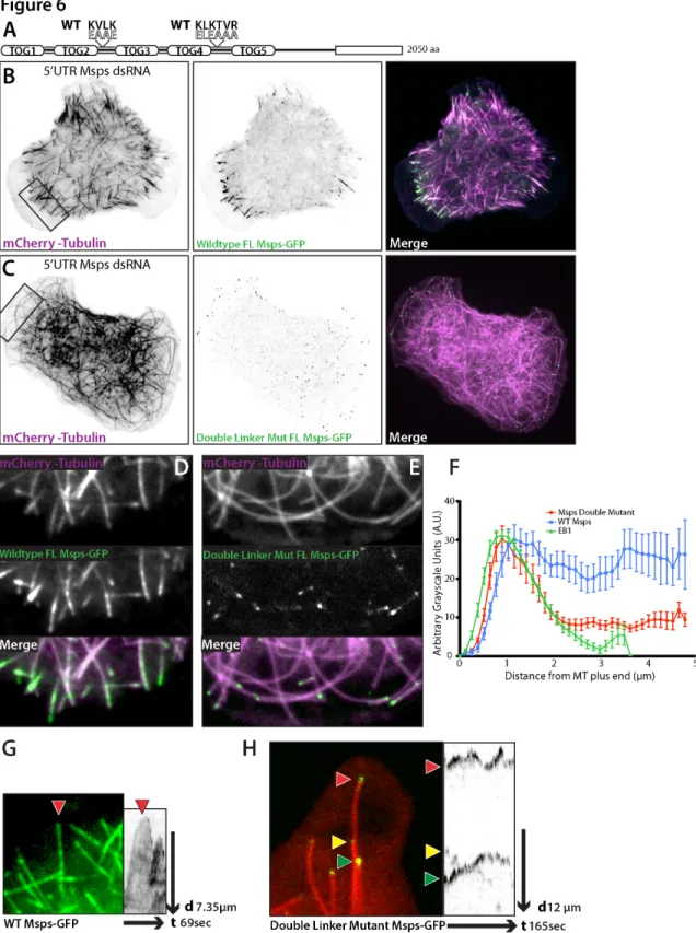
Figure 3-1. Msps localizes to both microtubule plus ends and to the lattice of peripheral microtubules. ... 89
Figure 3-2. Msps-GFP dynamics in S2 cells is EB1 dependent. ... 95
Figure 3-3. Expression of Msps TOG domains are sufficient to partially rescue Msps microtubule polymerization and EB1 dynamics. ... 104
Figure 3-4. Structure/function analysis of Mini spindles reveals two microtubule lattice-binding sites. ... 108
Figure 3-5. Msps has a novel microtubule lattice binding site that spans the linker region between TOGs 4 and 5 and TOG5 itself. ... 113
Figure 3-6. Mutation to the linker regions of Msps abrogates interaction with the lattice of peripheral microtubules. ... 119
Figure 3-S1. Expression levels of endogenous and exogenous Drosophila Msps using a novel antibody raised against TOG domain 2. ... 139
Figure 3-S3. Representative EB1::EB1-GFP kymographs of S2 cells treated with control or Msps dsRNA and transfected with Msps fragments. ... 143
Figure 3-S4. Automated microtubule tracking algorithm. ... 145
Figure 3-S5 Conserved motifs within the linker2 and linker4 of Drosophila
Msps and other Dis1/XMAP215 members. ... 147
Figure 4-1. Sentin RNAi affects the localization to microtubules of
endogenous Msps, but not EB1. ... 154
Figure 4-2. Sentin RNAi affects the velocity and lifetime of EB1-GFP comets .. 160
Figure 4-3. Sentin is responsible for the plus end accumulation of Msps-GFP and negatively regulates the microtubule lattice association Msps-GFP for interior microtubules ... 161
Figure 4-4. Msps 1707-1852 is the minimal plus end association domain of Msps. ... 163
Chapter 1 INTRODUCTION
Summary
Microtubules are a dynamic cytoskeleton that is essential for intracellular traffic, cell migration, and chromosome segregation. The dynamic nature of microtubules is regulated in cells by a diverse group of proteins called plus end proteins or +TIPs. It is the function and interplay between these +TIPs that produces the the normal dynamic instability found in vivo. In this chapter, I will outline our current understanding of microtubule dynamics in cells and how this is thought to be regulated by +TIPs. I will briefly introduce the major families of +TIP proteins and how they interact in interphase to regulate microtubule dynamics. Finally, I will examine the role for +TIPs in cell migration using a novel
Drosophila cell line called D17.
The Dynamic Microtubule Cytoskeleton
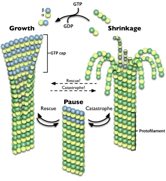
of the cell, microtubules are able to rapidly switch between phases of growth and shrinkage. This allows them to probe the cellular environment for vesicular cargo, contact sites of cortical signaling that require the delivery of key regulatory factors, or capture sister chromatids at the kinetochore during mitosis, in a process aptly termed microtubule “search and capture” (for review see Odde, 2005). The stochastic switching in microtubule behavior between phases of polymerization, depolymerization, and pause has been termed dynamic instability (Mitchison and Kirschner, 1984).
3
1974), termed the plus end, while the other minus end is stable and often anchored at the microtubule organizing center (MTOC) in animal cells.
It is the process of β tubulin GTP hydrolysis that is thought to signal a switch from polymerization to depolymerization. Shortly after incorporation, GTP is hydrolyzed and is thought to change the conformation of the tubulin heterodimer from a “straight” conformation that facilitates lateral interactions between subunits to that of a “kinked” conformation that disrupts lateral interactions between adjacent molecules (Krebs et al., 2005). Polymerization is thought to persist based on the existence of a cap of GTP bound monomers that maintain strong lateral interactions among protofilaments at the growing plus end (Caplow and Shanks, 1996) (Figure 1).
intermediate structure that microtubules must transition through to progress from growth to shrinkage, termed catastrophe, or from shrinkage toward growth, know as rescue.
5
Figure 1. Microtubules Are A Dynamic Polymer Network.
7
microtubule ends with relevant cellular sites (Akhmanova and Steinmetz, 2008). +TIPs play a key role in enhancing and regulating microtubule dynamic instability and will be the focus of the rest of this manuscript.
Plus end Proteins (+TIPs)
9
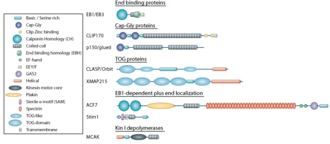
Figure 2. +TIP Families and Their Domain Structure.
End Binding (EB) Proteins
EB1 and other End Binding family members represent the core plus end protein conserved in all eukaryotic cells. All species possess at least one family member (Akhmanova and Hoogenraad, 2005), and in animals there is a ubiquitous isoform, commonly EB1 or EB3, and several tissue specific isoforms (Komarova et al., 2005). EB1 is thought to form the nucleus of the +TIP protein complex (Akhmanova and Steinmetz, 2008; Vaughan, 2005). Nearly all +TIPs have either a direct or indirect interaction with EB1, and this interaction may represent the primary plus end localization signal for other +TIPs.
11
This highlights a key question: How do +TIPs such as EB1 recognize the plus end? While the exact mechanism remains a mystery several models have arisen to explain the preference for the plus end by +TIPs such as EB1. One model postulates that EB1 recognizes a unique structure at the edges of the plus end sheet and facilitates its closure into a tube. This is based on in vitro evidence by cryo-electron microscopy that EB1 was preferentially localized to the microtubule seam where presumably protofilaments converge to close the microtubule (Vitre et al., 2008). This is also consistent with the types of microtubules that form in the presence of EB1. In vitro polymerized microtubules often deviate from the normal 13 protofilament structures found in cells, with tubes containing anywhere from 13-16 protofilaments. Under the same conditions, the addition of EB1 promotes the assembly of 13 protofilament-microtubules with a decrease in the number of higher protofilament tubes (Vitre et al., 2008). Another more prevalent model is that EB1 recognizes a specific conformation of GTP-bound tubulin that specifically labels the plus end.
GDP-bound forms. This recognition of tubulin seems to be specific to incorporated tubulin, as EB1 exhibits relatively low affinity for soluble tubulin (Slep and Vale, 2007; Niethammer et al., 2007).
13
modifications are made in vivo to EB family proteins and if it can affect their function and interaction with other +TIPs.
The majority of EB1’s interactions with other +TIPs occur through a recently characterized “SKIP” motif that binds to EB1’s dimerization domain (Slep et al., 2005, Honnappa et al., 2009). The inclusion of this motif is sufficient to target proteins to the plus end, and it is found in a wide range of proteins that possess additional microtubule association domains (e.g. CLIP170, CLASP) as well as proteins with no characterized microtubule regulatory function (e.g. RhoGEF2, STIM1). EB1 association via SKIP motifs even exists within +TIPs that antagonize microtubule growth, as in the depolymerizing kinesin 13 member MCAK (Honnappa et al., 2009). It seems that this SKIP motif is a general microtubule plus end localization signal that is a characteristic of almost every +TIP identified to date.
rescue (Komarova et al., 2009, Vitre et al., 2008). The latter agrees the most with in vivo examination of EB proteins. In Drosophila S2 cells, depletion of EB1 by RNAi lowers the frequencies of both catastrophe and rescue and results in paused, non-dynamic microtubules (Rogers, 2002). In mammalian cells, depletion of EB proteins results in a loss of persistent microtubule growth and a large increase in catastrophe frequency that is not mediated through EB1’s SKIP interacting C-terminal tail (Komarova et al., 2009). Overall, it seems that EB proteins are responsible for promoting growth through their CH domains as well as through interactions with other +TIP binding partners. Whether independently or through interactions with other +TIPs, EB proteins exert an important role in enhancing the normal dynamic instability of microtubules.
Cytoplasmic Linker Proteins (CLIPs)
15
Rich (CAP-Gly) domains necessary for binding both EB1 and tubulin’s EEY terminus (Steinmetz and Akhmanova, 2008), a serine rich microtubule-lattice binding domain, a homo-dimerizing coil-coil, a zinc-binding regulatory domain, and a C-terminal ETF motif. As mentioned previously, CLIP170, but not the shorter CLIP115 isoform, exhibits a head to tail auto-inhibition by binding of the CAP-Gly domains to the zinc binding domain and ETF motif at the C-terminus (Lansbergen et al., 2004) (Hayashi et al., 2007). It is the CAP-Gly domains that bind tubulin and EB1 and target CLIPs to the plus end (Dixit et al., 2009) (Wittmann, 2008), but the serine-rich domain also seems essential for plus end localization. It is likely that it mediates some form of microtubule lattice diffusion (Hoogenraad et al., 2000). The N-terminal CAP-Gly domains and serine-rich domains also have potent microtubule polymerization-stimulating abilities in bulk in vitro assays (Gupta et al., 2010).
the cell’s leading edge (Komarova et al., 2002). It is not entirely clear if this is due to the activity of CLIP170 or its ability to recruit CLASPs to the plus end (see below). It seems likely that the multimerization of CAP-Gly domains and a serine rich region allow CLIPs themselves to exert affects on microtubule dynamics. This question has not been definitively answered in vitro to determine the mechanism of CLIP170’s affect on dynamic instability.
Cytoplasmic Linker Associated Proteins (CLASPs)
17
2002, Al-Bassam et al., 2010). At their N-terminus, CLASPs have a series of three domains designated as TOG(1) and TOG-like(2) domains (Slep, 2009). TOG domains bind tubulin (Slep and Vale, 2007) and were first characterized in another family of +TIPs, the Dis1/XMAP215 family of proteins (see below). Between the first and second TOG-like (TOGL) domain is a serine and threonine-rich region that acts as a high affinity microtubule association domain (Wittmann and Waterman-Storer, 2005). Within this region is an EB1-binding “SKIP” motif (Mimori-Kiyosue et al., 2005). Finally, at the extreme C-terminus of the protein is the CLIP interaction domain. Although all of these domains have been elucidated and functionally tested separately, we have little knowledge about how these domains interact to affect CLASP function within the +TIP hierarchy. In mammalian fibroblasts, CLASP localizes at the plus end of leading edge microtubules, where it is associated with a stable, cortical complex of proteins including the microtubule/actin crosslinker MACF7/Shortstop, the posphatidylinositol (3,4,5)-binding (PIP3) protein LL5β, and the scaffolding protein ELKS (Lansbergen et al., 2006). These membrane bound cortical patches appear to be important in anchoring microtubules and preventing their catastrophe away from the leading edge. Additionally, cortical patches often localize near cortical actin and focal adhesions, but do not overlap perfectly. One complication is that cortical patches appear to be cell type specific, as they were observed in Swiss 3T3 fibroblasts and Hela cells, but not epithelial COS7 cells.
19
on either signaling gradients or cell type specific binding partners to modulate CLASP activity in the periphery to stabilize microtubules.
Dis1/XMAP215 TOG Proteins
The first representative of one of the most highly conserved +TIP families
was Xenopus XMAP215. It was the first +TIP identified, isolated from a fraction of
21
proteins, Dis1 and Alp14 (Garcia, 2001), all other species seem to possess only one XMAP215 homolog (Kinoshita et al., 2002). While other proteins also possess TOG domains (e.g. CLASP family members), there is little redundancy in their functions as a core regulators of microtubule dynamics. Dis1/XMAP215 family members interact with the plus end through their TOG domains, which bind tubulin using a tandem pair of TOG domains. It is thought that the TOG domains act to rapidly add or subtract tubulin from the plus end through their TOG domains (Slep and Vale, 2007; Al-Bassam et al., 2007). Several in vitro studies have focused on XMAP215’s ability to contribute to individual facets of microtubule dynamics such as growth or disassembly (Popov et al., 2002; Brouhard et al., 2008; Shirasu-Hiza, 2003), but the exact mechanism by which XMAP215 functions to increase overall microtubule dynamics has yet to be fully determined. One model postulates that because TOG domains bind tubulin from solution and facilitate addition to the plus end, and the local concentration of soluble tubulin heterodimer is what dictates whether TOG proteins contribute to growth or shrinkage (Brouhard et al., 2008). When the local concentration of tubulin drops below a certain level, TOG proteins bind to tubulin at the plus end, stripping monomers from the end and promoting depolymerization.
et al., 2001), and amoeba (Gräf et al., 2003) have arrays of five TOG domains, but lack the dimerization domain of their yeast homologues. As mentioned previously, TOG domains are comprised of six adjacent pairs of α helices connected by inter helical loops (Slep, 2009). By sequence analysis, the most conserved feature between TOG domains of separate species is the inter-helical loops that form a linear interface that is hypothesized to interact with tubulin. Of particular note is a conserved tryptophan (or phenylalanine in the case of TOG5) within the first inter-helical loop. Using a bacterially purified TOG1-2 construct, a binding shift can be observed with purified tubulin using gel filtration. Mutation of the either one or both tryptophans results in a stepwise loss of tubulin interaction in this assay (Slep and Vale, 2007).
23
vitro study using single molecules of XMAP215 to measure microtubule dynamics using total internal reflective fluorescence (TIRF) microscopy (Brouhard et al., 2008). Examining single molecules, the study’s authors observed processive movement of XMAP215 with the growing or shrinking microtubule end. In addition, the authors observed XMAP215 formed a 1:1 complex with tubulin by gel filtration, suggesting that this was the normal stoichiometry at the plus end. The authors proposed an alternate model where by XMAP215 family members processively move with the plus end, rapidly adding or subtracting single heterodimers to the plus end. One key question is how these observations correlate with the in vivo functions of XMAP215 family members. Exploration of this topic will be the subject of later chapters.
Kinesin 13, or Kin I Depolymerase Motor Proteins
localization domain, a Kin I-specific neck domain, a conserved core motor domain, and a C-terminal homodimerization domain.
The motor core domain of Kin I proteins has conserved ATP-binding features compared to other Kinesin motors, but also has unique features that are thought to aid its function of microtubule depolymerization. In particular, the α4 helix is the main energy transducer of the motor domain. This helix is thought to protrude from the arrow-like configuration of the motor domain into the intra-dimer cleft between α and β tubulin (Niederstrasser et al., 2002; Ogawa et al., 2004; Mulder et al., 2009). Based on this structural modeling, it is hypothesized that Kin I proteins use ATP-driven motor activity to induce a “bent” confirmation to the tubulin heterodimer that destabilizes lateral interactions and promotes the peeling “rams head” depolymerizing plus end structure. Kin I proteins are competent to form these microtubule end structures in vitro even in the presence of microtubule stabilizing reagents such as paclitaxel or the non-hydrolyzable GTP analog, GMPCPP (Desai et al., 1999; Hertzer et al., 2006). This suggests that Kin I proteins exert a measurable force to affect microtubules even in the presence of stabilizing agents that inhibit normal dynamic instability.
25
observed rapid diffusion of the molecule in vitro toward microtubule ends (Cooper and Wordeman, 2009). The discrimination of the Kin I motor for the microtubule ends versus the lattice seems to be a property of the N-terminus and neck domain, aided by dimerization through the C-terminus (Cooper et al., 2010).
The neck domain is a large stretch of highly charged residues. This appears to have multiple roles in the process of Kin I depolymerization. Kin I constructs missing the neck or with mutations that neutralize the neck’s charged amino acids have lower microtubule association rates, suggesting that the neck is vital for the initial attachment to microtubules (Cooper et al., 2010). These same mutants also display lower rates of microtubule depolymerization, even at saturating concentrations when microtubule association and diffusion have negligible affects on depolymerization. This suggests that the neck has an additional role in Kin I removal of tubulin heterodimers at the plus end. The addition of the N- and C-terminus appear to offer specific advantages and disadvantages as a microtubule depolymerase. A construct encoding only the neck domain and motor core displays the most potent depolymerization activity in vitro (Cooper et al., 2010).
What purpose then do the other domains add to Kin I function? In the case of the N-terminal localization domain, it seems that this domain acts to specifically enrich Kin I motors at the growing end. In human MCAK and
Drosophila KLP10A this is accomplished through EB1 SKIP motifs (Mennella et
polymer shrinkage. It is not yet clear if this is a constitutively active process or if these proteins become active through some regulation such as phosphorylation (Jiang et al., 2009). At the C-terminus, the dimerization domain exerts the most negative effect on the total Kin I depolymerization activity in vitro (Cooper et al., 2010). From in vitro assays, monomeric constructs that retain the neck and N-terminus, associate and dissociate with microtubule lattice at elevated rates and seem to target to the plus end much faster than wildtype constructs. Surprisingly however, these constructs are ten fold less efficient at removing tubulin from the plus end in vitro. Dimerizing two full length motors may enhance the ability of Kin I motors to processively peel heterodimers off of the plus end as well as enhance their ability to dissociate from their heterodimer substrate to begin another cycle of depolymerization (Cooper et al., 2010).
The activities of Kin I motors have often been viewed as antagonistic to other +TIPs that contribute to microtubule growth and stability such as CLASPs (Laycock et al., 2006) and Dis1/XMAP215 (Hyman et al., 1999) family members. This simplistic model of polar antagonism toward either growth or shrinkage is largely based on the interpretation of opposing mitotic phenotypes in Drosophila
27
degree of microtubule depolymerization as control treated cells. Overexpression of ch-TOG however, caused a surprising destabilization of microtubules that was entirely dependent on MCAK expression (Holmfeldt et al., 2004). This suggests that the role for mammalian ch-TOG and MCAK may be more specialized to regulate specific parameters of dynamic instability and not overall polymer and organization. This is in contrast to interphase Xenopus extracts, which have lower microtubule growth and shrinkage rates when depleted of XMAP215. In this experimental system, XKCM1 (the Xenopus MCAK) seems to specifically influence catastrophe frequency which can be elevated by depletion of XMAP215 (Hyman et al., 1999; Kinoshita, 2001), suggesting that the relationship between these proteins is much more complex than an antagonism between polymerase and depolymerase. This topic will be be further expanded upon in later chapters.
Novel +TIPs
non-plus end microtubule interactions domains, to be coupled to the plus end to spatially regulate cellular behavior or microtubule organization.
One example of a non-microtubule protein that relies on an EB1 SKIP motif to localize to plus ends is the Drosophila Rho guanine nucleotide exchange factor, RhoGEF2. Immunofluorescence or GFP-tagged RhoGEF2 in Drosophila
S2 cells revealed a localization identical to that of EB1, i.e. as a “comet” associated with the growing plus end (Rogers et al., 2004b). In Drosophila
29
also provides an additional mechanism for translocating and remodeling the endoplasmic reticulum.
One example of a MAP that uses EB1 plus end localization to affect microtubule behavior at the plus end is the ACF7 spectraplakin family of microtubule-actin crosslinkers (Kodama et al., 2003). These protein are enormous polypeptides with internal coil-coil plakin and spectrin-like repeats. At their N-terminus are a pair of actin binding calponin homology (CH) domains. At their C-terminus is GAS2 microtubule lattice binding domain. The Drosophila
Interfacing at the Plus End
Above, I have outlined the major +TIP families and how they contribute to regulate interphase microtubule dynamics. One of the enduring questions concerning +TIPs is how these proteins are integrated, regulated, and functionally balanced to produce the dynamics and behaviors at the microtubule plus end. This is an intriguing question, as the plus end is a somewhat nebulous cellular organelle, dynamic and below the diffraction limit of light microscopy. Added to that are the diverse set of molecules with contradictory functions that are all acting, seemingly simultaneously, on this same substrate, the plus end. Many studies have examined the individual contribution of proteins in vivo through depletion or overexpression techniques. Furthermore, structure-function studies at both the molecular and atomic level have yielded detailed domain maps of the major +TIP families. The next step will consist of taking these data and beginning to functionally test how these domains contribute to both microtubule behavior (dynamics and organization) as well as to the +TIP hierarchy among interacting partners, both known and unknown.
31
33
Figure 3. Microtubules are Essential for Key Cellular Processes.
The Role of +TIPs in Cell Migration
Cell migration is a multi-step process that involves the coordination of distinct cellular pathways to produce net cell locomotion (for review see Ridley et al., 2003). Cell motility begins with an initial polarization of the cell that identifies a direction of migration via a chemotactic, haptotactic, or durotactic signal. This can happen before the cell undergoes any morphological changes, but once initiated, the unique morphological subdomains that are established give the cell an asymmetry that reinforces polarity through the positive feedback of master regulatory proteins.
35
translocation and the final step of retraction of the cell rear. Cell retraction requires the de-adhesion of focal adhesions through endocytosis of integrin, disassembly of intracellular focal adhesion components, and myosin-II based constriction to pull up adhesions from the substrate. These multiple steps happen more or less sequentially in cycles and must be tightly coordinated between leading and lagging edge of the cell to couple protrusion and retraction to produce movement.
and GAPs to the leading edge via interactions with +TIPs, helps to regulate the balance between antagonistic Rho GTPases to create cycles of protrusive lamellipodia (Fukata et al., 2002; Pegtel et al., 2007).
37
down. In the same way that microtubules activate Rho at focal adhesions toward the direction of migration, microtubules similarly target the lagging edge of the cell to activate myosin II-based contractility downstream of Rho to facilitate retraction of the cell rear . This is also thought to be facilitated by endocytosis of the integrins at the cell rear to disassembly substrate attachments (Batchelder and Yarar, 2010).
+TIPs in Drosophila Cell Migration
Drosophila melanogaster represents an ideal model organism in which to study +TIP function. In terms of gene redundancy, Drosophila has only one isoform of most of the +TIP families, with the exception of EB proteins. In this regard, EB1 seems to be the major end binding protein and although similar proteins have been identified, they do not seem exhibit major overlap functionally with EB1. In addition to a more concise number of +TIPs, embryogenesis during the blastula stage of Drosophila development is dependent on the integrity of syncytial mitotic divisions, leading to the initial identification and characterization of many +TIPs through mutagenesis screens (Cullen et al., 1999; Lemos et al., 2000; Rogers et al., 2004a). In addition to being a developmental in vivo model organism, immortalized Drosophila cell lines such as Schneider’s line 2 (S2) cells have provided an in vitro system to examine the molecular behavior of
Drosophila +TIPs in cells (Rogers, 2002; Brittle and Ohkura, 2005; Sousa et al.,
2007; Applewhite et al., 2010). S2 cells not only offer a concise genome and the ability to observe intracellular dynamics at high resolution, but they are also highly susceptible to RNA interference (RNAi) gene knockdown. Despite these advantages, S2 cell lines are limited in the number of cellular behaviors that can be analyzed ex vivo. S2 cells exist as single cells and do not form cell-cell contacts or exhibit any form of motility.
39
wound assay over 16 hours. To perform this assay, D17 cells were cultured to monolayer confluency and then wounded along a line of approximately 300-400µm with a sterile pipet tip. Cells on the wound margin sense the loss of cell contact and migrate in to fill the wound. Mammalian fibroblasts or epithelial cells often close the wound as a contiguous line of cells. In the case of D17 cells, the cells sometimes retain cell contacts as they move, but primarily populate the wound as single cells and then regain cell-cell contacts as their confluency increases. By manually tracking cells as they migrate into this scratch wound assay, I calculated parameters of migration such as velocity, distance traveled, and directionality for single cells as they migrated in to the wound.
From this initial screen, I was able to identify +TIPs that displayed specific loss and gain of function phenotypes upon RNAi depletion (Figure 4). RNAi of the
Drosophila CLASP homologue, Orbit and CLIP190 resulted in an increase in
migration velocity. In contrast, RNAi of EB1 or Mini spindles (Msps) the
Drosophila XMAP215 homologue, caused a decrease in the overall velocity of
41
Using these initial results, I decided to more carefully examine the
interplay between +TIPs in S2 cells. My goal has been to understanding the
complex hierarchy of protein interactions at the plus end. To this end, I have
focused on the core plus end proteins: EB1 and XMAP215. EB1 is the primary
plus end hub molecule (Akhmanova and Steinmetz, 2008) and acts as a primary
localization signal for other +TIPs (Honnappa et al., 2009). XMAP215, in
contrast, is one of the few +TIPs that has been shown to localize to plus ends
independently of EB1 (Brouhard et al., 2008) and is thought of as a master
regulator of microtubule growth (Kinoshita et al., 2002). It is both the interplay
between EB1 and XMAP215 and other +TIPs, as well as the contribution of
XMAP215ʼs unique domain structure that sculpt in vivo dynamic instability
CHAPTER 2
D17-c3, A NOVEL DROSOPHILA MELANOGASTER CELL CULTURE SYSTEM FOR STUDYING CELL MOTILITY
This chapter is an initial characterization and protocol for using the D17 cell line that I developed in conjunction with my graduate advisor, Stephen Rogers. This manuscript is currently under secondary review for publication and the formatting
of this chapter is based on the journal’s protocol format.
Summary
Cultured Drosophila melanogaster cell lines such as S2 or S2R+, have
become an important tool in uncovering fundamental aspects of cell biology as
well in gene discovery. Despite their utility, these cells lines are non-motile and
cannot build polarized structures or cell-cell contacts. Here we outline a
previously isolated, but uncharacterized Drosophila cell line named Dm-D17-c3
(or D17). These cells spread and migrate in culture, form cell-cell junctions, and
are susceptible to RNA interference (RNAi). Using this protocol, we will outline
how investigators, upon receiving cells from the Bloomington stock center, can
culture cells, and prepare the necessary reagents to plate and image migrating
phenotypes through RNAi. From first thawing frozen ampules of D17 cells,
investigators can expect to begin assaying RNAi phenotypes in D17 cells within
roughly two to three weeks.
Introduction
Cultured Drosophila melanogaster cells have become a powerful genetic tool for the study of numerous cell biological processes. Established lines such as S2 and S2R+ offer high resolution cytology, simple culture conditions, a fairly homogeneous morphology when seeded on the lectin concanavalin A, and most importantly, a potent susceptibility to RNA interference (RNAi) that can be applied on a genomic level(Schneider, 1972; Yanagawa et al., 1998; Clemens et al., 2000; Rogers, 2002; Ramadan et al., 2007). These benefits are additionally advantageous due to the succinct nature of the fly genome(Adams et al., 2000) (roughly ~14,000 genes) and the potential to easily move cell culture observations into a model organism (Rogers et al., 2008). The end result has been a cell culture system that has been applied to disciplines as diverse as developmental biology (Wheeler et al., 2009; Johnston et al., 2009; Jiang et al., 2007), microbiology (Dorer et al., 2006), and high-throughput functional genomics (Wagner et al., 2007; D'Ambrosio and Vale, 2010).
45
cells that lack characteristics such as cell-cell junctions, which are necessary structures for organismal development.
The Drosophila Genomics Resource Center (DGRC,
https://dgrc.cgb.indiana.edu/) in Bloomington, IN is a repository for dozens of
Drosophila cells lines derived from various fly tissues at different developmental
stages. Many of these cell lines have remained uncharacterized with respect to their ability to respond to RNAi or their real-time dynamics in culture.
One such line, Dm-D17-c3 (hereafter referred to as D17), was isolated in addition to roughly two dozen other cell lines in the lab of the late Tadashi Miyake at the Mitsubishi-Kasei Institute of Life Sciences in Tokyo. Although only half of these lines were ever published(Ui et al., 1987), the entire collection of lines isolated from third instar larvae is currently available at the DGRC (https://dgrc.cgb.indiana.edu/cells/store/catalog.html?category=2). The D17 and their sibling lines were cultured from dissected imaginal discs, pools of multipotent cells in the developing Drosophila larvae responsible for creating the vast array of adult tissue needed during metamorphosis.
Characteristics of D17
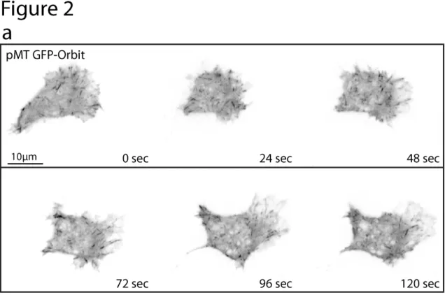
a robust motility of roughly 25-30µm/hour (Fig. 1, panel b), similar to the initial speeds of Drosophila hemocytes in vivo as they migrate along the ventral midline(Wood et al., 2006) (~24µm/hour). Additionally, when migrating, these cells adopt a morphology reminiscent of mammalian cells migrating in culture; producing a polarized, triangular organization with a large fan-like lamella at the leading edge and trailing retraction fibers in the rear of the cell (Fig. 2a).
Although the D17 cells were originally believed to be epithelial cells due to the nature of the tissue they were derived from(Ui et al., 1987), the cells display properties reminiscent of Drosophila hemocytes which are migratory Drosophila
47
suggesting that these two cell lines may have comparable expression profiles. Recent work from the modENCODE project has further characterized the transcriptional profile of 25 established Drosophila cell lines, including the D17 cell line(Cherbas et al., 2011). Expression profile clustering demonstrated that D17 cells share broad expression similarities to embryonic S2R+ cells. However, based on the expression of the homeotic transcription factor, Teashirt(Fasano et al., 1991), in D17 cells, the authors conclude that D17 cells are indeed imaginal haltere disc cells. This conclusion is somewhat confounded by the fact that embryonic S2 and S2R+ cells also express relatively high levels of Teashirt based on modENCODE expression data and it seems that this fact alone does not explicitly reveal the identity of the D17 cell line. Further work will be necessary to establish the identity of the D17 cell line based on gene expression, cell biological observations, and biochemical analysis between various
Drosophila cell lines. Hopefully, continued use of the D17 cell line further open up
intriguing questions about the mechanisms of cell identity, cell morphology, and cell motility.
Advantages and key applications of D17
depletion(Clemens et al., 2000) without the use of lipid-based transfections or virus infection. This methodology has been greatly enhanced by commercial and academic resources that provide online tools for designing dsRNA primers (Harvard Drosophila RNAi Screening Center, http://www.flyrnai.org), obtaining cDNA templates (Open Biosystems, http://www.openbiosystems.com), and procuring genome-wide as well as custom sub libraries (Harvard DRSC) of RNAi targets.
The utility of the D17 cell line should augment the studies of many in the cell biology and Drosophila communities that wish to study target genes and pathways in the context of biological processes such as cell migration and cell-cell interactions (Fig. 3a). In addition, many of the protocols already established for S2 cell culture(Rogers and Rogers, 2008) can easily be adapted for D17 cell culture. The D17 cell line represents an untapped resource to enhance existing questions in various fields as well as provide a model to ask new questions about the genetic basis of cell migration, chemotaxis, and cell differentiation.
Overview of the Procedure
49
to previously demonstrate the negative regulation of D17 migration by the microtubule severing enzyme, Katanin(Zhang et al., 2011).
EXPERIMENTAL DESIGN
RNAi: D17 cells can be treated in much the same way as common S2 and S2R+ lines (Steps 9-13). For any RNAi experiment, it is important that users include important RNA interference controls to ensure that RNAi phenotypes are penetrant and effectively affect migration. We recommend using the small GTPase Rho as a positive control for penetrant RNAi phenotypes in S2 cells as well as D17 cells as it can be assayed by western blot (Fig. 1e) and produces a easily visible multinucleate phenotype (Fig. 1d)(Rogers and Rogers, 2008; Drechsel et al., 1997). As a positive control for RNAi migration phenotypes, we suggest using the Drosophila Arp2/3 activator SCAR. SCAR exists as a single isoform in Drosophila and significantly inhibits D17 migration (Fig. 1c). RNAi against the small GTPase Rac can also be used to inhibit D17 migration, but investigators should note the compensatory effects of the two Drosophila Rac isoforms and related GTPase Mtl(Paladi and Tepass, 2004; Stramer, 2005). Control treated cells can be treated with dsRNA made against the Bluescript plasmid(Rogers and Rogers, 2008).
D17 cells express different levels of both proteins. Seven day treatment of dsRNA using the protocol outlined here results in a similar degree of knockdown between cell lines (Fig. 1e)., We suspect that the kinetics of protein depletion may be slightly slower in D17 cells due to their longer doubling time of 48-72 hours versus a 24 hour doubling time in S2 cells (JDC and SLR, unpublished observations). In our hands, potent depletion (greater than 80% knockdown) in D17 cells can usually be obtained within 7-9 days. We recommend that investigators empirically optimize the length of dsRNA treatment in D17 cells for each RNAi target to achieve the optimal knockdown.
51
Because the concentration of ECM components can vary from batch to batch, it is important that investigators test newly made ECM to ensure consistency across many experiments. To do this investigators should plate and image D17 cells at a range of ECM concentrations (diluted in PBS, see step 14, generally ranging from 1:10 to 1:300). Analyzing the velocity and other parameters of migration (see step 21) for each concentration should allow investigators to find an ECM concentration that either replicates results from a previous ECM batch or displays the desired parameters of migration for a given experimental approach.
Transient transfection: D17 transfection will allow the user to image the dynamics and localization of exogenous transgenes through high resolution immunofluorescence or in real time using bioluminescent tags such as EGFP. D17 cells are amenable to most of the expression constructs currently used for S2 cells. One caveat is that D17 cells seem to be somewhat insensitive to the commonly used Metallothionein promoter constructs (referred to as pMT vectors). These reagents can be used, but require significantly higher doses of copper sulfate to induce gene expression (between 500µM-5mM concentrations). Based on our experience, constitutive promoters such as the actin promoter or
the OpIE2 promoter, give medium to high levels of expression and can be used
only expresses an affinity or bioluminescent tag in D17 cells can both troubleshoot problems with the transfection protocol and control for the effects of toxic gene expression with a given assay. Users can also test transgenes by transfection in S2 cells or other non-motile Drosophila cell lines to determine the effects of their transgene on cell morphology. Using this protocol we normally achieve 10-40% transfection efficiency in D17 cells after 48 hours. The transfection protocol outlined here differs from protocols for transecting S2 cells by which we can commonly achieve 20-70% transfection efficiency, but both cell types seem to respond poorly to the transfection protocol of the other cell type.
53
measure both wound closure as well as the migratory properties of single cells moving into the wound.
When imaging D17 cells as single cells or within the context of a scratch wound assay, it is important that investigators consider: 1) the resolution of observation, 2) the temporal resolution of time series acquisition, and 3) maintaining consistency between conditions.
The magnification used for acquiring images of D17 migration should encompass the entire wound when using a scratch wound assay. When tracking single cells, the resolution for image acquisition should also be adequate to provide the necessary contrast and resolution to faithfully track cells as well as determine differences in morphology. We recommend using between 10 and 20x magnification for scratch wound assay, and increasing magnification to between 20-40x when imaging and tracking single D17 cells.
the cell’s exposure to heat and evaporation caused by light illumination. Typically, intervals of 3-7 minutes should be an adequate rate of acquisition to alleviate these effects.
Finally, users should attempt to maintain consistent assay conditions across various experimental conditions. This includes the number of cells treated and plated for each condition, although this can difficult to avoid when treatments affect cell viability. Performing a wound healing assay with a pipette tip can also cause inconsistencies between conditions. Users should attempt to create the most uniform scratches possible between wells by maintaining a constant angle and pressure on the pipette tip while wounding cell monolayers. Custom and commercial apparatus can also be used to aid in wounding multiwell plates of D17 cells (Vitorino and Meyer, 2008; Yarrow et al., 2004).
55
57
(a) D17 cells plated to monolayer confluency and scratched with a 200µl pipet tip (left) and allowed to migrate into the the wounding area over 16 hours (right). Representative migration tracks from two cells are indicated on the right in blue and green. The full movie is can be found in Movie S1. (b) Measure
instantaneous or step velocity of individual D17 cells from either a control or SCAR dsRNA treatment. N=3, 10 cells tracked for each experiment. Error bars indicate SEM. (c) D17 cells treated with SCAR dsRNA and wounded at 0 hours (top) and after 16 hours (bottom). (d) D17 cells after seven days of either control dsRNA (left) or Rho dsRNA (right). Depletion of Rho results in cytokinesis
59
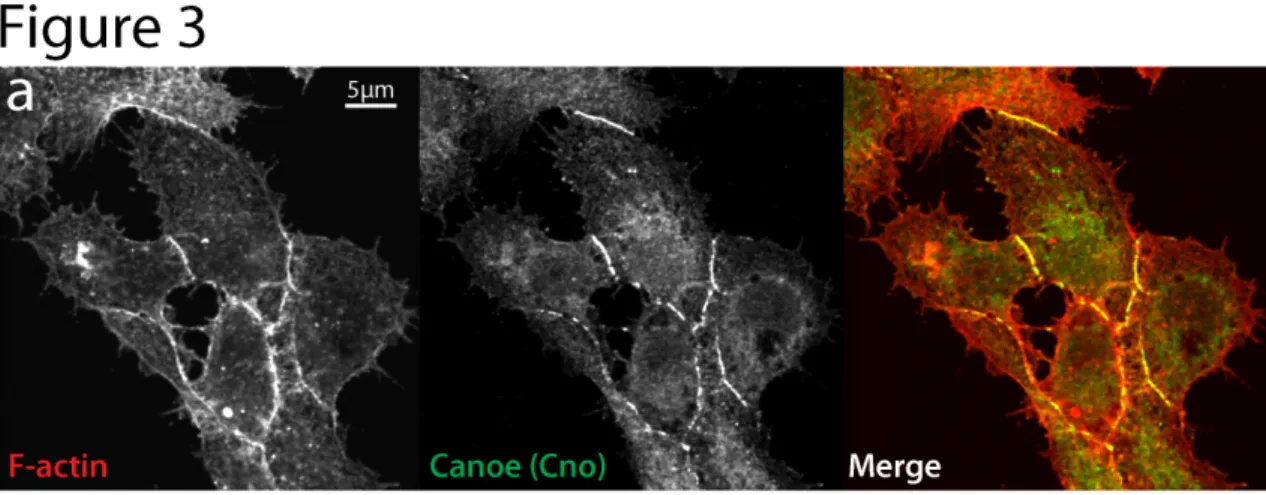
Figure 2-3. Immunofluorescence localization of cell-cell junction protein Canoe (Cno) in D17 cells.
MATERIALS
Reagents:
· ML-DmD17-c3 cell stocks are available from the Drosophila Genomics
Resource Center (https://dgrc.cgb.indiana.edu/)
· Schneider’s Insect medium (Invitrogen, cat. no. 11720-034) · Antibiotic/Antimycotic (Invitrogen, cat. no. 15240-096) · Fetal Bovine Serum (FBS) (Invitrogen, cat. no. 10099-141) · Human Insulin (Invitrogen, cat. no. 12585-014)
*CRITICAL* Many vendors sell FBS that is already heat-inactivated, usually at temperatures exceeding 65°C. This heat-inactivated serum is not suitable for D17 cell culture as it does not support long term cell passage, presumably because of the inactivation of important trophic factors. Non-heat-inactivated serum can be purchased and heat-inactivated by submerging the thawed serum in a 55°C water bath for one hour with occasional inversions to mix the serum.
· Cell Dissociation Buffer, PBS based (Invitrogen, cat. no. 13151-014) · D17 growth medium (see REAGENT SETUP)
61
· Hydrogen Peroxide (Fisher Scientific, cat. no. H325-100)*CAUTION*
Irritant.
· Sterile Phosphate buffered solution (PBS) (Invitrogen, cat. no. 10010-023) · Saturated Ammonium Sulfate solution (Sigma-Aldrich, cat. no. A6387)
(see REAGENT SETUP) *CAUTION* Irritant. Users should wear gloves when handling ammonium sulfate.
· Conditioned D17 growth medium (see Box 1)
· Fugene HD Transfection reagent (Roche, cat. no. 04709705001) · sterile water
· Suitable transfection vectors e.g. Metallothionein promoter pMT vectors
(Invitrogen, cat. no. V4120-20), OpIE2 promoter pIZ vectors (Invitrogen, cat. no. V800001) or Actin promoter pAc5.1 vectors (Invitrogen, cat. no. V4110-20). Vector backbones can be modified or purchased to
accommodate bioluminescent probes and other user-specific transgenes.
· D17 freezing medium (see Reagent Setup)
· dsRNA. These are user-defined and created based on standard
methods(Rogers and Rogers, 2008)
Equipment
· UV transilluminator (such as Fisher Scientific, cat. no. PBDLT88AQ) or
· Polystyrene petri dishes, 35 x 10 mm2 (Becton-Dickinson, cat. no. 351008)
· Glass bottom multi-well plate (Greiner, cat. no. 662892)
· Optical grade polystyrene/Polymer base multiwell plate (Nunc, cat. no.
165305)
· Glass coverslips, no. 1.5, 22mm2 (Corning, cat. no. 2940-225)
· Coverslip rack, porcelain (Coors, via Thomas Scientific cat. no. 8542E40)
· swinging -bucket tabletop centrifuge
· tissue culture flask vessels, 25cm2 (T25) (Becton-Dickinson, cat. no.
353082)
· tissue culture flask vessels, 75cm2 (T75) (Becton-Dickinson, cat. no.
353135)
· Sterile, laminar flow hood
· 500ml sterile bottle-top filter flask (Corning, cat. no. 431117)
· 25cm cell scraper (Becton-Dickinson, cat. no. 353086)
· Dialysis tubing (Pierce cat. no. 68035)
· Dialysis clips (Pierce cat. no. 68011)
· Inverted microscope with CCD camera
· Microscope image acquisition software e.g. MetaMorph (Molecular
Devices) or NIS Elements (Nikon)
63
Coverslip preparation (Timing - 1.5 hours) Before ECM-treating glass coverslips, coverslips should be cleaned by immersion in a strong acid for at least one hour. Before treatment, load untreated coverslips into porcelain racks. To prepare the acid solution, add two parts sulfuric acid to one part hydrogen peroxide. Typically this volume is 200ml of sulfuric acid added to 100ml of hydrogen peroxide within a one liter glass beaker. Carefully lower the porcelain rack(s) into the acid solution and ensure that the coverslips are fully immersed. After one hour acid treatment, coverslips should be rinsed continuously with sterile running water for 15 minutes and dried. Once dry, place in 35mm dishes either individually or together to prevent dust accumulation on their surface.
Reagent Setup
D17 freezing medium: (Timing - 10 minutes) D17 freezing medium can be made by mixing 4ml of conditioned D17 growth medium, 4ml of fresh D17 growth medium, and 1ml of sterile DMSO.
65 PROCEDURE
Thawing D17 cells *Timing - 1.5-2.5 hours*
1. In a sterile laminar flow hood, add 7 mls of D17 growth medium to a T25 tissue culture flask; this should be carried out prior to thawing the cells. *CRITICAL STEP* Because the delivery conditions of dry ice are suboptimal for the storage of frozen cells, it is important that the ampule of cells be plated as soon as it arrives from Bloomington.
2. Remove the vial of cells from the dry ice delivery container and immerse the bottom of the ampule in a 37°C water bath, shaking often to thaw the ampule as quickly as possibly to room temperature.
*CRITICAL STEP* All attempts should be made to keep the cryo vial lid from coming into contact with the water bath water. This will prevent contamination of the D17 cells.
3. Once the ampule contents are liquid, remove from the water bath and sterilize the ampule by wiping the outside with a 70% ethanol solution.
5. Leave the cells for 1-2 hours at room temperature (25 oC) to attach and spread on the bottom of the culture flask. Then, remove the medium and replace it with a fresh 7 mls of growth medium to remove any DMSO that was present in the frozen cell solution. D17 cultures can be grown in a sterile incubator or on a shelf at 25°C. Flasks do not require gas exchange vents or special atmospheric considerations.
Passaging D17 cultures *Timing - 20-60 minutes*
6. Remove the D17 growth medium from the culture flask and store this conditioned medium at 4°C for extracellular matrix production (See Box 1).
7. Split D17 cells using either Cell Dissociation Buffer (option A) or using a cell scraper (option B).
(A) Passaging using Dissociation Buffer
67
magnification, the cells should be seen to round up and lose cell-cell adhesion.
*Troubleshooting*
(iii) Gently remove the Cell Dissociation Buffer with a pipette and add 5 mls of fresh D17 growth medium. Vigorously pipette the medium up and down several times to detach the cells from the bottom of the culture flask.
(B) Passaging using a cell scraper
(i) Add 6mls of fresh D17 growth medium to the flask. Insert the cell scraper into the flask and move the blade across the flask
bottom, trying to remove a maximum amount of cells with the least blade strokes.
vessels as the cells contained within the foci should be competent to repopulate a new vessel. Aliquots of D17 cells can be frozen in D17 freezing medium using previously published protocols for S2 cells (Rogers and Rogers, 2008).
*CRITICAL STEP* The growth rate and survival of D17 cells is very
sensitive to plating density. The most common error when culturing D17 is to plate them at low densities which can result in extremely slow growth or cell death. An optimal density of 40-60% is essential for healthy cells and confluent passaging between 3-6 days.
CRITICAL STEP: The user should keep track of passage iterations as D17 cells will reach a terminal state after 25-30 passages where they may respond poorly to RNAi or transfection and may have slow growth or abnormal morphology.
*Troubleshooting*
RNA interference treatment *Timing*: 7-9 days
69
10. Allow the cells to adhere to the vessel bottom for one hour at room temperature (25°C). Cells can be transfected (see Box 2) within 30 minutes after plating and at anytime during RNAi treatment.
11. Remove media and replace with culture medium containing approximately 10µg/ml dsRNA. Treated cells should be incubated as normally passaged cells at room temperature (25°C).
12. Repeat dsRNA treatment (step 11) every two days for 7-9 days of treatment.
13. At 6-9 days of dsRNA treatment, harvest cell lysates for western blot analysis or transfer to ECM-coated vessels to assay for loss of function phenotypes e.g. migration phenotypes can be assayed as described in steps 18-21 below. The user should empirically determine the optimal length of treatment for each dsRNA construct and gene product. *Troubleshooting*
Plating D17 cells on ECM-coated surfaces *Timing- 1 hour*
14. Using a stock solution of D17 cell ECM (see Box 1), dilute the ECM solution in sterile PBS to the desired concentration. The exact
based on the optimal assay conditions and the yield of ECM purified per batch (see Experimental Design). As a general starting point, dilutions of ECM to PBS ranging from 1:50 - 1:200 seem to support robust attachment and migration in our hands.
*Troubleshooting*
15. Apply enough of the ECM/PBS solution to cover the surface of the coverslip or glass-bottom plate intended for culture.
16. Crosslink the ECM solution to the glass surface by plasma treatment for several minutes or high energy UV treatment for 45 minutes. For many users, placing the dish or coverslip onto the surface of a UV
transilluminatior used to visualize Ethidium bromide-stained agarose gels is sufficient, provided it can produce enough energy (approximately 8000 µw/cm2) to crosslink the ECM to the glass surface.
71
hour. Cells are viable on these crosslinked surfaces for several days and can proliferate to form confluent monolayers if plated in sub-confluent number. Generally, for sub-confluent plating, cell densities of 4,000-5,000 cells/mm surface are sufficient while densities of 7,000-8,000 cells/mm when cell monolayers are desired.
Wound Migration Assay *Timing - 18-24 hours*
18. Seed cells at monolayer densities on either tissue-culture treated vessels or ECM-coated glass surfaces at room temperature (25°C). Cells can be allowed to settle anywhere from 2-24 hours before wounding cell
monolayers. Additionally, if too few cells are initially seeded, users can apply additional cells to increase the confluency of D17 cells to monolayer density.
19. Once monolayers are obtained, use a 200µl pipette tip or other pointed implement to scratch though the monolayer, creating a wound. Wounds should be at least 200µm in size without visible obstructions of cell debris or substrate etching.
21. Mount the vessel on an inverted light microscope with phase contrast or DIC filters and begin imaging. D17 wounded monolayers can be imaged over the course of 18-24 hours using either discrete timepoints (option A) or a timelapse series (option B).
(A) Imaging D17 wounded monolayers using discrete timepoints
(i) Keeping the vessel stationary on the inverted microscope throughout the migration assay, acquire images of the wound at regular intervals or a acquire a set of images representing the beginning and end of the migration assay.
(ii) After acquiring all timepoint images, ImageJ software can be used to outline the scratch area and measure the area of the wound at each timepoint. By comparison of the total area between the initial timepoint and later timepoints, investigators can obtain the percentage of wound closure over a given time.
73
images at regular intervals of between 3-7 minutes (see Experimental Design).
(ii) Using ImageJ software, timelapse intervals can be assembled into a multi-image tiff file.
(iii) Apply the Manual Tracking plugin (Fabrice Cordelières,
http://rsbweb.nih.gov/ij/plugins/track/track.html) to the multi-image tiff files to track individual cells as they
migrate into the wounded area. Parameters such as
instantaneous and mean velocity, distance, and
directionality can be obtained using this plugin. Analysis
of D17 single cell motility at subconfluent densities can
also be performed using steps i-iii.
TROUBLESHOOTING
Troubleshooting advice can be found in Table 1
TIMING
Equipment Setup: coverslip preparation ~ 1.5 hours
Reagent Setup: D17 growth medium ~ 15 minutes once all ingredients are thawed
Reagent Setup: Saturated ammonium sulfate ~ 30 minutes Steps 1-5: Thawing D17 cells, 1.5-2.5 hours
Step 7(A) Cell Dissociation buffer: 25-60 minutes Step 7(B) cell scraper: 20 minutes
Steps 9-11: RNAi plating and initial treatment, 1.5 hours Step 12: Subsequent dsRNA treatment, 30 minutes Step 13: Full dsRNA treatment, 7-9 days
Steps 14-17: Plating D17 cells on ECM surfaces, 1 hour
Steps 18-21: Wound healing assay, 18-24 hours and can be performed concurrently with the end of dsRNA treatment
Box 1: Preparing D17 ECM, 18-24 hours
Box 2: D17 transient transfection, 30 minutes and can be performed concurrently with dsRNA treatment, wound healing treatment, or both.
Box 1 Preparing D17 Extracellular Matrix *Timing - 18-24 hours*
75
2. Once a sufficient amount has been collected (at least 100 mls), spin the media at 200xg for 10minutes to remove any cells or large precipitants. *CRITICAL STEP* Because there is some variability between lots of prepared ECM, in an effort to maintain consistency it is important that a large enough batch of conditioned media is processed to accommodate the user’s aims. To this end we would recommend processing between 200-400ml of conditioned medium at any one time which will produce enough ECM to last the user over one hundred experiments depending on the final concentration it is used at.
3. Pour the cell-free medium into a glass beaker and stir slowly on a stir plate.
4. Add approximately 40% volume of saturated ammonium sulfate dropwise to the slowly stirring medium (i.e. 40 mls ammonium sulfate added to 100 mls conditioned medium). The key to success at this step is to continue adding ammonium sulfate until the medium is cloudy and opaque (which indicates the precipitation of large proteins out of solution) rather than adding a precise volume.
centrifugation, the precipitated proteins should be pelleted and the supernatant should again be clear.
6. Discard the supernatant and resuspend the pellet in 1/100th of the original medium volume (i.e. for 100 mls conditioned medium, resuspend the pellet in 1ml) in cold, sterile PBS.
7. Pipette the resuspend pellet into a dialysis bag and dialyze in 1L of PBS at 4°C, changing the PBS out 2 to 3 times over the course of 16 to 24 hours to remove the ammonium sulfate from the ECM solution.
*Pause Point* The resuspended ECM pellet can be left to dialyze in fresh PBS overnight at 4°C.
8. Aliquot the dialyzed ECM into 1.7ml microcentrifuge tubes and spin in a tabletop centrifuge at 4°C at speeds in excess of 16,000xg for 10 minutes and transferring the supernatant to another microcentrifuge tube to remove any remaining precipitate.
77
Box 2 Transient Transfection of D17 cells *Timing - 30 minutes*
1. Plate D17 cells in either an ECM-coated glass vessel or a tissue culture-treated polystyrene multi-well dish to a confluency of 50-80% and leave them to adhere at room temperature (25°C) for 30 minutes to 24 hours.
2. After the cells have adhered to the culture vessel surface, replace the medium to remove cell debris.
3. Make up the Fugene HD transfection mixture and treat cells according to the manufacturer’s protocol at a ratio of 3µl Fugene HD to 2µg total DNA (3:2) diluted in sterile water, although we recommend that each user empirically determine their optimal transfection conditions for each transgene. The transfection reagent can remain on the plated D17 cells indefinitely.
4. Assess gene expression; this can be carried out within 24 hours depending on the type of promoter used to induce transgene expression.
ANTICIPATED RESULTS
Using either a wound healing assay (Fig. 1a) or by imaging single cells (Fig. 2a) over various time periods, the investigator can acquire timelapse data sets from which they may derive various parameters of cell migration including distance, velocity (Fig. 1b), and directional persistence.. In a wound healing assay, an investigator can expect control-treated cells to migrate from both sides, not necessarily as a single cell sheet, to fill in approximately 150-250µm of the wound area over 18-24 hours.
By optimizing the length of dsRNA treatment to obtain greater than 85% knockdown by western blot analysis, the user should expect a highly penetrant RNAi migration phenotype. Combining transfection of D17 cells with a wound healing assay
or single cell migration assay will also allow the user to determine the affects of overexpression on cell migration. Investigators can also take advantage of dsRNA against the 5‘ or 3‘ untranslated region (UTR) of an endogenous gene, allowing them to express exogenous cDNA transgenes to examine the
sufficiency of a particular gene to rescue RNAi migration phenotypes. In these cases, the lower transfection efficiency of 10-40% expressing cells can be
79
The authors would like to thank members of the Rogers lab for helpful discussions regarding the application of this protocol. We would also like to acknowledge M. Peifer for the generous gift of Canoe antibody and the
Drosophila Genomics Resource Center for cell lines and helpful protocols.
Author Contribution
JDC performed the protocol under the supervision of SLR. JDC and SLR wrote the protocol manuscript.
Competing financial interests
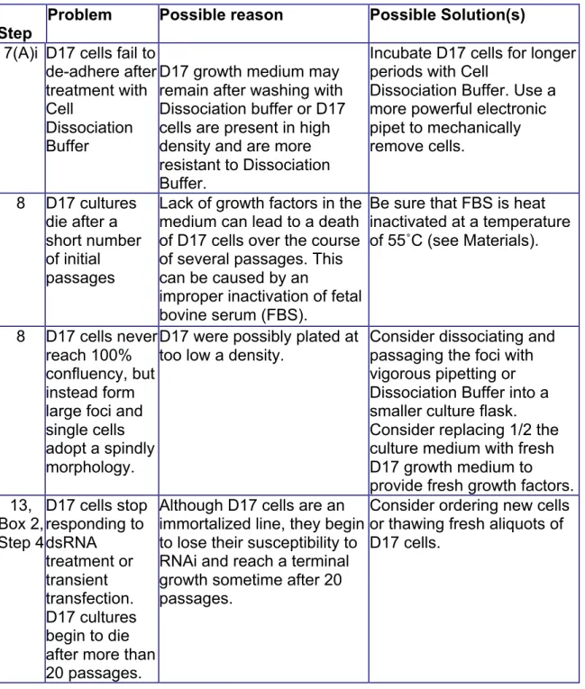
Table 2.1: Troubleshooting
Step
Problem Possible reason Possible Solution(s)
7(A)i D17 cells fail to de-adhere after treatment with Cell
Dissociation Buffer
D17 growth medium may remain after washing with Dissociation buffer or D17 cells are present in high density and are more resistant to Dissociation Buffer.
Incubate D17 cells for longer periods with Cell
Dissociation Buffer. Use a more powerful electronic pipet to mechanically remove cells.
8 D17 cultures die after a short number of initial passages
Lack of growth factors in the medium can lead to a death of D17 cells over the course of several passages. This can be caused by an
improper inactivation of fetal bovine serum (FBS).
Be sure that FBS is heat inactivated at a temperature of 55˚C (see Materials).
8 D17 cells never reach 100% confluency, but instead form large foci and single cells adopt a spindly morphology.
D17 were possibly plated at too low a density.
Consider dissociating and passaging the foci with vigorous pipetting or Dissociation Buffer into a smaller culture flask. Consider replacing 1/2 the culture medium with fresh D17 growth medium to provide fresh growth factors. 13,
Box 2, Step 4
D17 cells stop responding to dsRNA treatment or transient transfection. D17 cultures begin to die after more than 20 passages.
Although D17 cells are an immortalized line, they begin to lose their susceptibility to RNAi and reach a terminal growth sometime after 20 passages.
81 14, 16 D17 cells do
not adhere and migrate on ECM-crosslinked glass surfaces
This could be due to an inefficient crosslinking process or inefficacious ECM.
If using a UV crosslinking treatment, determine the power output of the UV source or use a plasma treatment system. D17 migration can be impaired by too low or too high
concentrations of ECM. The user should empirically test a range of ECM
concentrations to determine the optimal concentration per ECM batch (see