PATHOGENICITY AND EPITOPE SPECIFICITY OF IgG4 AUTOANTIBODIES IN ENDEMIC PEMPHIGUS FOLIACEUS
Flor de María Evangelista Montoya
A dissertation submitted to the faculty of the University of North Carolina at Chapel Hill in partial fulfillment of the requirements for the degree of Doctor of Philosophy in the
Department of Microbiology and Immunology
Chapel Hill 2012
Approved by:
Stephen H. Clarke, Ph.D. Luis A. Diaz, M.D.
Zhi Liu, Ph.D.
Stefanie Sarantopoulos, M.D., Ph.D.
ii ©2012
iii ABSTRACT
FLOR DE MARIA EVANGELISTA MONTOYA: Pathogenicity and Epitope Specificity of IgG4 autoantibodies in Endemic Pemphigus Foliaceus
(Under the direction of Dr. Zhi Liu)
Endemic pemphigus foliaceus, also known as Fogo Selvagem (FS), is an autoimmune
blistering skin disease that exhibits geographic clustering and 100% mortality without treatment. An active focus of FS is located in the rural reservation of Limao Verde (LV), Mato Grosso do Sul, Brazil. It is widely accepted that the autoantibodies that mediate FS are
predominantly of the IgG4 subclass against Desmoglein 1 (Dsg1) and that healthy individuals living in LV posses a mixture of circulating IgG anti-Dsg1 autoantibodies. It has
been suggested that an unknown environmental trigger present in LV may induce an anti-Dsg1 autoimmune response, which in certain genetically predisposed individuals leads to a pathogenic IgG4 response and clinical disease. The transition from preclinical to clinical
disease in FS has been associated with subclass switching and epitope spreading within the extracellular domain of Dsg1.
The N-terminal region of Dsg1 has been reported to be the target for pathogenic FS IgG autoantibodies. However, the fine epitope specificity for FS IgG4 autoantibodies remains unknown. The work in this dissertation refines the identification of epitopes in Dsg1
that are recognized specifically by IgG4 autoantibodies from FS patients. Two dominant conformational epitopes were identified using epitope excision and MALDI-MS/MS. One
iv
the EC2 domain (Q201-R213). Moreover, residues M133 and Q135 are required to achieve
the proper conformation of the epitope recognized by pathogenic IgG4 autoantibodies in FS. This study also reports that IgG4 FS antibodies recognize LJM11, a protein found in
the saliva of Lutzomya longipalpis. In addition, mice immunized with LJM11 generate anti-Dsg1 antibodies. Thus, insect bites may deliver salivary antigens that initiate a cross-reactive IgG4 autoimmune response in genetically susceptible individuals, which subsequently leads
to development of FS.
The findings of this dissertation may uncover targets such as the epitope in EC1 for
v
This dissertation is dedicated to my husband Kevin, for his unconditional love, support and
for being a blessing in my life. To my mother Pilar and to the memory of my father Félix, for
all their love and for providing me with the support to get to this moment. To my brother
Félix for all his love, for being my inspiration to pursue an intellectual career, and for taking
care of our mother during my “adventure” away from home.
vi
ACKNOWLEDGEMENTS
I would like to thank my advisor Dr Zhi Liu for the opportunity to work in his lab as well as for his guidance and support during graduate school. I have learned from him how to
be an independent scientist, as well as how to balance a successful career and family. I also thank him for accommodating and supporting me during stressful and challenging times in my life outside of the lab.
I am thankful to the current and past members of the Liu Lab, it has been great to work with you all. Most especial way to my graduate school “sister” Dr. Lisa Heimbach, who
has being a wonderful friend and listener, especially when things got challenging for our projects and we thought we would “graduate in 10 years”. I am deeply grateful to Lisa for her scientific and technical advice and for her help outside the lab, especially when I had
surgery and the times she took care of “Bubu”. I would also like to thank Kayla Shumate, who has been extremely helpful during my last semester in the lab. Kayla is a great friend
and lab manager. Thanks to Lin, Bin-Jin, Peng, Jaime, Megan, Ying-Ching and Lan for their help, friendship and for making the lab a fun place to work. I will miss you all.
I am deeply thankful to the UNC Dermatology Department, for being my “home”
before and during graduate school. Especially to the chairman, Dr Luis A. Diaz, who introduced me to the life of a researcher in US and has been a wonderful mentor throughout
vii
scientific advice and precious friendship, they always had a smile for me during hard times
the lab and reminded me to breathe and keep going. I am also thankful to Dr Ning Li, Phillip, Paula, Joseph, Aileen, Marilia, Melanie, Cindy and all the Dermatology family, for
all their help, ideas and friendship.
I would like to thank my dear thesis committee: Drs. Stephen Clarke, Stefanie Sarantopoulos, Barbara Vilen and Luis Diaz. They have been amazingly supportive in the
preparation of my dissertation. I appreciate their constant availability to answer my questions and discuss my research during single conversations or committee meetings. I am thankful
for all their questions, by which they were teaching me how to think and be a better scientist. I am also thankful to our collaborators, Drs. Aleeza Roth, David Klapper and Bahjat Qaqish, for their scientific expertise and technical contributions that were key for the completion of
my dissertation.
I would like to acknowledge other mentors and friends from the Microbiology and
Immunology Department, especially Dixie Flannery, who is an amazing Student Services Manager, she guided me in every step, form, requirement I had to fulfill during graduate school, she became my friend and second “mother” who was always checking how my
graduate school and personal life was going. Drs. Marcia Hobbs, Lorraine Cramer, Laura White, Roland Tisch, Glenn Matsushima and Silva Markovic-Plese were key resources for
my doctoral training and I am grateful for their wise advice and precious time.
Finally, I want to recognize the memory of Dr. Angel Quintanilla, my former mentor in Peru, who shared with me his enthusiasm for research and discovery. He believed in me on
viii
TABLE OF CONTENTS
LIST OF TABLES ………... x
LIST OF FIGURES ………....…... xi
LIST OF ABBREVIATIONS ………... xii
Chapter I. INTRODUCTION……….. 1
1.1. PEMPHIGUS.……… 1
1.2. INTERCELLULAR JUNCTIONS AND THE CADHERINS ……... 2
1.3. THE PEMPHIGUS GROUP: Clinical Manifestations and Desmosomal Targets ……….………...……….. 8
1.4. IMMUNOPATHOGENESIS OF AUTOANTIBODY-INDUCED BLISTERING IN PEMPHIGUS ………..……….. 10
1.5. ENVIRONMENTAL FACTORS IN ENDEMIC PEMPHIGUS FOLIACEUS ……….. 14
1.6. ANIMAL MODELS IN FS .………...………... 16
1.7. CONCLUDING REMARKS ………….…………...………... 16
REFERENCES ………... 18
II. DISSECTING THE CONFORMATIONAL EPITOPES RECOGNIZED BY PATHOGENIC IgG4 ANTI-DESMOGLEIN 1 AUTOANTIBODIES IN ENDEMIC PEMPHIGUS FOLIACEUS …...……….. 30
2.1. INTRODUCTION ………... 30
ix
2.3. RESULTS ………...……….... 40
2.4. DISCUSSION ………. 54
REFERENCES ………...………... 59
III. INDUCTION OF IgG4 ANTI-DESMOGLEIN 1 ANTIBODIES BY HEMATOPHAGUS INSECTS BITES .………... 63
3.1. INTRODUCTION ………...………... 63
3.2. MATERIAL AND METHODS………... 64
3.3. RESULTS ………... 67
3.4. DISCUSSION ………...………. 74
REFERENCES …………...………...… 76
IV. SUMMARY AND FUTURE DIRECTIONS …….…………... 79
x
LIST OF TABLES
Table
2.1. Primers used for construction of swapped-domain recombinant proteins and site directed mutagenesis ………....……….. 35
2.2. Conformational epitopes in the extracellular domain of human Dsg1 targeted by IgG4 autoantibodies from 20 FS patients ………... 46
2.3. Frequency of IgG4 anti-Dsg1 autoantibodies targeting conformational
xi
LIST OF FIGURES
Figure 1.1. : Molecular components of intercellular junctions and pemphigus variants ... 4 Figure 2.1. : Domain swapped hDsg1/Dsc1 recombinant proteins produced in
baculovirus expression system …... 41 Figure 2.2. : Analysis of IgG4 and non-IgG4 autoantibodies from FS patient serum by IP
and passive transfer ………... 42 Figure 2.3. : Prevalence of IgG4 autoantibodies against Dsg1(EC1) by ELISA ……... 43
Figure 2.4. : Epitope mapping by epitope excision and mass spectrometry ………... 44 Figure 2.5. : Location of 2 conformational epitopes recognized by FS IgG4
autoantibodies in a predicted model of hDsg1 extracellular domain ……... 48 Figure 2.6. : Site directed mutagenesis in the extracellular domains of hDsg1 and hDsg4.... 51
Figure 2.7. : Mutation of residues M133 and Q135 in EC1 domain of Dsg1 dramatically reduces the binding of IgG4 autoantibodies to Dsg1 extracellular domain... 52
Figure 2.8. : Detection of IgG4 autoantibodies against Dsg4-WT and Dsg4-mutants by ELISA…………... 53
Figure 3.1. : FS patients have high levels of anti-SGLL IgG4 antibodies ……….... 68 Figure 3.2. : The cross-reactivity of two IgG4 anti-Dsg1 monoclonal antibodies from FS
to SGLL by ELISA ………... 69 Figure 3.3. : IgG4 antibodies from FS patients and two IgG4 monoclonal anti-Dsg1
antibodies derived from FS patients recognize LJM11, a protein
component from SGLL ………... 71 Figure 3.4. : FS autoantibodies recognize LJM11 and sera from mice immunized with
xii
LIST OF ABREVIATIONS
AFM atomic force microscopy
Anti-HA anti-influenza hemagglutinin antibody Anti-His anti-histidine antibody
A129 alanine at position 129
A130 alanine at position 130 Bcl-2 B-cell lymphoma 2
BSA bovine serum albumin CaCl2 calcium chloride
CAR cell-adhesion recognition site
cDNA complementary Deoxyribonucleic acid CH1 constant domain 1
CNBr cyanogen bromide
DEAE diethylaminoethyl cellusose
DP desmoplakin
Dsc desmocollin
Dsg desmoglein
EC extracellular domain
EDTA ethylenediaminetetraacetic acid ELISA enzyme-linked immunosorbent assay
xiii FAT phenylalanine, alanine, threonine
FBS fetal bovine serum
Fc fragment crystallizable region
FS Fogo Selvagem (endemic pemphigus foliaceus) GST tag glutathione S-transferase tag
HAV histidine, alanine, valine
HC human control
hDsg1 human desmoglein 1
hIgG4 human immunoglobulin class G subclass 4 His-tag histidine-tag
HLA human leukocyte antigen
HRP horseradish peroxidase HSP heat shock protein
IB immunoblot
IEN intraepidermal neutrophilic IgA immunoglobulin class A
IgG immunoglubulin class G
IgG4 immunoglobulin class G subclass 4
IP immunoprecipitation
IRB Institutional Review Boards
kD kilodalton
LV Limao Verde (rural reservation in Brazil)
xiv
MALDI-MS/MS short version of MALDI-TOF/TOF MS/MS MAPK mitogen-activated protein kinase
MEC mutated extracellular domain mPV mucosal Pemphigus Vulgaris
mcPV mucocutaneous Pemphigus Vulgaris
µg microgram
µL microliter
mM millimolar
M133 methionine at position 133
NCBI National Center for Biotechnology Information
ng nanogram
NHS normal human serum
NIAID National Institute of Allergy and Infectious Diseases Ni-NTA nickel-nitriloacetic acid
O.D. optical density
PBS phosphate buffered saline PCR polymerase Chain Reaction
PF pemphigus foliaceus
PG plakoglobin
PKP plakophilin
PNP paraneoplastic pemphigus PV pemphigus vulgaris
xv Q201 glutamine at position 201
RAL arginine, alanine, leucine
ROC receiver-operating-characteristic analysis
RT room temperature
R144 arginine at position 144 R213 arginine at position 213
scFv single-chain variable fragment
SDS-PAGE sodium dodecyl sulfate polyacrylamide gel electrophoresis
SFM serum-free medium
SGLL salivary gland from Lutzomyia longipalpis SPD subcorneal pustular dermatosis
TBS tris buffered saline TFA trifluoroacetic acid
TPCK L-1-tosylamido-2-phenylethyl chloromethyl ketone UNC University of North Carolina at Chapel Hill
US United States
VHH heavy-chain single variable domain antibody
WT wild type
W51 tryptophan at position 51 YAG yttrium-aluminum garnet YAS tyrosine, alanine, serine
CHAPTER I INTRODUCTION
1.1. PEMPHIGUS
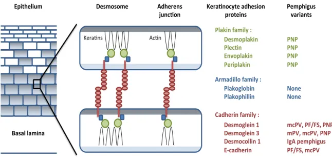
Pemphigus is a group of rare, autoimmune blistering diseases of the skin and mucous membranes mediated by autoantibodies against desmosomal members of the cadherin superfamily. There are several variants of pemphigus, each with unique clinical, histologic, and immunologic features. Interestingly, when different desmosomal proteins are targeted by the autoimmune response, different clinical and histologic features are seen [1, 2].
2
cloning techniques and cDNA libraries, the isolation of cDNA for pemphigus antigens demonstrated that desmogleins are indeed the target antigens in pemphigus.
1.2. INTERCELLULAR JUNCTIONS AND THE CADHERINS:
Intercellular junctions in the epidermis allow keratinocytes to adhere to one another and maintain the integrity of the epithelium. Morphological and biochemical studies have defined two major types of intercellular junctions in epithelial cells: adherens junctions and desmosomes [8, 9]. These intercellular junctions are composed of cadherins, a superfamily of calcium-dependent adherent proteins, which play an important role in the dynamic regulation of intercellular adhesion [10]. The cadherin superfamily is characterized by multiple “cadherin repeat” sequences of about 110 aminoacids in their extracellular domains (EC) and comprised of two major groups: Classical cadherins (E-, P-, N-cadherin) and desmosomal cadherins (desmogleins and desmocollins).
3
!- and "-catenin [15, 16], and desmosomal cadherins are linked to the intermediate keratin filaments through plakoglobin and desmoplakins I and II [9, 17].
cadherin and P-cadherin are classical cadherins expressed in the epidermis. E-cadherin is expressed in all layers of the epidermis, whereas P-E-cadherin is limited to the basal cell layer [18].
THE DESMOSOME:
4 a) Desmosomal cadherins:
Desmosomes were originally isolated from cow nose epidermis [21] and chemically characterized [22]. High levels of glycosylated proteins were found to be the main component of the desmosomes, and they were postulated to mediate cell-cell adhesion [23, 24]. Antibodies against a particular desmosomal glycoprotein were developed and used to screen a cDNA library. In this manner, the first desmosomal cadherin was cloned, sequenced, and named desmoglein derived from the greek “glein” (glue) [25]. Since then, the desmosomal cadherins family has been found to consist of seven different proteins: four
!"#$%#%$"&
!"#$%&'( '()*+,&-)$+(.&/&&!"#$%0()*+,& '("12,& 3,4%0()*+,& '"5+0()*+,& & 65$)7+((%&-)$+(.&/&& '()*%8(%9+,& '()*%0:+((+,& & ;)7:"5+,&-)$+(.&/& !"#$%8("+,&<& !"#$%8("+,&=& !"#$%1%((+,&<& 3>1)7:"5+,& & 30+?:"(+@$& A)#)(&()$+,)& B"5)2,%1.?"&)7:"#+%,&
05%?"+,#& '"$0:+8@#&4)5+),?#&
'C'& 'C'& 'C'& 'C'& & C%,"& C%,"& & $1'DE&'FGFHE&'C'& $'DE&$1'DE&'C'& I86&0"$0:+8@#& 'FGFHE&$1'D& & 67:"5",#& J@,12%,& )*%&(
5
desmoglein isoforms (Dsg1, Dsg2, Dsg3 and of Dsg4) and three desmocollin isoforms (Dsc1,
Dsc2 and Dsc3), all encoded by separate genes located on chromosome 18 in humans [26,
27].
Dsg1 and Dsc1 are the major desmosomal cadherins in the skin where they are
expressed throughout the epidermis, but most prominently in the upper layers. Dsg3 and
Dsc3 expression is predominant in the lower epidermis and decreased toward the upper
layers. Dsg1/Dsg3 and Dsc1/Dsc3 are mostly restricted to stratified epithelial tissues, such
as the skin. Dsg2 is expressed in a wide range of other simple epithelial tissues and
myocardia in the heart, whereas Dsg4 is mostly expressed in hair follicles [28].
Desmocollins and desmogleins are always found as a pair in the desmosomes; however, the
precise nature of their interaction is still under debate. Several studies indicate that
desmosomal cadherins contain a CAR site within their N-terminal EC1 extracellular domain and that this site is critical for maintenance of desmosomal adhesion [11]. The CAR site
sequence for Dsg1 and Dsc1 are RAL and YAT, respectively [13]. Interestingly, peptides
derived from these sequences were able to block the homophilic adhesion mediated by Dsg1
and Dsc1 upon incubation [12, 13]. Based on predictions from the C-cadherin crystal
structure, recent cryo-electron microscopy studies in human epidermis showed cis- and trans-
interactions of the EC1 domains, possibly via insertion of the tryptophan in position 51
(W51) into the hydrophobic pocket of the CAR site [29]. However, they also found some
cis-interactions of the EC 4-5 domains and it has been suggested that desmosomal cadherins may
show periodically zipper-like arrangements similar to classical cadherins [30].
6
homophilic and heterophilic trans-interactions. Using EC1-2 fragments of Dsg2, Dsc2 or Dsg3, it was shown that homophilic interactions occur in vitro [31, 32]. In addition, Waschke et al. have utilized atomic force microscopy (AFM) to estimate the strength of interaction between purified Dsg1 ectodomains [33] and showed that, in fact, Dsg1 could form homo-dimers and that the molecular binding properties of this homophilic adhesion may be comparable to other cadherins. Thus, mutation of key amino acid residues such as tryptophan in position 51(W51) and alanine at position 130(A130) that are involved in adhesion mediated by classical cadherins, abolished homophilic adhesion of desmosomal cadherins [34]. Recent studies using a similar AFM approach have shown Dsc3 homophilic binding and heterophilic interaction with Dsg1, but not with Dsg3 [35]. Heterophilic binding of Dsg2 with Dsc1 or Dsc2 have also been demonstrated [32, 36]. Furthermore, these heterophilic interactions also form in a calcium-dependent manner.
b) The armadillo proteins in the desmosome:
In the desmosomal plaque, the armadillo proteins plakoglobin and plakophilin associate with the cytoplasmic domains of desmosomal cadherins.
7
with "-catenin. PG can substitute for "-catenin in adherens junctions because both bind E-cadherin with similar affinity. However, PG has higher affinity for desmogleins, which explains the exclusion of "-catenin from desmosomes [40]. The arm domain of PG associates with both the intracellular domain of desmosomal cadherins and the N-terminus of desmoplakin [41-43]. Thus, PG is a critical linker in desmosomal adhesion.
-Plakophilins (PKP). PKP are members of a subfamily of armadillo proteins related to p120ctn. Three desmosomal PKPs (1, 2 and 3) have been described, and the genes encoding them are located on chromosomes 1, 12 and 11 respectively [44]. PKP1 and PKP2 have 2 isoforms (“a” and “b”) that result from alternative splicing [45, 46]. There is a fourth plakophilin : PKP4, also known as p0071, which is highly related to p120ctn and $-catenin [47]. PKPs (1-4) have been shown to bind directly to the intracellular domain of desmosomal cadherins. This interaction is mediated by their amino-terminal head domain and the functions for their armadillo repeats remain unknown. PKPs can also bind PG in order to facilitate clustering of desmosomal cadherins through lateral stabilizing interactions, which increases desmosome strength [41].
8
proteins (PG and PKP), its central coiled-coil rod domain mediates dimerization, and its carboxy-terminal tail is the linker for intermediate filaments [41, 51]. Other members such as plectin, envoplakin and periplakin are also found in desmosomes; however, their roles are still unclear. Genetic studies have shown that desmoplakin is the only indispensable plakin in intercellular junctions. Thus, inactivating plectin, which is present in desmosomes and hemidesmosomes, did not affect cell-cell adhesion in mice or humans [52, 53]. In addition, when using DP-deficient mice, skin blistering was observed and the desmosomes were not anchored to the intermediate filaments [54].
1.3. THE PEMPHIGUS GROUP: Clinical Manifestations and Desmosomal Targets
The pemphigus group consists of many disease variants, each with unique clinical, histologic, and immunologic features [55]. These disease variants include pemphigus vulgaris (PV), pemphigus foliaceus (PF), endemic pemphigus foliaceus (also known as Fogo Selvagem: FS), paraneoplastic pemphigus (PNP), and IgA pemphigus.
9
acantholysis upon passive transfer in a neonatal mouse model of disease (discussed in Section 1.6), thereby establishing pathogenicity [58-60].
PF presents clinically with superficial blisters or erosions over the trunk and extremities. In many patients, the blisters rupture spontaneously prior to presentation and the clinical exam reveals only superficial crusting and erosions. There is no mucosal involvement in PF. Histologically, PF shows a subcorneal split with acantholysis. Patients with PF classically harbor autoantibodies to Dsg1 alone [2, 55, 56]. Similar to PV, the establishment of Dsg1 as the target autoantigen was defined by affinity purified anti-Dsg1 autoantibodies in the neonatal passive transfer model [61].
FS represents an endemic form of PF with identical clinical, histologic, and immunologic features. The endemic nature of this disease makes it an ideal population to study the fine aspects of genetic susceptibility and environmental triggers of autoimmune disease [62]. Autoantibodies to Dsg1, particularly those of the IgG4 subclass, are a serologic indicator of disease in this patient population [63]. E-cadherin has also been shown to be a potential antigenic target in FS [64]. In addition, a recent serological analysis reveals that FS patients and many healthy controls living in endemic areas also harbor autoantibodies to other Dsg and Dsc family members, in addition to the anti-Dsg1 antibodies previously reported [65].
10
acantholysis, as removal of the Dsg3 specific antibodies from PNP sera abrogates the pathogenicity in the neonatal passive transfer model [68]. These patients also have autoantibodies to the plakin family of proteins, which are thought to be responsible for the dyskeratosis/necrotic keratinocytes [66, 67]. Interestingly, patients with erythema multiforme, a condition classically felt to be a non-immunologically mediated reaction pattern, can have a falsely positive indirect immunofluorescence on the classic PNP rat bladder substrate. There are recent reports of patients with erythema multiforme harboring anti-desmoplakin antibodies, though the pathogenicity of these autoantibodies has not yet been established [69, 70].
Finally, IgA pemphigus is distinguished by the presence of autoantibodies of the IgA subclass present on direct immunofluorescence in a classic intercellular space staining pattern. Histologically, IgA pemphigus can be of the intraepidermal neutrophilic (IEN) variant or can be of the subcorneal pustular dermatosis (SPD) variant [71]. Dsc1 has been shown to be the target antigen in the IEN variant of IgA pemphigus, whereas the target antigen in the SPD variant of IgA pemphigus is likely a non-desmosomal antigen as shown by immuno-electron microscopy studies [72]. Pathogenicity studies are currently lacking.
To date, there are no descriptions of isolated anti-desmoplakin (outside of what is seen in PNP and erythema multiforme) or anti-plakoglobin mediated cutaneous disease.
1.4. IMMUNOPATHOGENESIS OF AUTOANTIBODY-INDUCED BLISTERING IN PEMPHIGUS
11
association of susceptibility to pemphigus with certain HLA-DR and -DQ alleles has been
suggested [73]. However, the exact mechanism and whether pemphigus autoantibodies
induce loss of epidermal cell adhesion directly or indirectly is controversial. Different
mechanisms have been proposed such as: direct interference (steric hindrance), activation of
transmembrane signaling that downregulates cell-cell adhesion, proteinase activation
(plasminogen activator), and desmoglein internalization. There are two major theories that
are the most accepted but still under debate: a) direct interference of desmogleins
transinteraction by pemphigus autoantibodies, and b) activation of transmembrane signaling
pathways and/or apoptosis by pemphigus autoantibodies that indirectly results in
acantholysis.
a) Direct interference of desmogleins transinteraction by pemphigus autoantibodies (steric
hindrance theory): Accumulated evidence from several epitope mapping studies indicates
that pathogenic pemphigus autoantibodies target the amino-terminal end of Dsg1 and/or
Dsg3 ectodomains [74-76]. Data based on the crystal structure of classical cadherins suggest
that this N-terminal region harbors the adhesive interface of desmosomal cadherins [30, 77].
In addition, the pathogenicity of pemphigus IgG autoantibodies has been consistently
demonstrated by passive transfer studies in neonatal mice [60, 78]. Moreover, not only the
whole IgG molecule, but also the F(ab)2 and Fab fragments were found to be pathogenic,
independently of complement or plasminogen activator [79-82], suggesting that due to their
inability to cross-link cell-surface molecules, it is possible that they interfere directly with
adhesion. Furthermore, monoclonal antibodies (AK23) derived from a PV mouse model that
12
lesions in mice, whereas monoclonal antibodies recognizing other regions of Dsg3 ectodomain did not cause lesions in mice [83]. Using single-molecule atomic force microscopy (AFM), it has been shown that PV-IgG and AK23 monoclonal antibody directly inhibit Dsg3 homophilic binding under cell-free conditions, suggesting that direct inhibition of Dsg3 binding occurs in PV [84, 85]. Direct blocking of Dsg1 binding wasn’t observed by AFM, however, keratinocyte dissociation and loss of Dsg1- and Dsg3- coated microspheres to cultured keratinocytes were observed when using laser tweezer trapping [86], suggesting that acantholysis may not be solely dependent on direct interference of Dsg1-Dsg1 binding by pemphigus autoantibodies. Recent studies using peptides against the desmoglein adhesive interface as well as tandem peptides (obtained by dimerization of two of the initial peptides) added evidence that direct inhibition contributes to acantholysis when the tandem peptide prevented acantholysis induced by PV-IgG, yet this was not the case when using PF-IgG. Thus, some investigators proposed that PV and PF acantholysis may involve different mechanism [87].
[89-13
94]. Thus, activation of intracellular signaling within the target keratinocyte induced by
binding of pemphigus IgG has been proposed to contribute to the loss of cell-cell adhesion.
Previous studies have shown that phosphorylation of p38MAPK and HSP25 (the
murine homolog of human HSP27) occurs rapidly after exposure of cultured keratinocytes to
pemphigus IgG and in the skin of mice injected with pathogenic IgG [95]. Also,
phosphorylation of both p38MAPK and HSP27 has been observed in perilesional epidermis
of pemphigus patients [96]. Furthermore, p38MAPK inhibitors block both histological and
gross blister formation in the PF passive transfer model [95], suggesting that activation of
p38MAPK is an early and key step in PF IgG-induced acantholysis. p38MAPK signaling has
been implicated in other cellular responses such as desmosome assembly, cytoskeleton
reorganization, changes of the cell cycle and apoptosis [97, 98]. Moreover, there is evidence
that p38MAPK is involved in keratinocyte apoptosis [99, 100] and that DNA fragmentation
and caspase activation are induced in the epidermis of PF IgG-treated mice [101].
Keratinocyte-derived and local production of apoptotic inducers such as nitric oxide
synthase, Fas and Bcl-2, have also been detected in lesional skin of PF patients [102, 103]
along with increased levels of Fas ligand in serum of pemphigus patients [104]. Futhermore,
a biphasic activation of p38MAPK after the binding of pemphigus IgG has been recently
demonstrated where the first activation peak is linked to acantholysis and the second peak
coincided with apoptosis, suggesting that apoptosis occurs downstream to acantholysis in
pemphigus [99]. However, other studies suggests that apoptosis occurs before acantholysis
develops [101]. There is also the hypothesis that apoptotic signaling could precede
acantholysis in the absence of apoptotic cell death [98]. More importantly, caspase inhibitors
14
present, it is not clear which process precedes the other, but there is evidence that both are involved in pemphigus pathogenesis.
Another signaling pathway suggested to be involved is mediated through plakoglobin [105]. The observation was made that keratinocytes from plakoglobin-deficient mice were resistant to keratinocyte dissociation induced by PV IgG, suggesting that direct inhibition of Dsg binding may not be sufficient to cause acantholysis, and that plakoglobin could be part of a complex responsible for transferring the signal upon autoantibody binding from outside into the keratinocyte: “outside-in” signaling [106]. In addition, it has been shown that plakoglobin is involved in c-Myc repression, and c-Myc was also shown to be elevated in keratinocytes exposed to pemphigus autoantibodies [107, 108]. Nonetheless, the role of c-Myc signaling in pemphigus acantholysis remains unclear.
1.5. ENVIRONMENTAL FACTORS IN ENDEMIC PEMPHIGUS FOLIACEUS
The endemic form of pemphigus foliaceus, known as Fogo Selvagem (FS) which translates to “Wild Fire” in English because of the burning sensation that the disease causes, was first described in the early 1900s in certain areas of Brazil [109]. FS is highly endemic in the rural reservation of Limao Verde, in Mato Grosso do Sul, Brazil, where the prevalence is 3.4% and an incidence of 1-4 new cases per year. Other less-characterized forms have been also reported in Colombia, Peru and Tunisia [62, 110, 111].
15
as the decreasing prevalence of FS upon improvement of living conditions, suggest the presence of an environmental antigen that may exhibit molecular mimicry with Dsg1. Circulating anti-Dsg1 autoantibodies in the serum of 42% of healthy individuals living in endemic Brazilian areas has been reported [65]. Moreover, anti-Dsg1 antibodies are also detected in FS patients before onset of disease as a mixture of IgG1 and IgG4 autoantibodies that recognize the EC5 portion of Dsg1 [75, 114]. An additional report showed that patients with disease transmitted by hematophagus vectors (black flies, sand flies and kissing bugs) possess antibodies against the extracellular EC5 domain of Dsg1 [115]. These findings suggest that saliva of these vectors may contain Dsg1 homologs that induce a non-pathogenic response to Dsg1.
Another feature of FS is the increased frequency of familial cases and the association to certain HLA-DR alleles [116]. These alleles are HLA-DRB1-0102, 0404, 1402 and 1406. The hypervariable region of the DRB1 gene of these alleles share the sequence LLEQRRA at positions 67-74, suggesting that this sequence may be critical for susceptibility to FS. It is possible that an environmental antigen induces an initial cross-reactive anti-Dsg1 non-pathogenic response and only in individuals sharing these alleles is FS later developed.
IgG, IgM and IgE antibodies against Dsg1 are present not only in FS patients but also in healthy individuals living in Brazilian endemic areas [63, 117, 118]. Analysis of the hypervariable V genes of FS autoantibodies showed that they are antigen selected and this selection occurs before the onset of clinical FS [119].
16
predisposed individuals living in FS endemic areas. Furthermore, insect bites may deliver
antigens/allergens that drive the production of cross-reactive anti-Dsg1 autoantibodies.
1.6. ANIMAL MODELS IN FS
A passive transfer mouse model was developed to test in vivo pathogenicity of autoantibodies from FS patients [78]. Serum or IgG fraction from FS patient is given to neonatal mice by intradermal injection. The animals develop skin blisters and typical histological and ultrastructural features of the human disease, such as subcorneal vesicles [5]. The extent of disease correlates well with the indirect IF titers of human autoantibodies detected in the mouse skin. FS was also induced in these animals by injecting monovalent Fab fractions from FS autoantibodies [82], thus, the blistering in this animals is independent of complement activation. Despite the availability of recombinant human and mouse Dsg1, development of an active model for FS remains unsuccessful.
1.7. CONCLUDING REMARKS
17
FS provides a good model to study human autoimmune disease in which the
auto-antigen Dsg1 is fully characterized and the relevant autoantibodies are known to be
pathogenic. In addition, the epidemiology of FS provides an opportunity to study the
development of autoimmune disease in a well-defined, geographically limited population
with a high prevalence of the disease. The saliva of hematophagus insects such as black flies
have been suggested to harbor the cross-reactive antigen that induces a non-pathogenic
anti-Dsg1 response in healthy individuals living in FS endemic areas. However, the association of
an environmental trigger with the pathogenic anti-Dsg1 response in FS remains unknown.
The objective of this dissertation is to identify the principal antigenic determinant in the
extracellular domain of Dsg1 that is recognized by pathogenic FS autoantibodies and their
association with a potential environmental agent. Chapter 2, describes the identification of
conformational epitopes for pathogenic IgG4 autoantibodies from FS patients living in LV,
Brazil. Chapter 3 reports a non-infectious environmental agent that induces a potentially
pathogenic anti-Dsg1 response in FS patients. Finally, Chapter 4 discusses the implications
18 REFERENCES:
1. Joly, P. and N. Litrowski, Pemphigus group (vulgaris, vegetans, foliaceus, herpetiformis, brasiliensis). Clinics in dermatology, 2011. 29(4): p. 432-6.
2. Lever, W.F., Pemphigus and pemphigoid. 1965, Springfield, Illlinois: Charles C. Thomas Publisher.
3. Rock, B., C.R. Martins, A.N. Theofilopoulos, R.S. Balderas, G.J. Anhalt, R.S. Labib, S. Futamura, E.A. Rivitti and L.A. Diaz, The pathogenic effect of IgG4 autoantibodies in endemic pemphigus foliaceus (fogo selvagem). N Engl J Med, 1989. 320(22): p. 1463-9.
4. Emery, D.J., L.A. Diaz, J.A. Fairley, A. Lopez, A.F. Taylor and G.J. Giudice, Pemphigus foliaceus and pemphigus vulgaris autoantibodies react with the extracellular domain of desmoglein-1. The Journal of investigative dermatology, 1995. 104(3): p. 323-8.
5. Futamura, S., C. Martins, E.A. Rivitti, R.S. Labib, L.A. Diaz and G.J. Anhalt, Ultrastructural studies of acantholysis induced in vivo by passive transfer of IgG from endemic pemphigus foliaceus (Fogo Selvagem). The Journal of investigative dermatology, 1989. 93(4): p. 480-5.
6. Eyre, R.W. and J.R. Stanley, Identification of pemphigus vulgaris antigen extracted from normal human epidermis and comparison with pemphigus foliaceus antigen. The Journal of clinical investigation, 1988. 81(3): p. 807-12.
7. Stanley, J.R., V. Klaus-Kovtun and S.A. Sampaio, Antigenic specificity of fogo selvagem autoantibodies is similar to North American pemphigus foliaceus and distinct from pemphigus vulgaris autoantibodies. J Invest Dermatol, 1986. 87(2): p. 197-201.
8. Amagai, M., Adhesion molecules. I: Keratinocyte-keratinocyte interactions; cadherins and pemphigus. J Invest Dermatol, 1995. 104(1): p. 146-52.
9. Green, K.J. and C.A. Gaudry, Are desmosomes more than tethers for intermediate filaments? Nat Rev Mol Cell Biol, 2000. 1(3): p. 208-16.
10. Patel, S.D., C.P. Chen, F. Bahna, B. Honig and L. Shapiro, Cadherin-mediated cell-cell adhesion: sticking together as a family. Curr Opin Struct Biol, 2003. 13(6): p. 690-8.
19
12. Runswick, S.K., M.J. O'Hare, L. Jones, C.H. Streuli and D.R. Garrod, Desmosomal adhesion regulates epithelial morphogenesis and cell positioning. Nat Cell Biol, 2001. 3(9): p. 823-30.
13. Tselepis, C., M. Chidgey, A. North and D. Garrod, Desmosomal adhesion inhibits invasive behavior. Proc Natl Acad Sci U S A, 1998. 95(14): p. 8064-9.
14. Garrod, D., M. Chidgey and A. North, Desmosomes: differentiation, development, dynamics and disease. Curr Opin Cell Biol, 1996. 8(5): p. 670-8.
15. Hirano, S., A. Nose, K. Hatta, A. Kawakami and M. Takeichi, Calcium-dependent cell-cell adhesion molecules (cadherins): subclass specificities and possible involvement of actin bundles. J Cell Biol, 1987. 105(6 Pt 1): p. 2501-10.
16. Yap, A.S., W.M. Brieher and B.M. Gumbiner, Molecular and functional analysis of cadherin-based adherens junctions. Annu Rev Cell Dev Biol, 1997. 13: p. 119-46. 17. Bornslaeger, E.A., C.M. Corcoran, T.S. Stappenbeck and K.J. Green, Breaking the
connection: displacement of the desmosomal plaque protein desmoplakin from cell-cell interfaces disrupts anchorage of intermediate filament bundles and alters intercellular junction assembly. J Cell Biol, 1996. 134(4): p. 985-1001.
18. Hirai, Y., A. Nose, S. Kobayashi and M. Takeichi, Expression and role of E- and P-cadherin adhesion molecules in embryonic histogenesis. II. Skin morphogenesis. Development, 1989. 105(2): p. 271-7.
19. Delva, E., D.K. Tucker and A.P. Kowalczyk, The desmosome. Cold Spring Harb Perspect Biol, 2009. 1(2): p. a002543.
20. Franke, W.W., Discovering the molecular components of intercellular junctions--a historical view. Cold Spring Harb Perspect Biol, 2009. 1(3): p. a003061.
21. Skerrow, C.J. and A.G. Matoltsy, Isolation of epidermal desmosomes. J Cell Biol, 1974. 63(2 Pt 1): p. 515-23.
22. Skerrow, C.J. and A.G. Matoltsy, Chemical characterization of isolated epidermal desmosomes. J Cell Biol, 1974. 63(2 Pt 1): p. 524-30.
23. Skerrow, C.J., I. Hunter and D. Skerrow, Dissection of the bovine epidermal desmosome into cytoplasmic protein and membrane glycoprotein domains. J Cell Sci, 1987. 87 ( Pt 3): p. 411-21.
20
25. Koch, P.J., M.J. Walsh, M. Schmelz, M.D. Goldschmidt, R. Zimbelmann and W.W. Franke, Identification of desmoglein, a constitutive desmosomal glycoprotein, as a member of the cadherin family of cell adhesion molecules. Eur J Cell Biol, 1990. 53(1): p. 1-12.
26. Dusek, R.L., L.M. Godsel and K.J. Green, Discriminating roles of desmosomal cadherins: beyond desmosomal adhesion. Journal of dermatological science, 2007. 45(1): p. 7-21.
27. Cowley, C.M., D. Simrak, M.D. Marsden, I.A. King, J. Arnemann and R.S. Buxton, A YAC contig joining the desmocollin and desmoglein loci on human chromosome 18 and ordering of the desmocollin genes. Genomics, 1997. 42(2): p. 208-16.
28. Kljuic, A., H. Bazzi, J.P. Sundberg, A. Martinez-Mir, R. O'Shaughnessy, M.G. Mahoney, M. Levy, X. Montagutelli, W. Ahmad, V.M. Aita, D. Gordon, J. Uitto, D. Whiting, J. Ott, S. Fischer, T.C. Gilliam, C.A. Jahoda, R.J. Morris, A.A. Panteleyev, V.T. Nguyen and A.M. Christiano, Desmoglein 4 in hair follicle differentiation and epidermal adhesion: evidence from inherited hypotrichosis and acquired pemphigus vulgaris. Cell, 2003. 113(2): p. 249-60.
29. Al-Amoudi, A., D.C. Diez, M.J. Betts and A.S. Frangakis, The molecular architecture of cadherins in native epidermal desmosomes. Nature, 2007. 450(7171): p. 832-7.
30. Boggon, T.J., J. Murray, S. Chappuis-Flament, E. Wong, B.M. Gumbiner and L. Shapiro, C-cadherin ectodomain structure and implications for cell adhesion mechanisms. Science, 2002. 296(5571): p. 1308-13.
31. Amagai, M., S. Karpati, V. Klaus-Kovtun, M.C. Udey and J.R. Stanley, Extracellular domain of pemphigus vulgaris antigen (desmoglein 3) mediates weak homophilic adhesion. J Invest Dermatol, 1994. 102(4): p. 402-8.
32. Syed, S.E., B. Trinnaman, S. Martin, S. Major, J. Hutchinson and A.I. Magee, Molecular interactions between desmosomal cadherins. Biochem J, 2002. 362(Pt 2): p. 317-27.
33. Waschke, J., P. Bruggeman, W. Baumgartner, D. Zillikens and D. Drenckhahn, Pemphigus foliaceus IgG causes dissociation of desmoglein 1-containing junctions without blocking desmoglein 1 transinteraction. J Clin Invest, 2005. 115(11): p. 3157-65.
21
35. Spindler, V., W.M. Heupel, A. Efthymiadis, E. Schmidt, R. Eming, C. Rankl, P. Hinterdorfer, T. Muller, D. Drenckhahn and J. Waschke, Desmocollin 3-mediated binding is crucial for keratinocyte cohesion and is impaired in pemphigus. J Biol Chem, 2009. 284(44): p. 30556-64.
36. Chitaev, N.A. and S.M. Troyanovsky, Direct Ca2+-dependent heterophilic interaction between desmosomal cadherins, desmoglein and desmocollin, contributes to cell-cell adhesion. J Cell Biol, 1997. 138(1): p. 193-201.
37. Aberle, H., C. Bierkamp, D. Torchard, O. Serova, T. Wagner, E. Natt, J. Wirsching, C. Heidkamper, M. Montagna, H.T. Lynch and et al., The human plakoglobin gene localizes on chromosome 17q21 and is subjected to loss of heterozygosity in breast and ovarian cancers. Proc Natl Acad Sci U S A, 1995. 92(14): p. 6384-8.
38. Palka, H.L. and K.J. Green, Roles of plakoglobin end domains in desmosome assembly. J Cell Sci, 1997. 110 ( Pt 19): p. 2359-71.
39. Peifer, M., P.D. McCrea, K.J. Green, E. Wieschaus and B.M. Gumbiner, The vertebrate adhesive junction proteins beta-catenin and plakoglobin and the Drosophila segment polarity gene armadillo form a multigene family with similar properties. J Cell Biol, 1992. 118(3): p. 681-91.
40. Choi, H.J., J.C. Gross, S. Pokutta and W.I. Weis, Interactions of plakoglobin and beta-catenin with desmosomal cadherins: basis of selective exclusion of alpha- and beta-catenin from desmosomes. J Biol Chem, 2009. 284(46): p. 31776-88.
41. Kowalczyk, A.P., E.A. Bornslaeger, J.E. Borgwardt, H.L. Palka, A.S. Dhaliwal, C.M. Corcoran, M.F. Denning and K.J. Green, The amino-terminal domain of desmoplakin binds to plakoglobin and clusters desmosomal cadherin-plakoglobin complexes. J Cell Biol, 1997. 139(3): p. 773-84.
42. Troyanovsky, S.M., R.B. Troyanovsky, L.G. Eshkind, V.A. Krutovskikh, R.E. Leube and W.W. Franke, Identification of the plakoglobin-binding domain in desmoglein and its role in plaque assembly and intermediate filament anchorage. J Cell Biol, 1994. 127(1): p. 151-60.
43. Troyanovsky, S.M., R.B. Troyanovsky, L.G. Eshkind, R.E. Leube and W.W. Franke, Identification of amino acid sequence motifs in desmocollin, a desmosomal glycoprotein, that are required for plakoglobin binding and plaque formation. Proc Natl Acad Sci U S A, 1994. 91(23): p. 10790-4.
22
45. Mertens, C., C. Kuhn and W.W. Franke, Plakophilins 2a and 2b: constitutive proteins of dual location in the karyoplasm and the desmosomal plaque. J Cell Biol, 1996. 135(4): p. 1009-25.
46. Schmidt, A., L. Langbein, S. Pratzel, M. Rode, H.R. Rackwitz and W.W. Franke, Plakophilin 3--a novel cell-type-specific desmosomal plaque protein. Differentiation, 1999. 64(5): p. 291-306.
47. Hatzfeld, M. and C. Nachtsheim, Cloning and characterization of a new armadillo family member, p0071, associated with the junctional plaque: evidence for a subfamily of closely related proteins. J Cell Sci, 1996. 109 ( Pt 11): p. 2767-78.
48. Jefferson, J.J., C. Ciatto, L. Shapiro and R.K. Liem, Structural analysis of the plakin domain of bullous pemphigoid antigen1 (BPAG1) suggests that plakins are members of the spectrin superfamily. J Mol Biol, 2007. 366(1): p. 244-57.
49. Armstrong, D.K., K.E. McKenna, P.E. Purkis, K.J. Green, R.A. Eady, I.M. Leigh and A.E. Hughes, Haploinsufficiency of desmoplakin causes a striate subtype of palmoplantar keratoderma. Hum Mol Genet, 1999. 8(1): p. 143-8.
50. Hatsell, S. and P. Cowin, Deconstructing desmoplakin. Nat Cell Biol, 2001. 3(12): p. E270-2.
51. Green, K.J., D.A. Parry, P.M. Steinert, M.L. Virata, R.M. Wagner, B.D. Angst and L.A. Nilles, Structure of the human desmoplakins. Implications for function in the desmosomal plaque. J Biol Chem, 1990. 265(5): p. 2603-12.
52. Andra, K., H. Lassmann, R. Bittner, S. Shorny, R. Fassler, F. Propst and G. Wiche, Targeted inactivation of plectin reveals essential function in maintaining the integrity of skin, muscle, and heart cytoarchitecture. Genes Dev, 1997. 11(23): p. 3143-56.
53. Pulkkinen, L., F.J. Smith, H. Shimizu, S. Murata, H. Yaoita, H. Hachisuka, T. Nishikawa, W.H. McLean and J. Uitto, Homozygous deletion mutations in the plectin gene (PLEC1) in patients with epidermolysis bullosa simplex associated with late-onset muscular dystrophy. Hum Mol Genet, 1996. 5(10): p. 1539-46.
54. Vasioukhin, V., E. Bowers, C. Bauer, L. Degenstein and E. Fuchs, Desmoplakin is essential in epidermal sheet formation. Nat Cell Biol, 2001. 3(12): p. 1076-85.
55. Joly, P. and N. Litrowski, Pemphigus group (vulgaris, vegetans, foliaceus, herpetiformis, brasiliensis). Clin Dermatol, 2011. 29(4): p. 432-6.
23
57. Ding, X., V. Aoki, J.M. Mascaro, Jr., A. Lopez-Swiderski, L.A. Diaz and J.A. Fairley, Mucosal and mucocutaneous (generalized) pemphigus vulgaris show distinct autoantibody profiles. J Invest Dermatol, 1997. 109(4): p. 592-6.
58. Amagai, M., S. Karpati, R. Prussick, V. Klaus-Kovtun and J.R. Stanley,
Autoantibodies against the amino-terminal cadherin-like binding domain of pemphigus vulgaris antigen are pathogenic. J Clin Invest, 1992. 90(3): p. 919-26.
59. Ding, X., L.A. Diaz, J.A. Fairley, G.J. Giudice and Z. Liu, The anti-desmoglein 1 autoantibodies in pemphigus vulgaris sera are pathogenic. J Invest Dermatol, 1999. 112(5): p. 739-43.
60. Anhalt, G.J., R.S. Labib, J.J. Voorhees, T.F. Beals and L.A. Diaz, Induction of pemphigus in neonatal mice by passive transfer of IgG from patients with the disease.
N Engl J Med, 1982. 306(20): p. 1189-96.
61. Amagai, M., T. Hashimoto, K.J. Green, N. Shimizu and T. Nishikawa, Antigen-specific immunoadsorption of pathogenic autoantibodies in pemphigus foliaceus. J Invest Dermatol, 1995. 104(6): p. 895-901.
62. Culton, D.A., Y. Qian, N. Li, D. Rubenstein, V. Aoki, G.H. Filhio, E.A. Rivitti and L.A. Diaz, Advances in pemphigus and its endemic pemphigus foliaceus (Fogo
Selvagem) phenotype: a paradigm of human autoimmunity. J Autoimmun, 2008.
31(4): p. 311-24.
63. Qaqish, B.F., P. Prisayanh, Y. Qian, E. Andraca, N. Li, V. Aoki, G. Hans-Filho, V. dos Santos, E.A. Rivitti and L.A. Diaz, Development of an IgG4-based predictor of endemic pemphigus foliaceus (fogo selvagem). J Invest Dermatol, 2009. 129(1): p. 110-8.
64. Evangelista, F., D.A. Dasher, L.A. Diaz, P.S. Prisayanh and N. Li, E-cadherin is an additional immunological target for pemphigus autoantibodies. J Invest Dermatol, 2008. 128(7): p. 1710-8.
65. Flores, G., D.A. Culton, P. Prisayanh, B.F. Qaqish, K. James, M. Maldonado, V. Aoki, G. Hans-Filho, E.A. Rivitti and L.A. Diaz, IgG Autoantibody Response against
Keratinocyte Cadherins in Endemic Pemphigus Foliaceus (Fogo Selvagem). The
Journal of investigative dermatology, 2012. 132(11): p. 2573-80.
66. Anhalt, G.J., Paraneoplastic pemphigus. J Investig Dermatol Symp Proc, 2004. 9(1): p. 29-33.
67. Anhalt, G.J., S.C. Kim, J.R. Stanley, N.J. Korman, D.A. Jabs, M. Kory, H. Izumi, H. Ratrie, 3rd, D. Mutasim, L. Ariss-Abdo and et al., Paraneoplastic pemphigus. An
autoimmune mucocutaneous disease associated with neoplasia. N Engl J Med, 1990.
24
68. Amagai, M., T. Nishikawa, H.C. Nousari, G.J. Anhalt and T. Hashimoto, Antibodies against desmoglein 3 (pemphigus vulgaris antigen) are present in sera from patients with paraneoplastic pemphigus and cause acantholysis in vivo in neonatal mice. J Clin Invest, 1998. 102(4): p. 775-82.
69. Cozzani, E., G. Di Zenzo, V. Calabresi, M. Caproni, D. Schena, P. Quaglino, A.V. Marzano, P. Fabbri, A. Rebora and A. Parodi, Anti-desmoplakin antibodies in erythema multiforme and Stevens-Johnson syndrome sera: pathogenic or epiphenomenon? Eur J Dermatol, 2011. 21(1): p. 32-6.
70. Fukiwake, N., Y. Moroi, K. Urabe, N. Ishii, T. Hashimoto and M. Furue, Detection of autoantibodies to desmoplakin in a patient with oral erythema multiforme. Eur J Dermatol, 2007. 17(3): p. 238-41.
71. Tsuruta, D., N. Ishii, T. Hamada, B. Ohyama, S. Fukuda, H. Koga, K. Imamura, H. Kobayashi, T. Karashima, T. Nakama, T. Dainichi and T. Hashimoto, IgA pemphigus. Clin Dermatol, 2011. 29(4): p. 437-42.
72. Hashimoto, T., A. Komai, Y. Futei, T. Nishikawa and M. Amagai, Detection of IgA autoantibodies to desmogleins by an enzyme-linked immunosorbent assay: the presence of new minor subtypes of IgA pemphigus. Arch Dermatol, 2001. 137(6): p. 735-8.
73. Loiseau, P., L. Lecleach, C. Prost, V. Lepage, M. Busson, S. Bastuji-Garin, J.C. Roujeau and D. Charron, HLA class II polymorphism contributes to specify desmoglein derived peptides in pemphigus vulgaris and pemphigus foliaceus. J Autoimmun, 2000. 15(1): p. 67-73.
74. Sekiguchi, M., Y. Futei, Y. Fujii, T. Iwasaki, T. Nishikawa and M. Amagai, Dominant autoimmune epitopes recognized by pemphigus antibodies map to the N-terminal adhesive region of desmogleins. J Immunol, 2001. 167(9): p. 5439-48. 75. Li, N., V. Aoki, G. Hans-Filho, E.A. Rivitti and L.A. Diaz, The role of intramolecular
epitope spreading in the pathogenesis of endemic pemphigus foliaceus (fogo selvagem). J Exp Med, 2003. 197(11): p. 1501-10.
76. Futei, Y., M. Amagai, M. Sekiguchi, K. Nishifuji, Y. Fujii and T. Nishikawa, Use of domain-swapped molecules for conformational epitope mapping of desmoglein 3 in pemphigus vulgaris. J Invest Dermatol, 2000. 115(5): p. 829-34.
25
78. Roscoe, J.T., L. Diaz, S.A. Sampaio, R.M. Castro, R.S. Labib, Y. Takahashi, H. Patel and G.J. Anhalt, Brazilian pemphigus foliaceus autoantibodies are pathogenic to BALB/c mice by passive transfer. J Invest Dermatol, 1985. 85(6): p. 538-41.
79. Anhalt, G.J., H.P. Patel, R.S. Labib, L.A. Diaz and D. Proud, Dexamethasone inhibits plasminogen activator activity in experimental pemphigus in vivo but does not block acantholysis. J Immunol, 1986. 136(1): p. 113-7.
80. Anhalt, G.J., G.O. Till, L.A. Diaz, R.S. Labib, H.P. Patel and N.F. Eaglstein, Defining the role of complement in experimental pemphigus vulgaris in mice. J Immunol, 1986. 137(9): p. 2835-40.
81. Mahoney, M.G., Z.H. Wang and J.R. Stanley, Pemphigus vulgaris and pemphigus foliaceus antibodies are pathogenic in plasminogen activator knockout mice. J Invest Dermatol, 1999. 113(1): p. 22-5.
82. Rock, B., R.S. Labib and L.A. Diaz, Monovalent Fab' immunoglobulin fragments from endemic pemphigus foliaceus autoantibodies reproduce the human disease in neonatal Balb/c mice. J Clin Invest, 1990. 85(1): p. 296-9.
83. Tsunoda, K., T. Ota, M. Aoki, T. Yamada, T. Nagai, T. Nakagawa, S. Koyasu, T. Nishikawa and M. Amagai, Induction of pemphigus phenotype by a mouse monoclonal antibody against the amino-terminal adhesive interface of desmoglein 3. J Immunol, 2003. 170(4): p. 2170-8.
84. Heupel, W.M., D. Zillikens, D. Drenckhahn and J. Waschke, Pemphigus vulgaris IgG directly inhibit desmoglein 3-mediated transinteraction. J Immunol, 2008. 181(3): p. 1825-34.
85. Stanley, J.R. and M. Amagai, Pemphigus, bullous impetigo, and the staphylococcal scalded-skin syndrome. N Engl J Med, 2006. 355(17): p. 1800-10.
86. Waschke, J., C. Menendez-Castro, P. Bruggeman, R. Koob, M. Amagai, H.J. Gruber, D. Drenckhahn and W. Baumgartner, Imaging and force spectroscopy on desmoglein 1 using atomic force microscopy reveal multivalent Ca(2+)-dependent, low-affinity trans-interaction. J Membr Biol, 2007. 216(2-3): p. 83-92.
87. Waschke, J., The desmosome and pemphigus. Histochem Cell Biol, 2008. 130(1): p. 21-54.
26
89. Aoyama, Y. and Y. Kitajima, Pemphigus vulgaris-IgG causes a rapid depletion of
desmoglein 3 (Dsg3) from the Triton X-100 soluble pools, leading to the formation of Dsg3-depleted desmosomes in a human squamous carcinoma cell line, DJM-1 cells. J Invest Dermatol, 1999. 112(1): p. 67-71.
90. Aoyama, Y., M.K. Owada and Y. Kitajima, A pathogenic autoantibody, pemphigus
vulgaris-IgG, induces phosphorylation of desmoglein 3, and its dissociation from plakoglobin in cultured keratinocytes. Eur J Immunol, 1999. 29(7): p. 2233-40.
91. Calkins, C.C., S.V. Setzer, J.M. Jennings, S. Summers, K. Tsunoda, M. Amagai and
A.P. Kowalczyk, Desmoglein endocytosis and desmosome disassembly are
coordinated responses to pemphigus autoantibodies. J Biol Chem, 2006. 281(11): p. 7623-34.
92. Kitajima, Y., Y. Aoyama and M. Seishima, Transmembrane signaling for adhesive
regulation of desmosomes and hemidesmosomes, and for cell-cell datachment induced by pemphigus IgG in cultured keratinocytes: involvement of protein kinase C.
J Investig Dermatol Symp Proc, 1999. 4(2): p. 137-44.
93. Sato, M., Y. Aoyama and Y. Kitajima, Assembly pathway of desmoglein 3 to
desmosomes and its perturbation by pemphigus vulgaris-IgG in cultured keratinocytes, as revealed by time-lapsed labeling immunoelectron microscopy. Lab Invest, 2000. 80(10): p. 1583-92.
94. Shu, E., Y. Yamamoto, M. Sato-Nagai, Y. Aoyama and Y. Kitajima, Pemphigus
vulgaris-IgG reduces the desmoglein 3/desmocollin 3 ratio on the cell surface in cultured keratinocytes as revealed by double-staining immunoelectron microscopy. J Dermatol Sci, 2005. 40(3): p. 209-11.
95. Berkowitz, P., M. Chua, Z. Liu, L.A. Diaz and D.S. Rubenstein, Autoantibodies in the
autoimmune disease pemphigus foliaceus induce blistering via p38 mitogen-activated protein kinase-dependent signaling in the skin. Am J Pathol, 2008. 173(6): p. 1628-36.
96. Berkowitz, P., L.A. Diaz, R.P. Hall and D.S. Rubenstein, Induction of p38MAPK and
HSP27 phosphorylation in pemphigus patient skin. J Invest Dermatol, 2008. 128(3): p. 738-40.
97. Cuenda, A. and S. Rousseau, p38 MAP-kinases pathway regulation, function and role
in human diseases. Biochim Biophys Acta, 2007. 1773(8): p. 1358-75.
98. Schmidt, E. and J. Waschke, Apoptosis in pemphigus. Autoimmun Rev, 2009. 8(7): p.
27
99. Lee, H.E., P. Berkowitz, P.S. Jolly, L.A. Diaz, M.P. Chua and D.S. Rubenstein, Biphasic activation of p38MAPK suggests that apoptosis is a downstream event in pemphigus acantholysis. J Biol Chem, 2009. 284(18): p. 12524-32.
100. Nys, K., A. Van Laethem, C. Michiels, N. Rubio, J.G. Piette, M. Garmyn and P. Agostinis, A p38(MAPK)/HIF-1 pathway initiated by UVB irradiation is required to induce Noxa and apoptosis of human keratinocytes. J Invest Dermatol, 2010. 130(9): p. 2269-76.
101. Li, N., M. Zhao, J. Wang, Z. Liu and L.A. Diaz, Involvement of the apoptotic mechanism in pemphigus foliaceus autoimmune injury of the skin. J Immunol, 2009. 182(1): p. 711-7.
102. Gniadecki, R., G.B. Jemec, B.M. Thomsen and M. Hansen, Relationship between keratinocyte adhesion and death: anoikis in acantholytic diseases. Arch Dermatol Res, 1998. 290(10): p. 528-32.
103. Rodrigues, D.B., S.A. Pereira, M.A. dos Reis, S.J. Adad, J.E. Caixeta, A.M. Chiba, R.A. Sousa and V. Rodrigues, Jr., In situ detection of inflammatory cytokines and apoptosis in pemphigus foliaceus patients. Arch Pathol Lab Med, 2009. 133(1): p. 97-100.
104. Puviani, M., A. Marconi, E. Cozzani and C. Pincelli, Fas ligand in pemphigus sera induces keratinocyte apoptosis through the activation of caspase-8. J Invest Dermatol, 2003. 120(1): p. 164-7.
105. Muller, E., R. Caldelari, A. De Bruin, D. Baumann, C. Bierkamp, V.V. Balmer and M.M. Suter, Pathogenesis in pemphigus vulgaris: A central role for the armadillo protein plakoglobin. J Invest Dermatol, 2000. 115(2): p. 332.
106. Muller, E.J., L. Williamson, C. Kolly and M.M. Suter, Outside-in signaling through integrins and cadherins: a central mechanism to control epidermal growth and differentiation? J Invest Dermatol, 2008. 128(3): p. 501-16.
107. Williamson, L., T. Hunziker, M.M. Suter and E.J. Muller, Nuclear c-Myc: a molecular marker for early stage pemphigus vulgaris. J Invest Dermatol, 2007. 127(6): p. 1549-55.
108. Williamson, L., N.A. Raess, R. Caldelari, A. Zakher, A. de Bruin, H. Posthaus, R. Bolli, T. Hunziker, M.M. Suter and E.J. Muller, Pemphigus vulgaris identifies plakoglobin as key suppressor of c-Myc in the skin. EMBO J, 2006. 25(14): p. 3298-309.
28
foliaceus (Fogo Selvagem): II. Current and historic epidemiologic studies. The
Journal of investigative dermatology, 1989. 92(1): p. 4-12.
110. Galarza C, R.G., Mendoza D, Sanchez G, Vilcarromero M, Raez E, Penfigo foliaceo
endemico en el departamento de Ucayali-Peru. An de la Fac de Medicina UNMSM,
2002. 63: p. 19-24.
111. Kallel Sellami M, B.A.M., Mouquet H, Drouot L, Zitouni M, Mokni M, Cerruti M, Turki H, Fezza B, Mkhtar I, Ben Osman A, Zahaf A, Kamoun MR, Joly P, Masmoudi H, Makni S, Tron F, Gilbert D., Anti-desmoglein 1 antibodies in Tunisian healthy subjects: arguments for the role of environmental factors in the occurrence of
Tunisian pemphigus foliaceus. Clin Exp Immunol., 2004. 137(1): p. 195-200.
112. Warren, S.J., M.S. Lin, G.J. Giudice, R.G. Hoffmann, G. Hans-Filho, V. Aoki, E.A. Rivitti, V. Santos and L.A. Diaz, The prevalence of antibodies against desmoglein 1 in endemic pemphigus foliaceus in Brazil. Cooperative Group on Fogo Selvagem
Research. N Engl J Med, 2000. 343(1): p. 23-30.
113. Aoki, V., R.C. Millikan, E.A. Rivitti, G. Hans-Filho, D.P. Eaton, S.J. Warren, N. Li, J. Hilario-Vargas, R.G. Hoffmann and L.A. Diaz, Environmental risk factors in
endemic pemphigus foliaceus (fogo selvagem). J Investig Dermatol Symp Proc, 2004.
9(1): p. 34-40.
114. Warren, S.J., L.A. Arteaga, E.A. Rivitti, V. Aoki, G. Hans-Filho, B.F. Qaqish, M.S. Lin, G.J. Giudice and L.A. Diaz, The role of subclass switching in the pathogenesis of
endemic pemphigus foliaceus. J Invest Dermatol, 2003. 120(1): p. 104-8.
115. Diaz, L.A., L.A. Arteaga, J. Hilario-Vargas, J.G. Valenzuela, N. Li, S. Warren, V. Aoki, G. Hans-Filho, D. Eaton, V. dos Santos, T.B. Nutman, A.A. de Mayolo, B.F. Qaqish, S.A. Sampaio and E.A. Rivitti, Anti-desmoglein-1 antibodies in onchocerciasis, leishmaniasis and Chagas disease suggest a possible etiological link
to Fogo selvagem. J Invest Dermatol, 2004. 123(6): p. 1045-51.
116. Moraes, M.E., M. Fernandez-Vina, A. Lazaro, L.A. Diaz, G.H. Filho, H. Friedman, E. Rivitti, V. Aoki, P. Stastny and J.R. Moraes, An epitope in the third hypervariable region of the DRB1 gene is involved in the susceptibility to endemic pemphigus
foliaceus (fogo selvagem) in three different Brazilian populations. Tissue Antigens,
1997. 49(1): p. 35-40.
117. Diaz, L.A., P.S. Prisayanh, D.A. Dasher, N. Li, F. Evangelista, V. Aoki, G. Hans-Filho, V. dos Santos, B.F. Qaqish and E.A. Rivitti, The IgM anti-desmoglein 1 response distinguishes Brazilian pemphigus foliaceus (fogo selvagem) from other
forms of pemphigus. J Invest Dermatol, 2008. 128(3): p. 667-75.
29
endemic pemphigus foliaceus (fogo selvagem). The Journal of investigative dermatology, 2011. 131(4): p. 985-7.
CHAPTER II
DISSECTING THE CONFORMATIONAL EPITOPES RECOGNIZED BY PATHOGENIC IgG4 ANTI-DESMOGLEIN 1 AUTOANTIBODIES IN ENDEMIC PEMPHIGUS
FOLIACEUS
2.1. INTRODUCTION
Fogo Selvagem (FS), the endemic form of Pemphigus Foliaceus (PF), is a cutaneous autoimmune blistering disease that exhibits geographic clustering and a mortality of 100% without treatment. FS is highly endemic in the rural reservation of Limao Verde (LV), Brazil, where the prevalence is 3.5%, over 30 times higher than the non-endemic form seen in the US [1, 2].
FS and PF are mediated by autoantibodies against the extracellular domain of the epidermal cell adhesion protein, Desmoglein 1 (Dsg1) [3]. The binding of these autoantibodies causes cell detachment, a process known as acantholysis, and leads to blister formation. The autoantibodies that mediate FS are predominantly of the IgG4 subclass [4, 5]. Serum or IgG autoantibodies from FS patients are pathogenic when passively transferred into neonatal Balb/c mice, eliciting blisters in the injection site and thus resembling the human disease [4]. Moreover, F(ab)2 and monovalent Fab fragments also reproduce human disease in the passive transfer model, indicating an Fc-independent mechanism in FS pathogenesis [6].
31
in the passive transfer model [8] Thus, it appears that an environmental antigen(s) present in LV may trigger anti-Dsg1 autoantibody production, which in certain genetically predisposed individuals subsequently leads to pathogenic IgG4 secretion and clinical disease. Our recent report (see Chapter 3) strongly suggests that salivary antigens from sand flies and Dsg1 contain cross-reactive epitopes that may be relevant in the progression of the pathogenic autoimmune response in FS [9].
Previous reports demonstrate that the progression from preclinical to clinical stage of disease is associated with a dramatic rise of IgG4 anti-Dsg1 autoantibodies [5]. In fact, detection of IgG4 anti-Dsg1 autoantibodies in serum of normal individuals living in LV are predictors of clinical disease in approximately 49% of the cases [10]. It has been suggested that the transition from preclinical to clinical disease in FS is associated with a switch from a mix of IgG1 and IgG4 anti-Dsg1 autoantibodies during pre-clinical stage to a predominantly IgG4-mediated anti-Dsg1 response at the onset of clinically active disease [5]. Thus, polarization of the humoral response to a predominantly IgG4 mediated response is a key step in the development of FS.
32
onset of clinical FS, a pathogenic response against the EC1 and/or EC2 domains of Dsg1
arises [8].
The majority of previous epitope mapping studies in pemphigus have been performed
using domain swapped molecules and whole serum or IgG autoantibodies from PF or FS
patients. These studies consistently suggested that clinically active PF/FS is driven by IgG
autoantibodies specific for EC1 and/or EC2 domains of Dsg1 [8, 14]. However, the fine
epitope specificity of IgG4 anti-Dsg1 autoantibodies has never been defined. In this study we
have identified for the first time the conformational epitopes recognized by pathogenic IgG4
autoantibodies from FS patients. Employing recombinant human Dsg1 (hDsg1) extracellular
domain, highly purified FS IgG4 from 20 FS patients and the combination of Epitope
Excision with Matrix Assisted Laser Desorption/Ionization Mass Spectrometry
(MALDI-MS/MS), we identified two dominant conformational epitopes on hDsg1. One located within
the EC1 domain and another within the EC2. Moreover, site-directed mutagenesis of the
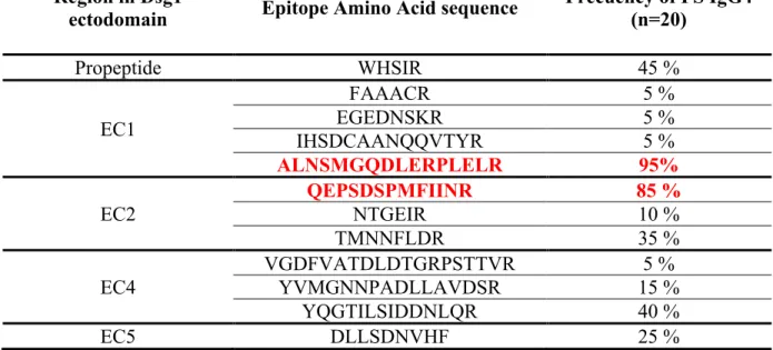
residues M133 and Q135 in hDsg1 (EC1), demonstrate that these residues are crucial in the
binding of pathogenic IgG4 autoantibodies from FS patients.
2.2.MATERIAL AND METHODS
Sources of human FS sera:
Well-characterized sera from patients with FS (n=20) from Brazil, as well as sera
from clinically healthy individuals from Limao Verde (n=1) and from the US (n= 1) were
used for epitope mapping studies. The diagnosis of FS in these patients was established by
33
other FS patients and sera from normal donors from the UNC Blood Bank were also available in our laboratory. Sera were collected following IRB policy from UNC-Chapel Hill (US) and University of Sao Paulo (Brazil).
Purification of IgG4 autoantibodies from FS sera:
IgG4 autoantibodies were purified from sera by affinity chromatography using Capture Select® human IgG4 affinity matrix (BAC BV, Leiden, The Netherlands). The Capture Select human IgG4 affinity matrix contains a 12kD llama antibody fragment that specifically recognizes human IgG4 without cross-reacting with other human IgG subclasses 1,2 or 3. Briefly, FS serum was loaded onto the matrix and later washed with PBS, pH=7.4. The matrix was eluted with 0.1M Glycine, pH=3.0. The bound fraction (elution) contained 97% IgG4 and the unbound fraction contained 92% IgG1, with small amounts of IgG2 and IgG3. Both fractions were dialyzed against PBS pH=7.4, concentrated by ultrafiltration and stored at -20°C. In addition, total IgG was purified from a normal individual living in LV by Protein G-affinity chromatography using HiTrap Protein G HP cartridge (GE Healthcare).
Levels of total IgG and IgG subclasses in sera or purification fractions were measured by a sandwich quantitative ELISA established in our lab for this study, using goat F(ab)2 anti-human IgG (Fab specific) as capture antibody, and monoclonal anti-human IgG or IgGsubclass horseradish-peroxidase conjugates as detecting antibodies.
Plasmid constructs: