Cellular and molecular mechanisms underlying srGAP2 function during neuronal development.
Sabrice Guy Guerrier
A dissertation submitted to the faculty of the University of North Carolina at Chapel Hill in partial fulfillment of the requirements for the degree of Doctor of Philosophy in the
Department of Pharmacology.
Chapel Hill 2009
Approved by:
Franck Polleux, Ph.D.
John Sondek, Ph.D.
Eva Anton, Ph.D.
James Bear, Ph.D.
Keith Burridge, Ph.D.
Sabrice Guy Guerrier
Cellular and molecular mechanisms underlying srGAP2 function during neuronal development.
(Under the direction of Franck Polleux, Ph.D.)
During brain development, proper neuronal migration and morphogenesis is critical for the
establishment of functional neuronal circuits. I have identified that srGAP2 negatively
regulates neuronal migration and induces both neurite outgrowth and branching through the
ability of its F-BAR domain to induce filopodia-like membrane protrusions resembling those
induced by I-BAR domains in vivo and in vitro. Previous work has suggested that in
non-neuronal cells, forced expression of proteins that promote filopodia decrease the rate of cell
migration and the persistence of leading edge protrusions. srGAP2 knockdown reduces
leading process branching and increases the rate of neuronal migration in vivo.
Overexpression of srGAP2 or its F-BAR domain has the opposite effects, increasing leading
process branching and dynamics and blocking migration. Finally, expression of a truncated
form of the F-BAR domain that localizes to the membrane but fails to elicit filopodia-like
membrane protrusions does not inhibit neuronal migration. This work highlights the
functional importance of proteins directly regulating membrane deformation for proper
iii Dedication
To my loving wife, Dreka, I could not have done this without your great strength, love and
Acknowledgements
James Bear, John Sondek, Keith Burridge, Eva Anton, Klaus Hahn, Sharon Milgram, Pat
Brennwald, Vytas Bankaitis, Gary Johnson, Lisa Plummer, Marie Rougié, Julien Courchet,
Rocky Cheung, Eldon Peters, Melody Lee, Janet Berrios, Jaqueline de Marchena-Powell,
Ashton Powell, Meghan Morgan, Takayuki Sassa, Jaeda Coutinho-Budd, Dante Bortone,
Randal Hand, Aurélie Gresset, Brenda Temple, Dionne Glast, Tiana Garrett, Ginnie Hench,
Mike Lee, Anthony DePass, Rick McGee, Michelle Smith, Robert Sago, Adlar Simmons,
Kerby Coulanges, Joseph Guerrier, Philippe Guerrier, Stephane Guerrier, Michele Guerrier,
v
TABLE OF CONTENTS
LIST OF FIGURES ... vii
LIST OF ABBREVIATIONS ... ix
CHAPTER ONE: General Introduction ... 1
Overview of cortical development ... 2
Reelin regulates both glia dependent and independent neuronal migration in the cortex . ... 7
CDK5 and radial migration ... 8
Rho family GTPases and radial migration ... 8
Microtubule dynamics and microtubule associated proteins and their roles in radial migration ... ..11
Regulators of actin dynamics and radial migration ... 15
Membrane deforming proteins and their potential role in neuronal migration ... 17
CHAPTER TWO: SrGAP2 regulates neuronal migration and morphology
through the ability of its F-BAR domain to induce filopodia-like membrane
protrusions ... 24
Introduction ... 24
Results ... 27
Expression of srGAP2 in the Developing Cortex ... 27
The F-BAR domain of srGAP2 deforms membrane like an I-BAR domain
... 39
srGAP2 regulates neurite formation and branching through the ability of its F-BAR domain to form filopodia ... 44
Reduction of srGAP2 expression promotes neuronal migration ... 50
The F-BAR domain is necessary and sufficient for srGAP2- mediated inhibition of radial migration ...53
srGAP2 inhibits migration by increasing leading process dynamics and branching ... 56
srGAP2 partially requires its RhoGAP and SH3 domains to inhibit migration ... 63
CHAPTER THREE: Discussion ... 75
Summary of results ... 76
srGAP2 is a novel F-BAR domain-containing protein ... 76
The role of srGAP2 during cortical development ... 80
Regulation of srGAP2: GAP and SH3 domains ... 82
Future Directions ... 85
CHAPTER FOUR: General Methods ... 87
vi
LIST OF FIGURES
Figure 1.1: Overview of mammalian cortical development. ... 3
Figure 1.2: Overview of cortical lamination ... 6
Figure 1.3: Nucleokinesis ... 13
Figure 1.4: Mammalian BAR superfamily of membrane-deforming proteins ... 17
Figure 1.5. Pattern of expression of srGAP1-3 in the developing telencephalon. ... 22
Figure 2.1: srGAP2 is expressed in neuronal progenitors and post-mitotic neurons and localizes to sites of membrane protrusion. ... 29
Figure 2.2: SrGAP2 induces filopodia formation in a F-BAR-dependent manner in COS7 cells.. ... 33
Figure 2.3: Expression of the F-BAR domain of srGAP2 in COS7 cells does not inhibit endocytosis. ... 35
Figure 2.4: SrGAP2 is an F-BAR domain containing protein ... 36
Figure 2.5: F-BAR induced filopodia required F-actin for their dynamic formation but not for their structural maintenance ... 42
Figure 2.6: Control FBP17 F-BAR tubulates liposome ... 44
Figure 2.7: Knockdown of srGAP2 in cortical neurons reduces axonal and dendritic branching ... 47
Figure 2.8: srGAP2 promotes filopodia formation and neurite outgrowth in an F-BAR dependent manner ... 49
Figure 2.9: Knockdown of srGAP2 promotes neuronal migration and reduces leading process branching... 52
Figure 2.10: SrGAP2 mediated inhibition of migration requires F-BAR mediated membrane deformation ... 55
Figure 2.11: srGAP2 increases leading process branching in F-BAR dependent manner ... 59
Figure 2.12: Expression of srGAP2 in post mitotic neurons inhibits radial migration. ... 61
Figure 2.13: NeuroD drives expression gene expression in post mitotic neurons ... 63
Figure 2.15: The GAP and SH3 domains participate in srGAP2’s ability to
promote filopodia formation in neurons ... 69
Figure 2.16: srGAP2 expressing cells accumulate in Stage 2 ... 71
Figure 2.17: The GAP and SH3 domains participate in srGAP2’s ability to
inhibit migration ... 72
viii
LIST OF ABBREVIATIONS
MGE
Medial Ganglionic Eminence
VZ
ventricular zone
CP
cortical plate
PP
preplate
MZ
marginal zone
dCP
deep cortical plate
SVZ
subventricular zone
IZ
intermediate zone
CDK5
cyclin dependent kinase 5
Ngn2
neurogenin 2
PI3K
phosphoinositide 3 kinase
JNK
c-jun N-terminal kinaseMTOC
microtubule organizing center
Lis-1
lissencephaly-1
DCX
doublecortin
FILIP
Filamin A interacting proteinBAR
Bin, Amphiphysin, RvsN-BAR
amphipathic (BAR)
FCH
Fer/Fes CIP4 homology
CC
coiled-coil
F-BAR
FCH- (BAR)
GAP
GTPase activating/accelerating protein
GEF
guanine nucleotide exchange factor
srGAP
slit-robo GAP
Chapter 1:
Overview of Cortical Development
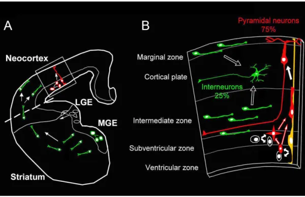
The microcircuit underlying the function of the cerebral cortex is composed of two
main neuronal sub-populations. The first, GABAergic interneurons are born in the medial
ganglionic eminence (MGE) and migrate tangentially to the neocortex where they will
provide inhibition to neuronal circuits (Fig. 1.1). The second, glutamatergic neurons are born
in the ventricular zone (VZ) of the neocortex and migrate radially to the cortical plate (CP)
where they will terminally differentiate displaying the proper dendritic morphology and extending axons to the proper locations (Fig. 1.1). These neurons will act as the excitatory
neurons of the cortex. Defects in the ratio of excitatory to inhibitory neurons, results in
severe developmental neuropathologies including from schizophrenia (Di Cristo, 2007) and
epilepsy (Guerrini and Marini, 2006). As a result much work has been focused on identifying
the genes that regulate the migration and differentiation of these two neuronal
sub-populations during the development of the cerebral cortex.
In order to understand the consequences of impairment of the proper ratio of
excitatory to inhibitory neurons in the cortex, one must first appreciate the manner in which
the neocortex develops. The neocortex is a layered structure: neurons sharing a given
function including axonal projection patterns, dendritic morphology, electrophysiological
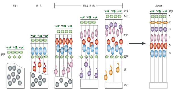
properties, etc... are grouped in separate layers. Between E10 and E11, the mouse
neocortex consists of two layers, the ventricular zone (VZ) where progenitors undergo
divisions and the preplate (PP) composed of the first postmitotic neurons present in the
mammalian cortex (Fig. 1.2). At approximately E12, neurons migrate out of the VZ and split
the PP into two layers (the marginal zone (MZ) and subplate (SP)) and thereby form the
cortical plate (CP). The MZ contains a key population of neurons called Cajal-Retzius cells
3
Figure 1.1: Overview of mammalian cortical development. A) Coronal view of developing
large secreted glycoprotein playing a key role in cortical layer formation. These early
postmitotic pyramidal neurons that form the immature CP constitute the future layer 6. This
will be the deepest cortical layer. These cells migrate primarily via somal translocation
whereby, their leading process remains attached to the basal membrane at the pial surface
while its soma translocates from the VZ to the CP. The next wave of neurons born at E13
will generate layer 5 will then migrate from the VZ using radial glial progenitors as a
substrate for migration. These layer 5 neurons will migrate passed the neurons that make up
the CP and this process of the newest born neurons bypassing their predecessors will
continue until about E18 (Fig. 1.2). This results in an “inside-out” development of the cortex
where the youngest neurons constitute the most superficial layers and the oldest neurons
constitute the deepest layers in the cortex. In the adult, this results in six cortical layers
where layer 1 cells are most superficial and layer 6 cells are the deepest. Clearly, the
cellular and molecular mechanisms controlling the precise timing of cell cycle exit, initiation
of migration and proper translocation of neurons to the individual cortical layer are critical for
the proper functional maturation of the cerebral cortex.
While somal translocation is thought to be the predominent mode of migration before
E14 (Gupta et al., 2002), radial migration underlies the majority of layer formation during
cortical development (E14-18; (Gupta et al., 2003)). Radial glial cells act as neuronal
progenitors giving rise to neurons in the ventricular zone. Once the neuron exits the cell cycle, it migrates to the sub-ventricular zone (SVZ) where it undergoes a short multipolar
phase, extending dynamic neurites in multiple directions (Noctor et al., 2004) before
polarizing by forming a single leading process. The neurons then attach to and migrate
along radial glia processes towards the CP using leading process attachment to pull the cell
soma forward. This process is known as nucleokinesis. Once they reach the CP, radially
5
dendrites. As one might expect much work has outlined the signaling pathways and
Figure 1.2. Overview of cortical lamination. During early cortical development (E11) the cortex
7
Reelin regulates both glia independent and dependent forms of neuronal
migration in the cortex
Reelin is an extracellular cue secreted by Cajal-Retzius cells resident in the marginal zone
of the cortex (Curran and D'Arcangelo, 1998). Reeler mutant mice, show defective cortical
lamination due to a mutation in the reelin gene (D'Arcangelo et al., 1995). Specifically these
mice develop an inverted cortex, due to defective preplate splitting, resulting in the formation
of the cortical plate beneath the neurons of the subplate and the appearance of an inverted
cortex (Curran and D'Arcangelo, 1998). Interestingly, 2 additional mouse and rat lines with
two spontaneously occurring mutations in mDAB1 (scrambler and yotari) share this
phenotype with the Reeler mice (Sheldon et al., 1997) suggesting that reelin and Dab1 may
act in the same pathway. In addition, the observation that loss of the very low density
lipoprotein receptor (VLDLR) and apolipoprotein E receptor 2 (ApoER2) also shared this
phenotype of no preplate splitting allowed for the identification of the reelin signaling
pathway (Trommsdorff et al., 1999). Indeed, reelin binds to VLDLR and ApoER2
(Hiesberger et al., 1999) and this binding leads to tyrosine phosphorylation of mDAB1 by
Src-family kinases (Benhayon et al., 2003). Taken together, this inability to split the preplate suggested that there is a defect in the ability of early somally translocating neurons to
migrate. It should be noted however, that reelin signaling does not only affect somally
translocating neurons. Reelin signaling also seems to affect radial glia-mediated neuronal
migration since ectopic expression of reelin in the ventricular zone rescued the preplate
splitting defect of the Reeler mouse but continued to present abnormal layer formation
(Magdaleno et al., 2002). This suggested that reelin signaling could regulate radial migration
independent of its function in preplate splitting. This data was later supported by work from
Chris Walsh’s lab demonstrating that acute reduction of Dab1 by RNA interference
(Olson et al., 2006).
CDK5 and radial migration
Cyclin dependent kinase 5 (CDK5) was shown to have no role in cell cycle
progression, compatible with its expression and activity in postmitotic neurons (Lew et al.,
1994). The activity of CDK5 is regulated by its coactivator p35 (Tsai et al., 1994), which is
also highly expressed in postmitotic neurons but not neuronal progenitors (Chae et al.,
1997; Tsai et al., 1994). Loss of p35 or CDK5 causes severe defects in neuronal migration and ultimately cortical lamination (Chae et al., 1997; Gilmore et al., 1998). The major defect
here appears to be impairment of radial migration following preplate splitting, so that the
newly born neurons cannot bypass their predecessors resulting in an inverted cortex with
proper preplate splitting (Chae et al., 1997; Gilmore et al., 1998; Gupta et al., 2002). This is
interesting since prior to preplate splitting neurons translocate by somal translocation
whereas, post preplate splitting, neurons migrate primarily by nucleokinesis, suggesting that
CDK5 primarily regulated radial glia-dependent migration. As a result, many subsequent
studies have aimed to identify molecular substrates downstream of CDK5 that mediate
these effects. In the subsequent sections I will discuss the molecules that regulate radial
migration and describe those that are regulated by CDK5.
Rho family GTPases and radial migration
The role of Rho family GTPases in non-neuronal cell types has been studied
extensively (Raftopoulou and Hall, 2004). Rho family GTPases are known to regulate a
myriad of processes from cell polarity and migration to membrane trafficking and
9
during neurodevelopment. As a result many recent studies have sought to understand the
role of Rho-GTPases during development of the cortex.
RhoA is thought to primarily play role in cell contraction at the rear (Ridley et al.,
2003). GTP bound RhoA activates rho-kinase, which leads to increase myosin light chain
phosphorylation and finally increased myosin contractility (Ridley et al., 2003). This activity is
thought to propel the cell rear forward during the migration cycle (Ridley et al., 2003). It
appears that this RhoA pathway may be conserved in neurons (Ge et al., 2006; Hand et al.,
2005; Heng et al., 2008). Hand et al. recently demonstrated that Neurogenin 2 (Ngn2), a
bHLH proneural transcription factor, specificies the radial migration properties of cortical
neurons. Mutation of tyrosine 241 of ngn2 to phenylalanine impaired migration and cells
showed a defect in nuclear translocation. Interestingly, this defect could be partially rescued
by dominant negative RhoA, suggesting the importance of RhoA activity in radial migration
(Hand et al., 2005). Further work expanded on these observations by demonstrating that the
rho effector, rho-kinase (ROCK) is required for radial migration (Nguyen et al., 2006a).
Finally, it was recently shown that myosinII activity was required for forward nuclear
movement during radial migration, since treatment with myosin inhibitor, blebbistatin
inhibited nuclear movement (Tsai et al., 2007). These data suggest that the
RhoA-ROCK-MyosinII pathway may be at work in radial migration.
While RhoA is thought to regulate contraction at the cell rear, Rac1 and Cdc42 are thought to act primarily by regulating actin dynamics at the leading edges of migrating cells
(Ridley et al., 2003). Cdc42 in particular, has been shown to act both at the leading edge
during migration, but also at the establishment of polarity before initiation of migration
(Raftopoulou and Hall, 2004). Indeed, the ability of cdc42 to regulate cell polarity is also
conserved during cortical development. Before radial migration occurs neurons are
and the ability for these cells to divide is dependent upon the polarized localization of
specific proteins (Costa et al., 2008). It turns out, that proper regulation of cdc42 is required
for this polarization. Specifically, two groups indepently demonstrated that loss of cdc42
disrupted cell division and the localization of β-catenin and atypical protein kinase C (aPKC),
two other proteins that are essential for neuronal polarity (Cappello et al., 2006; Chen et al.,
2006). Because cdc42 conditional deletion reported in these studies were obtained through
Cre-mediated recombination in neuronal progenitors, it is difficult to distinguish the function of cdc42 in cell migration from its function in cell division. However, recent work suggests
that cdc42 may be directly involved in radial migration. Konno et al. recently showed using
dominant-negative approaches that phosphoinositide 3 kinase (PI3K) activation was
required for radial migration (Konno et al., 2005). Moreover, they showed that dominant
negative cdc42 impaired radial migration after PI3K activation suggesting that cdc42
activation may be required for this activity.
Rac1 has also been implicated in regulating radial migration. Inhibition of Rac1
downstream of both PI3K and c-jun N-terminal kinase (JNK) blocked radial migration
(Kawauchi et al., 2003; Konno et al., 2005). Moreover, the Rac GEF, P-Rex1 was also
shown to be involved in radial migration (Yoshizawa et al., 2005). In addition, rac1/cdc42
effector p21 activated kinase (PAK)-1 was recently shown to be involved in radial migration.
Expression of consituitively active form of PAK-1 severely impaired radial migration and
reduction of PAK-1 had similar effects (Causeret et al., 2008).
While, RhoA, Rac1, and Cdc42 are the most commonly studied Rho-GTPases, it
was recently shown that Rnd2 plays an important function in radial migration (Heng et al.,
2008). Specifically, knockdown of Rnd2 transiently reduced radial migration and expression
of low levels of Rnd2 was sufficient to significantly rescue the migration defect observed in
11
Taken together these data highlight the potential importance of Rho family GTPase
regulation in radial migration. However, much of this data comes from genetic studies and
the use of constituitively active and dominant negative approaches. While these techniques
provide useful information, they lack the resolution and specificity needed to truly analyze
the specific role of these proteins in real time and with subcellular resolution. Future
experiments should determine in real-time the subcellular activation of Rho family proteins
using biosensors during neuronal migration. Moreover, shRNA studies would allow the study
of cell autonomous effects of these proteins. Finally, we have developed tools to study the
effects of gene manipulation specifically on neurons (Guerrier et al., submitted and (Heng et
al., 2008)) that would allow one to bypass the effects of these proteins on cell division in
order to observe the phenotypic consequences on radial migration.
Microtubule dynamics and microtubule associated proteins and their roles in
radial migration
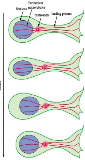
Microtubules play a key role in radial migration. This is due to the organization of
microtubules which form a “cage” around the nucleus on one end of the microtubule organizing center (MTOC) and extend from the MTOC to the extreme end of the leading
process (Fig. 3) and (Lambert de Rouvroit and Goffinet, 2001; Tsai and Gleeson, 2005). As
a result, many groups have sought to identify proteins that regulate microtubule dynamics
and as well as MTOC organization during radial migration. One such protein is
lissencephally-1 or Lis-1. As the name suggests, loss of Lis-1 causes type 1 lissencephaly
leading to a disorganization of the cortical layers during development as well as a
lissencephalic (smooth, absence of gyrification) cortex (Guerrini and Marini, 2006). shRNA
mediated reduction of Lis-1 has helped explain the lamination defect seen in patients with
development including cell division (Tsai et al., 2005). However, there were also significant
effects on the ability of immature neurons to undergo transition from multipolar to bipolar
(Tsai et al., 2005). Moreover, Lis-1 reduction led to impaired migration due to the inability of
the cell soma to translocate (Tanaka et al., 2004a; Tsai et al., 2007; Tsai et al., 2005).
Interestingly, the leading process of these cells continued to grow suggesting that Lis1 may
be required to couple somal translocation with leading process growth. Indeed, a
subsequent study demonstrated that Lis-1 knockdown impaired the movement of the
centrosome, which could explain why the nucleus does not move forward when Lis-1 levels
are reduced (Tanaka et al., 2004a). So how might Lis-1, a microtubule binding protein
regulates the movement of the nucleus? Lis-1 associates with the minus end directed motor
protein, dynein. The interaction between dynein and Lis1 is mediated by a protein called
Nudel. Interestingly, Nudel function is highly conserved since it is critical for the proper
migration of fungi (Tsai and Gleeson, 2005). Moreover, loss of 14-3-3 epsilon, which
regulates Nudel localization, causes lissencephaly similarly to lis1 and the compound
mutant (Lis1/14-3-3 double knockout) is more severe than the single knockout, again
suggesting the functional importance of this pathway for neuronal migration (Toyo-oka et al.,
2003). It was shown that like reduction of Lis1, reduction of dynein also impaired
centrosome movement (Tanaka et al., 2004a; Tsai et al., 2007). It was demonstrated that
dynein localized to the swelling present in front of the nucleus that forms prior to nuclear translocation and colocalized with centrosome. These data suggested that Lis1 may
regulate forward movement of the nucleus by regulating dynein localization to the
centrosome. Interestingly, it was shown that CDK5 can phophorylate Nudel (Niethammer et
al., 2000) and that 14-3-3 epsilon could bind to phosphorylated Nudel to properly localize
with Lis1 and dynein (Toyo-oka et al., 2003), thus providing one mechanism by which CDK5
13
Figure 1.3. Nucleokinesis. Neuronal migration in the cortex is mediated by the centrosome’s ability
Doublecortin (DCX), another microtubule associated protein, was identified as
mutated in a form of X-linked lissencephally (des Portes et al., 1998; Gleeson et al., 1998).
Its name comes from the fact that a sub-population of neurons accumulate in the white
matter (band heterotopia) resulting in ‘two gray matters’ or “double cortex” (Guerrini and
Marini, 2006). Like Lis1, shRNA studies revealed that reduction of DCX lead to a bona fide
defect in radial migration (Bai et al., 2003). How does DCX regulate migration? DCX was
shown to stabilize microtubules in vitro (Horesh et al., 1999) and to associate with
microtubules in neurons (Francis et al., 1999; Gleeson et al., 1999). Its ability to associate
with microtubules is phosphorylation dependent (Tanaka et al., 2004b). Interestingly, CDK5
was shown to phosphorylate DCX, and mutation of this phosphorylation site (serine 297)
reduced interaction between DCX and microtubules. Moreover phosphorylated DCX
localization to the microtubule cage around the nucleus in cortical neurons (Tanaka et al.,
2004b) suggesting that DCX may act to regulate microtubule pulling of the nucleus forward.
Moreover it was shown that DCX can interact with Lis1 biochemically (Caspi et al., 2000)
and DCX expression can rescue the migration defect caused by lis1 reduction in cerebellar
granule neurons (Tanaka et al., 2004a). More specifically DCX expression rescued the
dissociation of centrosome/nucleus coupling caused by Lis1 and dynein reduction
suggesting that DCX could facilitate their association with the cage around the nucleus and
thereby regulate nucleokinesis and neuronal migration (Tanaka et al., 2004a). These data provide a mechanism whereby Lis1 couples with DCX to regulate nucleokinesis.
Regulators of actin dynamics and radial migration
As one might expect, the regulation of actin polymerization is required for proper
15
that causes accumulation of neurons in the ventricular zone (Fox et al., 1998). Filamins are
thought to act as actin bundling proteins and allow the association of F-actin with plasma
membrane (Stossel et al., 2001). Careful studies in migrating cortical neurons have begun to
determine exactly how filamins might regulate migration during cortical development. First,
the actin binding activity of filamin A is required for its ability to regulate radial migration
(Nagano et al., 2004). Interestingly, these cells displayed a round shape with no leading
process extending from the cell body suggesting that f-actin bundling and stabilization may
be required for proper leading process formation (Nagano et al., 2004). In addition,
time-lapse analyisis of neurons expressing this actin binding deficient mutant of filamin a, showed
that indeed leading processes were produced however they were extremely dynamic and
cells never transitioned to a bipolar state (Nagano et al., 2004). Since impairment of filamin
A activity inhibited leading process formation, it was plausible to think that increased filamin
A may cause more efficient leading process formation. Cortical neurons seem to tightly
regulate the levels of filamin A present in the cells since it was found that Filamin A
expression levels in cortical neurons was tightly regulated by the protein FILIP (Filamin A
interacting protein) (Nagano et al., 2002). FILIP expression was shown to decrease Filamin
A levels in cells and expression of FILIP siRNA or overexpression Filamin A lead to
increased radially oriented leading processes (Nagano et al., 2004). Moreover, time-lapse of
FILIP siRNA treated neurons showed increased transition from multipolar to bipolar state suggesting that increased filamin A promotes a radially oriented leading process. In addition
to filamin A, the actin severing protein, cofilin, was recently shown to be required for radial
migration (Bellenchi et al., 2007). The genetic deletion of cofilin resulted in cortical
laminaiton defects (Bellenchi et al., 2007). These defects were due to effects both on cell
division and migration. Moreover, Kawauchi et al, showed that expression of dominant
Finally, Ena/Vasp proteins have recently been shown to regulate cortical
development. Deletion of Ena/Vasp proteins inhibited neurite initiation in cortical neurons. In
addition, these mutants demonstrated thicker cortical layers where Ena/Vasp null cells
migrated past wild type neurons, suggesting that migration is enhanced in these cells
(Kwiatkowski et al., 2007). This is consistent with the role of Ena/Vasp proteins in fibroblast
migration since reduction of Ena/Vasp actiivty in fibroblast results in increase leading edge
persistence and an increase rate of cell migration (Bear et al., 2000; Bear et al., 2002).
Taken together, these data suggest an important role for the regulation of actin dynamics in
migrating neurons, however there is much work needed to understand exactly how the
dynamics are regulated and the specific role that actin is playing in radial migration.
Membrane deforming proteins and their potential role in neuronal migration
Recently, in addition to the well-established role of the cytoskeleton in producing
forces to generate plasma membrane protrusions and invaginations, many
membrane-associated proteins have also been shown to directly sculpt and deform biological
membranes. It is now generally accepted that there are many different forms of membrane
remodeling controlled by specialized families of proteins which regulate a vast array of
important biological processes such as (1) endomembrane trafficking, exocytosis and
membrane fusion as well as (2) plasma membrane deformation including membrane
protrusions(lamelipodia and filopodia dynamics) and membrane invaginations (endocytosis,
17
Figure 1.4. Mammalian BAR superfamily of membrane-deforming proteins. (A) Diagram
19
These proteins generate membrane curvature through insertion of hydrophobic or
amphiphatic motifs into the membrane to induce bilayer asymmetry and through the
formation of membrane-bound protein scaffolds with intrinsic curvature (Itoh and De Camilli,
2006). I will now review the current knowledge on one of the protein superfamiles, the BAR
domain-containing proteins.
BAR domain superfamily: regulators of membrane curvature involved in
membrane invagination and membrane protrusion
• The BAR (Bin, Amphiphysin, Rvs) domain superfamily is a large family (>30) of
proteins playing central roles in membrane remodeling in all eukaryotes (Fig. 1.4B).
Mutations in genes encoding BAR domain proteins have been linked to several diseases
(Billuart et al., 1998; Endris et al., 2002), and inactivation of these proteins in cells and
animals is often characterized by severe phenotypes resulting from altered membrane
dynamics (reviewed in (Itoh and De Camilli, 2006; Scita et al., 2008)). Based on
structural features and phylogenetic relationships, the BAR domains can be divided into
distinct subfamilies (Frost et al., 2007). The canonical BAR domain is a dimeric module,
where three kinked antiparallel α-helices of each monomer form a banana-shaped
dimeric 6-helix bundle (Peter et al., 2004). BAR domains interact with cellular
membranes through their concave surface, which typically contains charged amino
acids. A subset of BAR domains (N-BARs) also contain an N-terminal amphiphatic helix
that folds upon membrane interaction and penetrates into the bilayer. In a number of
proteins, the BAR domain is also functionally linked to other membrane-binding motifs
such as PH or PX domains (Itoh and De Camilli, 2006). Thus, although the curved
shape of BAR domains appears to be critical for membrane tubulation, in many cases
lipid-binding motifs (Itoh and De Camilli, 2006).
• F-BAR domains (Fig. 1.4C) were originally identified as a FER-CIP4 homology
(FCH) domain in the N-terminal region of many actin-regulating proteins. Subsequent
studies by Pietro DeCamilli’s group and others revealed (1) most FCH-containing
proteins contain a coiled-coil (CC) region at variable distance from the C-terminal end of
the FCH domain and (2) overall secondary structure homology between [FCH+CC] and
BAR domains and demonstrated that the combination of FCH+CC domains or F-BAR
(FCH and BAR) domains tubulate membranes in vitro and in vivo like BAR domains (Itoh
et al., 2005; Tsujita et al., 2006). The structure of F-BAR domain differs from the
canonical BAR domain by containing five α-helices per monomer. Importantly, being
more elongated and gently curved (Fig. 2E), F-BAR domains induce larger-diameter
membrane tubules in comparison to BAR domains (Henne et al., 2007; Shimada et al.,
2007). A recent cryo-EM study demonstrated that F-BAR domains self-assemble into a
helical coat around the membrane tubules, providing evidence that these domains use a
combination of scaffolding and cooperative assembly to induce membrane curvature
(Frost et al., 2008).
• The I-BAR domain (Fig. 1.4D), which is also known as IM (IRSp53/MIM
homology) domain, was first identified as an F-actin crosslinking domain at the
N-terminal region of mammalian IRSp53 and missing-in-metastasis (MIM) proteins
(reviewed in (Mattila and Lappalainen, 2008; Scita et al., 2008)). However, subsequent
studies suggested that I-BAR/IM domains do not significantly crosslink actin filaments
under physiological conditions and revealed that the domain displays structural
homology to BAR domains. I-BAR/IM monomer consists of three α−helices that dimerize
into an antiparallel structure, which resembles a zeppelin or inverse BAR (I-BAR)
21
IRSp53 directly bind and deform membranes into tubules in vitro. However, in contrast to
the concave-shaped lipid-binding interface of BAR and F-BAR domains, the positively
charged lipid-binding surface of I-BAR domains displays a convex geometry. This
provided a possible structural explanation for why I-BAR domains induce membrane
protrusions rather than invaginations when expressed in cells. However, direct evidence
for this ‘‘inverse mechanism’’ has not been demonstrated yet. Furthermore, possible
differences in the membrane deformation properties within the I-BAR domain family are
likely.
What is the evidence that membrane-deforming proteins may be involved in neuronal
migration? Interestingly, FBP17, a F-BAR containing protein regulates neurite branching
downstream of Rnd2 activation (Heng et al., 2008). In addition, a screen for downstream
transcriptional targets of Ngn2 identified slit-robo GAP2 (srGAP2) as a potential
downstream transcriptional target of Ngn2 (Mattar et al., 2004). srGAP2 belongs to the
srGAP family of Rho GTPase activating proteins identified by Yi Rao’s group as cytoplasmic
interactors of the Robo receptor using a yeast-two-hybrid approach (Wong et al., 2001).
Analysis of the domain organization of the srGAP family showed that srGAPs possess an
N-terminal F-BAR domain, a RhoGAP domain and a C-N-terminal SH3 domain. srGAP1-3
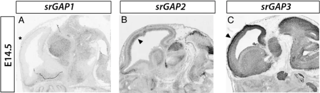
expression is enriched in the developing CNS where they show partially overlapping patterns of expression (Fig. 1.5). Interestingly, in the embryonic cortex, srGAP1 is not
detected whereas srGAP2 and 3 are expressed in progenitors and post-mitotic regions. The
SH3 domains of srGAPs were shown to associate with mouse formin 1 (Chan et al., 1996).
The SH3 domain of srGAP2 and srGAP3 have been shown to interact with WASP and
WAVE respectively (Chan et al., 1996; Linkermann et al., 2009; Soderling et al., 2002)
membrane and the actin cytoskeleton. Finally, recent work has implicated this family in the
regulation of cell migration since srGAP3 was recently shown to regulate cell migration in
cancer cells (Simpson et al., 2008). However the role of this family of proteins in the
regulation of radial migration remains poorly understood. As a result, the focus of this thesis
is to determine the function of srGAP2 during cortical development and specifically to
determine its effects on radial migration.
Figure 1.5. Pattern of expression of srGAP1-3 in the developing telencephalon. In situ
Chapter 2
(
Manuscript Re-Submitted to Cell for publication (3/19/2009).)srGAP2 regulates neuronal migration through the ability of its
F-BAR domain to induce filopodia-like membrane protrusions
Sabrice Guerrier 1,2, Jaeda Coutinho-Budd 2, Takayuki Sassa 2, Aurelie Gresset1, Nicole Vincent Jordan1, Adam Frost3, and Franck Polleux 1,2 #
1
Department of Pharmacology - School of Medicine- UNC-Chapel Hill
2
Neuroscience Research Center- UNC-Chapel Hill
3
Department of Molecular Biophysics and Biochemistry, Interdepartmental
Neuroscience Program, Yale University- School of Medicine
#
Address correspondence to:
Franck Polleux, Ph.D.
University of North Carolina- Chapel Hill
Neuroscience Center
Department of Pharmacology
115 Mason Farm Road
Room 8109C
CB-7250
Chapel Hill, NC
USA
Phone 919-966-1449 (office)
Fax 919-966-9605
Email [email protected]
INTRODUCTION
During brain development, neural progenitor proliferation, neuronal migration and
differentiation require considerable changes in cell shape involving coordinated cytoskeletal
and membrane remodeling (Ayala et al., 2007; Luo, 2002). In particular, during mammalian
cortical development, neurons born through asymmetric division of radial glial progenitors
have to migrate over long distances to reach their final destination in the cortical plate where
they will undergo terminal differentiation, which includes axonal and dendritic growth as well
as synapse formation. The cellular mechanisms by which cortical neurons migrate involve
the coordinated extension and adhesion of the leading process (LP) along radial glial
processes with the forward translocation of the nucleus (Ayala et al., 2007; Lambert de
Rouvroit and Goffinet, 2001; Tsai and Gleeson, 2005). These events are currently thought to
primarily depend on centrosome and microtubule dynamics involving the function of proteins such as Lis1, Doublecortin, and Nudel among others (Niethammer et al., 2000; Reiner et al.,
1995; Tanaka et al., 2004b; Tsai et al., 2005). However, recent genetic studies have
identified molecules that regulate leading process morphology and as a consequence, cell
migration. Specifically, deletion of cyclin dependent kinase 5 (CDK5) (Ohshima et al., 2007)
or its activator p35 (Gupta et al., 2003) resulted in an increase in LP branching and impaired
migration. These studies demonstrate a functional link between LP morphology and proper
neuronal migration.
The basis of neurite initiation, outgrowth and branching is rooted in the ability of the
cytoskeleton to undergo dynamic changes (Luo, 2002). The actin cytoskeleton, in particular,
has been shown to regulate axon and dendritic outgrowth and branching (Luo, 2002). While
several different actin structures contribute to these activities, bundled, filamentous actin
present in filopodia seem to be particularly important for neurite morphogenesis (Gupton
25
in growth cone morphology (Burnette et al., 2007; Gallo and Letourneau, 2004) and neurite
outgrowth (Luo, 2002) and branching (Dent et al., 2004; Gallo and Letourneau, 1998). In
addition, recent evidence suggests that filopodia formation is required for neurite initiation
(Dent et al., 2007; Kwiatkowski et al., 2007). Downregulation of the actin anti-cappers,
ENA/VASP proteins, which are potent inducers of filopodia using either functional
interference or a genetic approach, resulted in loss of filopodia and failed neurite initiation.
Interestingly, loss of ENA/VASP proteins also resulted in defects in cortical lamination
(Kwiatkowski et al., 2007) suggesting a complex functional relationship between filopodia
formation, neurite initiation and neuronal migration. However, at this point, the molecular
mechanisms underlying the function of filopodia dynamics in neurite initiation and branching
during neuronal migration are still poorly understood.
Classically, filopodia formation is thought to be dependent on mechanisms that
regulate actin polymerization at the barbed end of actin filaments and proteins that act to
bundle branched actin in order to form parallel bundles (Gupton and Gertler, 2007).
However, recent work has demonstrated that proteins that act on lipids at the plasma
membrane seem to play a role in filopodia formation as well (Gupton and Gertler, 2007).
LPR1, a lipid phosphatase related protein, was shown to induce filopodia independent of
many of the classical filopodia pathways including ENA/VASP proteins and Cdc42 and its
effectors (Sigal et al., 2007). In addition, IRSp53, a protein known to bind to and deform phospholipids membrane has also been shown to induce filopodia formation independent of
its ability to bundle F-actin (Mattila et al., 2007). These and other recent results strongly
suggest that changes in cell shape including filopodia dynamics or membrane invagination
and endocytosis require the function of membrane binding proteins that couple membrane
deformation and F-actin dynamics. However, the functions of proteins that deform
Here we identify slit-robo GTPase Activating Protein (srGAP2) as a potent inducer of
filopodia in neurons and an important regulator of neuronal migration and morphogenesis.
srGAP2 is expressed throughout the cortex during and after radial migration (Mattar et al.,
2004; Yao et al., 2008). It contains three functional domains: a predicted
FCH-Bin/Amphiphysin/Rvs domain (F-BAR domain), Rho GTPase accelerating/activating (GAP)
domain, and a Src Homology 3 (SH3) domain. Crystal structures of the F-BAR domains of
FBP17, CIP4, and FCHo2 demonstrated that these domains are elongated homodimers
characterized by a shallow curvature formed by the anti-parallel interaction of two
alpha-helical coiled coils (Henne et al., 2007; Shimada et al., 2007). In addition to sharing the
general fold and quaternary organization of the BAR domain superfamily as a whole, these
domains were found to share functional properties with ‘classical’ BAR domains, most
notably the ability to bind and deform membranes in vitro and in living cells (Frost et al.,
2008; Kakimoto et al., 2006; Shimada et al., 2007). However, to date, the in vivo functions of
F-BAR domain-containing proteins, including the srGAPs, have not been assessed.
RhoGAP domains inactivate Rho family GTPases by increasing their relatively slow
intrinsic rate of GTP hydrolysis. RhoGAP containing proteins are known to regulate cell
polarity, morphology, and migration in many cell types (Billuart et al., 1998; Moon and
Zheng, 2003; Ng et al., 2002). Finally, SH3 domains are polyproline-biding motifs mediating
protein-protein interactions. Interestingly, the SH3 domain of the related proteins, srGAP1 and srGAP3, have been shown to interact with the Robo1 receptor, a known axon guidance
receptor (Li et al., 2006; Wong et al., 2001), and the WAVE-1 complex, an
actin-polymerizing complex (Soderling et al., 2002). In addition the SH3 domain of srGAP2 was
shown to interact with N-WASP (Linkermann et al., 2009) placing the srGAP family of
27
Here we show that srGAP2’s ability to regulate neuronal migration and morphology
requires the unexpected ability of its N-terminal F-BAR containing domain to induce
filopodia-like membrane protrusions resembling those induced by the I-BAR domains of
IRSp53 and MIM. Interestingly, the RhoGAP and SH3 domains also participate in srGAP2’s
regulation of neuronal migration. Taken together, these results highlight the functional
importance of proteins directly regulating membrane deformation for proper neuronal
migration and morphogenesis.
RESULTS
Expression of srGAP2 in the Developing Cortex
To begin our study of the role of srGAP2 in cortical development, we first examined its pattern of expression. srGAP2 mRNA is expressed throughout the developing cortex and
is found both in proliferative zones (ventricular and subventricular zones, VZ and SVZ
respectively) at E13 and E15 and in postmitotic regions (cortical plate, CP) at E15 and P1
(Fig. 2.1A). In order to determine the pattern of srGAP2 protein expression, we obtained a
polyclonal antibody raised against the C-terminus of srGAP2 (Fig. 2.1B-C; (Yao et al., 2008).
srGAP2 protein is expressed throughout cortical development culminating at postnatal day 1
(P1) corresponding to the peak of neuronal migration in the cortex. Its expression is
maintained at P15 and reduced, but still present, in adult cortex (Fig. 2.1C).
srGAP2 expression pattern was examined by immunofluorescent staining showing
that it is ubiquitously expressed in the cortical wall (Fig. 2.1D) being found both in
Nestin-positive neuronal progenitors in the VZ (Fig. 1H-J) and MAP2-Nestin-positive post-mitotic neurons
in the CP (Fig. 2.1E-G). Finally, we determined the subcellular localization of endogenous
periphery of immature cortical neurons (Fig. 2.1K-M arrows) and was often localized to
29
Figure 2.1. srGAP2 is expressed in neuronal progenitors and post-mitotic neurons and
localizes to sites of membrane protrusion.
31
Full-length srGAP2 and its F-BAR Domain Induce Filopodia Formation
Over-expression of F-BAR domain-containing proteins such as FBP17 or CIP4 have
been shown to cause membrane invagination and tubulation in cell lines (Itoh et al., 2005;
Tsujita et al., 2006). To determine if srGAP2 and its predicted F-BAR domain had similar
properties, we expressed srGAP2 in COS7 cells. Surprisingly, expression of srGAP2 did not induce any membrane invaginations but instead, induced filopodia formation (Fig. 2.2D-F,
2.2P). This effect requires its F-BAR domain since deletion of the F-BAR domain (srGAP2•∆F-BAR-EGFP) resulted in normal COS7 cells morphology (Fig. 2.2G-I, 2.2P).
Interestingly, expression of the F-BAR domain of srGAP2 did not inhibit endocytosis,
assessed using Alexa546-Transferrin uptake assay (Fig. 2.3), as do the F-BAR domains of
FBP17 and CIP4 (Itoh et al., 2005). Furthermore, expression of the isolated F-BAR domain
fused to EGFP induced filopodia formation just as full-length srGAP2 (Fig. 2.2J-K, 2.2P). Of
note, the F-BAR domain is a very potent membrane-targeting motif (Fig. 2.2J). These data
suggest that the F-BAR domain of srGAP2 is necessary and sufficient for membrane
localization and the induction of filopodia-like membrane protrusions.
In order to distinguish the membrane targeting function of the F-BAR domain from its membrane deformation activity, we identified a small truncation of the C-terminal 49
amino-acids of the F-BAR domain (corresponding to two short alpha-helices in the C-terminal part
of the F-BAR domain) that we called F-BAR⊗∆49 (Fig. 2.4A). Expression of F-BAR⊗∆49-EGFP in COS7 cells results in membrane targeting but fails to elicit filopodia formation in COS7
cells (Fig. 2.2M-O, 2.2P). We do not know the structural basis for the inability of this
truncation to elicit filopodia formation but it may be due to an effect on oligomer formation
since it is thought that the c-terminal end of F-BAR domains are required for this activity.
Interestingly, these 49 amino acids reside in α6-8 (Fig. 2.4A) within the srGAP family that is
suggesting that the F-BAR domain of srGAP2 may contain additional sequences. Indeed,
we were unable to obtain stable protein expression of the minimal F-BAR domain.
Furthermore, as shown for other F-BAR domains (Frost et al., 2008; Itoh et al., 2005;
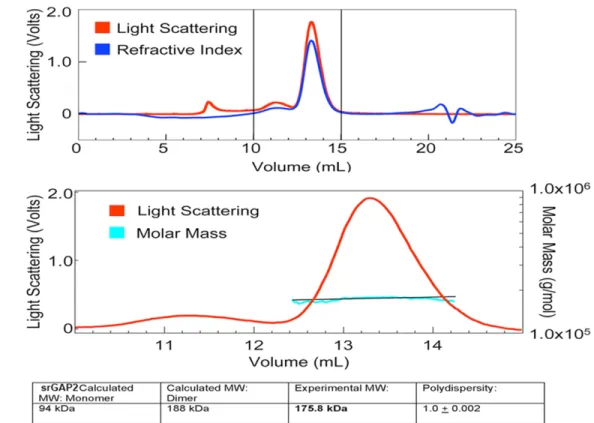
Kakimoto et al., 2006; Shimada et al., 2007), srGAP2 forms a stable dimer in solution as
assessed by light scattering assays (Fig. 2.4C) and deletion of the FCH domain (green box
Fig. 2.4A) which represents a significant portion of the dimerization interface, destroys the ability of srGAP2 to induce filopodia formation in COS7 cells (data not shown). Altogether,
these data suggest that all 8 alpha helices are likely to be required for formation of the
33
Figure 2.2. SrGAP2 induces filopodia formation in a F-BAR-dependent manner in COS7 cells.
(A-C) COS7 cell expressing EGFP counter stained with phalloidin for F-actin (red).
(G-I) Expression of srGAP2∆∆F-BAR
-EGFP fusion protein does not promote filopodia formation. (J-L) Expression of the F-BAR-EGFP fusion protein is sufficient to promote filopodia formation in COS7 cells. Note the significant increase in membrane targeting extreme periphery of the cell (J-L) and induces the formation of long actin rich protrusions (J-L) like full-length srGAP2. Thus expression of the F-BAR domain of srGAP2 is sufficient to induce filopodia. Moreover this activity is not simply dependent on localization to the plasma membrane since expression F-BAR∆∆49
-EGFP (M-O), which localized nicely to the plasma membrane, did not cause a significant increase in filopodia.
35
Figure 2.3. Expression of the F-BAR domain of srGAP2 in COS7 cells does not inhibit
endocytosis.
(A-D) COS7 cells expressing the F-BAR-EGFP fusion protein were incubated with Alexa 647-conjugated transferrin then fixed and permeabilized and stained with Alexa546-phalloidin to label F-actin (B). This transferrin-uptake assay reveals no significant difference in the level of endocytosis between F-BAR-EGFP-expressing cells (white arrowheads in D) and untransfected cells (blue arrowheads in D).
Figure 2.4. SrGAP2 is an F-BAR domain containing protein.
(A) Sequence alignment of the srGAP family from various species. Residues labeled in white on black background are identical. Red residues represent groups of conserved amino acids. SrGAP2-specific insertion is boxed in red. Predicted alpha-helices are depicted as red bars (secondary structure prediction was obtained using hhpred (Soding et al., 2005) (http://toolkit.tuebingen.mpg.de/hhpred) and Bioinfobank metaserver (http://meta.bioinfo.pl). The F-BAR domain is defined by the alpha helices 2-4. However, three additional alpha-helices are predicted C-terminal of the ‘minimal’ F-BAR domain and precede the GAP domain.
(B) Structural alignment of mouse srGAP2 with representative mouse F-BAR domains was performed using PromalS3D (Pei et al., 2008) (http://prodata.swmed.edu/promals3d/promals3d.php) and
(/smart.embl-39
heidelberg.de/). Purple boxes represent predicted coiled coil. Red box identifies srGAP-specific insertion.
(C) Top panel: purified full-length srGAP2 protein (aa 1-786 containing F-BAR, GAP and SH3 domains) (300 g) was loaded onto a Superose 6 column and separated by size exclusion
The F-BAR domain of srGAP2 deforms membrane like an I-BAR domain
The ability of srGAP2 or its F-BAR domain to induce filopodia in COS7 cells is
reminiscent of the activity of the structurally related, Inverse (I)-BAR domain-containing
proteins such as IRSp53 and Missing-in-Metastasis (MIM) (Mattila and Lappalainen, 2008;
Mattila et al., 2007; Saarikangas et al., 2009; Scita et al., 2008). The filopodia induced by
I-BAR domains are dependent upon their ability to interact with and to deform the membrane
(Mattila et al., 2007). Interestingly, F-actin depolymerization prevents the dynamics and
formation of new filopodia but does not affect the maintenance of pre-existing filopodia induced by the I-BAR domains of IRSp53 or MIM (Mattila et al., 2007). We tested if this
property of I-BAR proteins is shared by srGAP2 and its F-BAR domain. Untreated COS7
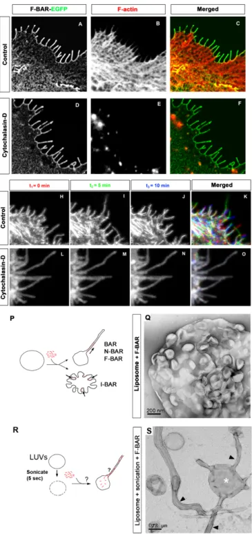
cells expressing the F-BAR domain of srGAP2 developed F-actin rich filopodia (Fig.
2.5A-C). While COS7 cells treated with cytochalasin-D were depleted of F-actin, this treatment had no effect on membrane localization of the F-BAR domain or on the maintenance of
filopodia-like protrusions (Fig. 2.5D-F).
We next wanted to determine if the dynamics of the filopodia induced by srGAP2
were dependent on F-actin. F-BAR-induced filopodia were highly dynamic in COS7 cells
protruding and retracting within less than a minute (Fig. 2.5H-2K, and Movie S1). In
contrast, F-BAR-induced filopodia formed prior to cytochalasin-D treatment were totally
resistant to F-actin depolymerization (Fig. 2.5L-O, and Movie S2). These data suggest that
the F-BAR domain of srGAP2 functions as an I-BAR domain in living cells, by inducing
filopodia that require F-actin for their dynamics but is independent of F-actin for their
structural maintenance.
In order to directly test the membrane deformation properties of the F-BAR domain of
41
negative stain transmission electron microscopy (TEM), this did not result in liposome
outward tubulation as has been reported for other F-BAR domains (see control experiment
using F-BAR domain of FBP17 added to an intact, preformed liposome Fig. 2.6). Rather, the
F-BAR domain of srGAP2 induced an inward dimpling or “scalloping” of the liposome
surface (Fig. 2.5P-Q), which is reminiscent of the activity of I-BAR domains in the same
conditions (Suetsugu et al., 2006), suggesting that F-BAR domain of srGAP2 can induce
“inverse” membrane tubulation.
This suggested the possibility that if the purified F-BAR domain of srGAP2 could be
exposed to the inside surface of liposomes, then protrusive tubules would form (Fig. 2.5R).
To test this hypothesis, mixtures of the F-BAR domain with intact, large unilamellar vesicles
(LUVs) were briefly sonicated (5sec) which presumably resulted in transient pore formation
in liposomes and introduction of the recombinant F-BAR inside LUVs. Following a wash,
liposomes were fixed, negatively stained and imaged using TEM. As predicted by the I-BAR
model, this resulted in numerous long tubular extensions emerging from LUVs (Fig. 2.5S).
Consistent with the dimensions of tubules induced by other members of the F-BAR and
I-BAR families (Frost et al., 2008; Mattila et al., 2007), the srGAP2 F-I-BAR-induced tubules
were 83 nm +/- 15 nm (average +/- SD, n=38) in diameter when imaged after variable
degrees of “flattening” in negative stain conditions. Importantly, at higher magnification, the
tubules observed by negative staining electron microscopy after sonication do no have an obvious protein coat surrounding the liposomes (Fig. 2.5S). This is in contrast with tubules
observed by other F-BAR and BAR domains that are known to coat the outer surface of the
tubule as shown in Fig. 2.6 (Frost et al., 2008; Shimada et al., 2007). Together, these
results suggest that unlike previously characterized F-BAR domains, the F-BAR domain of
srGAP2 induces tubular extensions, not invaginations, of the membrane and is therefore a
characterized I-BAR domain-containing proteins MIM and IRSp53 (Mattila et al., 2007;
Suetsugu et al., 2006).
Figure 2.5. F-BAR induced filopodia required F-actin for their dynamic formation but not for
43
(A-C) COS7 cell expressing the F-BAR-EGFP fusion protein not treated with cytochalasin D (control). Note the cortical localization of the F-BAR domain and the numerous F-actin-rich filopodia (phalloidin in B and C).
(D-F) COS7 cell expressing the F-BAR-EGFP fusion protein incubated with 400µM cytochalasin D for 30 minutes. Note that the complete loss of F-actin (phalloidin; E) had no effect on the localization of the F-BAR domain or on the structure of the F-BAR mediated protrusions.
(H-K) Time series showing the dynamics of F-BAR-EGFP-induced filopodia in COS7 cells. Time 0, 5, and 10 minutes are pseudo-colored in red, green, and blue respectively. Note there is little
colocalization of filopodia at the cell periphery (K). This is in stark contrast to COS7 cells expressing F-BAR-EGFP treated with cytochalasin- D (30 minutes) (L-O) where the protrusions remain static and do not grow or retract for the same period of time shown in control cells.
(P) Schema depicting tubulation assay in Q.
Figure 2.6. Control FBP17 F-BAR tubulates liposome.
45
srGAP2 regulates neurite formation and branching through the ability of its
F-BAR domain to form filopodia
The effects of srGAP2 on COS7 cells and liposomes as well as its localization to the
cell periphery in dissociated cortical neurons prompted us to ask if srGAP2 is required for
proper neuronal morphology. Therefore we first tested the function of srGAP2 in neuronal
morphogenesis by designing short-hairpin interfering RNA (shRNA) in order to acutely
knockdown srGAP2 expression (Fig. 2.7A). We found that srGAP2 knockdown in E15
cortical neurons led to a significant decrease in both axonal (Fig. 2.7C-D and 2.7F) and
dendritic branching after 5 div (Fig. 2.7G-H and 2.7J). Both of these effects were rescued by
co-transfection of an untargetable version of srGAP2 (Fig. 2.7E and 2.7I; 2.7F and 2.7J)
demonstrating that this is not an off-target effect. The fact that srGAP2 knockdown reduced
branching in cortical neurons, a process previously shown to require filopodia formation
(Dent et al., 2004), suggest that srGAP2 may promote neurite branching through its ability to
induce filopodia in neurons.
In order to determine if srGAP2 promoted filopodia formation and neurite initiation and
branching through its F-BAR domain, we performed structure/function analysis using electroporation of E15 cortical progenitors with various srGAP2 constructs followed by
dissociation, which induces rapid differentiation. First, we restricted our analysis to Stage 1
neurons (Dotti et al., 1988), which corresponds to the time point when immature neurons
produce a significant number of filopodia-like protrusions (Dent et al., 2007; Kwiatkowski et
al., 2007). Our analysis confirmed our previous results in COS7 cells, showing that
expression of full-length srGAP2 induced a significant increase in filopodia-like protrusions
in Stage 1 cortical neurons compared to control EGFP (Fig. 2.8A-B and 2.8F). This effect
As in COS7 cells, expression of the F-BAR domain alone potently induced filopodia
formation (Fig. 2.8D and 2.8F). Again, the effect of the F-BAR domain requires its
membrane deformation properties and not simply its membrane targeting property since
expression of F-BAR∆49 does not induce filopodia in Stage 1 cortical neurons (Fig. 2.8E and 2.8F) and instead induces large lamellipodia (arrowhead in Fig. 2.8E). These data suggest that srGAP2, through its F-BAR domain, induces filopodia in primary cortical neurons.
We then analyzed Stage 2 neurons defined by the presence of short neurites prior
the polarized emergence of a single axon (Dotti et al., 1988) in order to test if srGAP2 and its F-BAR domain were sufficient to promote the transition between filopodia-like membrane
protrusions and elongating neurites. As shown in Figure 2.8G-K, both full-length srGAP2
and the F-BAR domain significantly increased the total number of primary neurites emerging
from the cell body as well as the number of primary branches per neurite (Fig. 2.8L).
Expression of srGAP2∆F-BAR as well as F-BAR∆49 fail to increase primary neurite number and neurite branching compared to control EGFP (Fig. 2.8G and 2.8K and 2.8L) showing that (i)
the F-BAR domain is sufficient to increase neurite initiation and branching and (ii) that the
membrane deformation properties of the F-BAR domain are required for srGAP2’s ability to
47
Figure 2.7. Knockdown of srGAP2 in cortical neurons reduces axonal and dendritic branching.
(A) Western blot probed with ant-GFP and anti-actin antibodies from COS7 cells co-transfected with either control shRNA plus srGAP2-EGFP (lane 1), srGAP2 shRNA plus srGAP2-EGFP (Dha2, lane 2) or (Dha5, lane3) (B) Western blot probed with anti-GFP and anti-actin antibodies from COS7 cells co-transfected with either control shRNA plus EGFP (lane 1), srGAP2 shRNA plus srGAP2-EGFP (lane 2), a mutated form of srGAP2*-srGAP2-EGFP (resistant to srGAP2 shRNA) plus control shRNA (lane 3), or srGAP2*-EGFP plus srGAP2 shRNA (lane 4). srGAP2 shRNA significantly knocks down srGAP2 expression compared to control shRNA which can be rescued by expression of srGAP2*-EGFP (compare lanes 3 and 4).
neurons display frequent primary branches from the axon (arrowheads in B) and the primary dendrite (arrowheads in F). Both effects were markedly reduced in srGAP2 shRNA transfected neurons (D and H) and rescued by co-transfection of srGAP2 shRNA with srGAP2*-EGFP (E and I).
49
Figure 2.8. srGAP2 promotes filopodia formation and neurite outgrowth in an F-BAR
dependent manner.
(A-E) Stage 1 cortical neurons expressing various srGAP2 constructs. All cells are stained with β-III tubulin to indicate that it is a neuron and phalloidin to visualize F-actin.
the F-BAR domain since srGAP2∆F-BAR
-EGFP (C) expressing cells displayed filopodia at control levels whereas expression of F-BAR-EGFP (D) displayed a significant number of filopodia. This was not simply sue to the F-BARs ability to localize to the membrane (D) since F-BAR∆49 (E) localized nicely to the plasma membrane but did not cause an increase in the number of filopodia.
(F) Quantification of filopodia per micron in all conditions. Note srGAP2 and F-BAR are potent inducers of filopodia formation while srGAP2∆F-BAR and F-BAR∆49 are not. (EGFP n= 20 cells; srGAP2-EGFP n= 21 cells; srGAP2∆F-BAR-EGFP n= 20 cells; F-BAR-EGFP n= 20 cells; F-BAR∆49-EGFP n= 20 cells. Cells were taken from 3 independent experiments and analyzed using Mann-Whitney Test * p<0.05; ** p<.001; *** p<0.001. Green color indicates comparison to EGFP and blue color indicates comparison to srGAP2-EGFP)
(G-K) Stage 2 cortical neurons expressing various srGAP2 constructs. All cells are stained with β-III tubulin to indicate that it is a neuron and phalloidin to visualize F-actin. EGFP (G) expressing neurons and stage 2 display several neurites (white arrows) and some branching. Expression of srGAP2 (H) caused a significant increase in both neurite formation (white arrows) and branching (white
arrowheads). Similar to stage 1, expression of the F-BAR domain alone (J) also increased neurite formation (white arrows) and branching (white arrowheads), while deletion of the F-BAR domain in full length srGAP2 (I) or expression of F-BAR∆49 (K) did not increase primary neurite number or branching.
51
Reduction of srGAP2 expression promotes neuronal migration
To determine more generally the function of srGAP2 during cortical development, we
introduced our shRNA constructs directed against srGAP2 (Dha2 and Dha5; Fig. 2.7A) into
radial glial progenitors at E15 using ex vivo electroporation coupled with organotypic slice
culture (Hand et al., 2005). This assay represents a powerful tool to test the role of specific
genes in neuronal migration and differentiation. Interestingly, after 3 days in culture, at a
time point when only few control-shRNA electroporated neurons have migrated (Fig. 2.9A
and 2.9C-D), slices expressing srGAP2 shRNA showed a significant increase in the percentage of neurons that have reached the dense cortical plate (dCP) and a
corresponding decreased percentage of neurons in the intermediate zone (IZ) (Fig. 2.9B, and 2.9C-D) suggesting that reduction of srGAP2 expression promoted radial migration.
We next wanted to directly determine if srGAP2 knockdown increased neuronal
migration by regulating the rate of neuronal translocation. To do this we used time-lapse
confocal microscopy to visualize neurons coexpressing nuclear-(n) EGFP (to ease cell
tracking) and control (Fig. 2.9E-H and Movie S3) or srGAP2 shRNA (Fig. 2.9E-L and
Movie S4) in slice culture. We observed that srGAP2 shRNA expressing neurons migrated approximately 23% faster than those expressing control shRNA (Fig. 2.9M) suggesting that
reduction of srGAP2 increased cell speed.
Excessive LP branching in migrating cortical neurons can have strong inhibitory
effects on neuronal migration (Gupta et al., 2003; Ohshima et al., 2007). As a result we
wanted to determine if knockdown of srGAP2 reduced LP branching. Indeed, high
magnification reconstruction of the morphology of control shRNA or srGAP2 knockdown
neurons in layers 5/6 co-transfected with cytoplasmic EGFP, showed that their LP was
2.9N-P). Control neurons displayed approximately twice as many LP branches compared to srGAP2 shRNA-expressing neurons (Fig. 2.9P). These data strongly suggest that srGAP2
may negatively regulate the rate of radial migration by regulating LP branching and
53
Figure 2.9. Knockdown of srGAP2 promotes neuronal migration and reduces leading process
branching.
(A) E15 cortical slices cultured for 3 days after electroporation with EGFP + control shRNA. Slices were stained with anti-Nestin antibody revealing radial glial scaffold and Draq5 to illustrate
cytoarchitecture. Note that neurons have just begun to migrate but have yet to reach the cortical plate.
(B) E15 cortical slices cultured for 3 days after electroporation with EGFP + Dha2 (B, top panel) or Dha5 (B lower panel). Slices were stained with anti-Nestin antibody revealing radial glial scaffold and Draq5 to illustrate cytoarchitecture. Note that knockdown of srGAP2 using either Dha2 or Dha5 shRNA decreased the number of migrating neurons found in the IZ and compared to CP at 3div compared to control.
(E-L) E15 cortical slices cultured for 2 days ex vivo after electroporation with Nuclear EGFP (3NLS) along with control shRNA (E-H) or srGAP2 shRNA (I-L) were imaged using time-lapse confocal microscopy. Neurons transfected with srGAP2 shRNA undergo faster translocation within 4 hrs (I-L and no colocalization in L) than control shRNA-transfected neurons.
(M) Quantification of effects of srGAP2 knockdown on cell speed. SrGAP2 knockdown cells migrated approximately 23% faster (6.91 microns/h compared to 5.59microns/h) compared to control shRNA-transfected neurons. (ctl shRNA, n=95 cells; srGAP2 shRNA n=84 cells. Cells were taken from 3 independent experiments and analyzed using Mann-Whitney Test p<.05 = * , p<.01=**, p<.001=***). (N) High magnification images of control shRNA (left panel) or srGAP2 shRNA (right panel)
expressing neurons from layers 5/6. Note the branched morphology of the leading process of control shRNA expressing neurons (red arrowheads pointing to leading process tips) whereas srGAP2 shRNA expressing neurons displayed a much simpler, unbranched morphology (green arrowhead pointing to single leading process tip). This type of morphology could lead to more rapid cell migration.
(O) Computer-based reconstruction of representative quantified control and srGAP2 shRNA expressing neurons demonstrating the leading process branching effect.