THE IDENTIFICATION AND VALIDATION OF NEURAL TUBE DEFECTS IN THE GENERAL PRACTICE RESEARCH DATABASE
Scott T. Devine
A dissertation submitted to the faculty of the University of North Carolina at Chapel Hill in partial fulfillment of the requirements for the degree of Doctor of Philosophy in the School of Public Health (Epidemiology).
Chapel Hill 2007
Approved by
ABSTRACT Scott T. Devine
The Identification And Validation Of Neural Tube Defects In The General Practice Research Database
(Under the direction of Dr. Suzanne West)
Background: Our objectives were to develop an algorithm for the identification of
pregnancies in the General Practice Research Database (GPRD) that could be used to study
birth outcomes and pregnancy and to determine if the GPRD could be used to identify cases
of neural tube defects (NTDs).
Methods: We constructed a pregnancy identification algorithm to identify pregnancies in 15 to 45 year old women between January 1, 1987 and September 14, 2004. The algorithm was
evaluated for accuracy through a series of alternate analyses and reviews of electronic
records. We then created electronic case definitions of anencephaly, encephalocele,
meningocele and spina bifida and used them to identify potential NTD cases. We validated
cases by querying general practitioners (GPs) via questionnaire.
Results: We analyzed 98,922,326 records from 980,474 individuals and identified 255,400 women who had a total of 374,878 pregnancies. There were 271,613 full-term live births,
2,106 pre- or post-term births, 1,191 multi-fetus deliveries, 55,614 spontaneous abortions or
miscarriages, 43,264 elective terminations, 7 stillbirths in combination with a live birth, and
1,083 stillbirths or fetal deaths. A marker of pregnancy care was identifiable for 330,153
pregnancies, eighty-four percent of which had data available at least 180 days prior to the
first marker of pregnancy care. From the same population of 980,474 individuals, 217 NTD
questionnaires were returned. We validated a NTD diagnosis for 117 cases, giving our
electronic case definitions a positive predictive value of 0.71. The positive predictive value
varied by NTD type: 0.81 for anencephaly, 0.83 for cephalocele, 0.64 for meningocele, and
0.47 for spina bifida.
Conclusions: We were successful in identifying a large number of pregnancies in the GPRD. Our use of a hierarchical approach to identify pregnancy outcomes builds upon the methods
suggested in previous work, while implementing additional steps to minimize potential
misclassification of pregnancy outcomes. Our NTD identification algorithm was useful in
identifying three of the four types of NTDs studied. Additional information is necessary to
DEDICATION
To my wife Julie and my son Adam.
ACKNOWLEDGEMENTS
Thank you to Dr. Suzanne West for her leadership throughout this process. I would like to thank the Center for Education and Research of Therapeutics for their financial support of this project. Thank you to all the members of my dissertation committee for their time and effort for the past four years. Thank you to Susan Eaton from the General Practice Research Database for her guidance through the learning of the nuances of the
database. I would also like to thank Dr. Newell McElwee for his support and mentorship throughout my dissertation journey. Finally, I would like to thank Dr. Harry Guess who supported this project from the
TABLE OF CONTENTS
ABSTRACT...iii
DEDICATION... v
ACKNOWLEDGEMENTS ...vi
TABLE OF CONTENTS ...vii
LIST OF TABLES...xii
LIST OF FIGURES ...xiv
LIST OF ABBREVIATIONS...xvii
Chapter Page I. INTRODUCTION ... 1
II. REVIEW OF THE LITERATURE... 2
A. Neural Tube Defects ...2
1. Biology... 2
2. The Role Of Folic Acid ... 3
3. Genetic Risk Factors ... 7
4. Maternal Factors... 9
5. Medications ...11
6. Other Risk Factors...13
B. Clinical Definitions...13
1. Anencephaly ...14
C. Prenatal Diagnostic And/Or Screening Tests: ...18
1. Alpha-fetoprotein ...18
2. Amniocentesis ...19
3. Ultrasonography ...21
4. Current UK Guidelines For NTD Screening ...22
D. NTD Monitoring In The UK ...23
1. The National Congenital Anomaly System ...24
2. The European Concerted Action On Congenital Anomalies And Twins...25
3. Differences Between NCAS And EUROCAT Systems ...26
E. The GPRD ...28
1. Introduction ...28
2. The Mother-Baby Linkage ...30
3. Previous Birth Defect Research In The GPRD ...32
4. Previous Pregnancy Research Using The GPRD...35
III. STATEMENT OF SPECIFIC AIMS ...37
A. Hypotheses ...37
B. Rationale...37
C. Research Questions And Specific Aims...41
IV. METHODS ...43
A. Overview Of Methods Used...43
B. Data Used ...44
C. Procedure For Pregnancy Identification...47
1. Overview...47
2. Operational Procedure ...48
a. Step1 – Identification Of All Patients With Pregnancy Records...48
c. Step 3 – Remove Historical Event Records ...51
d. Step 4 – Identification And Ranking Of EOP Events ...53
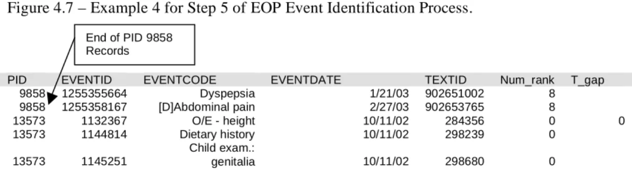
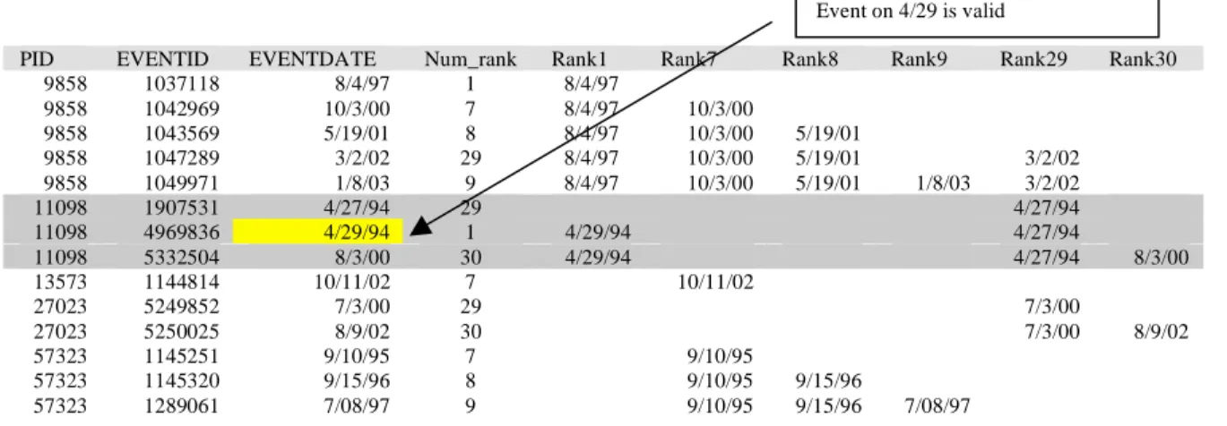
e. Step 5 – Removal Of Duplicate EOP Events Within Event Categories ...54
f. Step 6 – Removal Of Duplicate EOP Events Between Event Categories ...61
g. Step 7 – Identification Of All Patients With Complete Pregnancy Profiles...65
1. Sub-Step 1 – Identification Of Women With A PCM...66
2. Sub-Step 2 – Selecting Valid Occurrences Of PCMs...67
3. Sub-Step 3 – Create ID Variable For Events On 1/1/YEAR ...68
4. Sub-Step 4 – Identifying First PCMs ...69
D. Procedure For NTD Identification...83
1. Overview...83
2. Operational Procedure ...85
a. Step 1 – Universal Exclusions And Creation Of Mother And Child Files ...88
b. Step 2a – Identification Of Cases Within A Mother’s Records ...92
c. Step 2b – Identification Of Cases Within A Children’s Records ...97
d. Step 3 – Merging Mothers’ And Child’s Records And Eliminating Duplicates ...100
3. Validation Of Outcomes ...102
E. Quality Assurance/Quality Control ...103
1. GPRD Quality Assurance Practices ...103
2. Data Management Quality Assurance Practices ...104
3. Programming Quality Assurance Practices ...104
F. Analysis Plans ...105
1. Pregnancy Identification ...105
2. NTDs ...107
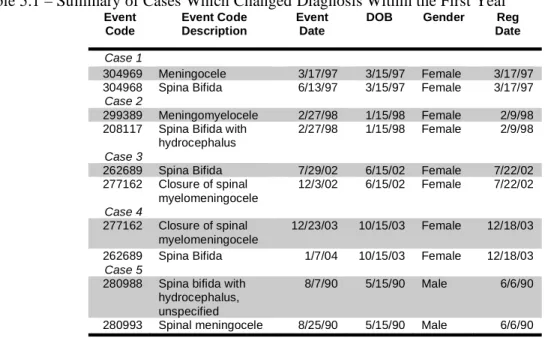
1. Review of NTD Cases With Changes In Diagnosis During 1st Year...110
a. Description Of Identified Cases With Changes In Diagnoses...111
b. Validation Results ...111
c. Conclusions...112
2. Pregnancy Identification Alternate Analyses ...113
3. NTD Identification Alternate Analyses ...121
a. Alternate Analyses On Time Searched Past Birth Criteria...122
b. Alternate Analyses On Registration Date Criteria ...125
4. Dataset Quality Assurance Analyses ...130
B. The Identification Of Pregnancies Within The GPRD...131
1. Introduction ...131
2. Methods ...131
a. Data And Study Population ...131
b. Pregnancy Medical Codes ...133
c. Identification Of Pregnancies ...134
1. Step 1 – Removal Of Duplicate Pregnancy Codes ...134
2. Step 2 – Selection Of Final Pregnancy Outcome ...135
3. Step 3 – Identifying The First PCM ...137
d. Evaluation Of Identification Algorithm...138
3. Results...139
4. Discussion...145
C. Validation Of NTD In The GPRD...150
1. Introduction ...150
2. Methods ...151
a. Identification And Validation Of NTD Cases ...152
3. Results...156
4. Discussion...163
VI. CONCLUSIONS ...168
A. Recapitulation Of Overall Study Aims, Findings And Degree To Which The Standards And Expectations For Doctoral Research Have Been Met. ...168
B. Strengths & Limitations ...170
1. Pregnancy Identification Strengths and Limitations...170
2. NTD Identification Strengths And Limitations...174
C. Future directions ...179
1. Pregnancy Identification ...179
2. NTD Identification ...180
APPENDICES...182
Appendix A – GP Questionnaires. ……….………182
Appendix B – NTD Codes. ………184
Appendix C – All Pregnancy Codes. ……….188
Appendix D – PCM Codes. ………...251
Appendix E – EOP Codes ……….272
LIST OF TABLES
Table Page
Table 2.1 – Data for NTDs (anencephaly, all spina bifida and encephalocele) from ONS
for the UK between 1999 and 2002. ...24
Table 2.2 – Data for NTDs (anencephaly, encephalocele, spina bifida and iniencephaly)
from EUROCAT for the UK between 2000 and 2003...26
Table 4.1 – Potential PCMs Used in the GPRD. ...66
Table 5.1 – Summary of Cases Which Changed Diagnosis Within the First Year...111
Table 5.2 – Summary of Validation Responses for Cases Which Changed Diagnosis
Within the First Year...112
Table 5.3 – Primary Results Of Sensitivity Analysis On Time Past Birth Criteria. ...122
Table 5.4 – Outcome Breakdown Of Additional Cases Added In Sensitivity Analysis
On Extension Of Time Past Birth To 730 Days. ...123
Table 5.5 – Gender Of Case Event Record In Sensitivity Analysis On Extension Of
Time Past Birth To 730 Days. ...124
Table 5.6 – Number Of Days Between Case Event Date And Estimated Birth Date In
Sensitivity Analysis On Extension Of Time Past Birth To 730 Days. ...124
Table 5.7 – Original Source File Of Case Event Record In Sensitivity Analysis On
Extension Of Time Past Birth To 730 Days. ...124
Table 5.8 – Year Of Case Event Record In Sensitivity Analysis On Extension Of Time
Past Birth To 730 Days. ...125
Table 5.9 – Primary results of Alternate Analysis on Registration Date Criteria. ...126
Table 5.10 – Outcome Breakdown Of Additional Cases Added In Sensitivity Analysis
on Registration Date Criteria. ...128
Table 5.11 – Gender of Case Event Record From Sensitivity Analysis On Registration
Date Criteria...128
Table 5.12 – Number Of Days Between Case Event Date And Estimated Birth Date
From Sensitivity Analysis On Registration Date Criteria. ...128
Table 5.13 - Number Of Days Between Case Event Date And Registration Date From
Sensitivity Analysis On Registration Date Criteria. ...129
Table 5.14 – Original Source File Of Case Event Record From Sensitivity Analysis On
Table 5.15 – Year Of Case Event Record From Sensitivity Analysis On Registration
Date Criteria...130
Table 5.16 – Comparison of record counts between received data and converted SAS
data sets. ...130
Table 5.17 – Distribution of pregnancy in women age 15 to 45 in the GPRD between
January 1, 1987 and September 14, 2004. ...141
Table 5.18 – Top Ten PCM Codes Identified Within The GPRD Between 1987 and
2004...142
Table 5.19 – Summary Statistics Of The Time Between First PCM And Matched EOP
Events In GPRD Between 1987 And 2004...143
Table 5.20 – Pregnancies With Between 30 And 360 Days Of Data Available Prior To
The First PCM In The GPRD Between 1987 And 2004. ...144
Table 5.21 – Neural Tube Defect Codes And Positive Predicative Values Based Upon
Validation of Records in The GPRD. ...159
Table 5.22 – Source Used To Confirm Or Refute NTD Diagnosis From Analysis Of
General Practitioner Questionnaires ...160
Table 5.23 – Diagnostics And Screening Tests Used In NTD Diagnosis From Analysis
Of General Practitioner Questionnaire...160
Table 5.24 – Neural Tube Defects by Linked Pregnancy Outcomes* for Validated
NTDs In The GPRD...162
Table 5.25 – Minimum Detectable Odds Ratio With 80 Percent Power Using 10:1
Control To Case Ratio...164
Table 6.1 – Minimum Detectable Odds Ratio With 80 Percent Power Using 10:1
LIST OF FIGURES
Figure Page
Figure 2.1 – The GPRD Mother-Baby Linkage ...30
Figure 4.1 – Steps 1 Through 3 Of EOP Event Identification Process. ...48
Figure 4.2 – Steps 4 Through 5 of EOP Event Identification Process. ...53
Figure 4.3 – Example For Step 4 of EOP Event Identification Process. ...54
Figure 4.4 – Example 1 for Step 5 of EOP Event Identification Process ...55
Figure 4.5 – Example 2 for Step 5 of EOP Event Identification Process ...57
Figure 4.6 – Example 3 for Step 5 of EOP Event Identification Process ...57
Figure 4.7 – Example 4 for Step 5 of EOP Event Identification Process. ...58
Figure 4.8 – Example 5 for Step 5 of EOP Event Identification Process ...58
Figure 4.9 – Step 6 Of EOP Event Identification Process. ...61
Figure 4.10 – Example 1 for Step 6 of EOP Event Identification Process ...62
Figure 4.11 – Example 2 for Step 6 of EOP Event Identification Process ...63
Figure 4.12 – Example 3 for Step 6 of EOP Event Identification Process ...64
Figure 4.13 – PCM Identification Process: Part 1 ...70
Figure 4.14 – PCM Identification Process: Part 2 ...71
Figure 4.15 – PCM Identification Process: Part 3 ...73
Figure 4.16 – PCM Identification Process: Part 4 ...75
Figure 4.17 – PCM Identification Process: Part 5 ...76
Figure 4.18 – PCM Identification Process: Part 6 ...78
Figure 4.19 – PCM Identification Process: Part 7 ...80
Figure 4.20 – PCM Identification Process: Part 8 ...82
Figure 4.21 – Initiation Of Case Identification Process. ...86
Figure 4.22 – Step 1 Of Case Identification Process...89
Figure 4.24 – Step 2a Of Case Identification Process In Mothers Records: Part 2...93
Figure 4.25 – Step 2b Of Case Identification Process In Children’s Records: Part 1...97
Figure 4.26 – Step 2b Of Case Identification Process Part 2...98
Figure 4.27 – Step 3 Of Case Identification Process...100
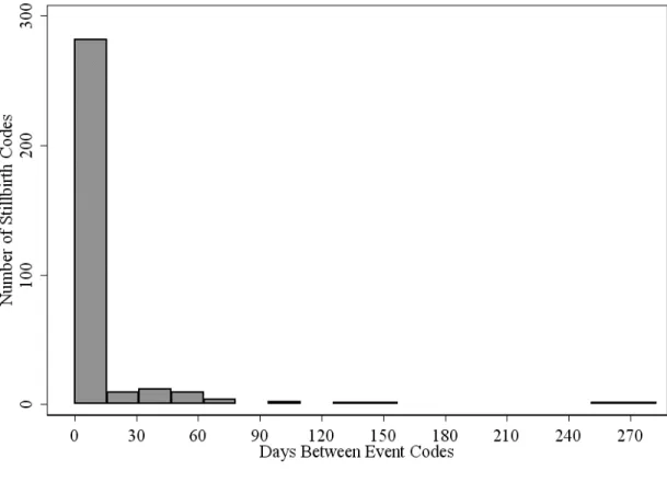
Figure 5.1 – Days Between Stillbirth Event Codes In The GPRD Between 1987 and 2004...114
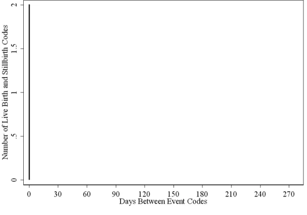
Figure 5.2 – Days Between Live Birth Stillbirth Event Codes In The GPRD Between 1987 and 2004. ...115
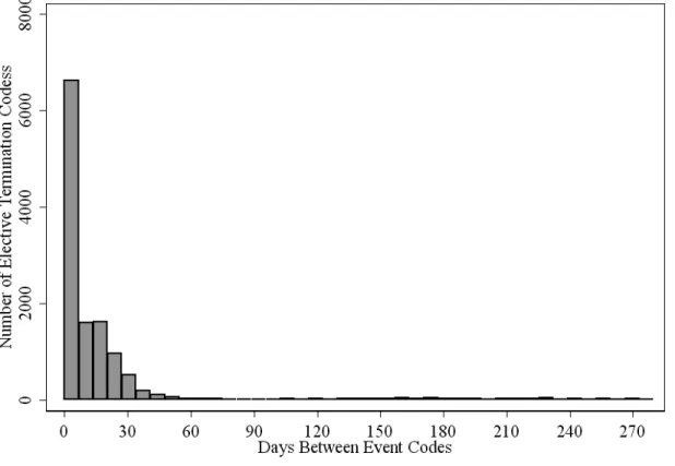
Figure 5.3 – Days Between Elective Termination Event Codes In The GPRD Between 1987 and 2004. ...116
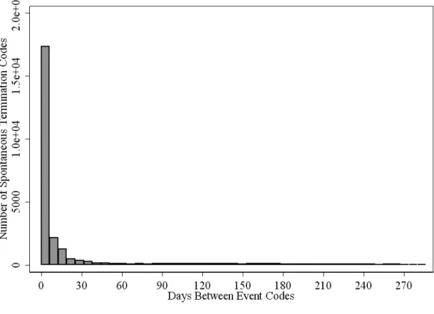
Figure 5.4– Days Between Spontaneous Termination Event Codes In The GPRD Between 1987 and 2004...117
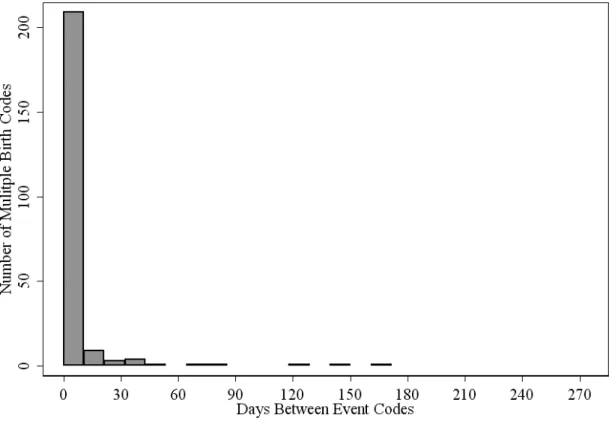
Figure 5.5 – Days Between Multiple Birth Event Codes In The GPRD Between 1987 and 2004...118
Figure 5.6 – Days Between Pre-term and Post-term Event Codes In The GPRD Between 1987 and 2004...119
Figure 5.7 – Days Between Live Birth Event Codes In The GPRD Between 1987 and 2004...120
Figure 5.8 – Number Of Days Between End Of Pregnancy Events For All Identified Pregnancies In The GPRD Between 1987 And 2004. ...121
Figure 5.9 – Example of Selection Challenges for Final Pregnancy Records in the GPRD. ...136
Figure 5.10 – Record Counts In GPRD Through Pregnancy Identification Process. ...140
Figure 5.11 – End of Pregnancy Event Counts By Outcome Type In The GPRD Between 1987 and 2004...141
Figure 5.12 – Time Between First PCM And Its Matched EOP Event Within The GPRD Between 1987 And 2004. ...143
Figure 5.13 – Progression of Records For Validated Neural Tube Defects in The General Practice Research Database Between 1987 and 2004. ...157
Figure 5.14 – Corrected Counts of Neural Tube Defect Cases in the General Practice Research Database Between 1991 and 2003. ...162
UK National Congenital Anomaly System Between Jan 1, 1991 And Dec 31,
2003...163
Figure 6.1 – Age Standardized Annual Prevalence Of NTDs From Corrected Case Counts Of NTDs From The GPRD And The UK National Congenital
Anomaly System Between Jan 1, 1991 And Dec 31, 2003. ...176
Figure 6.2 – Age Standardized Annual Prevalence Of NTDs From Event Type Specific Corrected Case Counts Of NTDs From The GPRD And The UK National
LIST OF ABBREVIATIONS
AChE Acetal Coline Esterase Inhibitor
AFP Alpha-Feto Protein
CI Confidence Interval
CNS Central Nervous System
EOP End-of-pregnancy
EUROCAT European Concerted Action on Congenital Anomalies and
Twins
FDA US Food and Drug Administration
GP General Practitioner
GPRD General Practice Research Database
hCG Human Chorionic Gonadotropin
ID Identification
ISAC Independent Scientific Advisory Committee
L&S Live and Stillborn
LMP Last Menstrual Period
MCA Medicines Control Agency
MHRA Medicines and Healthcare products Regulatory Agency
MoM Multiples of the Median
MSAFP Maternal Serum Alpha Feto Protein
NCAS National Congenital Anomaly System
NHS UK National Health Service
NIH National Institutes of Health
NTD Neural Tube Defect
ONS UK Office of National Statistics
OR Odds Ratio
OXMIS Oxford Medical Information System
PAPP-A Pregnancy-associated plasma protein A
PCM Pregnancy-care-marker
PID Patient Identification Number
PPV Positive Predictive Value
RR Risk Ratio/Relative Risk
RTI Research Triangle Institute
SEAG Scientific and Ethical Advisory Group
uE3 Unconjugated estriol
UK United Kingdom
US United States
UST Up to standard
VAMP Value Added Medical Products
I. INTRODUCTION
The purpose of this study is to develop methods for use in a comprehensive electronic medical records system that can identify neural tube defects (NTDs) for future
epidemiologic study. We use The General Practice Research Database (GPRD) to develop these methods. The GPRD is a large electronic medical record system use by general practitioners in the United Kingdom. Using this data, in combination with a specially designed General Practitioner (GP) questionnaire, we have developed and validated operational case definitions for the identification of NTDs.
In addition to this introduction, there are five primary chapters to this dissertation. We begin with Chapter II: a review of the literature. This review introduces the current state of knowledge on the biology of NTDs, the known risk factors and known
relationships between medications and NTDs, the current clinical practice for the
identification of neural tube defects, and the current prevalence and means of monitoring for NTDs in the United Kingdom. Finally, we introduce the GPRD and discuss some of the previous birth defect and pregnancy identification research using the data.
Chapter III discusses the specific aims for this dissertation. We introduce our working hypothesis and propose several specific aims meant to address these research questions. Chapter IV provides a detailed description of the methods used in this research. After an overview and discussion of the data used, we describe in detail the procedures used for the identification of pregnancies and neural tube defects. We end our discussion of methods with a description of the analyses used to assess the procedures.
Chapter V discusses the results of this dissertation. Results are divided into three sections: 1) The identification of pregnancies within the GPRD, 2) the identification and validation of NTDs within the GPRD and 3) the discussion of several additional analyses conducted beyond the primary results for the pregnancy and NTD identification
II. REVIEW OF THE LITERATURE A. Neural Tube Defects
1. Biology
NTDs are a group of severe central nervous system abnormalities that occur during early
embryonic development when the neural tube fails to close. The neural tube is an epithelial
tube formed from the neuroectoderm of the early embryo by the closure of the neural groove.
Through cell proliferation and organization, the neural tube develops into the central nervous
system.1 The neurulation process (neural tube closure) requires 10 days to complete and
occurs during the 3rd to 4th week post-fertilization.2, 3 Neural tube formation and neurulation
are the most complex phases of embryogenesis involving both extrinsic and intrinsic forces.2
These forces work together in the elevation and support of the neural plate, which then leads
to the folding and closure of the neural tube.2
Extrinsic forces involve cell structures and tissues outside of the neural plate. Defects
can occur in the neural tube when there is disruption of these extrinsic forces during initial
stages of neural plate elevation, or abnormal cell proliferation or inhibition of cell surface
glycoproteins of the neural and non-neural ectoderm.2 Intrinsic forces include the forces
occurring within the neural plate itself. Cytoskeletal elements, such as microtubules, actin
microfilaments and actin-binding proteins, are important during the process of transformation
of the neural plate epithelium.2 Disruption in these cytoskeletal elements has been shown to
The final stages of neural tube closure, particularly the actual site on the neural tube
where closure is initiated, remain controversial.2 Neural tube closure may occur following the
pattern of the mouse neural tube, whereby the tube closes in a zipper like fashion from the
cervical region to the posterior neuropore.6 Alternatively, neural tube closure may be more
similar to that of a chick, which closes at two initiation sites, a cervical site and a rostral
forebrain site.7 This controversy is partly due to the fact that mechanisms of closure and the
shape of the neural tube differ in different anatomic regions. Defects in different regions
may be the result of teratogenic agents that affect different mechanisms at a particular site
during the closure process.2 These delicate processes are of particular interest to researchers
as they occur during a period of time that a woman may not know she is pregnant, which
may present a critical window of exposure for medications to inflict damage to the growing
fetus.
2. The Role Of Folic Acid
In mammals, folate is an essential nutrient for cell function, division and differentiation.8
Folate is an important substrate in the formation of adenosine, guanine, and methionine
synthesis via homocysteine and serine and glycine interconversion. Adenosine, guanine and
methionine are involved in basic processes of cell formation and division, including
neurulation. There has been evidence of a relationship between folic acid deficiency and
malformations dating back to the middle of the 20th century. After early work in animal
models,9 the potent folic acid antagonist, aminopterin, was administered as an abortifacient
between 22 and 62 days of gestation.10 Spontaneous abortions resulted in 10 of the 12 cases.
hydrocephalus were seen. In a follow-up series of these 12 women that received
aminopterin early in pregnancy, one child with anencephaly was delivered.11, 12 These early
observations were the first evidence that chemical exposure to an embryo could result in a
malformation and provided early insight into folic acids role in neurulation.12
There are a number of ways folate deficiencies can develop including inadequate dietary
intake, malabsorption, altered metabolism and increased elimination.8 Proposed mechanisms
for folic acid’s role in the prevention of NTDs focus on overcoming these folate deficiencies
as well as decreasing the risks associated with genetic predisposition or metabolic
disturbances. Increased requirements, poor absorption or inadequate conversion of folate in
women with genetic predispositions for NTD have all been hypothesized as mechanisms for
the beneficial effect of folate in decreasing NTDs.8, 13-15 The ability of folic acid to surmount
metabolic disturbances of folate regulated metabolic pathways, primarily those involved in
the methylation process, has been demonstrated in a number studies.16-18 Impaired
methylation can cause errors in DNA synthesis and receptor molecule formation.8
Homocysteine metabolism is inter-related with folate status through the activities of
5-methyltetrahydrofolate, methionine synthase and the conversion of homocysteine to
methionine.8 Levels of homocysteine have been shown to be elevated in women who gave
birth to children with NTDs.19, 20 There are several possible disturbances in homocysteine
and folate metabolism that may lead to NTDs. Defects in enzymes involved in the
remethylation and transsulphuration of homocysteine can lead to elevated levels of
homocysteine. While common errors in transsulphuration do not appear to be associated
occurrence of NTDs.20, 22 Although still unclear, additional folate intake by the mother may
be enough to overcome elevated levels of homocysteine leading to a decrease in NTDs.
Clinical evidence for the role of folic acid in primary and secondary prevention dates
back to the 1960’s with some of the first work being done on the relationship between
multivitamin use and congenital abnormalities.23 Early theories focused on poor general
nutritional being the cause of NTDs. The hypothesis that multivitamin supplementation
(containing folic acid) prior to conception could decrease the number of central nervous
system defects was tested by the work of Smithells et al.24 The authors found that in a
secondary prevention cohort of mothers who were given the multivitamin supplement had
fewer subsequently affected newborns than mothers who were not given a multivitamin (0.6
versus 5.0 percent). Although this study had several methodological flaws,25, 26 it provided
important rationale to pursue data through randomized controlled trials.
Shortly after the Smithells study was published, Laurence et al published a double-blind
randomized controlled trial of secondary prevention of NTDs with folate treatment.27 The
treatment group was given 2 mg of folic acid twice daily pre-conceptually. In these high
risk women, the group on folic acid had no NTDs in 44 pregnancies, while the control group
had four in 51.12 Poor compliance and possible misclassification of outcomes left questions
as to the differences in impact of folic acid and placebo.12
Many of the shortcomings of these studies were meant to be addressed in the Medical
Research Council Trial.28 This randomized, multi-center, placebo controlled trial allocated
1817 women with a previous NTD pregnancy to either folic acid, a multivitamin, both or
placebo. Of 1195 pregnancies from this group, 6 of the 593 on folic acid had a NTD, while
0.71). Despite the criticisms of this study, including inadequate control for socioeconomic
status, uncertainty about the diagnosis of the initial NTDs and a failure to address primary
prevention, it was the strongest evidence to date concerning the protective nature of folic acid
supplementation and the prevention of secondary NTDs.12
The first study to address primary prevention of NTDs was conducted as part of the
Metropolitan Atlanta Birth Defects Program.29 In this case-control study, women were asked
about pre-conception vitamin use. Of the 14 percent of women who reported pre-conception
vitamin use, twice as many controls as cases reported that they took vitamin
supplementation. Criticisms of this study included the potential for recall bias, the failure to
ascertain stillbirths in the control group, and participation differences for whites and
non-whites.12 However, this study did offer the first evidence that not only high-risk mothers
could benefit from folic acid supplementation. Werler and colleagues extended these results
to include periconceptional folic acid exposure at differing doses.30 In women without a prior
history of NTD pregnancies who used folic acid containing vitamins 28 days before and after
their last menstrual period, the risk of NTD was 60% lower (RR=0.4, 95%CI: 0.2-0.6) than
those who did not take folic acid. When the dose of folic acid was 0.4mg the relative risk
estimate was 0.3 (95% CI: 0.1-0.6).
Several additional studies relying on patient self-report followed the Atlanta study, each
with mixed results. A case-control study sponsored by the NIH31 of women in California and
Illinois showed no major difference in the use of multivitamins or folate containing
supplements. A prospective cohort study conducted in Boston32 showed a substantial
Studies from Australia14, and Hungary33, each showed no definitive evidence of an
association between folic acid supplementation and primary prevention of NTDs.
In an attempt to summarize these data, Lumley et al conducted a systematic review of
the literature to assess the effects of increased consumption of folate on the prevalence of
NTDs.34 The authors identified four studies that met their inclusion criteria.27, 28, 33, 35
Pre-conceptual folate supplementation reduced the incidence of NTDs by 72 percent (relative risk
0.28, 95% CI: 0.13 to 0.58). The primary and secondary prevention of NTDs with folate
supplementation was associated with a 93 percent reduction (relative risk 0.07, 95%
confidence interval 0.00 to 1.32) and a 69 percent reduction (relative risk 0.31, 95% CI: 0.14
to 0.66) respectively.
3. Genetic Risk Factors
NTDs are thought to have both genetic and environmental determinants. Single gene
mutations and chromosomal abnormalities are associated with various NTDs. It is difficult to
determine what proportion of NTDs is due to these genetic causes or a combination of
environmental and genetic causes. In several studies, the proportion of fetuses with NTDs
that had chromosomal abnormalities ranged from 0 to 100 percent although not all studies
included spontaneous abortuses, stillborn and live born births.36-39 Autosomal recessive
disorders such as Meckel-Gruber syndrome and Walker-Warburg syndrome are associated
with encepholoceles.40 Trisomy 13 and 18 syndromes are also associated with anencephaly,
meningomyelocele and microencephaly (abnormal smallness of the brain).40 Pregnancies
NTDs in these cases are mostly genetic in origin.41 These cases may constitute a base NTD
prevalence.
Even in non-syndromal NTDs, there is evidence that genetic components are important.8
There are sex differences in the birth prevalence of certain NTDs.42 The sex distribution of
spina bifida is closer to birth proportions in most countries, with a slight female
predominance.43 There is also a reported excess of live born females with anencephaly,
however, it is unknown if this is actually a favoring of females, or selective loss of males
prior to birth.44, 45 Khoury et al evaluated data from the US national Birth Defects
Monitoring Program (1970-1978) and the Metropolitan Atlanta Congenital Defects Program
(1968-1979), and found that there was a female predominance among anencephaly and spina
bifida in cases of single NTDs.46 Hypotheses proposed to explain these sex distributions
include: 1) sex related differences in the rate of spontaneous abortions; 2) sex differences in
the process of early development of the embryo; 3) sex differences in susceptibility to
teratogens; and 4) genetic factors.42, 43
Studies of affected siblings have provided evidence for genetic risk factors. Elwood et al
estimated that the risk triples with each subsequent NTD-affected pregnancy after the first.43
Janerich and Piper conducted a review of New York State birth records looking at the
recurrence frequency of anencephaly and spina bifida in siblings of initial cases with
anencephaly or spina bifida.47 While the percentage of siblings of initial cases with
anencephaly or spina bifida was 1.8 percent, concordant twin pairs had a recurrence risk of
6.8 percent, indicating a genetic component. First, second and third-degree relatives of initial
cases with NTDs also appear to be at higher risk. In a study of spina bifida and anencephaly,
information about other affected family members.48 The occurrence of a NTD in first-degree
relatives was 3.2 percent, second-degree relatives were 0.5 percent and it was 0.17 percent in
third degree relatives. There is an increased prevalence of NTDs when the parents are
related. In a 1966 WHO study by Stevenson et al, the rates of NTDs in consanguineous
versus non-consanguineous marriages were 14.2 versus 5.7 per thousand total births
respectively, further adding to the evidence of a genetic component to NTD occurrence.44, 49
4. Maternal Factors
There are several maternal factors that appear to affect NTD prevalence. Ethnic
differences in the occurrence of NTDs are evident. Ethnicity as a risk factor was first
introduced to help explain the variability in prevalence of NTDs in the British Isles.41 In the
US, the risk among African Americans is low, while the risk among Hispanics, even after
controlling for diabetes and obesity, is high.50, 51 Maternal age appears to have only a minor
affect on the risk of NTD.41 When an association has been found, it appears to be in those
mothers under 20 and over 35.44 Parity may have a stronger effect than maternal age, with a
“modest risk in mothers of parity three or more”,43 however, other markers of maternal
fertility and the use of treatments for infertility do not appear to be associated with increased
risk.41, 52
Studies in Texas and California produced different results regarding the relationship
between previous pregnancy terminations and NTDs. In a case-control study of Hispanics in
Texas, Canfield et al. determined that women with a previous pregnancy termination had a
previous pregnancy termination.50 Todoroff and Shaw, in a study of prior spontaneous or
elective abortions, found a slightly decreased risk of NTDs.53
Obesity has been shown to increase the risk of NTD in several studies.51, 54, 55 Shaw and
colleagues investigated this risk using the data from a population-based case-control study
with cases identified from the California Birth Defects Monitoring Program.51 Women in the
highest pre-pregnancy weight group (pre-pregnancy weight of >100kg) had an increased risk
of having a child with a NTD (RR=2.1; 95% CI: 0.8 to 5.8) compared to those whose weight
was between 48 and 77 kg. Watkins et al conducted a similar study using data from the
Metropolitan Atlanta Birth Defects Case-Control Study.54 Obese women had an increased
risk 1.9 (95% CI: 1.1-3.4) times as high as average-weight women to give birth to an infant
with a NTD.
Because many obese women also have type II diabetes, Hendricks et al tried to establish
if obesity or underlying hyperinsulinemia was associated with an increased risk of NTD.56
They found that the presence of hyperinsulinemia and obesity yielded an odds ratio of 1.9
(95% CI: 1.2-3.0) compared to the absence of these two factors. Hyperinsulinemia adjusted
for obesity had a similar effect with an odds ratio of 1.8 (95% CI: 1.1-2.8). Obesity adjusted
for hyperinsulinemia had a more modest impact with an odds ratio of 1.4 (95% CI: 0.8-2.5).
Several maternal illnesses including “flu” or “cold” syndromes, and febrile illnesses have
been associated with increased risk of NTDs. In a study of records from the Finnish Register
of Congenital Malformations, Kurppa et al assessed the association between reported first
trimester maternal cold and anencephaly.57 In a case-control study of 393 mother-child pairs,
70 mothers with an anencephaly-affected child versus 17 control mothers reported a common
those without a fever were excluded, the result was similar but less precise (adjusted OR 4.7,
95% CI: 1.0-22.5).
Lynberg et al, using the Atlanta Birth Defects Case-Control Study data, evaluated the
association between NTDs and maternal exposure to flu, fever and medications taken for
illness.58 For mothers who reported episodes of flu with fever that lasted 2 or more days in
the time period from 1 month prior to 3 months post-conception, the risk of any NTD was 3.0
(95 percent CI: 1.9-4.7) times as high as the risk of any NTD in those mothers without an
episode. The risk of NTD when the mother had flu without fever prior to or after conception
was 2.0 (95% CI: 1.1-4.0) times as high as the risk in those who did not have the flu. The
risk of NTD for women who took medications for their flu episode was 4.3 (95% CI: 2.6-7.1)
times as high as the risk of NTD for women who did not take medications for the flu episode.
Shaw et al, using the California Birth Defects Monitoring Program data, evaluated the impact
of a variety of maternal illnesses on NTD occurrence.59 While still finding an association
between fever (OR 1.99, 95 percent CI: 1.37-2.90) or febrile illness (OR 1.99, 95 percent CI:
1.12-3.46) and NTDs, they failed to find the strong association between medication use and
NTD occurrence.
5. Medications
While the mechanism for medication induced NTDs are primarily unknown, those
that affect folic acid activity have been shown to be associated with an increase risk for
NTDs. There are a variety of pharmacodynamic mechanisms that medications can make use
of that can have this effect. Medications that can antagonize the effects of folic acid within
because they inhibit dihydrofolate reductase producing antifolate effects.60 Aminopterin and
methotrexate, antineoplastic agents, are potent folic acid antagonists and are known
teratogens.61 As mentioned above in the section on folic acid, aminopterin’s effects were
identified in early work on folic acid’s role in neural tube formation.10 By blocking the
conversion of folic acid to tetrahydrofolic acid, these agents can limit the formation of
important amino acids involved in cell formation.
A medication or its metabolite can also affect folic acid activity by impairing absorption
or altering hepatic metabolism.62-64 Colchicine, a common medication for gout, reduces blood
folate concentrations through an unknown mechanism.65 Cycloserine combined with
isoniazid for the treatment of tuberculosis results in lower serum folate levels compared with
isoniazid alone.66 Oral contraceptives have also been shown to lower folate concentrations.66
Colchicine, cycloserine, isoniazid and oral contraceptives have not been associated with
NTDs.
Several antiepileptic medications have been shown to increase the risk of NTDs.
Phenytoin reduces folic acid levels by affecting several enzymes involved in the metabolism
of folic acid or tetrahydrofolic acid.67-69 In a small study of multiple antiepileptic agents,
phenytoin use was associated with a case of anencephaly.62 Carbamazepine has been shown
to reduce serum folate levels by interfering with folate metabolism.70 Calandre et al found
that serum folate levels were lower in patients with higher serum carbamazepine levels.
Carbamazepine has been associated with spina bifida. In a review of literature, Rosa found a
1% incidence of spina bifida associated with carbamazepine treatment.71 Valproic acid
inhibits the metabolism of folic acid decreasing serum folic acid levels. Valproic acid is also
maternal valproic acid use.71 Combinations of these medications are also associated with
higher rates of birth defects. Lindhout found that pregnancies of women using
carbamazepine, valproic acid and phenobarbital (a barbiturate often used as for epilepsy)
with or without phenytoin resulted in a birth defect 58 percent of the time.72
6. Other Risk Factors
A variety of other risk factors have also been identified. Dietary, occupational and other
exposures have been examined as possible risk factors for NTDs. Tea use,73 lead exposure74
and high levels of organic matter in drinking water75 have been associated with increased
NTD occurrence. Some occupations with exposure to industrial chemicals and/or pesticides
have been associated with increased risk of NTDs.76-80 Parental socioeconomic status has
been associated with differing rates of NTDs, but contrary evidence leaves it a weak
predictor of risk.41
B. Clinical Definitions
The NTDs of interest in this study are anencephaly, craniorachischisis, encephalocele,
encephalomyelocele, meningocele and spina bifida. The clinical definitions for each of
these conditions are presented below. There are multiple reasons for choosing the
specific malformations to be studied in this project. The primary determinant is that
these are the most commonly occurring NTDs in the United Kingdom (UK). These
malformations, while relatively uncommon, carry a substantial burden in terms of
morbidity and mortality of the affected offspring. They also occur in sufficient numbers
Additionally, these malformations have clear clinical definitions that decrease the
likelihood of misdiagnosis. The potential does exist that some of the malformations
could be misclassified within a category of NTD (i.e. a meningocele is incorrectly
diagnosed as a spina bifida) however; this will not impact our primary results of total
NTDs.
The ultimate goal of this research is to provide validated case definitions identifying
new NTD cases and to further research of medications as risk factors. We are thus
interested in conditions that have unknown etiologies, not genetic syndromes. Although
some of these malformations do occur with certain genetic syndromes, none have a
syndrome as their sole cause.
1. Anencephaly
Anencephaly is the complete or partial absence of all or part of the brain, neurocranium
and the covering skin.40, 45, 81 When the cephalic neural tube fails to close, brain protrudes and
subsequently degenerates. Holo-anencephaly, or the complete absence of the brain, accounts
for 65 percent of cases in the US, with the remainder being cases of mero-anencephaly, or
partial absence of the brain.82 Because of the extreme nature of this disorder, anencephaly is
readily apparent at birth. In cases of anencephaly the failure of the cephalic neural tube
closure occurs on or about the 24th day post fertilization.45 The diagnosis of anencephaly can
occur upon routine obstetric ultrasound, generally based upon a coronal view that reveals the
absence of the brain. A diagnosis can be made as early as 11 weeks;83 however an
Craniorachischisis is similar to anencephaly in that the fetus is absent a developed brain,
and is associated with a contiguous spina bifida.45, 81, 84 The cervical spine is retroflexed to
the point that the head is set gazing upward.45 Craniorachischisis is often misdiagnosed as
iniencephaly, which is characterized by a closed cranium, enlarged foramen magnum as well
as a retroflexed spine with upward gaze.81 Cases of holo-anencephaly are associated with
craniorachischisis about 80 percent of the time.82 Diagnosis of craniorachischisis is similar to
that of anencephaly, occurring upon routine obstetric ultrasound at or after 14 to 15 weeks of
gestation.
The prognosis for the infant with anencephaly or craniorachischisis is uniformly fatal,
with a live born infant dying within hours to days after birth.45, 85, 86 Postnatal care is
supportive only, and generally not indicated. Prior to advances in prenatal diagnosis, the
ratio of stillbirth to live birth was approximately 50 percent.82 Between 1985 and 2000 in a
cohort of 171 cases of anencephaly in Utah, approximately 66 percent of all anencephaly
cases were terminated prior to delivery.87
2. Encephalocele
Cephaloceles are a group of anomalies due to a congenital defect of the skull resulting in
a skin covered herniation of the brain (encephalocele), brain and spinal cord
(encephalomyelocele) or a non-brain containing sac (cranial meningocele).45, 81 Most
cephaloceles occur along the midline of the cranium with lesions occurring in the occipital
region 74 percent of the time.45, 88, 89 Encephaloceles can have varying degrees of severity
with approximately 50 percent of infants with encephaloceles having additional congenital
cephaloceles are skin covered, alpha-fetoprotein (AFP) levels are generally not elevated, thus
most cases are identified from prenatal ultrasounds in low risk populations.45 The differential
diagnosis of cephalocele should include cystic hygroma, scalp edema, blebs, a normal ear,
brachial cleft cysts, amniotic band syndrome and cloverleaf skull, not just a paracranial
mass.45, 91, 92
Prognosis is determined by the content of the lesion rather than the size, with some small
lesions containing important brain tissue and/or signifying underlying CNS malformations.45,
93, 94
Prognosis is generally best with frontoethmoidal lesions and tend to be most grave with
rostral parietal lesions.45 Surgical repair is possible, and includes attempts to enlarge the
cranial cavity to preserve cerebral tissue and its vascular supply. However, when lesions
contain cortex, are associated with an absent corpus callosum, or other malformations, poor
survival and decreased intellect are often unavoidable.
3. Spina Bifida And Meningocele
Spina bifida is a defect of closure of the bones producing the spine due to failed fusion of
the caudal portion of the neural tube.45, 81 This defect may be covered by normal skin in the
case of spina bifida occulta. It may be a protruding sac in the case of spina bifida cystica. It
may also result in a completely open spine, in the case of rachischisis, which is often
incompatible with life.1, 90 Protrusion of neural tissue in a posterior spina bifida cystica is
readily apparent at birth with lesions occurring in varying sizes at any location along the
spine. Matson et al determined the location of lesions for a group of cases and found 42.2
percent lumbar lesions, 27.7 percent lubosacral, 9.9 percent thoracolumbar, 8.6 percent
90 percent of the time the protruding sac contains elements of spinal cord and/or nerves, also
known as meningomyelocele.45 The remaining cases of spina bifida are considered
meningoceles.
Meningocele, a form of spina bifida, occurs when a defect in the closure of vertebral
bones results in a protruding fluid filled sac containing abnormal meninges and cerebral
spinal fluid.45 The underlying spinal cord is usually intact; however, it may also protrude into
the sac, although not to the extent of a meningomyelocele. Normal skin usually covers the
sac. To distinguish this malformation from meningomyelocele, the sac should be
transilluminated or undergo magnetic resonance imaging at birth. Meningoceles are often
asymptomatic at birth, however it may be associated with serious co-morbidities including
diastematomyelia (a division of the spinal cord) and various tumors.45, 95
Cases of spina bifida are often difficult to diagnosis using direct sonographic
visualization. The sensitivity and specificity of these scans vary depending on the underlying
risk of the population.96 Indirect methods of visualization have been developed to help make
sonographic diagnoses. Infants with spina bifida who undergo ultrasound at 24 weeks of
gestation frequently have a bilateral, concave, frontal contour of the cranium (the lemon sign)
and cerebellar hemispheres with anterior curves with loss of the cisterna magna (the banana
sign).45 These signs have been shown in a large cohort of high risk pregnancies to have
positive and negative predictive values of 92 percent and 99.8 percent for the lemon sign and
100 percent and 99.7 percent for cerebellar anomalies for the banana sign.97
Prognosis for patients with spina bifida is variable. Significant morbidity and mortality
often depends on the severity and location of the lesion. Approximately 90 percent of infants
typically experience very high survival rates once beyond the first year of life.45 Loss of renal
function and shunt complications are the usual causes of death in older patients but many
patients can lead relatively normal lives.90 Population based data from British Columbia
found 1, 5 and 10 year survival to be 67, 65 and 64 percent respectively.98 Treatment with
primary closure of the lesion has been reported to increase long term survival rates in some
cohorts, while having only marginal impact in others.45, 99-102
C. Prenatal Diagnostic And/Or Screening Tests: 1. Alpha-fetoprotein
The primary method for screening for NTDs is testing for the presence of
alpha-fetoprotein (AFP) in maternal serum. AFP is the principal fetal plasma protein early in
gestation and remains so until the fetal liver matures and albumin becomes the primary
plasma protein.103 Amniotic fluid AFP (see below) passes through the placental barrier into
the maternal circulation and levels are measurable early in the first trimester. When a fetus
has an open neural tube lesion, high concentrations of AFP build up in amniotic fluid
subsequently leading to increased maternal serum concentrations.103, 104
Maternal serum AFP (MSAFP) levels rise through the first and second trimesters of
gestation in unaffected pregnancies, so gestational age must be considered when interpreting
results.104 MSAFP for prenatal screening purposes should be performed between the 15th and
20th weeks of gestation and results should be expressed in multiples of the median (MoM) for
gestational age.103 A result of 2.5 MoM in single gestations and 4.5 MoM in twin gestations
is considered elevated enough to perform additional diagnostic testing.103 Sensitivity of
MSAFP screening has been determined in a number of trials, but depends on the NTD under
88 and 92 percent depending on the underlying risk in the population.105, 106 Sensitivity to
MSAFP testing for spina bifida is lower, but depending upon the type of lesion involved, it is
still between 64 and 76 percent.105, 106
Because of the possibility of overlap of MSAFP level in affected and unaffected NTD
cases at different gestational ages, elevated MSAFP should not be considered diagnostic.104
However, elevated MSAFP levels have been shown to be highly predictive of NTDs in a
number of trials. In a series of studies by Drugan et al, MSAFP levels of 2.5 to 2.9 MoM are
associated with NTDs 3.4 percent of the time, while defects occurred 40.3% of the time with
a MoM of greater than 7. 107, 108 Other studies indicate that level of MSAFP greater than 5
MoM can be associated with ultrasound confirmed defects as much as 71 percent of the
time109, while levels greater than 8 MoM are most commonly associated with large structural
defects and/or fetal death prior to 20 weeks of gestation.110
The UK Collaborative AFP Study, the first major study to determine the parameters of
association between AFP and NTDs, produced detection rates for anencephaly of 98.2
percent and open spina bifida of 97.6 percent.111 False positives do occur when fetal blood
contaminates the sample. Fetal blood contains 100 to 200 times the AFP per milliliter that
amniotic fluid does at a given gestational age, and will thus give false results.104 The
diagnostic cut off for AFP varies by gestational age between 2.5 at 13-15 completed weeks to
4.0 for 22-24 completed weeks.104
2. Amniocentesis
Amniocentesis is the collection of amniotic fluid from the amniotic sac of a developing
performed between the 15th and 20th week of gestation to aide in the determination of fetal
karyotyping.113 Although the procedure is extremely accurate in the screening and diagnosis
of certain genetic disorders and NTDs, it is not without risk. Tabor et al conducted a
randomized controlled trial and found that the risk of spontaneous abortion after
amniocentesis was approximately 1 percent.114 Roper et al found a similar cumulative fetal
loss rate of 1.2 percent, but the rate was variable dependent upon the gestational age at
amniocentesis.115 When the amniocentesis was performed before 14 weeks, the fetal loss rate
was 1.0 percent, while the rate increased to 3.1 percent after 18 weeks of gestation. As these
rates are generally higher than some of the observed rates of the genetic disorders and NTDs
that amniocentesis is meant to detect, less invasive and less risky tests are preferred in
populations at low risk for the underlying defect.
Amniocentesis can also be used to detect the presence of acetylcholinesterase enzymes
(AChE) in the amniotic fluid. While non-specific cholinesterase enzymes are present in the
amniotic fluid, AChE are normally only found in the cerebrospinal fluid and within red blood
cell membranes.104 When an open NTD occurs, AChE can be detected in the amniotic
fluid.116 Between the 13th and 24th weeks of gestation, a group of confirmed open NTDs with
a high amniotic fluid AFP (>99.6 percentile) had a positive amniotic fluid AChE in 99.5
percent of cases.117 The Second Report of the Collaborative AChE Study recommended that
the best policy for use of the AChE test was in the analysis of amniotic fluid samples with
AFP results greater than 2.0 MoM.118 This approach was predicted to yield a true positive
3. Ultrasonography
Ultrasonography is considered the primary diagnostic technique for prenatal
identification of NTDs. This technology, first utilized in 1958, has been demonstrated to
provide accurate diagnostic information for gestational age and fetal anomalies.119-122
Prenatal ultrasonography is a complex technology which uses sound waves to produce
images of the developing fetus.113 These images of the developing fetus allow the direct
visualization of anencephaly and cephaloceles. 45, 103, 123 Anencephaly was the first
malformation to be diagnosed by ultrasound.124 Campbell et al determined that ultrasound
could be used between the 14th and 15th week of gestation to determine a diagnosis of
anencephaly.123, 125 Accuracy of diagnosis of anencephaly by prenatal ultrasound has been
shown to approach 100 percent.105, 126, 127
In the case of spina bifida, direct visualization is often difficult, thus indirect visualization
methods have been devised.45 Infants with spina bifida who undergo ultrasound at 24 weeks
of gestation frequently have a bilateral, concave, frontal contour of the cranium (the lemon
sign) and cerebellar hemispheres with anterior curves with loss of the cisterna magna (the
banana sign).45 One of the first studies to confirm the utility of ultrasonography in spina
bifida affected pregnancies was that of Nicolaides et al.128 The authors retrospectively
analyzed the ultrasounds of 70 fetuses between 16 and 24 weeks of gestation that were
diagnosed with open spina bifida lesions. Their work confirmed the use of indirect signs,
such as the “lemon” sign and the “banana” sign in the diagnosis of NTDs.45 Van den Hof et
al were able to detect 98 percent of spina bifida cases in a cohort of 1561 high risk mothers,
While the diagnostic ability of ultrasound for spina bifida may not approach that of
anencephaly, when combined with other maternal screening approaches, diagnostic ability is
improved. Nadel et al showed that the use of ultrasound in mothers with elevated MSAFP
decreased the need for amniocentesis to confirm the diagnosis of a NTD.130 Lennon et al
examined a group of 2257 patients at high risk for an open NTD either because of a family
history of NTDs or a positive MSAFP.131 2053 patients were given an ultrasound with 55
NTDs occurring in this cohort. All of the NTDs in this cohort were detected prenatally. The
sensitivity and specificity of ultrasound in the identification of NTDs was 97 and 100 percent
respectively. The positive predictive value was 100 percent and the negative predictive value
was 99.9 percent.
4. Current UK Guidelines For NTD Screening
The Royal College of Obstetricians and Gynecologists and the National Institute for
Clinical Excellence have proposed routine antenatal care for pregnant women.132 Women
should be scheduled for between seven and ten antenatal appointments for uncomplicated
pregnancies. Ultrasound testing is recommended for all pregnant women between the 10th
and 13th week of pregnancy to determine gestational age, detect multiple pregnancies, and
improve the performance of screening procedures for Down’s syndrome and other anomalies.
In addition, women should be offered an additional ultrasound scan between week 18 and 20
to detect congenital anomalies. The Guideline recommends standard screening for Down’s
syndrome between 11 and 20 weeks of gestation through the performance of nuchal
translucency as well as several combined tests. The recommendations are as follows: 1)
hCG and PAPP-A); 2) Gestational age from 14 to 20 weeks - the triple test (hCG, AFP and
uE3) or the quadruple test (hCG, AFP, uE3, inhibin A); 3) Gestational age from 11 to 14
weeks and14 to 20 weeks - the integrated test (NT, PAPP-A + hCG, AFP, uE3, inhibin A) or
the serum-integrated test (PAPP-A + hCG, AFP, uE3, inhibin A). The “Triple”,
“Quadruple”, “Integrated” and “Serum-integrated” tests all incorporate tests for
alpha-fetoprotein thus also serving as a screening tool for NTDs.
Nuchal translucency (NT) testing may have some utility in the detection of NTDs. NT
testing is conducted using a transvaginal ultrasound device to measure the normal
subcutaneous space between the skin and the cervical spine in the fetus early (12th to 14th
week) in pregnancy. A space less than 3 mm has been associated with increased risk for
Down’s syndrome, 18, 13 and triploidy and Turner syndrome.133 As this screening
ultrasound occurs much earlier than diagnostic ultrasounds for other abnormalities (NTDs for
example), researchers have assessed if these early ultrasound can be used to identify other
abnormalities. McAuliffe and colleagues determined that while NT can identify some serious
structural abnormalities (i.e. anencephaly), the 18 to 20 week ultrasound should remain the
gold standard.134
D. NTD Monitoring In The UK
Historically, the UK has had some of the highest recorded rates of NTDs. Prevalences of
anencephaly and spina bifida were as high as 60/10,000 births in the 1940’s and
1950’s.135, 136 Increased awareness of NTDs, folic acid supplementation and general UK
population trends (such as increased immigration, changes in birth rate) may be related to the
roughly 8/10,000 births in the 1990’s.137 Table 2.1 describes the current rates of NTDs in the
UK.138-141
Table 2.1 – Data for NTDs (anencephaly, all spina bifida and encephalocele) from ONS for the UK between 1999 and 2002.
1999 2000 2001 2002 Totals:
Live Births 67 88 71 83 309
Still Births 31 36 29 34 130
Induced Abortions 295 331 288 255 1,169
Total Cases 393 455 388 372 1,608
Total Live Births & Stillbirths 624,862 607,304 597,506 599,279 2,428,951 Prevalence/10,000 (L&S) 1.57 2.04 1.67 1.95 1.81
These historically high rates of occurrence, in addition to the thalidomide tragedy of the
1960s created the impetus within the UK for a continuous monitoring system for congenital
anomalies.
1. The National Congenital Anomaly System
The Office of National Statistics initiated the National Congenital Anomaly System
(NCAS) in 1964. The England and Wales National Congenital Anomaly System collects data
from birth registries throughout the UK and reports data to the Office of National Statistics
on a continuous basis. Data are collected from birth notifications by local health care
authorities in all regions of the UK using a standardized case report form that is completed
and sent to the Office of National Statistics. Data are collected on live and stillbirths, and
reporting is conducted on a voluntary basis. Some local congenital anomaly registries also
report information to the NCAS.141
Rather than attempt to estimate the prevalence of various anomalies, the goal of the
NCAS is to detect changes in the frequency of reporting of particular anomalies or groups of
because of the voluntary nature of data reporting, the reported rates may offer important
estimates of the prevalence of these conditions. The database is a valuable tool for detecting
possible signals for further investigation; however, increases in notification may be due to
changes in the reporting practices rather than true changes in prevalence.
Limitations of the data are due to the reliance on passive surveillance techniques for case
ascertainment, lack of collection of information on spontaneous abortions and limited
information on elective terminations of pregnancy. Although not collected directly through
the NCAS, data on elective terminations and their association with a potential congenital
anomaly is available through other National Health Service data. This data is captured and
presented along with the data from live and stillbirths collected through the passive reporting
system. The NCAS does collect some basic exposure information (such as mother’s and
father’s occupation), but the data have limited utility for research on risk factors for
congenital anomalies.
2. The European Concerted Action On Congenital Anomalies And Twins
The EUROCAT (European Concerted Action on Congenital Anomalies and Twins)
program was set up in 1974 to monitor epidemiologic information on congenital
anomalies.142 The aim of EUROCAT is to carry out epidemiologic surveillance of congenital
anomalies in Europe. As of 2003, data from 41 member registries from 20 countries are
collected and transmitted to a central registry in the UK. Eight registries from the UK are
currently full member registries, transmitting case data on all congenital anomaly cases in
their region. Each member registry transmits core variables that are recorded using a
anomaly subgroups between 1980 and 2003 are available to researchers through the
EUROCAT website (www.eurocat.ulster.ac.uk). Core variables reported to the EUROCAT
include date of birth, gender of fetus/infant, number of fetuses/infants delivered, type of birth
(including spontaneous abortions), gestational age, demographic information on the mother
and syndromal and malformation details. In EUROCAT prevalence calculations, numerators
include cases identified from live birth, fetal deaths from 20 weeks gestation (stillbirths and
spontaneous abortions) and induced abortions. A baby/fetus with several anomalies is
counted once within each class of anomaly. The number in different classes of cases cannot
be added to reach a total number of babies/fetuses. A baby is counted once only in any given
prevalence. Current data on NTD prevalence with denominators including live births, fetal
deaths and induced abortions are presented in Table 2.2.
Table 2.2 – Data for NTDs (anencephaly, encephalocele, spina bifida and iniencephaly) from EUROCAT for the UK between 2000 and 2003.
2000 2001 2002 2003 Totals:
Live Births 37 31 45 20 171
Fetal Deaths (>= 20 weeks) 9 8 14 8 52 Induced Abortions 212 207 201 134 960
Total Cases 258 246 260 162 1183
Total Live Births & Stillbirths 204,693 192,117 192,785 111,765 904,886 Prevalence/10,000 12.6 12.8 13.49 14.49 13.07
Produced using EUROCAT Website Database: http://eurocat.ulster.ac.uk/pubdata/report8tab.html (accessed 6/13/05)
3. Differences Between NCAS And EUROCAT Systems
The importance of the differences in the utility of each database for the surveillance of
congenital anomalies should not be overlooked. Boyd et al compared the NCAS to four of
the UK registries that report to the EUROCAT.143 Isolated cases were derived from similar
locales to the four UK registries and created a ratio of cases identified by the national register
percent of the cases identified by the four registries when terminations of pregnancy were
excluded, and 27 percent when terminations of pregnancies were included. The lowest
ascertainment was for NTDs, with only 11 percent ascertainment when terminations were
included and 68 percent ascertainment when terminations were excluded. The authors note
that although the stated goal of the NCAS is for signal monitoring, the degree to which it can
meet this goal is hindered by the uncertainty of the magnitude of under-ascertainment. If
under-ascertainment is constant, signals can be detected. If, however, it is not constant, there
is no way of knowing if any increase in reporting is due to increased ascertainment or to a
true increase in prevalence.
A primary advantage of the development of a cohort population in our proposed study is
the ability to determine various prevalence estimates. While the prevalence from the ONS
and the EUROCAT were determined using different methodologies, they are both likely
representative of the decline in occurrence compared to historical highs.41, 137, 144 The
disparity is indicative of a number of issues with the study of NTD prevalence. Improvement
in folic acid use, both from supplementation and fortification of foods, has been identified as
a cause for the decline in rates of NTDs.33, 87, 145-152 However, it is believed that only a portion
of NTDs are due to folic acid deficiency, thus the decline in rates is unlikely due entirely to
E. The GPRD 1. Introduction
The GPRD was initiated in 1987 and is the world’s largest anonymized patient electronic
medical records database. The Medicines and Healthcare products Regulatory Agency
(MHRA), formerly Medicines Control Agency (MCA), manages the GPRD in the UK.
Approximately 35 million patient years of data representing 8.9 million unique patients
are currently available from the database.154 Over 350 general practices are currently
submitting data to the GPRD on 3 million patients or five percent of the UK
population.154, 155
The GPRD has a makeup similar to the population of the UK. The practitioners are
geographically dispersed, with a tendency to be part of larger rather than smaller group
practices. The age distribution of the GPRD is similar to the UK’s distribution and is reported
in Table 3. Race within the GPRD is similar to that of the UK population. In 2001 the UK
population was predominantly white (92.1 percent of the total population) with Asian or
Asian British representing the largest portion of the minority population (50.2 percent of the
minority population).156 Sex is evenly distributed within the GPRD with 50.7 percent
female.157 This ratio is roughly equivalent to the UK population with 51.4 percent female.158
The age groups of less than or equal to 9 years old are less and greater than 80 years old are
more represented in GPRD, compared to national statistics. Combined, these aspects make
prevalences from the GPRD generalizable to the UK population and allow us to achieve our
second aim.
Extensive outcome information is available within the GPRD. Patient demographics,
comments by the physician using OXMIS and/or Read coding systems are recorded.
Records of referrals to hospitals, hospital treatment outcomes and hospital discharge reports
are also located within the patient records. Detailed information on pregnancies is essential
for epidemiologic study of congenital anomalies. The GPRD provides details on deliveries,
stillborn and live born births and elective pregnancy terminations. Spontaneous abortions are
also recorded in the database, although the completeness of this data and that for elective
terminations is limited to those events that the physician was aware of and recorded in the
database.
Since a long-term goal is to better identify potential teratogens, the information on
exposures is equally important. GP’s prescriptions, including details on formulation, strength,
quantity prescribed, dosing instructions, indication for treatment and events leading to
withdrawal of a drug treatment are recorded. Actual prescription dispensation is not
recorded. Although there is a moderate dispensing fee incurred by all patients for each
prescription, most patients fill their prescriptions indicated by a greater than 90 percent
concordance between the records of prescriptions in the GPRD and those of the UK’s
prescription Pricing Authority.159 Information on over the counter medications is not
recorded in the database. Lab test results, immunization records and alcohol and smoking
habits are also available for some patient records. This information will provide vital
information regarding exposures and confounders that are generally not available in US