To understand how Middle East respiratory syndrome coronavirus (MERS-CoV) transmitted from bats to humans, we com-pared the virus surface spikes of MERS-CoV and a related bat coronavirus, HKU4. Although HKU4 spike cannot mediate viral entry into human cells, two mutations enabled it to do so by allowing it to be activated by human proteases. These mutations are present in MERS-CoV spike, explaining why MERS-CoV infects human cells. These mutations therefore played critical roles in the bat-to-human transmission of MERS-CoV, either directly or through intermediate hosts.
S
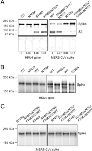
ince its emergence in 2012, Middle East respiratory syndrome coronavirus (MERS-CoV) has infected over 1,100 people, with a case fatality rate of⬃38% (1,2). Bats are considered the natural reservoir of MERS-CoV because several coronaviruses, including HKU4, have been isolated from bats and are genetically related to MERS-CoV (3–5). An envelope-anchored spike protein mediates coronavirus entry into host cells. It first binds a host receptor through its S1 subunit and then fuses membranes through its S2 subunit (6–8). For membrane fusion, the spike must be cleaved at the S1/S2 boundary by one or more host pro-teases (9–13). We recently showed that both HKU4 and MERS-CoV spikes recognize host receptor dipeptidyl peptidase 4 (DPP4) (14). Furthermore, only MERS-CoV spike, and not HKU4 spike, mediates viral entry into human cells because the former, but not the latter, can be activated by human endogenous proteases (14). Here we further identified two residue differences between the two spikes that account for their different capabilities to mediate viral entry into human cells. Our results have revealed a likely evolu-tionary pathway for the emergence of MERS-CoV as a human pathogen, either from bats directly or through intermediate hosts. By comparing the sequences of MERS-CoV and HKU4 spikes, we identified two regions in MERS-CoV spike that may serve as human protease target motifs but that differ from the correspond-ing regions in HKU4 spike (Fig. 1). The first region is a motif for human proprotein convertases (hPPC motif) (15, 16), but the critical Arg748 in MERS-CoV spike corresponds to Ser746 in the HKU4 spike, which deviates from the hPPC motif. The second region is a motif for human endosomal cysteine proteases (hECP motif) (17,18), but the motif Ala763-Phe764-Asn765 in MERS-CoV spike corresponds to Asn762-Tyr763-Thr764 in HKU4 spike, which likely introduces an N-linked glycosylation site and blocks the access of human endosomal cysteine proteases. These residue differences in the two human protease motifs between MERS-CoV and HKU4 spikes may affect the capabilities of the spikes to mediate viral entry into human cells.To evaluate the potential genetic changes required for HKU4 to infect human cells, we reengineered HKU4 spike, aiming to build its capacity to mediate viral entry into human cells. To this end, we introduced two single mutations, S746R and N762A, into HKU4 spike. The S746R mutation was expected to restore the
hPPC motif in HKU4 spike, whereas the N762A mutation likely disrupted the potential N-linked glycosylation site in the hECP motif in HKU4 spike. To confirm that the S746R mutation re-stored the hPPC motif, we produced retroviruses pseudotyped with HKU4 spike (referred to as HKU4 pseudoviruses) in human cells and showed that HKU4 spike containing the S746R mutation was partially cleaved during the molecular maturation process, whereas wild-type HKU4 spike remained intact (Fig. 2A). Con-firming that the N762A mutation disrupted the N-linked glycosy-lation site in the hECP motif was technically challenging because of the large size and heavy glycosylation of HKU4 spike (the trim-eric HKU4 spike has 78 predicted N-linked glycosylate sites and a total molecular mass of⬃530 kDa). Nevertheless, we managed to identify a slight downward shift of HKU4 spike with the N762A mutation using Western blot analysis (Fig. 2B), consistent with successful removal of the N-linked glycosylation site. We do not currently have direct evidence to show that the spikes are cleaved right at the hPPC and hECP motifs by human proteases. However, both of the hPPC and hECP motifs in the spikes have been well documented to be the cleavage sites for human proteases (16–18). Moreover, mutations in these motifs in coronavirus spikes have demonstrated dramatic effects on viral entry into human cells (see below). Thus, the identified hPPC and hECP motifs are likely to be cleaved by human proteases, although we cannot completely rule out the possibility that alteration of these motifs may affect protease cleavages at a spatially adjacent site on the spikes. In either case, our study revealed that both of these motifs play
crit-Received15 May 2015Accepted7 June 2015
Accepted manuscript posted online10 June 2015
CitationYang Y, Liu C, Du L, Jiang S, Shi Z, Baric RS, Li F. 2015. Two mutations were critical for bat-to-human transmission of Middle East respiratory syndrome coronavirus. J Virol 89:9119 –9123.doi:10.1128/JVI.01279-15.
Editor:S. Perlman
Address correspondence to Fang Li, [email protected].
Y.Y. and C.L. contributed equally to this article.
Copyright © 2015, American Society for Microbiology. All Rights Reserved.
ical roles in HKU4- and MERS-CoV-spike-mediated entry into human cells (see below).
We examined the capability of the mutant HKU4 spike to mediate viral entry into three types of human cells (Fig. 3Afor HEK293T cells; data not shown for Huh-7 and MRC-5 cells), using a pseudovirus entry assay as previously described (14). In the absence of exogenous protease trypsin, HKU4 pseudovi-ruses bearing either the reengineered hPPC motif or the reen-gineered hECP motif were able to enter human cells, whereas HKU4 pseudoviruses bearing both of the reengineered human protease motifs entered human cells as efficiently as when ac-tivated by exogenous trypsin (Fig. 3A). In contrast, wild-type HKU4 pseudoviruses failed to enter human cells. Therefore, the reengineered hPPC and hECP motifs enabled HKU4 spike to be activated by human endogenous proteases and thereby allowed HKU4 pseudoviruses to bypass the need for exogenous proteases to enter human cells. These results reveal that HKU4 spike needs only two single mutations at the S1/S2 boundary to gain the full capacity to mediate viral entry into human cells.
To confirm the functions of the reengineered human pro-tease motifs in HKU4 spike, we used propro-tease inhibitors to probe the human proteases that activate HKU4-spike-medi-ated viral entry into human cells. Human proprotein conver-tase (PPC) inhibitor and endosomal cysteine protease (ECP) inhibitor both blocked human cell entry of HKU4 pseudovi-ruses bearing the reengineered hPPC motif and the reengi-neered hECP motif, respectively (Fig. 3B). This result verified that the gained capability of HKU4 pseudoviruses to enter hu-man cells was due to the reengineered huhu-man protease motifs in HKU4 spike.
To investigate the functions of the two human protease motifs in MERS-CoV-spike-mediated entry into human cells, we reengi-neered MERS-CoV spike in the opposite way, aiming to reduce or eliminate its capability to mediate viral entry into human cells. To this end, we introduced an R748S single mutation and A763N/ F764Y/N765T triple mutations into MERS-CoV spike (Fig. 1).
The R748S mutation destroyed the hPPC motif in MERS-CoV spike (Fig. 2A), whereas the A763N/F764Y/N765T triple muta-tions successfully added an N-linked glycosylation site to the hECP motif in MERS-CoV spike (Fig. 2C). MERS-CoV
pseudo-FIG 1Domain structure of MERS-CoV and HKU4 spike proteins. The spikes contain a receptor-binding S1 subunit, a membrane-fusion S2 subunit, a transmembrane anchor (TM), and an intracellular tail (IC). S1 contains the receptor-binding domain (RBD) that binds DPP4 receptor. S2 contains the fusion peptide (FP), heptad repeat 1 (HR1), and heptad repeat 2 (HR2), all of which are essential structural elements for the membrane fusion process. The S1/S2 boundary in MERS-CoV spike (defined as the region between the RBD and the fusion peptide) contains one established human protease motif that is recognized by proprotein convertases (hPPC) (15,16); it also contains one established human protease motif that is recognized by endosomal cysteine proteases (hECP) (17,18). Sequence alignments of these regions in MERS-CoV and HKU4 spikes (GenBank accession no.AFS88936.1for MERS-CoV spike;ABN10839.1for HKU4 spike) are shown, with the critical residue dif-ferences labeled in red. Arrows indicate the predicted sites of cleavage by hu-man proteases in MERS-CoV spike.
viruses bearing either the mutated hPPC motif or the mutated hECP motif demonstrated a decreased capability to enter three types of human cells (Fig. 3Cfor HEK293T cells; data not shown for Huh-7 and MRC-5 cells). MERS-CoV pseudoviruses bearing both of the mutated human protease motifs were unable to enter human cells. Exogenous trypsin was able to fully rescue the capa-bility of mutant MERS-CoV pseudoviruses to enter human cells. Moreover, PPC inhibitor and ECP inhibitor both blocked human cell entry of wild-type MERS-CoV pseudoviruses but had no effect on human cell entry of MERS-CoV pseudoviruses bearing the mutant hPPC motif and the mutant hECP motif, respectively (Fig. 3D). Thus, the mutations in the hPPC and hECP motifs together eliminated MERS-CoV-spike-mediated viral entry into human cells. These results demonstrate that the two functional human protease motifs in MERS-CoV spike played a critical role in the bat-to-human transmission of MERS-CoV.
After examining HKU4- and MERS-CoV-spike-mediated viral entry into human cells, we investigated how these spikes mediate viral entry into bat cells. Because of the low transfection efficiency of bat cells, we were unable to package pseudoviruses in bat cells.
Instead, we packaged HKU4 and MERS-CoV pseudoviruses in HEK293T cells and subsequently performed HKU4- and MERS-CoV-spike-mediated pseudovirus entry into two types of bat cells: RSKT (Rhinolophus sinicusbat kidney cells) and Tb1-Lu cells ( Ta-darida brasiliensisbat lung cells) (Fig. 4). Wild-type HKU4 pseu-doviruses entered bat cells efficiently, whereas HKU4 pseudovi-ruses bearing the reengineered hECP motif (which removed the N-linked glycosylation site) demonstrated an enhanced capability to enter bat cells (Fig. 4AandB). On the other hand, wild-type MERS-CoV pseudoviruses entered bat cells efficiently, whereas MERS-CoV pseudoviruses bearing the mutant hECP motif (which added the N-linked glycosylation site) demonstrated a re-duced, but still significant, capability to enter bat cells (Fig. 4Cand
D). Moreover, most of HKU4- and MERS-CoV-spike-mediated pseudovirus entry into bat cells could be blocked by the ECP in-hibitor, also suggesting that bat endosomal cysteine proteases ac-tivate coronavirus spikes bearing a glycosylated hECP motif (Fig. 4). These results indicate that, unlike human endosomal cysteine proteases, bat endosomal cysteine proteases are capable of recog-nizing and efficiently cleaving the hECP motif containing a
sylation site. The molecular and structural differences between human and bat hECPs accounting for their functional differences will be investigated in future research.
Understanding the molecular mechanisms for cross-species transmissions of viruses is critical for evaluating their emerging disease potentials and for preventing or controlling their spread in human populations. Here we examined the different cell entry activities of the spike proteins from human-infecting MERS-CoV and a closely related bat coronavirus, HKU4. Although MERS-CoV and HKU4 spikes share high sequence homology and recog-nize the same host DPP4 receptor, only MERS-CoV spike, and not HKU4 spike, mediates viral entry into human cells. Our study revealed that introduction of two single mutations, S746R and N762A, into HKU4 spike at the S1/S2 boundary fully instilled its capability to mediate viral entry into human cells. MERS-CoV spike already contained both of these mutations, explaining why MERS-CoV is capable of infecting human cells. Thus, these two mutations in the spike are essential for MERS-CoV to transmit from bats to humans by allowing MERS-CoV spike to be activated by human cellular proteases. Viral adaptation to human cellular proteases is critical for viral infection of human cells because hu-man cellular proteases, particularly endosomal proteases, are more reliable sources than some extracellular proteases to activate viral entry. Previous research also identified two mutations in SARS-CoV spike that led SARS-CoV to transmit from palm civets
to humans (19–22). These mutations increased the capability of SARS-CoV spike to bind human receptor angiotensin-converting enzyme 2. Thus, different entry factors appear to have played the most critical roles in the cross-species transmission of MERS-CoV and SARS-CoV: adaption to human cellular proteases by MERS-CoV and adaption to human receptor by SARS-MERS-CoV. Although MERS-CoV spike might also need to adapt to human DPP4 re-ceptor upon infecting human cells (14, 23), such adaptations might have only incremental effects on the infectivity of MERS-CoV in human cells. In contrast, the two mutations adaptive to human cellular proteases transformed MERS-CoV spike from completely lacking to fully possessing the capacity to mediate viral entry into human cells, and thus they likely played the most critical role in the bat-to-human transmission of MERS-CoV, either di-rectly or through intermediate hosts.
ACKNOWLEDGMENTS
This work was supported by NIH grants R01AI089728 (to F.L.) and R01AI110700 (to R.S.B. and F.L.).
REFERENCES
1.Zaki AM, van Boheemen S, Bestebroer TM, Osterhaus A, Fouchier RAM.2012. Isolation of a novel coronavirus from a man with pneumonia in Saudi Arabia. N Engl J Med367:1814 –1820.http://dx.doi.org/10.1056 /NEJMoa1211721.
Emerg Infect Dis19:456 – 459.http://dx.doi.org/10.3201/eid1903.121503. 4.Holmes KV, Dominguez SR. 2013. The new age of virus discovery: genomic analysis of a novel human betacoronavirus isolated from a fatal case of pneumonia. mBio4:e00548-12.http://dx.doi.org/10.1128/mBio .00548-12..
5.Lau SK, Li KS, Tsang AK, Lam CS, Ahmed S, Chen H, Chan KH, Woo PC, Yuen KY.2013. Genetic characterization of Betacoronavirus lineage C viruses in bats reveals marked sequence divergence in the spike protein of pipistrellus bat coronavirus HKU5 in Japanese pipistrelle: implications for the origin of the novel Middle East respiratory syndrome coronavirus. J Virol87:8638 – 8650.http://dx.doi.org/10.1128/JVI.01055-13. 6.Perlman S, Netland J.2009. Coronaviruses post-SARS: update on
repli-cation and pathogenesis. Nat Rev Microbiol7:439 – 450.http://dx.doi.org /10.1038/nrmicro2147.
7.Li F.2015. Receptor recognition mechanisms of coronaviruses: a decade of structural studies. J Virol89:1954 –1964.http://dx.doi.org/10.1128/JVI .02615-14.
8.Li WH, Wong SK, Li F, Kuhn JH, Huang IC, Choe H, Farzan M.2006. Animal origins of the severe acute respiratory syndrome coronavirus: in-sight from ACE2-S-protein interactions. J Virol80:4211– 4219.http://dx .doi.org/10.1128/JVI.80.9.4211-4219.2006.
9.Belouzard S, Millet JK, Licitra BN, Whittaker GR.2012. Mechanisms of coronavirus cell entry mediated by the viral spike protein. Viruses4:1011– 1033.http://dx.doi.org/10.3390/v4061011.
10. Heald-Sargent T, Gallagher T.2012. Ready, set, fuse! The coronavirus spike protein and acquisition of fusion competence. Viruses4:557–580. http://dx.doi.org/10.3390/v4040557.
11. Simmons G, Zmora P, Gierer S, Heurich A, Pohlmann S.2013. Pro-teolytic activation of the SARS-coronavirus spike protein: cutting en-zymes at the cutting edge of antiviral research. Antiviral Res100:605– 614. http://dx.doi.org/10.1016/j.antiviral.2013.09.028.
12. Simmons G, Gosalia DN, Rennekamp AJ, Reeves JD, Diamond SL, Bates P.2005. Inhibitors of cathepsin L prevent severe acute respiratory syndrome coronavirus entry. Proc Natl Acad Sci U S A102:11876 –11881. http://dx.doi.org/10.1073/pnas.0505577102.
13. Simmons G, Reeves JD, Rennekamp AJ, Amberg SM, Piefer AJ, Bates P.
2004. Characterization of severe acute respiratory syndrome-associated
coro-reduce viral infectivity. J Infect Dis211:889 – 897.http://dx.doi.org/10 .1093/infdis/jiu407.
16. Millet JK, Whittaker GR.2014. Host cell entry of Middle East respiratory syndrome coronavirus after two-step, furin-mediated activation of the spike protein. Proc Natl Acad Sci U S A111:15214 –15219.http://dx.doi .org/10.1073/pnas.1407087111.
17. Bosch BJ, Bartelink W, Rottier PJ.2008. Cathepsin L functionally cleaves the severe acute respiratory syndrome coronavirus class I fusion protein upstream of rather than adjacent to the fusion peptide. J Virol82:8887– 8890.http://dx.doi.org/10.1128/JVI.00415-08.
18. Biniossek ML, Nagler DK, Becker-Pauly C, Schilling O.2011. Proteomic identification of protease cleavage sites characterizes prime and non-prime specificity of cysteine cathepsins B, L, and S. J Proteome Res10:
5363–5373.http://dx.doi.org/10.1021/pr200621z.
19. Li F.2013. Receptor recognition and cross-species infections of SARS coronavi-rus. Antiviral Res100:246–254.http://dx.doi.org/10.1016/j.antiviral.2013.08.014. 20. Li F, Li WH, Farzan M, Harrison SC.2005. Structure of SARS corona-virus spike receptor-binding domain complexed with receptor. Science
309:1864 –1868.http://dx.doi.org/10.1126/science.1116480.
21. Li WH, Zhang CS, Sui JH, Kuhn JH, Moore MJ, Luo SW, Wong SK, Huang IC, Xu KM, Vasilieva N, Murakami A, He YQ, Marasco WA, Guan Y, Choe HY, Farzan M.2005. Receptor and viral determinants of SARS-coronavirus adaptation to human ACE2. EMBO J24:1634 –1643. http://dx.doi.org/10.1038/sj.emboj.7600640.
22. Li F.2008. Structural analysis of major species barriers between humans and palm civets for severe acute respiratory syndrome coronavirus infec-tions. J Virol82:6984 – 6991.http://dx.doi.org/10.1128/JVI.00442-08. 23. Wang Q, Qi J, Yuan Y, Xuan Y, Han P, Wan Y, Ji W, Li Y, Wu Y, Wang
J, Iwamoto A, Woo PC, Yuen KY, Yan J, Lu G, Gao GF.2014. Bat origins of MERS-CoV supported by bat coronavirus HKU4 usage of human re-ceptor CD26. Cell Host Microbe16:328 –337.http://dx.doi.org/10.1016/j .chom.2014.08.009.