Address for correspondence Dr. Dwi Murtiatutik
Dermatology and Venereology Department, Faculty of Medicine Universitas Airlangga, Dr. Soetomo Teaching Hospital,
Jl. Prof. dr. Moestopo 6-8, Surabaya – Indonesia. Ph: +62 315501605
Email: [email protected]
excision of the nodules from the scrotal skin was done without any complications. And there has been no recurrence for the past six months at follow-up.
Key words
Idiopathic, scrotal calcinosis, scrotum, calcinosis cutis.
Introduction
Calcinosis cutis is a disorder of deposits of calcium in the dermis. Idiopathic scrotal calcinosis (ISC) is a common form of idiopathic calcinosis cutis.1 However, considerable debate exists as to whether this term accurately applies, as some researchers suggest that ISC is truly late presentation of epidermal inclusion cysts that have undergone dystrophic calcification.2,3
It is a rare and benign condition defined as the existence of multiple calcified and asymptomatic nodules within scrotal skin that occur without any anomaly of the phosphate/ calcium metabolism. The condition was first described by Lewinski in 1883. It often presents in the third and fourth decade of life. The condition may progress over several years before the patient consults a medical professional, due to
the indolent and painless nature of these tumors on the scrotum.4,5
The typical appearance includes rock-hard white nodules on the scrotal skin. Even though most of the cases are asymptomatic and patients often seek medical advice for cosmetic reasons, some complain with pruritus, soreness and white-chalky exudate from the nodules accompanied with secondary infection of the skin.1,6,7
The pathogenesis of ISC is still controversial. Some authors think that ISC is the result of dystrophic calcification of preexisting structures such as epidermal, pillar, hybrid, indeterminate and ductal cysts. It may also be due to the degeneration of the dartoic muscles. Some authors did not find any evidence of preexisting cystic structures and think this condition is idiopathic.2,3
(Figure 1) The dermatological state before treatment. (a,b) on region scrotalis, multiple, firm, painless, subcutaneous nodules
The golden standard for treatment is an elliptical excision of the nodule above dartos fascia, as the nodules are localized within the dermis. Primary closure of the wound is often achievable because of the skin laxity.8-11
The diagnosis of ISC is established by histopathological examination, which reveals the presence of calcified deposits of varying size in the dermis that may be surrounded by histiocytes and inflammatory giant cell reaction. Partial epithelial lining around the calcified deposits may be visible in some cases.3,5
Case report
A 45-year-old male came to outpatient clinic on 18 December 2017, presented with multiple, painless nodular lesions on the scrotum. Initially, nodular lesions were very soft in palpable and then became harder. The lesions also increased in size gradually during the last 2 decades without causing any symptoms or discomfort. On enquiry, he revealed that the first lesion appeared 25 years back with new nodules
appearing particularly in the last twenty years. Patient was married, and there was no preceding history suggestive of sexually transmitted disease (STD), trauma, inflammation or discharge from the scrotum. There was no lesion in other parts of the body. He was not a known diabetic and never taken any immunosuppressive drugs. There were no features suggestive of hypercalcaemia. There was no history of drugs intake before the lesions appeared, drug hypersensitivity, blood transfusion, injections or drug abuse. History of fever, headache, malaise, diarrhea, weight loss were denied. General physical and systemic examination did not reveal any abnormality.
Dermatological examination showed multiple nodular, mobile, firm, painless, subcutaneous nodules within the scrotal wall that measured from 3 to 5 cm in diameter (Figure 1). There were no areas of ulcerations, inflammation, discharge on the scrotum skin.
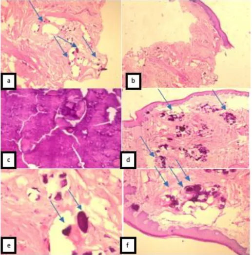
Figure 2 (a,bx4) Calcified intradermal nodule lacking an epithelial lining. (c,dx10) Original magnification of the same nodule composed of basophilic calcified material without epithelial lining showing amorphous form. (e,fx20) haematoxylin and eosin staining) Diffuse calcified cystic areas within the fibrohyalinated stroma
The histopathological evaluation revealed the presence of tissue pieces coated with epidermis. Stroma is fibrous connective tissue with wide calcification area. There were no sebaceous glands. There was no sign of malignancy. It was representative of the diagnosis (Figure 2).
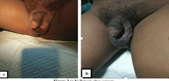
The patient was treated by surgical treatment. The involved scrotal skin was excised using an elliptical incision above the level of the dartos muscle and the defect was closed using 4-0 chromic suture in an interrupted fashion. Pathologic analysis revealed multiple calcified nodules of variable size in the dermis, some with focal giant cell reaction consistent with scrotal skin calcinosis. The patient has not experienced
any postoperative complications. Surgery based on excision of scrotal skin was performed successfully with a good aesthetic result. After a 6-month follow- up period post excision there was no reccurence
Discussion
Figure 3 (a,b) Patient after surgery
and phosphate metabolism and serum levels are normal, local tissue abnormalities, such as alterations in collagen, elastin, or subcutaneous fat may trigger calcification. The internal organs usually remain unaffected. Metastatic calcinosis is characterized by an abnormal calcium and/or phosphate metabolism, leading to the precipitation of calcium in cutaneous and subcutaneous tissue. Skin calcinosis in
iatrogenic calcinosis cutis is a side effect of therapy. Calciphylaxis presents with small vessel calcification mainly affecting blood vessels of the dermis or subcutaneous fat. Disturbances in calcium and phosphate metabolism and hyperparathyroidism can be observed.2,3
central calcium deposit dissolved during tissue preparation.(Hematoxylineeosin stain; original magnification: 325.)3
Figure 6 Photomicrograph showing giant cell and inflammatory cell (H and E ×100)5
which is the case with our patient. The youngest and the oldest reported patients were a 9 years old and an 85 years old respectively.5 Our
are marble like in appearance and easily palpable. Most patients are asymptomatic and present because of cosmetic concern. Few patients may present with pruritus, ulcerations, and discharge of chalky material. A case of ISC presented with prostatitis-like symptoms (chronic pelvic and perineal pain). The interval between the occurrence of the disease and therapy is often several years. Usually, scrotal nodules develop slowly over many years to decades. In most cases, there are no associated calcinosis elsewhere.13 Clinical manifestation of this patient showed asymptomatic hard yellowish nodules with variable size, 3-5cms.
Clinically ISC may be confused with epidermal inclusion cysts, steatocystoma, angiokeratoma, genital leiomyoma, scleroderma especially in patients with CREST syndrome (Calcinosis cutis, Raynaud’s phenomena, Esophageal dysfunction, Sclerodactyly and Teleangiectasia), and dermatomyositis.14
evidence of surrounding epithelial lining 3,5 We did excisional biopsy in one nodule for histological examination and it showed stroma i.e. fibrous connective tissue with wide calcification areas in dermis (Figure 5 & 6).
The pathogenesis of ISC is unclear and controversy exists as to whether the disease is idiopathic or the result of dystrophic calcification of preexisting structures including epidermal cyst, eccrine epithelial cyst, and degenerated dartoic muscle. In our patient, there was no evidence of epidermoid or pillar cystic structure and no epithelial lining around the calcified lesions. According to the histologic findings, the case was considered idiopathic.15,16
In addition, patients are slow to consult because of the "intimate" localization of the disease. The reason for consultation is most often an aesthetic issue affecting the sexual life of patients. Our patient had this complaint for twenty years ago before he came for treatment. The cosmetic improvement of the scrotum by the disappearance of the nodules and the correction of cutaneous ptosis had a major impact on patients' quality of life.
The etiology of scrotal calcinosis remains uncertain and controversial.17 The disease is limited to the dermis, leaving the dartos layer intact. 18 Therefore, surgical excision must be limited to the scrotal skin because the calcified nodules are localized in the dermis.11 In our case, there was multiple nodule and elliptian excision was performed. Recurrence after surgery is not usual in ISC and the risk of recurrence is also controversial, and some authors insist on the high probability of recurrence after primary excision.19 The surgical approach must be perfect and the extent of excision must include the whole lesion even the smallest ones to avoid rapid recurrence.11
References
1. Xia Lei1, Bo Liu2, MD, Qionghui Cheng1, Jinjin Wu1. Idiopathic Scrotal Calcinosis: report of two cases and review of literature. Int J Dermatol 2012; 51:199–203.
2. Janet A. Fairley, Cutaneous Mineralization and Ossification. In: L.A Goldsmith, S.I Katz, BA Gilcherest, AS Paller, DJ Leffel, K Wolf, editors, Fitzpatrick’s Dermatology in General Medicine, 8th edition. New York: The McGraw-Hill Companies, Inc; 2012. p.3086-96.
3. Nadine Reiter, MD,A Laila El-Shabrawi, MD,A Bernd Leinweber, MD,A Andrea Berghold, Phd,B, Elisabeth Aberer, Mda, Calcinosis cutis Part I. Diagnostic pathway. J Am Acad Dermatol 2011; 65:1-12. 4. Dubey S, Sharma R, Maheshwari V. Scrotal
calcinosis: Idiopathic or dystrophic?. Dermatol Online J 2010; 16:5.
5. Sheela Chaudhari, Sachan Bhat, Deepa Hatwal, Pawan Bhat, Neha Batra, Scrotal calcinosis - An Etiological Dilemma: A Prospective Study. Int J Sci Stud 2015; 3:1-7.
6. V. Shah, T. Shet, Scrotal calcinosis results from calcification of cysts derived from heir follicles: a series of 20 cases evaluating the spectrum of changes resulting in scrotal calcinosis. Am. J. Dermatopathol 2007; 29:172–175.
7. A. Pompeo, W. Molina, G. Pohlman, et al., Idiopathic Scrotal Calcinosis: a rareentity and a review of the literature. Can Urol Assoc J 2013; 7:439–41.
8. Nadine Reiter, MD,A Laila El-Shabrawi, MD,A Bernd Leinweber, MD,A Andrea Berghold, Phd,B, Elisabeth Aberer, Mda, Calcinosis cutis Part II. Treatment options. J Am Acad Dermatol 2011; 65:15-22.
9. R.A. Agha, A.J. Fowler, A. Saetta, I. Barai, S. Rajmohan, D.P. Orgill, for the SCARE Group, The SCARE statement: consensus-based surgical case report guidelines. Int. J. Surg 2016;1-3.
10. W. Noel, B. Hersant, J.-P. Meningaud, Traitement chirurgical de la calcinose scrotale en un temps One-staged surgical technique for scrotal calcinosis. Progrès en urologie 2016; 26:176—180.
Non-Shivani1, Usha Shinde1, Idiopathic Scrotal Calcinosis. Indian J Surg 2016; 78:329–330.