ORIGINAL ARTICLE
Iran J Allergy Asthma Immunol August 2019; 18(4):379-392.
In Vitro Evaluation of CMV Specific CD8+T Cells Function in
CMV+ Colorectal Cancer Patients Compared to Healthy Controls
Mona Shaker Ardakani1, Fatemeh Pak1,2, Parviz Kokhaei1,2, Mohammad Sadegh Fazeli3, Yadollah Shakiba4, Seyed Morteza Tabatabaei Yazdi5, Ali Abbasian5, and Maryam Nourizadeh6
1 Cancer Research Center, Semnan University of Medical Sciences, Semnan, Iran 2
Department of Oncology-Pathology, Cancer Center Karolinska, Karolinska Institutet, Stockholm, Sweden
3 Department of Surgery, Imam Khomeini Complex Hospital, Tehran University of Medical Sciences, Tehran, Iran 4
Regenerative Medicine Research Center, Kermanshah University of Medical Sciences, Kermanshah, Iran
5 Tehran Blood Transfusion Center, Tehran, Iran 6
Immunology, Asthma and Allergy Research Institute, Tehran University of Medical Sciences, Tehran, Iran
Received: 1 February 2019; Received in revised form: 9 April 2019; Accepted: 29 April 2019
ABSTRACT
The oncogenic role of human cytomegalovirus (HCMV) has been recently shown in
different cancers like colorectal cancer (CRC). According to the recent immunotherapy
approach to target the CMV-expressing tumor cells, we investigated the CMV
peptide-stimulated CD8+T cells functions in CRC patients compared to healthy individuals.
All sixteen patients and seven controls were CMV seropositive. Blood samples were
obtained from patients without chemotherapy and radiotherapy before surgery. Cytotoxic
CD8+ T cells were generated using 14-day culture of PBMCs in the presences of CMV
peptide epitopes and rhIL-2. In addition to the supernatant evaluations for TNF-α and
IFN-γ, the functionality of CD8+ T cells was examined by detecting CD107a and intracellular
IFN-γ using flow cytometry. CMV DNA was detected in tissues by Real Time PCR.
CMV DNA was found in 31% of tumor tissues, while it was not seen in the adjacent
non-tumor tissues. There was a close association between CMV in tumor tissue and tumor
grade. Surface expression of CD107a and intracellular IFN-γ in CMV-stimulated CD8+T
cells and the level of IFN-γ production in patient and control groups increased significantly
after culture. The number of functions increased in patients (
p
<0.05) and healthy individuals
after culture. Followingstimulation, expressions of CD107a and intracellular IFN-γ were
elevated in tumor CMV positive patients while the TNF-α secretion was decreased.
In vitro stimulation of PBMC in the presence of CMV peptide epitopes and IL-2 can be
an applicable method to generate cytotoxic CD8+ T cells in CRC patients for future T cell
therapy.
Keywords: Cytomegalovirus; CD107a; Interferon gamma (IFN-γ); Solid tumour
Corresponding Author: Maryam Nourizadeh, PhD;
Immunology, Asthma, and Allergy Research Institute, Tehran University of Medical Sciences, Tehran, Postal Code: 1419733154,
INTRODUCTION
Colorectal cancer (CRC) is known as the third leading cause of cancer-related deaths in the world.1 Despite increasing the longevity, routine treatment protocols including chemotherapy, radiotherapy, and surgery have severe side effects and lack the specific targeting of tumor cells.2 Therefore, new alternative immunotherapy approaches have been recently planned to potentiate the different arms of the immune system.3,4 It is critical to find the most immunogenic tumor specific antigens (TSAs) in designing the immunotherapy protocols.5,6 TSAs contain different groups of antigens which are not routinely expressed on normal cells, including viral antigens, mutated antigens, and cancer-testis antigens.7 It has been speculated that about 12-20% of human cancers worldwide are caused by oncogenic viruses worldwide considering that 80% of them occurs in developing countries.8 Oncoviruses have been programmed to cause mutations and irregular proliferation of host cells by integrating into the chromosomal DNA.9 Therefore, they are relevant targets as TSA for cancer immunotherapy.10 Human Cytomegalovirus (CMV) is a beta herpes virus which has not been considered as an oncogenic virus up to now.11 According to the reports, more than ninety percent of Iranians and 50-90% of people around the world have been infected with CMV.12,13 This virus is never completely removed from the body. It remains in latency phase and can be reactivated in various immune compromised conditions.14,15
A potential oncomodulary function of CMV has been reported following the detection of CMV peptides in tumor tissues, but not adjacent non-tumor tissues of seropositive cancer patients suffered from colon cancer, malignant glioma, breast cancer, salivary gland cancer, and prostate adjacent non-tumor.2,16 Furthermore, a negative correlation between the level of CMV infection in tumor tissue and the overall survival of the glioblastoma patients has been shown by Rahbar and et al.17 An in vitro study on CMV+ glioblastoma patients indicated a significant increase in CMV specific CD8+ T cell function of PBMCsstimulated by CMV peptidesplus IL-2.18 The researchers also found that four times injections of CMV specific CD8+ T cells to a patient with glioblastoma led to a relative improvement in clinical parameters like neuronal function.19,20
Among the studies on the relationship between CMV and colorectal cancer, a few of them have shown a lower disease-free survival rate in patients with CMV-positive than CMV-negative tumours.21,22 The most relative reason is the ability of the virus to induce the angiogenesis, resistance to apoptosis, cellular invasion, and metastasis.23 In contrast, there are some studies on negative association between CMV in tumor tissues and progression of colorectal cancer which may be related to the use of different detecting techniques with various levels of specificity and sensitivity.24-27
The results of recent investigations suggest that adoptive T cell therapy using CMV-specific CD8+ T cells can be a potential tool for future immunotherapy of cancer patients by targeting the CMV-infected tumor cells. Although, a comparative in vitro study on the induction of CMV-specific CD8+ T cells in CRC CMV+ patients and the healthy controls is lacking. In this study, we have evaluatedthe presence of CMV DNA in the tumor and the adjacent non-tumor tissues of CRC patients. Then, CMV-specific CD8+Tcells from CMV-seropositive CRC patients and healthy controls were generated and their potential cytotoxic activity was investigated.
MATERIALS AND METHODS
Samples
Sixteen CMV+ colorectal cancer patients (mean age 58 years, 5 males and 11 females) who were referred to the department of surgery in Imam Khomeini Hospital and seven CMV+ healthy controls (mean age 32 years, 1 male and 7 females) were included in this study. This study was evaluated and approved by the ethics committee of Semnan University of Medical Sciences (N. IR.SEMUMS.REC.1394.102). The informed consent forms were signed by participants.
CMV Peptide Epitopes
Quanti FERON-CMV Kit which applies for monitoring CMV specific T cell immune responses was used to cover all of the necessary CMV peptide epitopes. This kit includes CMV and MIT tubes. The CMV tube contains 22 types of common HLA class I restricted CMV-peptide epitopes (pooled CD8+ T-cell epitopes); so there was not essential for HLA-typing of the participants. MIT tube contains phytohemagglutinin (T-cell mitogen as positive control). CMV-peptide epitopes were coated on the wall of the tubes and dissolved by adding warm RPMI-1640 to the tubes and rotating for 30 minutes.
In vitro Expansion of CMV-specific CD8+ T Cells
Peripheral blood mononuclear cells (PBMCs) were isolated by layering of diluted blood onto Ficoll-hypaque (Inno-Train, Germany) using density gradient centrifugation. Approximately 14×106 cells were obtained from 8-10 mL peripheral blood; 4×10⁶ cells were used for initial phenotyping of the cells before stimulation and the remaining cells were divided into two groups as the stimulator and responder cells. In order to pulse the antigen presenting cells with CMV peptides, the stimulator cells were cultured in the presences of 1 µg/mL of pooled CD8+ T-cell CMV epitopes (obtained as described above) overnight. Then they were 30G irradiated and co-cultured with the respondent cells at a ratio of 2:1 in complete media containing RPMI-1640,10% fetal calf serum,1% penicillin-streptomycin at 37ºC for 14 days. PBMCs with PHA and without stimulation were considered as positive and negative controls, respectively. Recombinant human interleukin-2 (rhIL-2, 20 U/mL) was added on days 3, 7 and 10. Re-stimulation with CMV (1 µg/mL) and PHA (1 µg/mL) was done on days 7, 10 and immediately before assessing of CD8+ T cells. The supernatant was collected for evaluation of IFN-γ and TNF-α using the ELISA method.
Evaluation of CMV-specific CD8+ T Cells Function
CD8+ T cells function was further investigated by examining cell surface expression of CD107a (LAMP-1) as a hallmark of degranulating T lymphocytes before and after coculturing with responder cells. Intracellular IFN-γ assessment was also performed.
Flow Cytometry
For evaluating the CD8+T cells before and after
14-day co-culturing between the CMV-stimulated (irradiated) and unstimulated PBMCs, cells were harvested and stained for 30 minutes in incubator 37ºc, 5% Co2 with 1 µg/ml CMV peptides, 50 ng/mL phorbol myristate acetate (PMA), 500 ng/mL Ionomycin and Anti-CD107a PE-CY5. Thereafter, 5 µg/ml monensin (BD, GolgiStop) and 5 µg/mL brefeldin A were added to the flowcytometry tubes. Following 6-hour incubation, cells were washed once in FACS buffer and stained with anti-CD8a FITC. For intracellular staining of IFN-γ, cells were fixed and permeabilized according to the manufacturer’s instructions and incubated with anti-IFNγ PE, for 30 minutes (all materials excep monensin from eBioscience, USA). Mouse IgG1 PE-FITC (Dako) antibodies were used asan isotype control. Stained cells were resuspended in 500 μL FACS buffer with 1% paraformaldehyde and run by BD Accuri C6 Plus. Data were analyzed using FLOWJO software (10.4version, USA).
Enzyme-linked Immunosorbent Assay (ELISA)
ELISA technique was used to assess IFN-γ (Human TNF alpha ELISA Ready-SET-Go! Kit) and TNF-α cytokines (Human IFN gamma ELISA Ready-SET-Go! Kit) in supernatant before and after culture in the presence of CMV peptide epitopes and PBMC alone (as negative control). The sensitivity of detection was 4 pg/mL for TNF-α and 4 pg/ml for IFN-γ (both from eBioscience).
Real Time PCR for Detection of CMVDNA in Tumor and Adjacent Non-tumor Tissues
After DNA extraction (Qiagen kit), Real Time PCR was done to amplify IE(Immediate-Early) gene (translating to IE protein), using artus CMV RGQ MDx Kit containing primers and fluorogenic probe for IE gene detection. PCR was carried out with initial denaturation at 95ºC for 10 minutes, followed by 50 cycles of denaturation at 95ºC for 10s and annealing– extension at 60ºC for 1 min and the presence of CMV in the tissue was investigated by analyzing the Real Time PCR data.
Statistical Analysis
Non-parametric) were used. To compare a quantitative variable in two independent groups, Independent sample t-test or Mann-Whitney were applied. To assess the difference of a quantitative variable in more than 2 independent groups, one-way ANOVA was used. Considering the distribution of data, Paired t-test or Wilcoxon rank test were used to compare the level of a quantitative variable in a dependent group before and after treatment. Pearson or Spearman test was performed to assess the correlation of two quantitative variables. Chi-Square test was utilized to evaluate the relationship between two qualitative variables. To draw the graphs, GraphPad Prism 6 (Graphpad Software Inc., La Jolla, CA, USA) was used.
RESULTS
Demographic Features of CRC Patients
All 16 CRC patients and 7 healthy controls were seropositive. It means that the patients and controls had not an active CMV infection. Demographic features and the stage of cancer have been shown in Table 1.
Adenocarcinoma was confirmed in the patients without any signs of metastases.
Detection of CMV DNA in Tumor Tissue of Some CRC Patients
CMV DNA was detected in tumor tissue of 5 (31%) CRC patients, while the virus was not found in any of the adjacent non-neoplastics pecimens. Of five positive samples, four samples were in moderately differentiated grade (intermediate grade) and one sample was in poorly differentiated grade (high grade). Although there was a close association between CMV presence in tumor tissue and tumor grade, it was not statistically significant (p=0.05). Moreover, there was no correlation between the stage of tumor and detection of CMV DNA in tumor tissues.
Activation of CMV-specific CD8+T Cells
PBMC from CMV-seropositive CRC patients and healthy individuals were stimulated with 22 types of common HLA class I restricted CMV-peptide epitopes or with PHA (as the positive control).
Table 1. Demographic features of colorectal cancer (CRC) patients
Grade AJCC stage** M* N* T* CMV IgG Age Sex Patients No Not available Not available Not available Not available Not available + 86 Female 1 Well differentiated 0 M0 N0 Tis + 46 Female 2 Moderately differentiated 2C M0 N0 T4b + 45 Female 3 Well differentiated 1 M0 N0 T2 + 43 Female 4 Moderately differentiated 1 M0 N0 T1 + 77 Female 5 Poorly differentiated 3B M0 N1b T3 + 57 Female 6 Not available Not available Not available Not available Not available + 54 Female 7 Moderately differentiated 2A M0 N0 T3 + 61 Female 8 Well differentiated 3B M0 N1b T3 + 55 Male 9 Well differentiated 2A M0 N0 T3 + 71 Female 10 Well differentiated 2A M0 N0 T3 + 36 Male 11 Moderately differentiated 3C M0 N1a T4b + 78 Female 12 Well differentiated 3C M0 N2b T3 + 49 Male 13 Poorly differentiated 3B M0 N1b T3 + 39 Male 14 Moderately differentiated 2A M0 N0 T3 + 63 Male 15 Moderately differentiated 3A M0 N1 T1 + 76 Female 16
Cells in the medium were considered as a negative control. The function of CD8+ T cells was investigated by detecting the cell surface expression of CD107a and intracellular IFN-γ using flow cytometry before and after 14-day culture. As explained in the method section, the cells of stimulated and non-stimulated groups were exposed to PMA+Ionomycin (P/I) for 6 hours. It was done for stimulating the cytokine production and cytotoxicity function (CD107a expression).Representative flow cytometry data of one patient has been shown in Figure 1a.
In CRC patient group, the expression ofCD107a was significantly increased in CD8+ T cells after culture in all three groups of CMV peptides, PHA and no stimulation (p=0.001, p=0.029, and p=0.042, respectively). A potentiating effect of CMV peptides on cytotoxicity function of CD8+T cells was observed (Figure 1b). In healthy subjects, a significant increase in CD107a expression was detected only in CMV-stimulated CD8+ T cells after culture (p=0.037, Figure 1c).
(a)
a
Lymphocytes Singlet cells
0.8%
(b) (c)
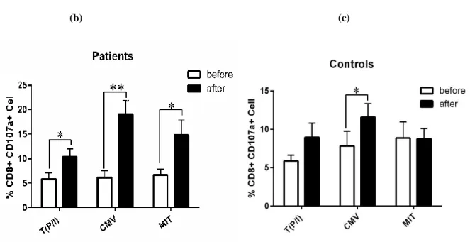
Figure 1. Flow cytometry analysis and examination of CD107a expression on CD8+ T cells
(a) Representative flow cytometry data of CMV-stimulated CD8+ T cells before and after culture in one CRC patient. Lymphocytes were identified from singlet cell. Dot blots show double staining of CD8 T cells for CD107a and IFN-γ in CMV-stimulated cells and isotype control or unstained cells.
Expression of CD107a before and after culture in CD8+ T cells generated from PBMCs was compared among cells stimulated with CMV peptides, phytohaemagglutinin-A (MIT) and unstimulated cells in (b) patients and (c) healthy controls. After stimulation, cells were exposed to PMA+Ionomycin (P/I) for 6 hours and then stained for anti-CD107a PE-CY5 antibody. Thereafter, cells were stained using anti-CD8a FITC and anti-IFNγ PE after adding monensin and brefeldin A. Mouse IgG1 PE-FITC antibody was used as an isotype control. Paired t-test was used for T (P/I) and MIT group and Wilcoxon rank test was applied for the CMV group to compare the level of quantitative variables before and after treatment. One-way ANOVA test was used for statistical analysis between the groups.
All experiments were done in duplicate. Data b & c in this panel represents the Mean±SEM. (* indicating p<0.05 and **
p<0.01).
Although CD107a expression following 14-day culture in the presence of CMV peptides was higher in CRC patient than healthy individuals, this difference was not statistically significant (Figure2a). Comparative analysis of CD107a expression between the patients with CMV positive and negative tumors showed a remarkable surge in both groups, but that was significant only in tumor CMV negative patients (CMV negative tumor p=0.004, CMV positive tumor p=0.05) (Figure 2b).
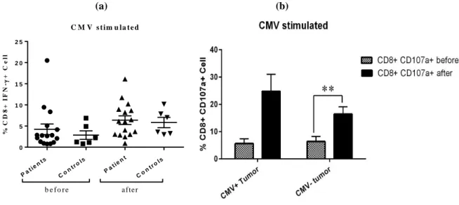
Considering the intracellular cytokine assay, the percentage of IFN-γ positive CD8+T cells in CMV-stimulated cells was increased after culture in CRC patients and healthy controls but the difference was not significant (Figure 3a, Figure 3b). While a comparison of Mean Fluorescence Intensity (MFI) in CMV
stimulated CD8+T cells of CRC patients and healthy controls revealed a significant rise of IFN-γ production after culture in both groups (CRC patients p=0.01, healthy controls p=0.01) (Figure 3c). There was no change in IFN-γ expression of mitogen-stimulated cells and cells without stimulation after culture. In addition, we could find a significant increase in IFN-γ expression among tumor CMV positive patients after culture (p=0.031), but it was unchanged in CMV negative tumor patients. In addition, higher IFN-γexpression inCD8+ T cells of patients with CMV positive tumor s than CMV negative tumor s after culture was detected (p=0.007, Figure 3d). Percentage of CD8+IFN-γ+ cells did not change before and after culture in both CRC patients and healthy controls.
b
(a) (b)
Figure 2. Comparison of CD107a expression between colorectal cancer (CRC) patients and healthy subjects and also between tumor CMV positive and negative CRC patients before and after culture
(a) Comparison of CD107a expression between CMV-stimulated CD8+ T cells of 16 CRC patients and 7 healthy subjects before and after culture (Mann-Whitney U test). (b) Analysis of CD107a expression in CMV-stimulated CD8+ T cells of tumor CMV positive and negative CRC patients before and after culture (Independent sample t-test). All experiments were done in duplicate. Data in this panel represents the Mean±SEM. (** indicating p<0.01).
Measurement of IFN-γ and TNF-α Production by ELISA Technique
The level of IFN-γ production in supernatants increased significantly after culture in both patients and controls’ CMV-stimulated (p=0.001, p=0.00) and non-stimulated cells (p=0.001, p=0.003). Although a higher level of IFN-γ was seen in CMV-stimulated compared to non-stimulated cells before culture in both patient and control subjects, this change was significant only in the patient groups (p=0.01) (Figure 4a, 4b). There was no difference in IFN-γ production between patient and control groups before and after culture (Figure 4c). Evaluation of this cytokine in CMV-stimulated cells between tumor CMV positive and negative patients showed a non-significant improvement in cytokine production after culture in both groups (Figure 4d).
TNFα secretion was also investigated in cell supernatant using ELISA method. Analysis of results showed that there was no difference in the level of TNF-α between stimulated and non-stimulated cells before and after culture in both patient and control groups (Figure 5a). The level of TNF-α from CMV-stimulated cells before culture in patients with CMV positive tumor was higher than those with CMV
negative, although this difference was not significant (Figure 5b).
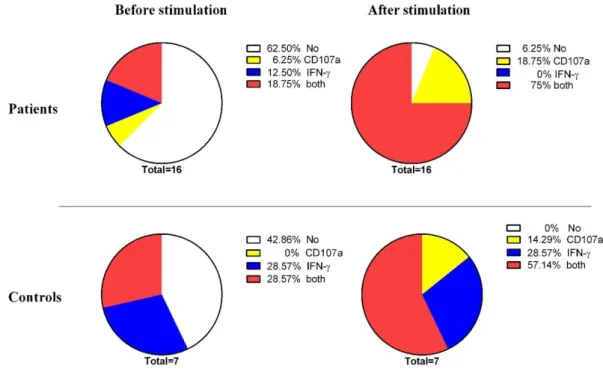
Poly-functional CD8+T Cell Analysis
The CD8+ T cells with the ability of CD107a expression and intracellular IFN-γ production upon CMV peptide stimulation were considered as poly-functional cells. Thus, a number of the CD8+ T cells with a simultaneous dual capacity was compared before and after culture in both patients and healthy individuals. The poly-functionality of CMV-stimulated CD8+ T cells was increased after culture in both patients and controls groups individually, however, it was noticeable in CRC patients (p=0.001). There is no significant difference between the two groups regarding CMV stimulated CD8+ T cells to function before and after culture (Figure 6). To summarize the results of this study, a schematic figure has been shown (Figure 7).
Pa t ien
ts
Co ntro
ls
Pa t ien
t
Co ntro
ls 0
5 1 0 1 5 2 0 2 5
%
C
D
8
+
I
F
N
-
+
C
e
ll
C M V s t im u la t e d
(a) (b)
(c) (d)
(e)
Figure 3. Intracellular IFN-γ in CD8+ T cells.
(a) Comparison of intracellular IFN-γ expression before and after 14-day culture in colorectal cancer (CRC) patients’ CD8+ T cells which was stimulated by CMV or phytohaemagglutinin-A (MIT) and without peptide stimulation (only stimulated with P/I). PMA+Ionomycin(P/I) was added to each of the 3 groups as co-stimulator for 6 hours (Paired t-test). (b) Comparison of intracellular IFN-γ expression before and after culture in healthy controls CD8+ T cells which was stimulated by CMV or phytohaemagglutinin-A (MIT) and without peptide stimulation (only stimulated with P/I). PMA+Ionomycin(P/I) was added to each of the 3 groups as co-stimulator for 6 hours (Paired t-test). (c) Representative IFN-γ MFI in CMV-stimulated CD8+ T cells before and after culture in CRC patients and healthy controls (Wilcoxon rank test). (d) Comparison of intracellular IFN-γ expression in CMV-stimulated CD8+ T cells of tumor CMV positive and negative CRC patients before and after culture. (e) Comparison of CD8+T IFN-γ+ cells in CRC patients and healthy controls before and after culture. All experiments were done in duplicate. Data in this panel represents the Mean±SEM. (* indicating p<0.05 and ** p<0.01).
a
c
d
Pa t ien t
s
Co n t ro l
s Pa t
ien t Co n
t ro l s
0 5 1 0 1 5 2 0 2 5
%
C
D
8
+
I
F
N
-
+
C
e
ll
C M V s t im u la t e d
(a) (b)
(c) (d)
Figure 4. Analysis of IFN-γ secretion (measured by ELISA technique) in cells supernatant of CMV-stimulated and non-stimulated cells in colorectal cancer (CRC) patients and healthy controls before and after culture
(a) Comparison of IFN-γ secretion in cell supernatant of CMV-stimulated and non-stimulated cells (T) of CRC patients before and after 14-day culture in the presence of IL-2 cytokine. (b) Analysis of IFN-γ secretion level in cells supernatant of CMV-stimulated and non-stimulated cells (T) of healthy controls before and after 14-day culture in the presence of IL-2 cytokine. (c) Comparison of IFN-γ secretion in cells supernatant of CMV-stimulated cells between patient and control groups before and after 14-day culture in the presence of IL-2 cytokine. (d) A difference shown the between tumor CMV positive and negative CRC patients in terms of IFN-γ secretion upon CMV-stimulation before and after 14-day culture in the presence of IL-2 cytokine. For analysis of non-parametric data, we used the Mann-Whitney U test for independent groups and Wilcoxon rank test for dependent groups. All experiments were done in duplicate. Data in this panel represents the Mean±SEM (**p<0.01, ***p<0.001)
(a) (b)
Figure 5. TNF-α secretion using ELISA technique in cells supernatant of CMV-stimulated and non-stimulated cells in colorectal cancer (CRC) patients and healthy controls before and after culture
(a) Comparison of TNF-α secretion in cells supernatant of CMV-stimulated and non-stimulated cells (T) of CRC patients and healthy controls before and after 14-day culture in the presence of IL-2 cytokine. (b) It was shown difference between CRC patients with CMV positive and negative tumours in terms of TNF-α secretion upon CMV-stimulation before and after 14-day culture in the presence of IL-2 cytokine. All experiments were done in duplicate. Data in graph a represents mean and in graph b represents the Mean±SEM.
Figure 6. Polyfunctional CD8+T cell analysis upon CMV-stimulation before and after culture in colorectal cancer (CRC) patients and healthy controls
T u m o r t is s u e s Ad ja c e n t n o r m a l t is s u e s 0
5 1 0 1 5 2 0 2 5
C M V + C M V
-F
re
q
u
e
n
c
y
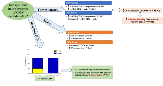
Poly-functionality CMV specific CD8+T cells (P=0.001) Flowcytometry
14-day culture in the presence
of CMV peptides +IL-2
Co-expression of CD107a & IFN-γ CMV+ Tumors
• % CD8+CD107a+ expression (P=0.05) • %CD8+ IFN-γ+ cells (P<0.05)
CMV- Tumors
• % CD8+CD107a+ expression (P<0.01) •Unchanged %CD8+ IFN-γ+ cells
CMV+ Tumors
• TNF-α secretion (P>0.05) • IFN-γ secretion (P>0.05)
CMV- Tumors
•Unchanged TNF-α secretion • IFN-γ secretion (P>0.05)
• CMV preferentially infects tumor tissue
• Close association between CMV presence in tumor tissue &tumor grade (P=0.05) 31% Tumor CMV+
Figure 7. Schematic procedure of CMV peptide-specific CD8+ T cells
DISCUSSION
In this study, we appliedreal time PCR method using sequence-specific TaqMan Probe to detect CMV in freshly frozen tissues. It was done based on previously published papers suggesting PCR techniqueinstead of or alongside the in situ hybridization (ISH) and immunohistochemistry (IHC) methods for detection of CMV in tumortissues.26,27 In addition, we decided to use fresh samplesbased on degradative effects of formalin on DNA(in formalin-fixed samples) and subsequent false negative results.27,28 According to our results, virus DNA was detected in tumorand not in adjacent non-tumortissues of five CRC patients (31%) which was in accordance with the report of Chen and et al.24 All CMV+tumorsamples, except one sample, were intermediate grade and there was a close but no significant relationship between the tumorgrade and the existence of CMVin tumortissues. Similarly,a recent study conducted on 103 CRC tumortissues and 98 adjacent non-tumortissues, CMV US28 proteinwas detected in 38% of the patients’ tumorsamples. However, they founda significant correlation between the presence of CMV in tumor tissue andtumor grade, metastasis, and survival of the patients.29
Considering the CMV peptides as
tumortumor-specific antigens, it has been the preliminary concept of cancer immunotherapy in some recent successful clinical trials in glioblastoma (Clinical Trials, NCT01205334). However, the possibility of using this treatment for CRC patients has not been studied yet. Evaluating the immunological responses of CMV-specific T cells in CRC patients would be requisite in vitro study for utilization in clinical trials. Therefore, in conjunction with comparing T cell function (evaluating CD107a and IFN-γ expression using flow cytometry) in patients and healthy subjects, we tried to improve the function of these cells by 14-day cell culture in the presence of IL-2 and common HLA class I restricted CMV-peptide epitopes. A similar study was conducted by Margaret Inokuma et al. They investigated the functional response of specific T-cells in the presence of CMV and influenza virus peptides as well as associated tumor antigens (TAAs) in patients with breast cancer. The results showed a more potentiate, effective and heterogeneous response of specific T cells against CMV and influenza peptides compared to TAAs alone.30
In the present study, the percentage of CD107a and MFI of IFN-γ in both patient and control groups increased significantly following 14-day culture in the presence of CMV peptides. CD107a expression in PHA-stimulated CD8+ T cells (MIT) and cells without
CD1
0
7
stimulation also improved after culture in both patients and controls, while it was not as much as CMV-stimulated group. One possible reason might be due to the presence of PMA+ionomycin or IL-2 cytokine in the culture media. IL-2 stimulates the expression of CD107a on cytotoxic T cells and PMA+Ionomycin upturns both of CD107a and intracellular IFN-γ expression in lymphocytes. Therefore, T cells may be de-granulated and produce IFNγ although not as potentiate as cells with specific stimulation.31
Except for releasing the granule components such as perforin and granzymes in the immunological synapse which leads to target cells death, cell surface expression of CD107a is also a sensitive marker for detection of cytotoxicity.32,33 Precopio ML et al demonstrated that MFI of interferon-gamma in polyfunctional CD8+T cells (stimulated by viral peptides) was 5-10 times greater than single-function CD8+T cells, while this association did not exist about CD107aMFI.34 Our analysis of CD8+ T cells function also revealed that the poly-functional CMV specific CD8+T cells (co-expression of CD107a and MFI of IFN-γ) increased in both CRC patients and healthy controls after culture.
IFN-γ raises the expression of MHC-I, MHC-II, and co-stimulator molecules on the surface of antigen-presenting cells (APC) which is very important to potentiate the MHC-restricted antigen-presenting function and host immune response especially by cytotoxic T cells (CTLs).35 IFN-γ stimulates differentiation of Th1 cells directly by inducing Th1-specific transcription factor (T-bet) and indirectly by activating mononuclear phagocytes to produce IL-12.36 CMV-stimulated CD8+ T cells of tumor CMV positive and negative patients had no difference in expression of CD107a and intracellular IFN-γ prior to culture, but after culture the expression of both CD107a and intracellular IFN-γ in tumor CMV positive patients was more than CMV negative, although the difference for CD107a expression was not significant. Other differences between tumor CMV positive and negative patients was in the level of TNF-α secretion from CMV-stimulated cells before culture which was higher in tumor CMV positive than negative patients. According to the literature, TNF-α has dual function against tumor cells. From one side it induces proliferation, metastasis, angiogenesis, and survival of tumor cells by activating NF-κB signaling pathway and from another side, it triggers the cell death by
activating the JNK pathway. The exchange between NF-κB and JNK determines the response of cells in exposure to TNF-α.37 Previous study showed that acute inflammation intensifies the risk of colorectal malignancies38 and there is a positive correlation between serum TNF-α level and CRC stage and elevated TNF-α levels in CRC patients is associated with a reduction of patients survival.39,40
Evaluation of IFN-γ production by ELISA method revealed significant growth in both the CMV-stimulated and non-CMV-stimulated cells of patients and healthy controls after culture. The reason why there was no discrepancy in the level of cytokine secretion between stimulated and non-stimulated cells may be due to the effect of IL-2 on cells. However, before culture, the level of this cytokine in CMV-stimulated cells was higher than those with no stimulation.
In the current study, the numbers of CMV-stimulated CD8+ T cells function (polyfunctional CD8+ T cells) were augmented following culture, on the other hand, the number of patients and healthy control who had non-functional CD8+ T cells (CD107a expression or intracellular IFN-γ) decreased after culture. Due to the fact that polyfunctional CD8+ T cells response is associated with better control of viral contamination,41 polyfunctional CD8+ T generated in this study must have the ability to eliminate infected-target cells. However, we could not find any correlation between the CMV positive tumor tissues and number of cell functions.
The limitation of this study was a small sample size and also lack of using the confirming tests for the cytotoxic function of CMV-specific CD8+ T cells against CMV positive tumor cells. In the present study, we evaluated CMV DNA in tumor and adjacent non-tumor tissues of CRC patients before chemotherapy and radiotherapy. Since the possibility of CMV re-activating exists following chemotherapy and radiotherapy, it is recommended to evaluate tissues in CMV negative tissues after chemo-radio therapy. Also, it would be valuable to investigate the association of antiviral treatments against CMV, such as Ganciclovir and Foscarnet, with the survival rate of colorectal cancer patients. Moreover, the CAR T cell technology will be especially useful for designing the CMV specific T-cells armed with transgenic TCRs for future cancer immunotherapy approaches.42
therapy) is similar to healthy subjects and in vitro stimulation of PBMC in the presence of CMV peptide epitopes and IL-2 can be an applicable method to generate potent cytotoxic CD8+ T cells for future T cell therapy in CRC patients.
ACKNOWLEDGEMENTS
This study has been deduced from MSc dissertation which has been granted by Semnan University of Medical Sciences and performed in Immunology, Asthma and Allergy Research Institute. We also would like to thank Dr. Raheleh Shokouhi for performing the statistical analysis.
REFERENCES
1. Rossi M, Jahanzaib Anwar M, Usman A, et al. Colorectal Cancer and Alcohol Consumption—Populations to Molecules. Cancers 2018; 10(2):38.
2. Chen H-P, Chan Y-J. The oncomodulatory role of human cytomegalovirus in colorectal cancer: implications for clinical trials. Front Oncol 2014; 4:314.
3. Birendra K, Hwang JJ, Farhangfar CJ, Chai SJ. Advances in Immunotherapy in the Treatment of Colorectal Cancer.
American Journal of Hematology/Oncology®.
2017;13(7).
4. Jäger D, Jäger E, Knuth A. Immune responses to tumour
antigens: implications for antigen specific
immunotherapy of cancer. J Clin Pathol 2001; 54(9):669-74.
5. Somers VA, Brandwijk RJ, Joosten B, Moerkerk PT, Arends J-W, Menheere P, et al. A panel of candidate tumor antigens in colorectal cancer revealed by the serological selection of a phage displayed cDNA expression library. J Immunol 2002; 169(5):2772-80. 6. June CH. Adoptive T cell therapy for cancer in the clinic.
J Clin Invest 2007; 117(6):1466-76.
7. Coulie PG, Van den Eynde BJ, van der Bruggen P, Boon T. Tumour antigens recognized by T lymphocytes: at the core of cancer immunotherapy. Nat Rev Cancer 2014; 14(2):135-46.
8. Mesri EA, Feitelson MA, Munger K. Human viral oncogenesis: a cancer hallmarks analysis. Cell Host Microbe 2014; 15(3):266-82.
9. Şevik M. Oncogenic viruses and mechanisms of oncogenesis. Turkish Journal of Veterinary and Animal Sciences 2012; 36(4):323-9.
10. Ramos JC, Lossos IS. Newly Emerging Therapies
Targeting Viral-Related Lymphomas. Curr Oncol Rep 2011; 13(5):416-26.
11. zur Hausen H. The search for infectious causes of human cancers: where and why. Virology 2009; 392(1):1-10. 12. Herbein G, Kumar A. The oncogenic potential of human
cytomegalovirus and breast cancer. Front Oncol 2014; 4:230.
13. Shaiegan M, Rasouli M, Zadsar M, Zolfaghari S. Meta-analysis of cytomegalovirus seroprevalence in volunteer blood donors and healthy subjects in Iran from 1992 to 2013. Iran J Basic Med Sci 2015; 18(7):627-34.
14. van Zuylen WJ, Hamilton ST, Naing Z, Hall B, Shand A, Rawlinson WD. Congenital cytomegalovirus infection: Clinical presentation, epidemiology, diagnosis and prevention. Obstet Med 2014; 7(4):140-6.
15. Breda G, Almeida B, Carstensen S, Bonfim CM, Nogueira MB, Vidal LR, et al. Human cytomegalovirus detection by real-time PCR and pp65-antigen test in hematopoietic stem cell transplant recipients: a challenge in low and middle-income countries. Pathog Glob Health 2013; 107(6):312-9.
16. Michaelis M, Doerr HW, Cinatl J. The story of human cytomegalovirus and cancer: increasing evidence and open questions. Neoplasia 2009; 11(1):1-9.
17. Rahbar A, Peredo I, Solberg NW, Taher C, Dzabic M, Xu X, et al. Discordant humoral and cellular immune responses to Cytomegalovirus (CMV) in glioblastoma patients whose tumors are positive for CMV. OncoImmunology 2015; 4(2):e982391.
18. Schuessler A, Smith C, Beagley L, Boyle GM, Rehan S, Matthews K, et al. Autologous T-cell therapy for cytomegalovirus as a consolidative treatment for recurrent glioblastoma. Cancer res 2014; 74(13):3466-76. 19. Crough T, Beagley L, Smith C, Jones L, Walker DG, Khanna R. Ex vivo functional analysis, expansion and adoptive transfer of cytomegalovirus-specific T-cells in patients with glioblastoma multiforme. Immunol Cell Biol 2012; 90(9):872-80.
20. Ghazi A, Ashoori A, Hanley P, Salsman VS, Shaffer DR, Kew Y, et al. Generation of polyclonal CMV-specific T cells for the adoptive immunotherapy of glioblastoma. J Immunother 2012; 35(2):159-68.
21. Chen HP, Jiang JK, Lai PY, Chen CY, Chou TY, Chen YC, et al. Tumoral presence of human cytomegalovirus is associated with shorter disease‐free survival in elderly patients with colorectal cancer and higher levels of intratumoral interleukin‐17. Clin Microbiol Infect 2014; 20(7):664-71.
and inflammatory bowel disease: Is there a link? World J Gastroenterol 2006; 12(30):4813-8.
23. Cinatl Jr J, Vogel J-U, Kotchetkov R, Doerr HW. Oncomodulatory signals by regulatory proteins encoded by human cytomegalovirus: a novel role for viral infection in tumor progression. FEMS Microbiol Rev 2004; 28(1):59-77.
24. Chen H-P, Jiang J-K, Chen C-Y, Chou T-Y, Chen Y-C, Chang Y-T, et al. Human cytomegalovirus preferentially infects the neoplastic epithelium of colorectal cancer: a quantitative and histological analysis. J Clin Virol 2012; 54(3):240-4.
25. Dimberg J, Hong TT, Skarstedt M, Löfgren S, Zar N, Matussek A. Detection of cytomegalovirus DNA in colorectal tissue from Swedish and Vietnamese patients with colorectal cancer. Anticancer Res 2013; 33(11):4947-50.
26. Tafvizi F, Fard ZT. Detection of human cytomegalovirus in patients with colorectal cancer by nested-PCR. Asian Pac J Cancer Prev2014; 15(3):1453-7.
27. Bai B, Wang X, Chen E, Zhu H. Human cytomegalovirus infection and colorectal cancer risk: a meta-analysis. Oncotarget 2016; 7(47):76735.
28. Rogers BB, Alpert LC, Hine EA, Buffone GJ. Analysis of DNA in fresh and fixed tissue by the polymerase chain reaction. Am J Pathol 1990; 136(3):541-8.
29. Cai Z-Z, Xu J-G, Zhou Y-H, Zheng J-H, Lin K-Z, Zheng S-Z, et al. Human cytomegalovirus-encoded US28 may act as a tumor promoter in colorectal cancer. World J Gastroenterol 2016; 22(9):2789-98.
30. Inokuma M, dela Rosa C, Schmitt C, Haaland P, Siebert J, Petry D, et al. Functional T cell responses to tumor antigens in breast cancer patients have a distinct phenotype and cytokine signature. J Immunol 2007; 179(4):2627-33.
31. Aktas E, Kucuksezer UC, Bilgic S, Erten G, Deniz G. Relationship between CD107a expression and cytotoxic activity. Cell Immunol 2009; 254(2):149-54.
32. Betts MR, Brenchley JM, Price DA, De Rosa SC, Douek DC, Roederer M, et al. Sensitive and viable identification of antigen-specific CD8+ T cells by a flow cytometric assay for degranulation. J Immunol Methods 2003; 281(1-2):65-78.
33. Trapani JA, Smyth MJ. Functional significance of the perforin/granzyme cell death pathway. Nat Rev Immunol 2002; 2(10):735-47.
34. Precopio ML, Betts MR, Parrino J, Price DA, Gostick E, Ambrozak DR, et al. Immunization with vaccinia virus
induces polyfunctional and phenotypically distinctive CD8<sup>+</sup> T cell responses. J Exp Med 2007; 204(6):1405-16.
35. Schroder K, Hertzog PJ, Ravasi T, Hume DA. Interferon-gamma: an overview of signals, mechanisms and functions. J Leukoc Biol 2004; 75(2):163-89.
36. Abbas AK, Lichtman AH, Pillai S. Cellular and Molecular Immunology: with STUDENT CONSULT Online Access: Elsevier Health Sciences; 2014.
37. Wang X, Lin Y. Tumor necrosis factor and cancer, buddies or foes? Acta Pharmacol Sin2008; 29(11):1275-88.
38. Kim S, Keku TO, Martin C, Galanko J, Woosley JT, Schroeder JC, et al. Circulating levels of inflammatory cytokines and risk of colorectal adenomas. Cancer Res 2008; 68(1):323-8.
39. Stanilov N, Miteva L, Dobreva Z, Stanilova S. Colorectal cancer severity and survival in correlation with tumour necrosis factor-alpha. Biotechnology, Biotechnological Equipment. 2014; 28(5):911-7.
40. Stanilov NS, Dobreva ZG, Stanilova SA. Higher TNF-Alpha Production Detected in Colorectal Cancer Patients Monocytes. Biotechnol Biotechnol Equip 2012; 26(sup1):107-10.
41. Harari A, Cellerai C, Enders FB, Köstler J, Codarri L, Tapia G, et al. Skewed association of polyfunctional antigen-specific CD8 T cell populations with HLA-B genotype. Proc Natl Acad Sci U S A 2007; 104(41):16233-8.


![Address [on annual developments] by Mr. Jean Monnet, President of the High Authority. Strasbroug, 12 May 1954](data:image/gif;base64,R0lGODlhAQABAIAAAP///wAAACH5BAEAAAAALAAAAAABAAEAAAICRAEAOw==)