Original Research Article
A comprehensive study on complications of endoscopic sinus surgery
Jude Anselm Shyras D.
1*, Mohana Karthikeyan S.
2INTRODUCTION
The nasal endoscope has revolutionized the diagnosis and treatment of diseases of the nose and paranasal sinuses. The use of nasal endoscope for the identification of sinus and nasal pathology within the narrow spaces and recesses of the nose and delicate management of the disease has benefited the patient by providing more accurate surgery, preservation of function, and faster healing.1 From its introduction, the concepts of endoscopic sinus surgerycontinue to evolve because of increased understanding of the anatomy, improved endoscopes and video equipment, newer instrumentation, and improved technology.2
Endoscopic surgery aims at maintaining the physiological function and anatomic structure. The extent of the operation is individualized according to each patient. It is focused on the osteomeatal complex in the middle meatus and the ethmoidal cells. The term functional endoscopic sinus surgery is used to draw attention to the potential for re-establishing sinus drainage and mucosal recovery.3
Because of highly variable individual anatomy and the intimate relationships to the orbit, anterior cranial fossa and vascular structures, sinus surgery has many potential complications. Excellent visualization by recent advances in endoscopic technology and detailed preoperative and intra operative analysis of complex anatomy by improved radiographic technology of computed tomography scan,
ABSTRACT
Background: In recent years, functional endoscopic sinus surgery has become the standard of care in the surgical management of chronic sinusitis and sino nasal polyposis. Because of highly variable anatomy and closely related vital structures, it has many potential complications. This study aims to evaluate the complications of FESS, factors influencing its occurrence and management of complications.
Methods: This is a prospective study done in a tertiary care hospital over a period of one year. The first 100 patients diagnosed as chronic sinusitis or sino nasal polyposis, planned for FESS were included in the study and they were followed up for three months, post operatively. The occurrence of complications and factors associated with that were studied.
Results: We had 1% of major complications and 12% of minor complications in this study. Major factors influencing the occurrence of complications are extension of the disease pathology and anatomical variations of the paranasal sinuses.
Conclusions: FESS is one of the commonly performed surgeries in Rhinology, and the occurrence of major complications is less and extensive disease with altered anatomy is the major factor in the occurrence of complications.
Keywords: FESS, Complication, Endoscopic sinus surgery
Department of Otorhinolaryngology, 1Kanyakumari Govt. Medical College & Hospital, 2Karpaga Vinayaga Institute of Medical Sciences, Tamil Nadu, India
Received: 10 April 2017 Revised: 20 April 2017 Accepted: 27 April 2017
*Correspondence:
Dr. Jude Anselm Shyras D., E-mail: [email protected]
Copyright: © the author(s), publisher and licensee Medip Academy. This is an open-access article distributed under the terms of the Creative Commons Attribution Non-Commercial License, which permits unrestricted non-commercial use, distribution, and reproduction in any medium, provided the original work is properly cited.
magnetic resonance imaging scan and image guidance navigation systems help in reducing the potential complications.
Most of the reported complications are minor. Experience of the surgeon and familiarity with the endoscopic anatomy and its variations play an extremely important part in reducing complications. With functional endoscopic sinus surgery, it is possible to achieve consistently good results, provided the surgery is done accurately and with care.
METHODS
It's a prospective study, done in the Department of Otorhinolaryngology, KMC, from January 2015 to December 2015. During the study period of one year, all the resistant cases of chronic sinusitis and sino nasal polyposis who were advised surgery were included in this study. These patients were explained about the study. Those who had given the consent were included in the study as per inclusion and exclusion criteria.
Inclusion criteria
Patients in the age group of 14 to 60 years with chronic sinusitis / sinonasal polyposis, not responding to intensive medical management (at least 6months) and with supportive diagnostic nasal endoscopy and radiological findings were selected for endoscopic sinus surgery
Exclusion criteria
Exclusion criteria were pathologies like lesions of the pituitary, orbit, lacrimal apparatus, intracranial complications of sinusitis and neoplasm were excluded from the study; gross septal deviation, in which endoscopic sinus surgery could not be performed without septal correction; patients with bleeding diathesis and other general conditions like diabetes and hypertension, which may complicate the intra operative and postoperative period.
Patient selection involves a detailed examination and a trial with medical management. Thorough history of complaints and their duration were obtained. Prior medical and surgical history was obtained. All the patients were subjected to a detailed ENT clinical examination. Diagnostic nasal endoscopy was done for all the patients. If there were findings suggestive of sinusitis, osteomeatal complex disease, the patients were subjected to radiological examinations like X-ray paranasal sinuses and computed tomography scan of paranasal sinuses. Patients with chronic sinusitis / sinonasal polyposis not responding to medical treatment and with supportive Diagnostic nasal endoscopy and radiological findings were selected for the endoscopic sinus surgery. Totally 100 patients were included in the study.
Diagnostic nasal endoscopy
All the patients were subjected to diagnostic nasal endoscopic examination under local anaesthsia with 4% lignocaine with oxymetazoline solution. Findings were marked as per the modified Lund and Kennady endoscopic appearance staging system.4
Computed tomography scan of paranasal sinuses Computed tomography of paranasal sinuses were evaluated and recorded as per the Lund and MacKay radiologic staging system.5 Computed tomography scans were thoroughly studied and all the anatomical variants were recorded as per the Lund and MacKay scoring system.
Surgical technique
The procedures were performed under hypotensive general anesthesia. The head end of the patient was elevated to15° in reverse trendelburg position. The nose was decongested and anesthetized with 4% lignocaine with oxymetazoline - soaked cotton pledgets placed in inferior meatus and middle meatus and left in place for 10 minutes. Throat packing was done.
All the cases were done by senior faculty members of the institute. Local infiltration was given with 1% lignocaine with 1:100000 adrenaline. Karl Storz 0°, 30° and 45° rigid Hopkins rod nasal endoscopes were used. Karl Storz endovision camera system with Karl Storz halogen bulb cold light source & light carrier cable were used.
The standard Meserklinger technique was followed in all the cases.6 Extent of surgery was based upon the pathology of the individual patient. Standard endoscopic sinus surgery instrument set was used and no power shaving systems were used.
Intraoperative bleeding and surgical field visibility were graded for all the cases and recorded with Boezaart Van Der Merwegrading.7
Postoperative follow-up
Intranasal packing was done routinely with Merocel sponges. Packs were removed on the first postoperative day. Patient was put on parentral antibiotics for 3 days and discharged on 2nd postoperative day, unless there was any complication. Antibiotics were used for 3 weeks. Either systemic or topical steroids were employed, depending on severity and nature of the disease. Patients were also given oral antihistamine therapy to reduce any allergic component of their disease, if applicable.
any debris, loose tissue or adhesions. At the second postoperative visit on 14th day, the patient was examined for the development of synechiae, which were opened at that time; stenting was done, for those who were at risk of synechiae reformation. The third visit was on 28th day, when the mucosal status was reassessed, and inhaled nasal steroids were prescribed as indicated in the patients with sinonasal polyposis and nasal allergy. Thereafter follow- up was individualized for each patient. Post operatively all the patients were followed up for three months.
RESULTS
Age of the patients participated in this study is from 14 years to 60 years. Out of 100 patients, 60 were males (60%) and 40 were females (40%) (Table 1).
Table 1: Age and sex distribution.
Age group Males Females Total
14 – 20 years 8 4 12 21 – 30 years 26 16 42 31 – 40 years 14 10 24 41 -50 years 8 7 15 51 -60 years 4 3 7 Total 60 40 100
Figure 1: Anatomical variations score.
Figure 2: CT scan score.
The common anatomical variant noted was concha bullosa which was found on 39% of the patients, followed by paradoxical middle turbinate in 21% of patients, haller cells in 16%, agger nasi in 15% and everted uncinate process in 5% of the patients. In the Anatomical variation score, maximum of 4 in 3% of cases, 3 in 13% of cases, 2 in 24% of cases and score 1 in 20% of cases were recorded (Figure 1).
According to the Lund – MacKay computed tomography scan scoring system, the score was ranging from 22 to 9 (Figure 2).
As per Modified Lund-Kennedy scoring system, diagnostic nasal endoscopy score was calculated. 21% of patients were having the score within 0 to 4, 49% of patients were between 5 to 8 and 30% of patients having 9 to 12.
Figure 3: Disease pathology age wise distribution.
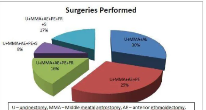
Table 2: Surgical procedures performed.
Surgery Percentage of
patients
Uncinectomy 100 Middle meatal antrostomy 100 Anterior ethmoidectomy 100 Posterior ethmoidectomy 70 Sphenoidectomy 25 Frontal recess surgery 33 Reduction of the middle turbinate 0
We had sixty cases of chronic sinusitis and forty cases of sinonasal polyposis, with involvement of various sinuses (Figure 3).
middle meatal antrostomy, anterior and posterior ethmoidectomy. 17% of the patients underwent uncinectomy, middle meatal antrostomy, anterior and posterior ethmoidectomy, frontal recess surgery and sphenoidotomy (Table 2, Figure 4).
Figure 4: Surgical procedures performed.
Intraoperative bleeding and surgical field visibility was graded by using Boezaart Van Der Merwe surgical field visibility grading. 8% of patients had cadaveric conditions like surgical field with minimal suction, 62% had grade 2 visibilities with infrequent suctioning, 26%
had grade 3 visibilities with frequent suctioning and 4% had grade 4 surgical field visibilities.
We encountered a cerebrospinal fluid leak post operatively in a case of extensive sinonasal polyposis, involving all the paranasal sinuses.8 The patient was taken up for the surgical correction of the defect. He underwent endoscopic cerebrospinal fluid leak closure after one week.9 He was discharged from the hospital on 14th day without any residual problems.
We had 12 cases of minor complications, most of them were adhesions that occurred in 8 patients, 3 patients presented on the 7th post-operative day and 5 others on 14th post-operative day. Out of these 8 patients, only 3 had symptomatic nasal block. All the cases were managed by release of adhesions under local anaesthesia, and careful frequent post-operative follow up.
We had one case each of periorbital ecchymosis and periorbital emphysema, which was managed conservatively. Anterior nasal packs were removed immediately. Vision and eye movements were normal. A course of steroids were given. Patient was discharged on 7th post-operative day without any sequelae.
Table 3: Complications.
Category Complication No. of patients Male Female Total Percentage Management
Major
Orbital haematoma (post septal) 0 0 0
Loss of vision 0 0 0
Diplopia 0 0 0
CSF leak 1 0 1 Endoscopic closure
Meningitis 0 0 0
Brain abscess 0 0 0
Focal brain damage 0 0 0
Haemorrhage requiring transfusion 0 0 0
Carotid artery injury 0 0 0
Epiphora 0 0 0
Blindness 0 0 0
CNS deficits 0 0 0
Death 0 0 0
Minor
Periorbital emphysema 1 0 1 Conservative Periorbital ecchymosis 1 0 1 Conservative Dental or lip pain or numbness 0 0 0
Adhesions requiring treatment 5 3 8 Release Epistaxis requiring packing 1 1 2 Packing
Bronchospasm 0 0 0
Sinus infection 0 0 0
Dental or lip pain or numbness or
anosmia 0 0 0
We had two cases of bleeding, one on the table which was controlled with the post nasal pack in addition to anterior nasal pack and another one which was on 4th
DISCUSSION
We had one case of CSF rhinorrhoea, which is a major complication, and was managed surgically and the patient was discharged without any sequelae. This complication occurred in a case of bilateral extensive polyposis involving all the paranasal sinuses. The incidence of major complication is 1% in this study. The overall incidence of major complications in the study of May was 1.2% and by Levine was 0.85%.10 The overall average of incidence in internationally published studies is 1.1%. We didn’t come across any major complications involving orbit in our study.
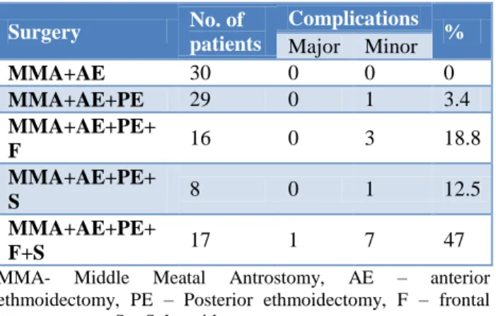
Table 4: Procedures done and occurrence of complications.
Surgery No. of
patients
Complications %
Major Minor MMA+AE 30 0 0 0 MMA+AE+PE 29 0 1 3.4 MMA+AE+PE+
F 16 0 3 18.8
MMA+AE+PE+
S 8 0 1 12.5
MMA+AE+PE+
F+S 17 1 7 47
MMA- Middle Meatal Antrostomy, AE – anterior ethmoidectomy, PE – Posterior ethmoidectomy, F – frontal recess surgery, S – Sphenoidotomy.
Table 5: Anatomical variations and occurrence of complications. Anatomical variations score No. of patients Complications %
Major Minor
0 40 0 3 7.5
1-2 44 0 6 13.7
3-4 16 1 3 25
Table 6: Types of pathology and occurrence of complications.
Pathology No. of
patients
Complications %
Major Minor Chronic Sinusitis 70 0 4 5.8 Sinonasal
Polyposis 30 1 8 30
In the minor complications, we had 12 cases of minor complications; most of them were adhesions that occurred in 8 patients that come to 8% of study population. All the cases were managed by release of adhesions under local anaesthesia, and careful frequent post-operative follow up. Most of the adhesions occurred in the cases of extensive disease (Table 4, 5 and 6).
We had one case each of periorbital ecchymosis and periorbital emphysema, which was managed
conservatively. Minor orbital complications occurred in 2% of study population. We had two cases of bleeding. Minor complication of bleeding occurred in 2% of cases. Total minor complications rate in our study is 12%. The incidence of minor complications as reported by May is 5.4% and by Levine is 8%.
In the first U.S. study that quantified complications related to endoscopic sinus surgery, Stankiewicz, reported a 6% major and 13% minor complication rate, the most common being synechiae.11 In a follow-up study, Stankiewicz reported on the complication rate of a subsequent group of 90 patients, and noted a rate of 2.4%, which compared favourably with previous reports of complications as reported by Freedman and Kern in 1979 using conventional intranasal methods. This significant drop in the complication rate was attributed to greater operative experience, concurrent cadaveric dissection, and the use of limited ethmoidectomy initially, with gradual progression to more extensive procedures. Several studies have subsequently demonstrated a further decline in the incidence of complications. Dessi noted a 1.2% complication rate for overall complications.
The complications both major and minor in this study are comparable to the international standards.
CONCLUSION
Though a lot of endoscopic sinus surgeries are being done nowadays, the incidence of major complications are low. Extension of the disease pathology and variations in the local anatomy are the major factors influencing the occurrence of complications. Pre-operative imaging of the patient to understand the extent of the disease and anatomical variations, thorough knowledge of anatomy, identification of key landmarks, preservation of normal sinus mucosa, meticulous intra operative tissue handling, periodic saline irrigation, proper haemostasis and using technologically advanced instruments are the major factors, which can definitely reduce the occurrence of complications and improve the patient outcome.
Funding: No funding sources Conflict of interest: None declared
Ethical approval: The study was approved by the Institutional Ethics Committee
REFERENCES
1. Messerklinger W. Endoscopy of the nose. Baltimore: Urban and Schwarzenberg; 1978;1(1)100-1.
2. Stammberger H. Endoscopic endonasal surgery – concepts in treatment of recurring rhinosinusitis. Part II. Surgical technique. Otolaryngol Head Neck Surg. 1986;94(2):147–56.
4. Lund VJ, Kennedy DW. Quantification for staging sinusitis. The Staging and Therapy Group. Ann Otol Rhinol Laryngol Suppl. 1995;167:17–21.
5. Lund VJ, MacKay IS. Staging in rhinosinusitis. Rhinol. 1993;31:183-4
6. Stammberger H. Functional endoscopic sinus surgery: the Messerklinger technique. Philadelphia: BC Decker; 1991.
7. Boezaart AP, van der Merwe J, Coetzee A. Comparison of sodium nitroprusside- and esmolol-induced controlled hypotension for functional endoscopic sinus surgery. Can J Anaesth. 1995;42(51):373-6.
8. Maniglia AJ. Fatal and major complications secondary to nasal and sinus surgery. Laryngoscope 1989;99:267-83.
9. Mattox DE, Kennedy DW. Endoscopic management of cerebrospinal fluid leaks and cephaloceles. Laryngoscope. 1990;100:857-62.
10. May M, Levine HL, Mester SJ, Schaitkin B. Complications of endoscopic sinus surgery: Analysis of 2108 patients – Incidence and prevention. Laryngoscope. 1994;104:1080-3. 11. Stankiewicz JA. Blindness and intranasal
endoscopic ethmoidectomy. Prevention and management. Oto laryngol Head Neck Surg. 1989;101:320-9.