421
Optimization of PCR-RFLP Method to Confirm the
Identification of Enterococcus faecalis and Enterococcus
Faecium Isolated from Food Samples
Miroslav Kročko, Michal Gábor, Monika Lavová, Martina Miluchová, Margita Čanigová, Jana Bezeková, Viera Ducková, Anna Trakovická
Slovak University of Agriculture in Nitra, Tr. A. Hlinku 2, 949 76, Nitra, Slovakia
Abstract
Commercial kits for species identification of Enterococcus faecalis and Enterococcus faecium are in some cases
(environmental isolates) incorrect to distinguish this species of enterococci. Also the positive results of multiplex PCR are subjected by amount and purity of genomic DNA. This study aimed to optimize the identification of
Enterococcus faecalis and Enterococcus faecium isolated from food samples and from the culture collection by
PCR-RFLP. The strains identified by conventional biochemical EN-COCCUS test (reactions to arginine, sorbose, arabinose, manitol, sorbitol, melibiose, raffinose, melezitose) were submitted to multiplex PCR. The PCR amplification of 16S rRNA with universal primers generated a 433 bp product. Restriction fragment length polymorphism (RFLP) was analysed by restriction endonuclease HinfI. The sizes of restriction fragments for Enterococcus faecalis were 285bp, 85bp and 63 bp and for Enterococcus faecium were 370 bp and 63bp.
Keywords: PCR-RFLP, 16S rRNA, multiplex PCR, Enterococcus faecalis, Enterococcus faecium.
1. Introduction
One of the uncertainties in environmental microbiology research is accurate identification of bacterial species. Without high-throughput, accurate identification of target organisms, studies in such important areas as epidemiology, antibiotic resistance assessment and microbial source tracking are compromised from the beginning [1, 2]. Commercially available kits are often used by clinical laboratories as an alternative to the numerous physiological tests needed to identify enterococcal species [3]; nevertheless, all commercial kits vary in their performance and persistently show many drawbacks, especially in cases of atypical strains, and at best need supplementary manual tests, which somewhat impair their usefulness. 1Species identification of
enterococci by phenotypical methods is time consuming [4], however, and misidentification of
* Corresponding author: Miroslav Kročko, [email protected]
Enterococcus faecium as Enterococcus gallinarum or Enterococcus casseliflavus and vice versa is a frequent problem [5]. Also Carvalho et al. (1998) [6] and Willey et al. (1999) [7], reported that in some cases identification of atypical Enterococcus faecium strains may be problematic, as is distinguishing Enterococcus gallinarum from Enterococcus faecium and also identifying Enterococcus durans, Enterococcus avium, Enterococcus raffinosus, Enterococcus hirae, and Enterococcus mundtii.
422 Their ubiquitous nature and resistance to adverse environmental conditions account for their ability to colonise different habitats and underlie their potential to easily spread through the food chain [11]. An increasing number of enterococci isolated from food production have development resistance to various therapeutic antibiotics, including vancomycin and tetracyclines. Enterococci belong to the lactic acid bacteria (LAB) group and are widely distributed in nature, but they are not generally recognized as safe (GRAS) [12, 13].
The aim of the work was to estimate the
use of relatively simple PCR-restriction
fragment length polymorphism (PCR-RFLP)
identification method of
Enterococcus
faecium
and
Enterococcus faecalis
in
comparison with biochemical test and
multiplex PCR.
2. Materials and methods
Sampling
Pork samples (n = 75) were obtained 24 h post mortem. Surface swabs of pork were taken from 25 cm2 of thigh (musculus semimembranosus). For the microbiological analysis, sampling of poultry was carried out 1 h post mortem (abdomen area of 25 cm2). The ewe‘s milk samples (n = 61) were collected from several areas of Slovakia. The unpasteurized cow milk samples (n = 32) were collected from different farms. Samples of Bryndza cheese (n = 39) were made by three different manufacturers and purchased in retail outlets. The homogenates of each samples and swabs from the meats were then submitted to serial 10-fold dilutions in sterile physiological saline and 1ml of each dilution were cultured on selective diagnostic Slanetz–Bartley agar at temperature 37 ± 1 °C for 48 ± 2 h (Biokar Diagnostic, France).
Species identification of enterococci by commercial biochemical tests
Typical colonies of enterococci were transferred to bile esculin azide agar (Biokar Diagnostic) for species identification. Based on positive growth (esculin hydrolysis), the following tests were carried out for presumptive identification of the isolates: microscopic characteristic of colonies (conformation, motility, cleanness of cultures), Gram staining, production of catalase and pyrolidonyl arylamidase (PYRAtest, Lachema,
Czech Republic), and pigmentation. Selected Gram positive, catalase negative, and PYRAtest positive isolates were submitted to a growth test in the presence of 6.5 % NaCl and pH 9.6. Isolates were identified at the species level using a biochemical EN-COCCUS test (Lachema). The reactions to arginine, sorbose, arabinose, manitol, sorbitol, melibiose, raffinose, melezitose were evaluated and according to manufacturer color key were identified species of Enterococcus spp. (E.). Genomic isolation of E. faecalis and E. faecium The isolation of genomic enterococci DNA from the overnight cultures at 37 °C was prepared. The bacteria were transferred to 200µl of Tris-HCl and EDTA (100mM Tris-HCl pH 8.0 and 10mM EDTA) and washed twice in the same solution. After centrifugation the bacterial pellet were incubated at 37 °C with intermitted shaking in conditions with 300µl of lysis buffer (0.1M NaCl, 0.02M Tris HCl, 0.001M EDTA, 1.2 % Trition X-100) and lysozyme solution (concentration 20 mg/ml) for 2 hours. The 5 µl proteinase K solution (Fermentas, Germany) and to remove RNA, 4 µl of Rnase-A solution (Fermentas) were added to the mix. The contents were mixed thoroughly and incubated at 58 °C for 2 hours. After incubation the mixtures were centrifuged at 10 000 rpm at 20 °C for 3 min. The 100 µl of 5 M NaCl (Merck, Germany) was added to upper layer and incubated on ice for 5 min and centrifuged at 13 000 rpm at 20 °C for 10 min. The upper aqueous phase was separated without disturbing the interphase containing cell debris and proteins. DNA in the supernatant (collected in sterile 1.5 ml tube) was precipitated out with 96 % freeze ethanol. The precipitated DNA was pelleted by centrifugation at 12 000 rpm for 10 min. The DNA pellet was washed once with freeze 70 % ethanol and dried at 37 °C. The final pellet was dissolved in 50 µl of Tris-HCl and EDTA (100mM Tris-HCl pH 8.0 and 10mM EDTA).
423 The PCR method for chosen species identification was performed using specific primers: E. faecium (215 bp) - F:5´GAAAAAACAATAGAAGAATT AT3´ R:5´ TGCTTTTTTGAAT TCTTCTTTA 3´ and E. faecalis (941 bp) F:5´ATCAAGTACAGTT AGTCTTTATTAG3´ R:5´ACGATTCAAAGCT AACTGAATCA GT3´ One microliter of DNA was added to a mixture containing 2.5 µl of 10 x PCR buffer (Fermentas), 0.5 µl of each 10 mM deoxynucleoside triphosphate (Fermentas), 2.0 µl 25 mM MgCl2 (Fermantas), 0.25µl 5 U of DreamTaq polymerase (Fermentas) and 0.5 µl of each 10 pmol primers (IDT, USA). The PCR amplification was performed in the thermal cycler C1000 (Biorad, USA). Samples were incubated for 3 min at 95 ºC to denature the target DNA and were maintained 30 cycles of 95 ºC for 30 s, 54 ºC for 40 s and 72 ºC for 60 s. The samples were then incubated at 72 ºC for 10 min for a final extension and were maintained at 4 ºC until they were tested. Gels were stained with GelRed (Biotium, USA) and visualized in UV light. Isolates producing an amplicon band of the appropriate size by agarose gel (1.5 %) electrophoresis were considered positive for species identification. Two references strains E. faecalis (CCM 4224) and E. faecium (CCM 2308) were obtained from the Czech Collection of Microorganisms at the Masaryk University in Brno.
Species identification of E. faecalis and E. faecium of specific fragment 16 S rRNA gene by PCR-RFLP method
The 16S rRNA gene was amplified by PCR with the following primers: forward primer U968 [14], 5’- AACGCGAAGAACCTTAC -3’; reverse primer L1401 [14], 5’- CGGTGTGTACAAGACC C -3’. PCR amplification was carried out in 25 μl volumes containing 1 μl of bacterial DNA and 24 μl of amplification mastermix, which contained the following components: 10 pmol each primer, 0,2 mM (each) dNTPs, 2.5 µl of 10 x PCR buffer (Fermentas), 2.0 µl 25 mM MgCl2 (Fermentas) and 1.25 U of Taq DNA polymerase (Fermentas). The PCR amplification was performed in a C1000 thermal cycler (Biorad). Samples were incubated
for 3 min at 95ºC to denature the target DNA and went through 30 cycles of 95ºC for 10 s, 58ºC for 20 s and 72ºC for 30 s. The samples were then incubated at 72ºC for 5 min for a final extension and were maintained at 4ºC until they were tested. An amplicon of 433 bp was expected.
Aliquots of 10 μL of each 433 bp amplified product were digested with restriction endonuclease FASTDigest HinfI (Fermentas) as recommended by the manufacturer. The specific fragments of restriction splitting for each species was detected on 3 % agarose gel (Serva, Germany) in 1 x sodium-borate buffer at 200 V for 10 min. The gel was stained with GelRed (Biotium) and visualized in UV light. Fragment sizes were assessed against a 100 bp DNA ladder (Fermentas).
Restriction maps were calculated from the amplicon regions of the GenBank sequences (E. faecalis – Y18293, E. faecium - HQ641333) using NEBCutter 2.0 [15]. The predicted restriction fragments from E. faecalis were 285 bp, 85 bp, 63 bp and from E. faecium were 370 bp, 63 bp.
3. Results and discussion
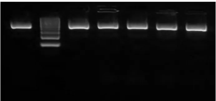
From the food isolates of enterococci it was identified 394 strains to genus Enterococcus. The combination of selective agars, negative catalase test and positive hydrolysis of L-pyrollidonyl-β -naphthyamine (PYR) test, performed well for isolation of Enterococcus genus, which was correlated with results obtained by using of 16S rDNA (433bp) gene (Figure 1). All tested isolates amplified the 16S rDNA fragment and sequence was appeared in the agarose gel. Results confirmed the useful of 16S rDNA for the identification of Enterococcus genus.
424 Figure 1. Amplification of 16S rDNA fragments
(433bp) isolated from Enterococcus spp.
1 E. faecalis CCM 4224, 2 Ladder 100bp, 3 E. faecium
CCM 2308, 4-7 enterococci isolated from food samples.
Enterococci isolated from food samples which were identified by commercial biochemical test as E. faecium and E. faecalis and Group 3 (E. durans, E. hirae and E. faecalis asaccharolytic var.) were submitted to confirmation by multiplex PCR and PCR-RFLP methods.
Enterococcus faecalis was the most frequently identified Enterococcus species (49 %) and all isolates of this species were identified with 100 % assurance by the commercial biochemical test. Twenty isolates determined by commercial biochemical test as Group 3, were difficult to accurately identified, which was due to variable reactions to raffinose, melibiose and melezitose. Eighteen isolates of Group 3 were identified by multiplex PCR method to E. faecalis species. In this study, E. faecalis became predominant species in every investigated samples type with the exception of pork, where predominated species E. faecium. Enterococcus faecalis-specific primers successfully amplified the expected 941 bp product from each of the E. faecalis isolates, demonstrating agreement between the commercial biochemical test and the confirmation at the molecular level by multiplex PCR method. The suitability of commercial biochemical tests for E. faecalis species identification tested also Harwood et al. (2004) [17]. Authors most frequently identified E. faecalis (42.2 %) from the freshwater, saltwater, sewage, seagull faeces, human faeces and dog faeces samples. All isolates identified with 90 % or greater assurance by the biochemical test system, which was in agreement with results of our study.
One isolate identified in our study by commercial biochemical test as E. faecium was not identified by multiplex PCR method. Six microbiological
and biochemical positive strains not identified by EN-COCCUS test, designated as Enterococcus spp., were by multiplex PCR method identified as E. faecium (Figure 2). Jurkovič et al. (2006) [18] found some discrepancies between results of enterococci identification from Bryndza cheese samples, obtained by commercial biochemical test and PCR method. Seven enterococci strains identified by commercial biochemical test were identified as E. faecium and by PCR method as E. faecalis. Three strains of E. casseliflavus were determined by PCR method as E. faecium (two strains) and E. faecalis (one strain).
Figure 2. Multiplex PCR identification of E. faecium
(215 bp) and E. faecalis (941 bp)
Ladder 100bp; 1 E. faecium isolated from cow milk; 2 E. faecalis isolated from cow milk; 3 E. faecium
isolated from pork; 4 E. faecalis isolated from pork; 5 E. faecium isolated from Bryndza cheese; 6 E. faecalis
isolated from Bryndza cheese; 7 E. faecium isolated
from ewe´s milk; 8 E. faecalis isolated from ewe´s
milk; 9-10 E. faecium isolated from poultry; 11 E. faecium CCM 2308; 12-14 E. faecalis isolated from
poultry; 15 E. faecalis CCM 4224.
Angeletti et al. (2001) [19] reported that the use of a PCR with primers for ddlE. faecalis and ddlE. faecium,
together with primers for vanC-1, -2, and -3, may be the most simple molecular approach for both rapid and precise identification of antibiotic resistant enterococci while avoiding the drawbacks of commercial kits. On the other hand, Monstein et al. 2000 [20]; Kariyama et al. 2000 [21]; Patel et al. 1998 [22]; Dutka-Mahlen et al. 1995 [23] found that the PCR with primers for ddlE. faecalis and ddlE. faecium, together with primers
425 include among the proposed molecular methods for enterococcal species identification the amplification and sequencing of the 16S ribosomal DNA (rDNA) gene.
Bacterial 16S rRNA is a common target for taxonomic purposes, largely due to the mosaic composition of phylogenetically conserved and variable regions within the gene [24].
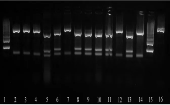
The chosen 16S rDNA product (433bp) was digested with enzyme HinfI to fragments for E. faecium 370 and 63 bp and for E. faecalis 285; 85 and 63bp (Fig 3). It was found the accurate separation of E. faecium species, but similar restriction fragments as E. faecalis were obtained also after E. hirae isolates digestion.
Williams et al. (1991) [25] reported that there is a low rRNA homology between E. faecalis and other enterococci and E. durans is phylogenetically closer to E. faecium and E. hirae. Also Leclerc et al. (1996) [26] identified several distinct phylogenetic groups (species groups) of enterococci, based on sequencing of the 16S rRNA gene. They found that the E. faecium group, which contains species whose 16S rDNA sequence is 99.3–99.7% similar, is comprised of E. faecium, E. durans, E. hirae and E. mundtii. Enterococcus faecalis, in contrast, represents a distinct lineage within the Enterococcus spp. Scheidegger et al. (2009) [27] found that thirty-seven isolates of the food enterococci exhibited atypical phenotypic traits. Among them, eleven E. faecalis strains presented atypical results in the methyl-β-D-glucopyranoside and one in the raffinose test. Five isolates of E. faecium presented atypical results in the potassium tellurite test. Their results shown that PCR-RFLP combined with a few biochemical tests was a relative simple, rapid and reliable technique to identify species of Enterococcus.
Figure 3. Restriction DNA fragments of E. faecium
(370; 63bp) and E. faecalis (285; 85; 63bp) obtained
with digestion of PCR product (16S rDNA 433bp) with enzyme HinfI.
1 Ladder 100bp; 2 E. faecium isolated from cow milk;
3 E. faecium isolated from pork; 4 E. faecium isolated
from Bryndza cheese; 5 E. faecalis isolated from cow
milk; 6 E. faecalis isolated from Bryndza cheese; 7 E. faecium isolated from poultry; 8-10 E. faecalis isolated
from poultry; 11 E. faecalis CCM 4224; 12 E. faecium
CCM 2308; 13 E. faecalis isolated from ewe´s milk; 14 E. faecium isolated from ewe´s milk; 15 Ladder 100 bp;
16 PCR product 16S rDNA.
Angeletti et al. (2001) [19] found that four isolates identified by multiplex PCR as E. faecalis and two identified as E. faecium out of 279 isolates were not enterococci. They were identified by 16S rDNA sequencing as two Streptococcus spp., one Leuconostoc sp., one Aerococcus sp. and one Streptococcus bovis strain. Among the other multiplex PCR negative strains and but phenotypically identified as E. faecium, four were identified by 16S rDNA sequencing as E. avium (three strains) and E. durans (one strain). Nine strains identified by commercial biochemical test as E. faecium were identified as E. faecalis by multiplex PCR, six of which were also confirmed by sequencing of amplified 16S rDNA. In agreement with our results, 16S rDNA sequencing confirmed all multiplex PCR identifications for available strains.
4. Conclusions
426 identified isolates by commercial biochemical test as E. durans, E. hirae, E. mundtii, E. casseliflavus, currently analyzed, could be confirmed by multiplex PCR or PCR-RFLP methods. It can be probably due to with phylogenetic distance of E. faecalis and E. faecium to other mentioned enterococci species and not reliably identified their phenotypic characteristics.
Acknowledgements
This work was supported by the VEGA grants from the Ministry of Education Science, Research and Sport of the Slovak Republic, grant No. 1/0410/09 and 1/0897/11 and by the Slovak Research and Development Agency under the contract No. LPP-0220-09.
References
1. Wiggins, B. A., Discriminant analysis of antibiotic resistance patterns in fecal streptococci, a method to differentiate human and animal sources of fecal pollution in natural waters, Applied and Environmental Microbiology, 1996, 62, 3997-4002
2. Harwood, V. J., Wiggins, B., Hagedorn, C., Ellender, R. D., Gooch, J., Kern, J., Samadpour, M., Chapman, A. C. H., Robinson, B. J., Thompson, B. C., Phenotypic library-based microbial source tracking methods: efficacy in the California collaborative study, Journal of Water and Health, 2003, 1, 153-166
3. Facklam, R. R., Elliott, J. A., Identification, classification, and clinical relevance of catalase-negative, gram-positive cocci, excluding the streptococci and enterococci, Clinical Microbiology Review, 1995, 8, 479-495
4. Moore, D. F., Zhowandai, M. H., Ferguson, D. M., McGee, C., Mott, J. B., Stewart, J. C., Comparison of 16S rRNA sequencing with conventional and commercial phenotypic techniques for identification of enterococci from the marine environment, Journal of Applied Microbiology, 2006, 100, 1272-1281
5. Eigner, U., Fahr, A., Weizenegger, M., Witte, W., Evaluation of a new molecular system for simultaneous identification of four Enterococcus species and their
glycopeptide resistance genotypes, Journal of Clinical Microbiology, 2005, 43, 2920-2922
6. Carvalho, M. S., Teixeira, L. M., Facklam, R. R., Use of tests for acidification of methyl-a-D-glucopyranoside and susceptibility to efrotomycin for differentiation of strains of Enterococcus and some
related genera, Journal of Clinical Microbiology, 1998, 36, 1584-1587
7. Willey, B. M., Jones, R. N., McGeer, A., Witte, W., French, G., Roberts, R. B., Jenkins, S. G., Nadler, H., Low, D. E. Practical approach to the identification of
clinically relevant Enterococcus species, Diagnostic
Microbiology and Infectious Disease, 1999, 34, 165-171
8. Menichetti, F., Current and emerging serious Gram-positive infections, In Clinical Microbiological Infection, 2005, 11, 22-28
9. Murray, B. E. 1990. The life and times of the
Enterococcus, Clinical Microbiology Review, 3, 46-65
10. Wellinghausen, N., Bartel, M., Essig, A., Poppert, S., Rapid identification of clinically relevant
Enterococcus species by fluorescence in situ
hybridization, Journal of Clinical Microbiology, 2007, 45, 3424-3426
11. Fracalanzza, S. A. P., Scheidegger, E. M. D., Santos, P.F., Cardarelli-Leite, P., Teixeira, L. M., Antimicrobial resistance profiles of enterococci isolated from poultry meat and pasteurized milk in Rio de Janeiro, Brazil, Memórias do Instituto Oswaldo Cruz, 2007, 102, 853-859
12. Tsai, C. C., Liu, T. H., Chen, M. H., Tsai, C. C., Tsen, H. Y., Toxicity evaluation for an Enterococcus faecium strain TM39 in vitro and in vivo, Food and
Chemical Toxicology, 2004, 42, 1601-1609
13. Gomes, B. C., Esteves, C. T., Palazzo, I. C. V., Darini, A. L. C., Felis, G. E., Sechi, L. A., Franco, B. D. G. M., De Martinis, E. C. P., Prevalence and characterization of Enterococcus spp. isolated from
Brazilian foods, Food Microbiology, 2008, 25, 668-675 14. Nubel, U., Engelen, B., Felske, A., Snaidr, J., Wieshuber, A., Amann, R. I., Ludwig, W., Backhaus, H., Sequence heterogeneities of genes encoding 16s
rRNAs in Paenibacillus polymyxa detected by
temperature gradient gel electrophoresis, Journal of Bacteriology, 1996, 178, 5636-5643
15. Vincze, T., Posfai, J., Roberts, R. J., NEBcutter: a program to cleave DNA with restriction enzymes, Nucleic Acids Research, 2003, 31, 3688-3691
16. Fortina, M. G., Ricci, G., Borgo, F., Manachini, P. L., Rapid identification of Enterococcus italicus by
PCR with primers targeted to 16S rRNA gene, Letters in Applied Microbiology, 2007, 44, 443-446
17. Harwood, V. J., Delahoya, N. C., Ulrich, R. M., Kramer, M. F., Whitlock, J. E., Garey, J. R., Lim, D. V., Molecular confirmation of Enterococcus faecalis
and E. faecium from clinical, faecal and environmental
sources, Letters in Applied Microbiology, 2004, 38, 476-482
18. Jurkovič, D., Križková, L., Dušinský, R., Belicová, A., Sojka, M., Krajčovič, J., Ebringer, L., Identification and characterization of enterococci from bryndza cheese, Letters in Applied Microbiology, 2006, 42, 553-559
427
20. Monstein, H. J., Johansson, Y., Jonasson, J., Detection of vancomycin resistance genes combined with typing of Enterococci by means of multiplex PCR
amplification and multiple primer DNA sequencing, Acta Pathologica et Microbiologica Scandinavica, 2000, 108, 67-73
21. Kariyama, R., Mitsuhata, R., Chow, J. W., Clewell, D. B., Kumon, H., Simple a reliable multiplex PCR assay for surveillance isolates of vancomycin-resistant enterococci, Journal of Clinical Microbiology, 2000, 38, 3092-3095
22. Patel, R., Piper, K. E., Rouse, M. S., Steckelberg, J. M., Uhl, J. R., Kohner, P., Hopkins, M. K., Cockerill III, F. R., Kline, B. C., Determination of 16S rRNA sequences of enterococci and application to species identification of nonmotile Enterococcus gallinarum
isolates, Journal of Clinical Microbiology, 1998, 36, 3399-3407
23. Dutka-Malen, S., Evers, S., Courvalin, P., Detection of glycopeptide resistance genotypes and identification to the species level of clinically relevant enterococci by PCR, Journal of Clinical Microbiology, 1995, 33, 1434
24. Gurtler, V., Stanisich, V. A. New approaches to typing and identification of bacteria using the 16S-23S rDNA spacer region, Microbiology, 1996, 142, 3-16 25. Williams, A. M., Rodrigues, U. M., Collins, M. D. Intrageneric relationships of Enterococci as determined by reverse transcriptase sequencing of small-subunit rRNA, Research in Microbiology, 1991, 142, 67-74 26. Leclerc, H., Devriese, L. A., Mossel, D.A., Taxonomical changes in intestinal (faecal) enterococci and streptococci: consequences on their use as indicators of faecal contamination in drinking water, Journal of Applied Bacteriology, 1996, 81, 459-466 27. Scheidegger, E. M. D., Fracalanzza, S. A. P., Teixeira, L. M., Cardarelli-Leite, P., RFLP analysis of a PCR-amplified fragment of the 16S rRNA gene as a tool to identify Enterococcus strains, Memórias do