POST-DIAGNOSIS CHANGES IN POLYCYCLIC AROMATIC HYDROCARBON SOURCES OF EXPOSURE AND
SURVIVAL FOLLOWING BREAST CANCER
Humberto Parada, Jr.
A dissertation submitted to the faculty at the University of North Carolina at Chapel Hill in partial fulfillment of the requirements for the degree of Doctor of Philosophy in the Department
of Epidemiology in the Gillings School of Global Public Health.
Chapel Hill 2016
© 2016
ABSTRACT
Humberto Parada, Jr.: Post-diagnosis Changes in Polycyclic Aromatic Hydrocarbon Sources of Exposure and Survival Following Breast Cancer
(Under the direction of Marilie D. Gammon)
In 2016, an estimated 246,000 women will be diagnosed with, and 40,000 deaths will be attributed to, breast cancer. Polycyclic Aromatic Hydrocarbons (PAHs), a group of over 100 different chemicals formed during the incomplete combustion of organic substances, may
influence survival after breast cancer. This dissertation examined whether the primary sources of PAH exposure, tobacco smoke and intake of grilled/smoked meat, and changes in exposure after diagnosis were associated with mortality after breast cancer. To address the dissertation aims, I utilized resources from the Long Island Breast Cancer Study Project (LIBCSP), a population-based cohort study of 1,508 women who were diagnosed with first primary breast cancer in 1996/1997. Women were interviewed at baseline, shortly after diagnosis, and again five years later and have been followed for 18+ years using the National Death Index.
Results of Aim 2 showed that at-diagnosis high intake of total grilled/barbecued and smoked meat was associated with a 23% increased risk of all-cause mortality. At-diagnosis intake of smoked beef/lamb/pork was positively associated with all-cause and breast cancer mortality, while intake of smoked poultry/fish was inversely associated with mortality. Women with continued high post-diagnosis intake of grilled/barbecued and smoked meat had a further elevated risk of all-cause mortality; risk increased from 23% to 31%. Consistent with the associations observed for at-diagnosis intake, risk of breast cancer-specific mortality was inversely associated with high post-diagnosis intake of smoked poultry/fish.
TABLE OF CONTENTS
LIST OF TABLES ... ix
LIST OF FIGURES ... xii
LIST OF ABBREVIATIONS ... xiii
CHAPTER I: BACKGROUND ... 1
Epidemiology of Breast Cancer ... 1
Breast cancer definitions ... 1
Breast cancer prevalence, incidence, and mortality ... 2
Established risk and prognostic factors... 4
Epidemiology of Polycyclic Aromatic Hydrocarbons (PAHs) and Breast Cancer ... 26
PAH Definition ... 26
Sources of Exposure and Metabolism... 26
Measurement of PAH exposure ... 29
PAH Exposure Prevalence ... 31
PAH adducts and breast cancer... 32
Outdoor/Indoor Air pollution and breast cancer ... 33
Smoking-related PAH exposures and breast cancer ... 35
Diet-related PAH exposures and breast cancer ... 39
Summary ... 42
REFERENCES ... 56
CHAPTER II: RESEARCH METHODS ... 79
Significance... 81
Innovation ... 84
Approach ... 86
Research Strategy... 86
Study Population ... 87
Exposure Assessment... 88
Covariate Assessment ... 93
Missing Data ... 95
Outcome Assessment ... 96
Data Analysis ... 97
Study Statistical Power ... 98
Summary ... 100
REFERENCES ... 110
CHAPTER III: POST-DIAGNOSIS CHANGES IN SMOKING AND SURVIVAL FOLLOWING BREAST CANCER ... 115
Overview ... 115
Introduction ... 116
Methods... 117
Study Population ... 118
Smoking Assessment ... 119
Covariate assessment ... 119
Outcome Assessment ... 120
Statistical Analysis ... 121
Results ... 122
Prevalence of smoking among women with breast cancer ... 122
At-/post-diagnosis smoking and survival after breast cancer ... 124
Discussion ... 125
Conclusions ... 127
REFERENCES ... 144
CHAPTER IV: POST-DIAGNOSIS CHANGES IN ENVIRONMENTAL TOBACCO SMOKE EXPOSURE AND SURVIVAL FOLLOWING BREAST CANCER ... 147
Overview ... 147
Introduction ... 148
Methods... 148
Environmental Tobacco Smoke Exposure Assessment ... 148
Covariate assessment ... 149
Outcome Assessment ... 149
Statistical Analysis ... 149
Results ... 150
At-Diagnosis Environmental Tobacco Smoke Exposure ... 150
At-/Post-Diagnosis Environmental Tobacco Smoke Exposure ... 150
Discussion ... 151
REFERENCES ... 156
CHAPTER V: POST-DIAGNOSIS CHANGES IN GRILLED, BARBECUED, AND SMOKED MEAT INTAKE AND SURVIVAL FOLLOWING BREAST CANCER ... 157
Overview ... 157
Introduction ... 158
Methods... 159
Study Population ... 159
Covariate assessment ... 161
Outcome Assessment ... 162
Statistical Analysis ... 162
Results ... 164
Pre-diagnosis intake of grilled/barbecued and smoked meat ... 164
Post-diagnosis changes in intake grilled, barbecued, and smoked meat ... 165
Discussion ... 166
REFERENCES ... 177
CHAPTER VI: DISCUSSION ... 180
Summary ... 180
Biologic Plausibility... 182
Study Advantages and Limitations ... 182
Future Directions ... 186
Public Health Impact... 189
Conclusions ... 190
REFERENCES ... 191
LIST OF TABLES
Table I-1. Summary of breast cancer risk and prognostic factors. ... 44 Table I-2. Evidence of estrogenic and anti-estrogenic activity of selected PAH
compounds by PAH exposure source. ... 46 Table I-3. Median Percentiles of OH-PAHs in NHANES 2011-2012 among participants
aged 20 years and older, by smoking status (N=1,703). ... 47 Table I-4. Studies examining the associations between PAH sources of exposure and
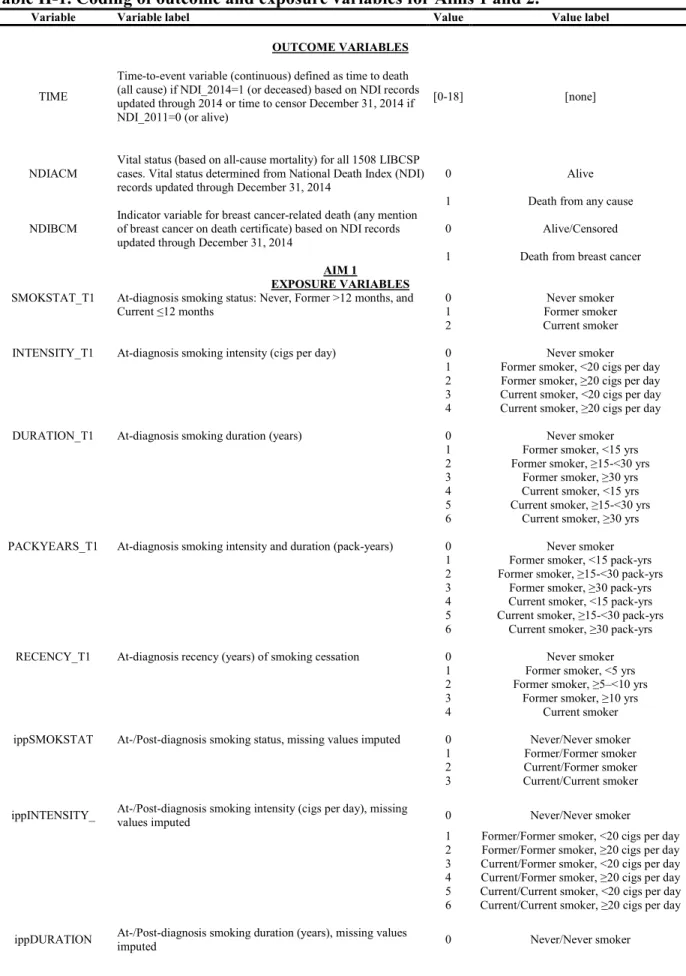
survival after breast cancer. ... 48 Table II-1. Coding of outcome and exposure variables for Aims 1 and 2. ... 102 Table III-1. Distribution of selected at-diagnosis participant and disease characteristics of
the LIBCSP women diagnosed with breast cancer in 1996-1997 (N=1,508), overall and
by pre- and at-diagnosis smoking status. ... 128 Table III-2. Cox regression hazard ratios (HRs) and 95% confidence intervals (CIs) for
the association between pre- and at-diagnosis cigarette smoking and mortality in the
LIBCSP women diagnosed with breast cancer in 1996-1997 (N=1,508). ... 130 Table III-3. Cox regression hazard ratios (HRs) and 95% confidence intervals (CIs) for
the association between pre- and at-diagnosis cigarette smoking and mortality in the
LIBCSP women diagnosed with invasive breast cancer in 1996-1997 (n=1,273). ... 132 Table III-4. Cox regression hazard ratios (HRs) and 95% confidence intervals (CIs) for
the association between pre- and at-diagnosis cigarette smoking and mortality in the LIBCSP women diagnosed with estrogen receptor-positive breast cancer in 1996-1997
(n=726). ... 134 Table III-5. Cox regression hazard ratios (HRs) and 95% confidence intervals (CIs) for
the association between at-/post-diagnosis cigarette smoking and mortality in the LIBCSP women diagnosed with breast cancer in 1996-1997 (n=1,332). ... 136 Table III-6. Cox regression hazard ratios (HRs) and 95% confidence intervals (CIs) for
the association between at-/post-diagnosis cigarette smoking and mortality in the LIBCSP women diagnosed with breast cancer in 1996-1997, using a complete-case analysis
(n=955). ... 138 Table III-7. Cox regression hazard ratios (HRs) and 95% confidence intervals (CIs) for
the association between at-/post-diagnosis cigarette smoking and mortality in the LIBCSP women diagnosed with invasive breast cancer in 1996-1997 (n=1,106). ... 140 Table III-8. Cox regression hazard ratios (HRs) and 95% confidence intervals (CIs) for
the association between at- and post-diagnosis cigarette smoking and mortality in the LIBCSP women diagnosed with estrogen receptor positive breast cancer in 1996-1997
Table III-9. Cox regression hazard ratios (HRs) and 95% confidence intervals (CIs) for the association between at- and post-diagnosis cigarette smoking and mortality among LIBCSP overweight and obese (BMI ≥25 kg/m2) women diagnosed with breast cancer in
1996-1997 (n=711). ... 143 Table IV-1. Cox regression hazard ratios (HRs) and 95% confidence intervals (CIs) for
the association between pre-diagnosis and at-diagnosis environmental tobacco smoke (ETS) exposure and mortality in the LIBCSP women diagnosed with breast cancer in
1996-1997 (N=1,508). ... 152 Table IV-2. Cox regression hazard ratios (HRs) and 95% confidence intervals (CIs) for
the association between pre-diagnosis and at-diagnosis environmental tobacco smoke (ETS) exposure and mortality in the LIBCSP women diagnosed with invasive breast
cancer in 1996-1997 (n=1,273). ... 153 Table IV-3. Cox regression hazard ratios (HRs) and 95% confidence intervals (CIs) for
the association between at-/post-diagnosis environmental tobacco smoke exposure (ETS) and mortality in the LIBCSP women diagnosed with breast cancer in 1996-1997
(n=1,339). ... 154 Table IV-4. Cox regression hazard ratios (HRs) and 95% confidence intervals (CIs) for
the association between at-/post-diagnosis environmental tobacco smoke exposure (ETS) and mortality in the LIBCSP women diagnosed with invasive breast cancer in 1996-1997
(n=1,111). ... 155 Table V-1. Distribution of participant characteristics at diagnosis among the LIBCSP
women diagnosed with first primary breast cancer in 1996-1997, overall and by
grilled/barbecued and smoked meat intake (N=1,508). ... 170 Table V-2. Cox regression hazard ratios (HRs) and 95% confidence intervals (CIs) for the association between pre-diagnosis lifetime and annual intake of grilled/barbecued and smoked meat and mortality in the LIBCSP women diagnosed with breast cancer in
1996-1997 and followed for 18+ years (N=1,508). ... 172
Table V-3. Cox regression hazard ratios (HRs) and 95% confidence intervals (CIs) for the association between pre-diagnosis lifetime and annual intake of grilled/barbecued and smoked meat and mortality in the LIBCSP women diagnosed with invasive breast cancer
in 1996-1997 and followed for 18+ years (N=1,273). ... 173
Table V-4. Cox regression hazard ratios (HRs) and 95% confidence intervals (CIs) for the association between pre-diagnosis/post-diagnosis annual intake of grilled/barbecued and smoked meat and mortality in the LIBCSP women diagnosed with breast cancer in
1996-1997 and followed for 18+ years (n=1,339). ... 174
Table V-5. Cox regression hazard ratios (HRs) and 95% confidence intervals (CIs) for the association between pre-diagnosis/post-diagnosis annual intake of grilled/barbecued and smoked meat and mortality in the LIBCSP women diagnosed with invasive breast cancer
Table V-6. Cox regression hazard ratios (HRs) and 95% confidence intervals (CIs) for the association between pre-diagnosis/post-diagnosis annual intake of grilled/barbecued and smoked meat and mortality in the LIBCSP women diagnosed with breast cancer in
LIST OF FIGURES
Figure I-1. Breast Cancer Incidence and Mortality Rates among Women of All-Races,
United States, 1975-2011. (National Cancer Institute 2016) ... 54 Figure I-2. Structures and nomenclatures of the 16 PAHs on the EPA priority pollutant
list. ... 55 Figure II-1. Pre/At-diagnosis and Post-diagnosis PAH Exposures and Survival Following
Breast Cancer in the LIBCSP. ... 105 Figure II-2. Directed Acyclic Graphs of the association between breast cancer survival
and at-diagnosis active smoking (A) and post-diagnosis changes smoking (B). ... 106 Figure II-3. Directed Acyclic Graphs of the association between breast cancer survival
and at-diagnosis ETS exposure (A) and post-diagnosis changes ETS exposure (B). ... 107 Figure II-4. Directed Acyclic Graphs of the association between breast cancer survival
and at-diagnosis grilled/barbecued and smoked meat intake (A) and post-diagnosis
LIST OF ABBREVIATIONS
ATSDR Agency for Toxic Substances and Disease Registry BMI Body Mass Index
CC Complete-Case
CDC Centers for Disease Control and Prevention CI Confidence Interval
COX-2 Cyclooxygenase-2 CYP Cytrochrome P450 DCIS Ductal Carcinoma In Situ DNA Deoxyribonucleic Acid
E1 Estrone
E2 Estradiol
EGFR Epidermal Growth Factor Receptor ER Estrogen Receptor
ETS Environmental Tobacco Smoke FCS Fully Conditional Specification FFQ Food Frequency Questionnaire HCAs Heterocyclic Amines
HER-1 Human Epidermal Growth Factor Receptor 1 HER-2 Human Epidermal Growth Factor Receptor 2
HR Hazard Ratio
HRT Hormone-Replacement Therapy
IARC International Agency for Research on Cancer ICD International Statistical Classification of Diseases LCIS Lobular Carcinoma in Situ
MAR Missing At Random
MCAR Missing Completely At Random MNAR Missing Not At Random
NDI National Death Index
NHANES National Health and Nutrition Examination Survey NSAIDs Non-Steroidal Anti-Inflammatory Drugs
OCs Oral Contraceptives OH-PAHs Monohydroxy-PAHs
OR Odds Ratio
PA Physical Activity
PAHs Polycyclic Aromatic Hydrocarbons PM2.5 Particulate matter <2.5 μm in diameter
PM10 Particulate matter <10 μm in diameter
PR Progesterone Receptor
RR Risk Ratio
SEER Surveillance, Epidemiology, and End Results SHBG Sex Hormone-Binding Globulin
US United States
US EPA United States Environmental Protection Agency WHI Women’s Health Initiative
CHAPTER I:BACKGROUND
This dissertation examined the role polycyclic aromatic hydrocarbon (PAH) sources of exposure before and after diagnosis in relation to breast cancer survival. The first aim examined the associations between active cigarette smoking and environmental tobacco smoke and
changes in cigarette smoke exposure and all-cause and breast cancer mortality after diagnosis. Similarly, the second aim examined the associations between intake of grilled and smoked meats and changes in intake of grilled and smoked meats and all-cause and breast cancer mortality after diagnosis. This background section first summarizes the epidemiology of breast cancer
incidence, which has been extensively studied, along with breast cancer survival, focusing on non-PAH factors. I first discuss these established risk and prognostic factors as they provide insight into the potential underlying biological mechanisms driving the hypothesized associations with PAH exposures. The discussion of risk and prognostic factors is followed by a discussion focused on the epidemiology of PAHs in relation to breast cancer incidence and survival, summarizes the literature on the studies conducted to date, and highlights the existing gaps in research that this dissertation addressed.
Epidemiology of Breast Cancer
Breast cancer definitions
the cells of the breast, are able to grow and invade surrounding tissues, and are able to
metastasize to distant areas of the body (American Cancer Society 2015b). The normal structures of the breasts include lobules – the milk-producing glands; ducts – which carry the milk from the lobules to the nipples; and stroma – the fatty and connective tissues that surround the lobules, ducts, and blood and lymphatic vessels of the breasts (American Cancer Society 2015b). Approximately 80% of invasive breast cancers arise from the ductal epithelial cells and are referred to as invasive ductal carcinomas (The Johns Hopkins University 2015b). Invasive lobular carcinomas arise from the epithelial cells that line the lobules and are the second most common invasive breast cancer diagnosed (10%-15%) (The Johns Hopkins University 2015c). A small number of breast cancers originate in other tissues of the breast. Ductal and lobular
carcinomas in situ (DCIS and LCIS, respectively) are considered non-invasive breast tumors contained within the duct or lobular basement membranes (Ellis et al. 1992; The Johns Hopkins University 2015a).
Breast cancer prevalence, incidence, and mortality
cancer statistics across population-based registries since 1973, an increase in the rate of new breast cancer cases diagnosed in the 1980’s is evident (Figure I-1) (National Cancer Institute 2016). This increase is attributed to the adoption and use of mammography screening as
evidenced by the increase in the number and the proportion of localized and small tumors and in situ tumors and the decrease in the incidence of large tumors diagnosed during this time period (Chu et al. 1996; White et al. 1990). From 1988 to 2002 the breast cancer incidence rate
stabilized until it declined in the year 2003. The decline is thought to be temporally related to the first report of the Women’s Health Initiative, which confirmed the findings of prior studies reporting an increased risk of coronary heart disease and breast cancer associated with the use of estrogen-progestin combination therapy (Writing Group for the Women’s Health Initiative Investigators 2002) that resulted in a decrease in the use of hormone-replacement therapy among postmenopausal women in the United States (Ravdin et al. 2007). Incidence rates have since once again stabilized with the most recent data indicating that the age-adjusted incidence rate is currently 125.0 per 100,000 women per year across women of all races (National Cancer Institute 2016).
Established risk and prognostic factors
Estrogens are a group of compounds that promote the development and maintenance of the female reproductive system. The physiologic functions of estrogens in women include development of secondary sexual characteristics, regulation of gonadotropin secretion for ovulation, preparation of tissues for progesterone response, maintenance of bone mass,
regulation of lipoprotein synthesis, and regulation of insulin responsiveness (Nelson and Bulun 2001). The two major endogenous estrogens include estrone (E1), estradiol (E2) and are
synthesized from androgens by the aromatase enzyme primarily in the ovaries and secondarily in adipose and skin tissues (Nelson and Bulun 2001). As outlined in a review by Yager and
Davidson (Yager and Davidson 2006), estrogens are hypothesized to be mammary gland
carcinogens via nuclear, mitochondrial, and plasma membrane estrogen receptor (ER)-mediated pathways. Through these pathways, in estrogen responsive tissues such as the ovaries and the mammary gland, the presence of estrogen results in altered gene expression leading to increased cell proliferation and decreased cell apoptosis (Katzenellenbogen 1996). By promoting the proliferation of cells with existing mutations or by increasing the opportunity for novel
mutations, estrogens contribute to breast carcinogenesis (Pike et al. 1993). Independent of ER-mediated pathways, estrogens also undergo extensive metabolism which leads to the production of genotoxic, mutagenic, and carcinogenic metabolites (Mueck and Seeger 2007).
This may be due to limitations of early studies that had small sample sizes, utilized timed urine samples (rather than 24-hour collection) – a less sensitive method of steroid hormone assessment than the use of serum samples (Riad-Fahmy et al. 1982), and employed a case-control study design in which samples were collected after the initiation of disease in cases (Toniolo 1997). Because disease, treatment, or behavioral changes after diagnosis may influence hormone levels, the biomarkers collected after diagnosis may not have been reflective of the etiologically
relevant time-period, thus leading to mixed results (Toniolo 1997). It was not until the 1990’s during which results of several prospective nested case-control studies (Berrino et al. 1996; Helzlsouer et al. 1994; Toniolo et al. 1991, 1995) specifically designed to address the role of endogenous hormones in breast cancer were published that the hypothesis regained momentum. Studies have since continued to further examine both hormones and these and other
epidemiologic risk factors, and prognostic factors, in relation to breast cancer. These include age at menarche, age at menopause, parity, and age at first and multiple pregnancies (Mcpherson et al. 2000; The Endogenous Hormones and Breast Cancer Collaborative Group 2002).
Additionally, exposure to exogenous sources of estrogens through exposure to oral
Age
Aging and the molecular, cellular, and physiologic processes that accompany it including increased genomic instability, increased oxidative stress, increased DNA damage, and decreased DNA repair capacity, are important in the etiology of all cancers, including breast cancers
(Anisimov 2007). The incidence rate of breast cancer rises rapidly with age, is highest during the reproductive years, and increases more slowly until menopause when the rate slows (Clemmesen 1948; Pike et al. 1983b). In the United States, the median age at diagnosis of breast cancer is 62 years (National Cancer Institute 2016).
Particularly poor survival has been observed among women diagnosed at a younger age, especially those diagnosed younger than 35 years of age (RR=2.18; 95% CI=1.64-2.89), and women diagnosed over the age of 80 (RR=1.80; 95% CI=1.45-2.25) compared to women diagnosed between the ages of 40 and 49 (Brandt et al. 2015; Kroman et al. 2000; Reeves et al. 2000). Although only approximately 2% of all breast cancers are diagnosed in women under the age of 35 (National Cancer Institute 2016), it is the most common cancer in women under 35 and young women generally present with more advanced and aggressive disease at diagnosis
skin) may also play an important role in survival, especially among women with hormone-sensitive tumors (Hemsell et al. 1974).
Reproductive factors
Menarche and menopause. Epidemiologic studies show that breast cancer incidence increases by 5% for each year younger at menarche and by 3% for each year older at menopause (Collaborative Group on Hormonal Factors in Breast Cancer 2012). An earlier initiation of menarche results in earlier exposure to hormones and regular menstrual cycles (Kelsey et al. 1993) and older age at menopause results in continued hormonal exposure. Because most (approximately 60-70%) breast cancers are estrogen-sensitive (Dunnwald et al. 2007), increased lifetime exposure to these hormones may raise a woman’s risk of developing breast cancer. Further evidence is provided by the observation of an inverse association between bilateral oophorectomy, or surgical removal of the ovaries, and breast cancer progression, which was recognized even before the importance of estrogens was understood (Love and Philips 2002). Recent studies show that when performed before the age of 40, bilateral oophorectomy reduces the risk of breast cancer by 20% to 50% (Nichols et al. 2011).
Age at menarche and menopausal status at diagnosis, however, do not appear to be independently associated with breast cancer mortality (Barnett et al. 2008; Giordano et al. 2004). Because chemotherapy can often lead to amenorrhea, an abnormal absence of menstruation, and premature initiation of menopause in pre- and peri-menopausal women (Ganz 2005; Goodwin et al. 1999; Morgan et al. 2012) post-treatment menopausal status may be more relevant.
L. Dellvigne 2011). Several studies report longer disease free survival for patients who developed drug-induced amenorrhea as compared with non-amenorrheic women (Aebi et al. 2000; Bianco et al. 1991; Del Mastro et al. 1997; Pagani et al. 1998), though not all studies are in agreement (Collichio and Pandya 1994; Del Mastro et al. 1997; Vanhuyse et al. 2005).
Parity and age at first and multiple pregnancies. Numerous epidemiologic studies have reported an increased risk of developing breast cancer among nulliparous women and a long-term reduced risk of breast cancer for increasing number of full-long-term births and early age at first birth (Kelsey et al. 1993). In a meta-analysis (Ma et al. 2006) of ten case-control and cohort studies, each additional birth was associated with a reduced the risk of developing ER+/PR+
breast cancer by 11% (RR=0.89, 95% CI=0.84-0.94) and women in the oldest age group at first birth had a 27% increased (RR=1.27, 95% CI=1.07-1.50) risk of ER+/PR+ breast cancer than
women in the youngest age group. Each pregnancy; however, is also associated with a transient increased risk of developing breast cancer for 5, but up to 15, years after childbirth (Lambe et al. 1994; Liu et al. 2002). Subsequently, women who are diagnosed with breast cancer at or shortly after a pregnancy experience particularly poor survival. These tumors are more likely to be hormone receptor-negative, high histologic grade, node positive, and have higher mitotic count, and higher stage compared to tumors of nulliparous women (Alsaker et al. 2011; Daling et al. 2002). Few studies have examined whether parity is associated with survival after breast cancer, but recent studies indicate that higher parity is associated with worse survival (Butt et al. 2009; Trivers et al. 2007a), which may be due to enhanced initiation or progression of malignant cells or delayed diagnosis and thus worse prognosis, among women with high parity (Butt et al. 2009).
exposure to endogenous hormones, and by increasing the differentiation of ductal cells, making them less susceptible to carcinogenic insult (Lipworth et al. 2000; Russo et al. 2001; Visvader and Stingl 2014). Breastfeeding also provides a route of excretion of many lipophilic and potentially carcinogenic chemicals as there is rapid assimilation of lipid-soluble chemicals during milk production (Sim and McNeil 1992). Before these underlying mechanisms were fully understood, animal studies, followed by epidemiologic studies, generated these hypotheses documenting an inverse association between breastfeeding and risk of breast cancer. The results of epidemiology studies show that for every 12 months of breastfeeding the risk of developing breast cancer decreases by 4%-5%, compared to no breastfeeding (Collaborative Group on Hormonal Factors in Breast Cancer 2002; Ma et al. 2006).
Studies examining whether breastfeeding influences prognosis after a breast cancer diagnosis have been limited and results have been mixed. One study (Phillips et al. 2009)
(Kwan et al. 2015). The inverse associations between a history of breast feeding and breast cancer mortality were more pronounced among hormonally-sensitive (ER+ or PR+) Luminal A and Luminal B tumors (HR=0.52, 95% CI=0.31-0.89 and HR=0.60, 95% CI=0.26-1.41), but also among non-hormonally sensitive (ER- and PR-) basal-like tumors (HR=0.64, 95% CI=0.24-1.72).
The authors hypothesized that the transformation of breast cells during pregnancy could lead to the development of more differentiated (ER+ and PR+) breast cancers which are better prognostic
tumor subtypes.
Exogenous hormone use
Oral contraceptive (OC) use. Use of oral contraceptives (OCs) is highly prevalent in the US with approximately 10.7 million women reporting current use (Mosher 2010). OC pills were first introduced in the US in 1960 and since the early 1970’s there has been an interest in
understanding the impact of OC use on breast cancer incidence. Results of very early studies reported no increased risk of breast cancer (Arthes et al. 1971; Fasal and Paffenbarger, Ralph S. 1975; Henderson et al. 1974; Ory et al. 1976), likely due to an insufficient amount of time
women who ever used oral contraceptives (Hunter et al. 2010; Kahlenborn et al. 2006). This risk is further elevated among women who use OCs before their first full-term pregnancy (OR=1.44, 95% CI=1.28-1.62) compared to women who use OCs after their first full-term pregnancy (OR=1.15, 95% CI=1.06-1.26), which is consistent with observations of inverse associations between pregnancy and breast cancer incidence (Kahlenborn et al. 2006).
The use of oral contraceptives after a diagnosis of breast cancer in considered
contraindicated (World Health Organization. 4th ed. 2001) due to the proliferative effects of estrogens on cancerous breast cells. Although most studies have failed to find an association between OC use and mortality (Ewertz et al. 1991; Greenberg et al. 1985; Holmberg et al. 1994; Lees et al. 1989; Millard et al. 1987; Mohle-Boetani et al. 1988; Rosner and Lane 1986;
Sauerbrei et al. 1998), these studies focused on ever/never use of oral contraceptives and all-cause mortality. In contrast to these studies, three studies have examined current OC use at breast cancer diagnosis (Lu et al. 2011; Trivers et al. 2007b; Wingo et al. 2007). In the first study, among women in the LIBCSP, Trivers and colleagues reported an increased HR of all cause-mortality (HR=1.97, 95% CI=1.15-3.38) among women who currently used OCs or who used OCs within 1 year of breast cancer diagnosis (Trivers et al. 2007b). Additionally, risk of breast cancer mortality was further elevated (HR=3.03, 95% CI=1.61-5.69) among women who used high-dose estrogen formulations (Trivers et al. 2007b). In the second study published in 2007 and in the third study published in 2011, both failed to reach the same conclusions finding no association between current OC use or recent high-dose estrogen OC use and breast cancer-specific mortality (Lu et al. 2011; Wingo et al. 2007). Additional studies examining recent and post-diagnosis use of oral contraceptives are needed to help clarify these associations.
effective for the treatment of menopausal symptoms. Therapies include estrogen alone (“estrogen therapy”) or estrogen plus progestin (“combined hormone therapy”) and are administered as pills, and more recently as skin patches, gels and sprays that are applied to the skin. These hormones can act systemically or locally. While effective in managing menopausal symptoms, HRT use also has health risks. In 2002, after a follow-up of 5 years, the Women’s Health Initiative (WHI) trial of estrogen plus progestin (1 daily tablet containing 0.625 mg of conjugated equine estrogen and 2.5 mg of medroxyprogesterone acetate) versus placebo was terminated early due to an observed increased risk of developing breast cancer (HR=1.26, 95% CI=1.00-1.59), heart disease (HR=1.29, 95% CI=1.02-1.63), and stroke (HR=1.41, 95% CI=1.07-1.85) among women assigned to the intervention (Writing Group for the Women’s Health Initiative Investigators 2002). Similar effect estimates had been previously reported in a meta-analysis of 51 case-control studies that included 52,705 women with invasive breast cancer and 108,411 women without breast cancer; a 35% increased risk of breast cancer incidence (RR=1.35, 95% CI=1.21-1.49) among women who had used HRT for 5 years or longer (Collaborative Group on Hormonal Factors in Breast Cancer 1997). The meta-analysis also showed that the risk of developing breast cancer increased with increasing duration of use (≥15 years of use versus never-use, RR=1.58, SE=0.121) with an apparent attenuation in risk after cessation of use of HRT, which largely disappeared after 5 years of cessation (current use versus never use, RR=1.21, SE=0.04; last use 1-4 years versus never use, RR=1.10, SE=0.06; last use 5-9 years versus never use, RR=1.01, SE=0.07) (Collaborative Group on Hormonal Factors in Breast Cancer 1997).
evidence supporting this hypothesis, however, has been mixed. In a meta-analysis (Col et al. 2001) of 11 studies published through May 1999 of post-diagnosis HRT use and breast cancer recurrence, a statistically non-significant inverse association (RR=0.82, 95% CI=0.58-1.15) was found between HRT use and breast cancer recurrence. In one study published shortly after the meta-analysis, the rate of recurrence was significantly lower (RR=0.50, 95% CI=0.30-0.85) among women who used HRT after diagnosis compared to nonusers (O’Meara et al. 2001). Similarly, reduced rates were also observed for breast cancer mortality (RR=0.34, 95% CI=0.13-0.91) and all-cause mortality (RR=0.48, 95% CI=0.29-0.78) (O’Meara et al. 2001) among women who used HRT after diagnosis. In the Stockholm trial of HRT use (n=188 women randomized HRT and n=190 randomized to no HRT) and breast cancer recurrence, after 10.8 years of follow-up a 30% increased (HR=1.3, 95% CI=0.9-1.9) risk was observed among HRT-users compared to non-HRT-users (Fahlén et al. 2013). Several possible explanations for these contradictory findings include unmeasured confounding, including confounding by indication, and more aggressive screening among breast cancer survivors who use HRT. In contrast to these findings, a randomized clinical trial (Holmberg and Anderson 2004) that investigated the safety of a 2-year HRT treatment in women who were previously treated for breast cancer was
Genetics
(O’Brien et al. 2010).
Diet
Although the relationship between diet and breast cancer incidence has received considerable scientific attention, few consistent associations are observed (Vera-Ramirez et al. 2013). This is possibly due to methodological issues including differences in measurement of dietary intake, potential misclassification of exposures, high correlations among nutrients, and insufficient time for follow-up (Moorman and Terry 2004; Vera-Ramirez et al. 2013). As reviewed below, of the dietary exposures that have been examined, only alcohol intake is consistently associated with increased risk of breast cancer incidence and inconsistently with mortality (Rock 2002; Vera-Ramirez et al. 2013). Several studies, including randomized trials, report an inverse association between fruit and vegetable intake and breast cancer incidence or mortality and a positive association between fat intake and breast cancer incidence and mortality, but results are inconsistent.
transport estrogens as biologically inactive forms, in premenopausal and postmenopausal women (Key et al. 2011; Rinaldi et al. 2006). Through these mechanisms, it is hypothesized that alcohol intake may also affect survival after breast cancer diagnosis. While several studies have found a positive association between the highest levels of intake of alcohol and breast cancer-specific mortality (HRs ranging from 1.51 to 4.32) when exposed before, at, and after diagnosis
(Allemani et al. 2011; Fuchs et al. 1995; Hebert et al. 1998; Kwan et al. 2010; McDonald et al. 2002; Vrieling et al. 2012) and an inverse association between moderate intake of alcohol and all-cause mortality (Barnett et al. 2008; Flatt et al. 2010; Reding et al. 2008; Saxe et al. 1999), results of most studies have been null as summarized in a meta-analysis of 25 follow-up studies of alcohol use (14 studies of pre-diagnosis drinking, 10 studies of post-diagnosis drinking, and 1 study of both pre- and post-diagnosis drinking) and breast cancer mortality published in 2013 (Gou et al. 2013). In the meta-analysis (Gou et al. 2013), neither pre- nor post-diagnosis alcohol consumption were associated with breast cancer mortality (HR=1.05, 95% CI=0.93-1.19 and HR=1.08, 95% CI=0.94-1.25); however drinking >20g/d of alcohol was associated a 14% (95% CI=2%-27%) increased hazard of breast cancer-specific mortality.
Study, the New York State Cohort, and the Netherlands Cohort Study, the highest quartile of total intake of fruits and vegetables was associated with only a 7% reduction (RR=0.93, 95% CI=0.86-1.00, PTrend = 0.12) in breast cancer risk.
In the few observational studies that have examined fruit and vegetable intake and breast cancer-specific mortality, higher intake of fruits and vegetables and intake of micronutrients such as beta carotene, calcium, vitamin A, vitamin C, vitamin E have been shown to be inversely associated with breast cancer mortality (Fink et al. 2006; Jain et al. 1994; Patterson et al. 2010). In the Long Island Breast Cancer Study Project, among post-menopausal women, intake of any fruits and vegetables at diagnosis was associated with a reduced (HR=0.68, 95% CI=0.42-1.09) risk of all-cause mortality; the association was reported to be similar for breast cancer-specific mortality although the estimates were not provided (Fink et al. 2006). In the survival cohort study within National Breast Screening Study in Canada, the HR of breast cancer mortality among women with the highest quartiles of intake of beta carotene was 0.48 (95% CI=0.23-0.99) and vitamin C was 0.43, (95% CI=0.21-0.86) relative to women with intake in the lowest
quartiles (Jain et al. 1994). Higher intake of fruits and vegetables is also inversely associated with all-cause mortality (Dal Maso et al. 2008; McEligot et al. 2006). In randomized clinical trials, fruit and vegetable intake has also been shown to be inversely associated with breast cancer mortality when combined with high physical activity (Pierce et al. 2007b), although modification of fruits and vegetable intake alone does not appear to reduced mortality from breast cancer (Pierce et al. 2007a, 2013). Given these suggestive findings, current guidelines recommend that women be encouraged to adopt a diet high in fruits and vegetables after breast cancer diagnosis (Runowicz et al. 2016).
saturated fat, can concentrate lipophilic carcinogenic chemicals including pesticides, and may contain growth factors such as insulin-like growth factor I – has also been extensively studied in relation to breast cancer incidence. Results, however, are inconclusive and the data available do not support they hypothesis that dietary fat increases the risk of breast cancer incidence
(Moorman and Terry 2004).
women assigned to a low-fat diet intervention compared to controls (Chlebowski et al. 2006), while the Women’s Healthy Eating and Living Trial found no association between the
intervention designed to reduce fat intake and the control groups and recurrence (Pierce et al. 2007a). Despite these mixed findings, current guidelines for breast cancer survivors recommend that women limit intake of saturated fats (Runowicz et al. 2016).
Other lifestyle factors
NSAID use. Non-steroidal anti-inflammatory drugs (NSAIDs) such as Aspirin, Acetaminophen, and Ibuprofen are chemically distinct compounds that share a common therapeutic action; they inhibit the cyclooxygenase-2 (COX-2) enzyme, which catalyzes the synthesis of prostaglandins from dietary arachidonic acid (Vane 1971). Prostaglandins increase aromatase gene expression in breast cells and in surrounding tissue resulting in estrogen
effects from treatment with NSAIDs, such as gastrointestinal bleeding and perforation (Agrawal and Fentiman 2008).
After breast cancer initiation, the primary prostaglandin produced by COX-2, PGE2, can transactivate EGFR leading to stimulation of migration of tumor cells (Wang and Dubois 2006). Intratumoral aromatase may also be an important source of estrogens available for tumor growth (Esteban et al. 1992). Therefore, inhibiting COX-2 and EGFR tyrosine kinase could block the spread of metastatic disease. Several observational studies have explored whether aspirin use is associated with survival after breast cancer. At least three have reported a reduced risk of breast cancer mortality among women who used aspirin at- (Holmes et al. 2010) and post- (Blair et al. 2007; Fraser et al. 2014) diagnosis with hazard ratios ranging from 0.53 to 0.57 for all cause-mortality and from 0.36 to 0.53 for breast cancer cause-mortality; however, not all have results have been in agreement (Holmes et al. 2014; Kwan et al. 2007; Li et al. 2012; Wernli et al. 2011). Additionally, three studies examining de novo post-diagnosis use of aspirin found no association with breast cancer mortality suggesting that the benefits of aspirin use may be attributable to pre-diagnosis use (Zhang et al. 2012).
Obesity. In postmenopausal women, with cessation of estrogen synthesis in the ovaries, the major pathway of estrogen production becomes the conversion of androstenedione into estrone in adipose tissue and skin (Grodin et al. 1973; Lønning et al. 1990). Additionally, postmenopausal obese women may have a higher proportion of bioavailable estrogen due to lower levels of SHBG (Key et al. 2011; Zhang et al. 1984). Adipose tissues also secrete
increased risk of breast cancer incidence among postmenopausal women with increased height and weight (de Waard and Baanders-Van Halewijn 1974; Valaoras et al. 1969). More recently, body mass index (BMI) – a surrogate for adiposity that incorporates both height and weight – and waist-to-hip ratio (WHR) are more consistently used measures of obesity. A recently published secondary analysis of the Women’s Health Initiative (WHI) randomized clinical trial reported that women in the highest categories of obesity (BMI ≥35 kg/m2) had a 58% increased
(HR=1.58, 95% CI=1.40-1.79) risk of breast cancer incidence compared to normal weight women and obesity was associated with more advanced disease, including larger tumors, lymph node involvement, and regional or distant stage at diagnosis (Neuhouser et al. 2015). These observations were consistent with most previous studies and meta-analyses of breast cancer incidence (Cheraghi et al. 2012; Endogenous Hormones Breast Cancer Collaborative Group 2003; Munsell et al. 2014), but not all (Cecchini et al. 2012). In contrast, among premenopausal women increased obesity is associated with a small reduced risk of developing breast cancer (Amadou et al. 2013; Munsell et al. 2014; Renehan et al. 2008; Ursin et al. 1995) possibly due to anovulation and lower levels of circulating estrogen levels (Potischman et al. 1996).
addition to the studies examining pre- and at-diagnosis weight and breast cancer survival, several studies have also shown that post-diagnosis weight gain after breast cancer, a common
occurrence after chemotherapy treatment (Demark-Wahnefried et al. 1997), negatively impacts survival. In the LIBCSP, Bradshaw and colleagues reported an increased (HR=2.84, 95%
CI=1.15-6.65) hazard of breast cancer-specific mortality among women who had more than 10% weight gain after diagnosis compared to women who maintained their weight within 5%
(Bradshaw et al. 2012), consistent with findings of several prior studies (Kroenke et al. 2005; Nichols et al. 2009), but not all (Caan et al. 2008, 2012; Chen et al. 2010). While the causal mechanism remains unresolved, the authors posit two hypotheses that could explain the poorer survival observed among obese women. The first is that obese patients may have more
biologically aggressive tumors thought to be a result of increased leptin production in adipose tissue which, among many functions, stimulates tumor cell mitogenesis, tumor cell migration and invasion, induces angiogenesis, and induces aromatase activity (Rose et al. 2002). The second is that obese women may be undertreated with regards to chemotherapy since doses of most chemotherapy drugs are based on body surface area and physicians may have concerns that obese women will experience toxic effects at high doses, thus, reducing doses (Griggs et al. 2005).
levels of inflammation (Gleeson 2007).
Epidemiologic studies examining the associations between physical activity and breast cancer incidence have been complicated by methodological issues in assessing PA including the potential for misclassification of exercise due to variation in sources of physical activity (e.g. recreational PA, occupational PA, and activities of daily living), use of varying definitions of physical activity (e.g. frequency, duration, and intensity), and inaccurate assessment of physical activity during etiologically relevant time period(s), which are unknown (Gammon et al. 1998), but studies have consistently observed an inverse association between increased levels of physical activity and breast cancer incidence. A systematic review (Monninkhof et al. 2007) of 19 cohort studies and 29 case-control studies published through February 2006 provides strong evidence for an inverse association between physical activity and incidence of breast cancer (15%-20% reduced risk) though the evidence was stronger for postmenopausal breast cancer (20%-80% reduced risk) than premenopausal breast cancer.
Physical activity is thought to influence the progression of breast cancer through the same mechanisms by which it is believed to influence incidence. Indeed, observational studies report an inverse association between PA and breast cancer survival. In a meta-analysis (Lahart et al. 2015) of 22 prospective cohort studies, inverse associations were observed between breast cancer-specific mortality and lifetime diagnosis (HR=0.73, 95% CI=0.54-0.98), recent pre-diagnosis (HR=0.84, 95% CI=0.73-0.97), and post-pre-diagnosis recreational PA (HR=0.59, 95% CI=0.45-0.78). In addition, meeting recommended PA guidelines post-diagnosis was associated with a HR of breast cancer mortality of 0.67 (95% CI=0.50-0.90) (Lahart et al. 2015). Substantial heterogeneity was found in several of the comparisons, but results were in agreement with
Several issues regarding the benefits of physical activity in relation to breast cancer prognosis remain unresolved including understanding the optimal frequency and duration of physical activity.
Disease characteristics and treatment
Epidemiology of Polycyclic Aromatic Hydrocarbons (PAHs) and Breast Cancer
PAH Definition
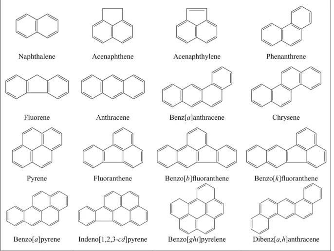
Polycyclic Aromatic Hydrocarbons (PAHs) include over 100 different chemicals that are formed during the incomplete combustion of coal, oil and gas, and other organic substances like tobacco and charbroiled/smoked meats (Agency for Toxic Substances and Disease Registry (ATSDR) 1995a). These molecules consist of two or more fused aromatic rings and are, by definition, composed of hydrogen and carbon (see Figure I-2 for the structures and
nomenclatures of the 16 PAHs on the US Environmental Protection Agency (EPA) priority pollutant list (Yan et al. 2004)). PAHs are generally lipophilic and this property increases with increasing complexity of the compounds (Boström et al. 2002). As pure chemicals PAHs are solid and range in appearance from colorless to white or pale yellow-green (US Environmental Protection Agency, 2008). The sources of exposure and metabolism, measurement of PAH exposure, PAH exposure prevalence and how PAH and PAH sources of exposure relate to breast cancer incidence and survival as reviewed in the following sections.
Sources of Exposure and Metabolism
Non-occupational PAH sources of exposure in the US include, primarily, cigarette smoking and, among non-smokers, diet; and, secondarily, outdoor and indoor air pollution (Skupińska et al. 2004). Among non-smokers, dietary sources account for up to 70% of
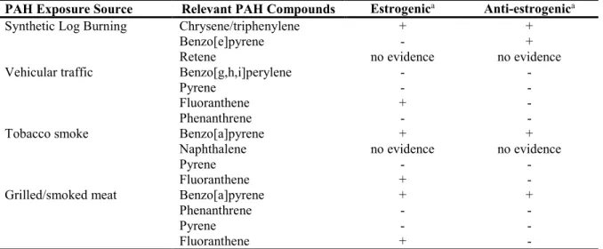
than 100 possible different PAHs that can be originate within a single source, PAHs can also co-occur with other chemicals some of which are known carcinogens. For example, in vehicular traffic air pollution, PAHs, including the estrogenic PAH fluoranthene (see Table I-2 adapted from White, 2015), are found as a mixture with ozone, carbon monoxide, nitrogen oxides, heavy metals, and other particulate matter (Wu et al. 2012). Synthetic logs can contain PAHs such as chrysene/triphenylene as well as polychlorinated biphenyls which show estrogenic and anti-estrogenic effects in vitro (Gullett et al. 2003; Wolff et al. 1997) (Table I-2). In cigarette smoke, PAHs, including benzo[a]pyrene which has been documented to exert both estrogenic and anti-estrogenic effects in vitro and naphthalene (Table I-2), are found with the carcinogens benzene, arsenic, heavy metals, formaldehyde, vinyl chloride, and N-Nitrosamines (IARC 2004). In meats and high-fat foods, PAHs can bioaccumulate along with dioxins and other persistent lipophilic pollutants such as organochlorine pesticides and polychlorinated biphenyls (Loomis et al. 2015). In high temperature-cooked foods, PAHs including benzo[a]pyrene, fluoranthene, pyrene, and phenanthrene (Table I-2), are formed along with heterocyclic amines (HCAs) depending on the method of food preparation – HCAs are formed when amino acids pyrolyze in meat juice, though pan-frying foods produces more HCAs than grilling and smoking foods (Knize et al. 1999). Although PAHs co-occur with many chemicals, PAHs are common across all sources and only PAHs and PAH sources of exposure have been consistently associated with breast cancer
incidence. Although dioxin has been found to be associated with breast cancer incidence, results have been mixed; in a study of the 1976 Seveso, Italy industrial disaster, dioxin exposure
a decreased (quartile 4 versus quartile 1 RR=0.65, 95% CI=0.45-0.96) risk of ER-/PR- breast
cancer was found (Danjou et al. 2015). In addition to PAHs being the most common contaminant across these sources of exposure, there is a plausible biologically mechanism linking PAHs and breast cancer (Gammon and Santella 2008).
Inhalation, ingestion, and dermal contact are the possible routes by which PAHs can enter the body. Once in the body, PAHs induce expression of Phase I and Phase II metabolizing
enzymes; the most important being CYPs 1A1, 1A2, 1B1, and 3A4 – the cytochrome P450 (CYP) superfamily of enzymes – and epoxide hydrolase (Luckert et al. 2013). During Phase I metabolism, PAH parent compounds are activated to potentially estrogenic reactive dihydrodiol intermediates by cytochrome p540 enzymes (Kummer et al. 2008; Luckert et al. 2013; Menzie et al. 1992). The dihydrodiols are further oxidized into diol epoxides, which are able to covalently bond to exocyclic amino groups of guanine and adenine, forming stable adducts on DNA (Lin et al. 2001). The metabolites of the PAHs benzo[j]fluoranthene, benzo[b]fluoranthene,
benzo[k]fluoranthene, benzo[a]pyrene and dibenzo[a,h]anthracene are known carcinogens able to form adducts in laboratory studies (Cavalieri et al. 1991; IARC 2010). The DNA adducts can cause mismatch in DNA replication and may alter promoter methylation or promoter binding, leading to inheritable DNA mutations or abnormal gene expression (Moorthy et al. 2015). Phase II metabolism includes conjugation of metabolites from Phase I – the hydroxyl-PAH metabolites – with small molecules catalyzed by specific enzymes such as sulfotransferases, UDP-glucoronyl transferases, and glutathione S-transferases. Sulfation and glucoronidation produces polar
Measurement of PAH exposure
Measures of exposure
Exposure to PAHs can be measured indirectly by questionnaire by querying study participants about exposures to the primary sources of PAH exposure. Questionnaires have the advantage of being able to elicit information about the lifetime while being relatively
inexpensive.
• Self-reported active smoking, including history, duration, and intensity of smoking, and exposure to environmental tobacco smoke are widely accepted and reliable measures of exposure (Krall et al. 1989). Smoking and ETS exposure can further be confirmed by measuring serum, hair, salivary, or urinary cotinine – the predominant metabolite of nicotine (Binnie et al. 2004).
• Intake of grilled and smoked foods and methods of food preparation via questionnaire can be used to estimate the dietary contribution of PAHs (Gammon et al. 2002b; Steck et al. 2007).
• Indoor air pollution can also be measured by questionnaire as a proxy for PAH exposure. In the LIBCSP, White and colleagues estimated exposure to PAHs by asking participants about their use of indoor stoves and fireplaces, which included the frequency of use, the type of material burned and the ages of participants during the time they lived in
residences (White et al. 2014).
specifically designed to measure particulate matter combined with meteorological data and participant residential information can be useful for predicting a person’s exposure to particulate matter, and thus PAHs (Hu et al. 2013). Personal air monitors can also provide information about personal exposure to particulate matter/PAHs (Binková et al. 1995).
Measures of internal dose
PAHs can be measured in body tissues and blood, but the high cost of measuring PAH parent compounds in these media makes their use in epidemiologic studies challenging (Agency for Toxic Substances and Disease Registry (ATSDR) 1995a). Instead, urinary monohydroxy PAHs (OH-PAHs) are a less expensive biomarker alternative for assessing exposure to PAHs which show high correlations with PAH exposures. For example, feeding studies report significantly increased concentrations of urinary 1-hydroxypyrene levels after consumption of charbroiled meat (Kang et al. 1995; van Maanen et al. 1994). Urinary metabolites have the advantage of being able to account for PAH exposures from all sources and all routes of exposure, but concentrations will be reflective of recent exposures unless there is chronic exposure with little variation (Agency for Toxic Substances and Disease Registry (ATSDR) 1995b). In the feeding studies, urinary metabolite concentrations returned to baseline within 24-72 hours of cessation of exposure (Kang et al. 1995). Often, one or several PAH metabolites can be used as a surrogate for assessing exposure to several PAHs. For example, 1-hydroxypyrene, the urinary metabolite of pyrene, is often measured since levels show strong positive correlations with several environmental PAHs (Binnie et al. 2004; Ciarrocca et al. 2014).
Measures of biologically effective dose
(Agency for Toxic Substances and Disease Registry (ATSDR) 1995b); however, in population studies it is often too invasive or infeasible to sample specific tissues, for example breast tissue. Therefore, adducts measured in blood are often used as a surrogate for tissue adduct levels (Santella 1999). These measures have the disadvantage that adduct levels measured in blood may not accurately reflect those of the tissue of interest, but studies show strong correlations between formation of PAH-DNA adducts in peripheral white blood cells and exposures such as charcoal-broiled beef consumption (Rothman et al. 1990; van Maanen et al. 1994) and ambient air
pollution in occupational settings (Santella et al. 1993). Also, like the urinary metabolites, DNA-adducts and protein-DNA-adducts reflect of short term exposures (Binková et al. 1995). These
biomarkers of biologically effective dose, however, are objective measures not subject to participant recall of past exposures, unlike self-reported questionnaire data, and they represent a biologically relevant end-point associated with carcinogenesis.
PAH Exposure Prevalence
PAHs are highly ubiquitous exposures that occur in mixtures; human exposure to PAHs occurs on a daily basis (Agency for Toxic Substances and Disease Registry (ATSDR) 1995a). The National Health and Nutrition Examination Survey (NHANES) conducted by the Centers for Disease Control and Prevention (CDC) conducts biomonitoring of several urinary PAH
metabolites. These measurements provide estimates of PAH exposure prevalence in a population based sample of the US. In one study (Xu et al. 2010) examining the associations between eight OH-PAHs (urinary metabolites of naphthalene, fluorene, phenanthrene, and pyrene) and
of detection (LOD) for >95% of participants. Median levels were lowest for the phenanthrene metabolite 2-hydroxyphenanthrene (61 ng/L in 2001-2002, 63.6 ng/L in 2003-2004) and highest for the naphthalene metabolite 2-hydroxynaphthalene (2,646 ng/L in 2001-2001 and 3,118.5 ng/L in 2003-2004) (Xu et al. 2010). Median concentrations of 1-hydroxypyrene measured 46 ng/L in 2001-2002 and 80.8 ng/L in 2003-2004 (Xu et al. 2010). For comparison, in (Table I-3), I provide the concentrations for adults (age ≥20 years) of the 10 measured OH-PAHs in National Health and Nutrition Examination Survey (NHANES) 2011-2012 (Centers for Disease Control and Prevention 2014a), the most recently available NHANES data, by smoking status. From these data, it is evident that all OH-PAH concentrations are elevated for smokers and those exposed to ETS compared to non-smokers/never-smokers. For 10 measured OH-PAHs, proportions above the LOD were at least 98% for all participants. Patterns of exposure are similar in 2011-2012 as compared to those reported by Xu and colleagues (Xu et al. 2010).
PAH adducts and breast cancer
tissue of breast cancer patients were higher than in the samples of the reduction mammoplasty controls (Li et al. 1996; Perera et al. 1995; Rundle et al. 2000). In the LIBCSP, after a second round of assays, the pooled (n cases = 873 and n controls = 941) OR for the association between detectable PAH-DNA adduct levels and breast cancer incidence was 1.29 (95% CI=1.05-1.58); the association was more pronounced among premenopausal (OR=1.56, 95% CI=1.09-2.23) than among postmenopausal (OR=1.14, 95% CI=0.88-1.47) women (Gammon et al. 2010).
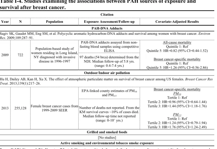
The LIBSCP has also been the first (and only) study to examine the associations between PAH-DNA adducts measured in peripheral blood and survival after breast cancer (Sagiv et al. 2009) (see Table I-4). The hazard ratio for breast cancer specific mortality was elevated
(HR=1.26, 95% CI=0.56-2.86) in relation to the highest quintile of PAH-DNA adduct levels, but estimates were imprecise and included the null (Sagiv et al. 2009). The magnitude of the
association between PAH-DNA adducts and survival is suggestive of an association. As it is possible that the LIBCSP was underpowered to adequately address this issue, additional studies with a larger sample sizes or longer follow-up are needed to help clarify this association.
Outdoor/Indoor Air pollution and breast cancer
found to be positively associated with breast cancer incidence (r=0.89, 0.82, 0.71, and 0.68, respectively) (Wei et al. 2012). In the LIBCSP, using a historical geographic exposure model, Mordukhovich and colleagues estimated the association between long-term, individualized residential traffic benzo[a]pyrene exposure estimates, as a proxy for exposure to particulate traffic PAHs, and breast cancer incidence. Consistent with prior studies of traffic-related air pollution (Crouse et al. 2010) including a study conducted on Long Island, NY (Lewis-Michl et al. 1996) and a study using similar methodology (Nie et al. 2007), in the LIBCSP women with vehicular traffic estimates in the top 5% had a 44% increased (OR=1.44, 95% CI=0.78-2.68) odds of breast cancer incidence and the magnitude of the association was more pronounced (OR=1.67, 95% CI=0.91-3.05) among women with ER-/PR- tumors (Mordukhovich et al. 2016).
The LIBCSP has been the first study to examine the association between use of indoor stoves/fireplaces, an indicator of indoor air pollution and breast cancer incidence (White et al. 2014). In their study, White and colleagues reported a 42% increased (OR=1.42, 95% CI=1.11-1.84) risk of breast cancer incidence among women who reported ever burning synthetic logs, but not among women who reported ever burning wood alone (White et al. 2014).
Only one study has examined the associations between outdoor air pollution and breast cancer survival (Hu et al. 2013) and no studies have examined the association between indoor air pollution in relation to survival (see Table I-4). In their study, using SEER and US EPA data from 1999-2009, Hu and colleagues observed an increased risk of breast cancer-specific
mortality among women exposed to high levels of particulate matter less than 10 μm in diameter (PM10) (PM10 ≥28.82 μg/m3 versus <23.09 μg/m3 HR=1.44, 95% CI=1.18-1.76) and high levels
of PM2.5 (PM2.5 ≥15.04 μg/m3 versus <11.64 μg/m3 HR=1.76, 95% CI=1.24-2.49) (Hu et al.
both breast cancer incidence and survival.
Post-diagnosis changes in outdoor/indoor air pollution and breast cancer
No studies have examined whether post-diagnosis changes in exposure to outdoor or indoor air pollution are associated with survival after breast cancer diagnosis. However, outdoor and indoor air pollution exposures, which account for a relatively smaller proportion of PAH exposure (Skupińska et al. 2004), are unlikely to change drastically after diagnosis. Additionally, in the LIBCSP, on which this dissertation is based, the prevalence of the outdoor measures that were most strongly associated with breast cancer incidence were comparatively low (<5% for outdoor air pollution) (White, 2015).
Smoking-related PAH exposures and breast cancer
Active smoking
of cigarette smoke constituents on breast epithelial cells (Meek and Finch 1999; Rodgman et al. 2000) and the anti-estrogenic effects of smoking on menstrual function which can result in an earlier initiation of menopause (Baron et al. 1990; Windham et al. 1999). In their meta-analysis (Gaudet et al. 2013) of 15 cohort studies totaling 991,100 women of which 31,198 developed breast cancer, however, Gaudet and colleagues showed that active cigarette smoking was
associated with a 1.12 (95% CI=1.08, 1.16) increase in breast cancer incidence. Additionally, the association was stronger among women who initiated smoking before a first birth (HR=1.21, 95% CI=1.14-1.28) and among women who developed ER+ tumors (HR=1.20, 95%: 1.00-1.45
(Gaudet et al. 2013).
Examining active cigarette smoking in relation to survival after breast cancer has
Environmental Tobacco Smoke (ETS)
Exposure to environmental tobacco smoke (ETS) (U.S. Department of Health and Human Services 2006)– which has a nearly identical qualitative composition, but a total PAH content lower than sidestream (released by the cigarette) smoke (Lodovici et al. 2004; U.S. Department of Health and Human Services 2006). – has also been extensively examined in relation to breast cancer (Rodgman et al. 2000). In a meta-analysis (Khuder and Simon 2000) of three cohort studies and eight case-control studies published from 1984-2000, a relative risk of breast cancer incidence of 1.41 (95% CI=1.14-1.75) was observed among women who reported ever exposure to environmental tobacco smoke, although there was significant heterogeneity between studies. In the LIBCSP, an increased OR (OR=2.10, 95% CI=1.47-3.02) of developing breast cancer was found among nonsmokers who lived with a smoking spouse for more than 27 years (Gammon et al. 2004). Additionally, among women who developed ER+/PR+ tumors, the OR of breast cancer incidence was 1.42 (95% CI=1.00-2.00) for women who reported ever exposure to both active and passive smoke compared to those who were never exposed. Bing ever exposed to passive smoke only was slightly, but not significantly associated with ER+/PR+ breast cancer incidence (OR=1.15, 95% CI=0.80-1.65) (Gammon et al. 2004). In a more recently published meta-analysis of 24 studies published through January 2008 and the Million Women Study, in the eight prospective studies, the relative risk of breast cancer incidence was not elevated (RR=0.99, 95% CI=0.93-1.05) in relation to ever exposure to passive smoke, but the 17 case-control studies showed a 21% (RR=1.21, 95% CI=1.11-1.32) elevated risk in breast cancer incidence suggesting that there could be systematic differences in the reporting of past exposures between cases and controls (Pirie et al. 2008).
Wartenberg et al. 2000) have examined whether exposure to ETS is associated with survival after breast cancer (Table I-4) and most (Kakugawa et al. 2015; Sagiv et al. 2007; Wartenberg et al. 2000) have found no increased risk of mortality. One group of collaborators (Boone et al. 2015) reported a two-fold (HR=2.12, 95% CI=1.24-3.63) increased risk of breast cancer-specific mortality among women with at-diagnosis moderate and/or high (>10 hours per week) recent ETS exposure among never smokers.
The paradoxical results of a no-to-weak association between active smoking and the stronger association between passive smoking and breast cancer incidence may be explained by the differences in routes of exposure. It is hypothesized that most of the breast carcinogenic damage of cigarettes may be coming from vapor phase constituents in cigarette smoke (Wells 1991). Because up to 70% of tar, a source of PAHs, in ETS is in the vapor phase, whereas all of the tar in direct smoking is in the particulate phase, ETS may be a more important source of exposure to carcinogens since particulate smoke is cleared into the mouth and swallowed, but vapor phase constituents are inhaled and absorbed into the bloodstream and into the lymph system (Wells 1991).
Post-diagnosis changes in smoking and breast cancer survival
95% CI=1.56-3.39, respectively) and breast cancer (HR=1.73, 95% CI=1.13-2.60, versus HR=1.60, 95% CI=0.79-3.23, respectively) mortality. No studies have examined whether ETS and changes in ETS are associated with survival following breast cancer.
Diet-related PAH exposures and breast cancer
Up to 70% of PAH exposure for a non-smoking person can be attributed to diet (Phillips 1999; Skupińska et al. 2004). PAH-containing foods include barbecued, grilled, broiled, and smoked meats; roasted, baked, or fried foods; and breads, cereals, and grains, and vegetables (IARC 2010). Thus, PAHs in food arise from two sources, food-preparation and environmental contamination – although food preparation methods such as charring or barbecuing meat over charcoal, wood, or an open flame introduces far more PAHs than contamination (Larsson et al. 1983). During grilling and barbecuing, PAHs are generated through pyrolysis of meat products when fat drips from the meat onto a heated surface and produces smoke that coats the food with the compounds (Larsson 1986). The type of cooking, cooking temperature, time, amounts of fat, and oil, and proximity to the flame influence the formation of PAHs (Larsson et al. 1983; Perez 2002). Drying techniques used for cereal preservation such as combustion gas heating and smoking leads to an increase in the concentration of PAHs (Ramesh et al. 2004). Environmental contamination of plant foods occurs through deposition on leafy plants with high surface area; contamination of livestock occurs through the consumption of contaminated pastures and
vegetation; contamination of fish and shellfish occurs through contamination of fresh and coastal waters.
cooked meat and cancer incidence, including breast cancer. Studies have consistently shown an increased risk of breast cancer incidence among women who consume the largest quantities of well-done meat (Dai et al. 2002; De Stefani et al. 1997; Iscovich et al. 1989; Knekt et al. 1994; Sinha et al. 2000; Steck et al. 2007; Zheng et al. 1998). For example, in the Iowa Women’s Health Study of more than 40,000 women aged 55-69 who completed a mailed questionnaire in 1986, women who consumed well-done hamburger, beef steak and bacon had a 4.62 increased risk of breast cancer incidence compared to women who consumed the meat rare or medium done (Zheng et al. 1998). In a study conducted in China, a 92% increased (OR=1.92, 95% CI=1.30-2.83) risk of breast cancer was observed among women with high intake of well-done red meat and a 52% increased (OR=1.52, 95% CI=1.05-2.22) risk for high intake of well-done freshwater fish (Dai et al. 2002). In hospital-based case-control study of Uruguayan women – a population exposed to a diet with large amounts of red beef – the OR of breast cancer incidence was 2.26 (95% CI=1.24-4.12) among women with meat intake in the highest quartile relative to women with meat intake in the lowest quartile (De Stefani et al. 1997).
Several studies have specifically examined whether the intake of grilled and smoked meat is associated with increased risk of breast cancer incidence (Han et al. 2004; Lee et al. 2012; Mourouti et al. 2015; Steck et al. 2007). Most studies report an elevated odds of breast cancer (ORs ranging from 1.47-2.58) among women who consume the highest levels of grilled and smoked meats compared to women who consume the lowest levels (Han et al. 2004; Lee et al. 2012; Steck et al. 2007). In the LIBCSP a modest increased risk of breast cancer was observed among postmenopausal, but not premenopausal, women consuming the most grilled or
observed in the study by Mourouti and colleagues and the ORs for the association between intake of grilled and smoked meats and breast cancer incidence were not reported, cases were more likely to consume grilled meat at least once per week (26% versus 21%). In their study, adjustment for BMI may have resulted in over-adjustment since BMI could be a potential mediator: grilling and smoking meats results in lower fat intake compared to other cooking methods such as pan-frying and deep-frying which often use hydrogenated cooking oils – one of the major sources of trans-fatty acids (WC et al. 1993). Additionally, intake of grilled and smoked meats could also result in increased fat intake depending on the fat content of the meat (Rock et al. 2012).
To date, no studies have examined whether food sources of PAH-containing foods, particularly those that have been grilled or smoked, influence survival after breast cancer.
Post-diagnosis changes in dietary intake of PAH-containing foods
Observational studies suggest that women with a prior diagnosis of breast cancer report more healthful diets including diets high in fruits and vegetables and fiber and low in high-fat foods after diagnosis (Salminen et al. 2000; Thomson et al. 2002). In the Women’s Healthy Eating and Living Study of 3,084 breast cancer survivors (women diagnosed, on average, in the past 24 months before study enrollment), 91% of women reported consuming grilled foods in the 12 months before diagnosis. Approximately 23% reported decreasing and 11% reported
Summary
Many of the established epidemiologic risk factors for breast cancer incidence are closely related to lifetime hormone exposures and in particular, estrogen exposure. These risk factors include, primarily, endogenous (reproductive factors such as parity, breastfeeding, menarche and menopause and obesity) as well as exogenous (oral contraceptive and hormone therapy use) sources of hormone exposure, which highlight the central role of estrogens and other hormones in directly and indirectly influencing the development of breast cancer. Other established factors such as age, family history, and genetics highlight the molecular, cellular, and biological
processes that lead to the development of cancer. Other factors such as obesity, physical activity, alcohol use and NSAID use underscore the importance of endogenous estrogen exposure, but also highlight other hypothesized mechanisms of carcinogenesis, including insulin resistance, oxidative stress and inflammation.
Several of these risk factors have also received considerable scientific attention in relation to survival after breast cancer diagnosis, though most have only received limited attention. Existing survival studies, however, provide support that exposure to estrogens and estrogen-like compounds shortly before diagnosis and after diagnosis also have the ability to influence prognosis since they have the potential to induce cell proliferation in