Ar ticle
The Rockefeller University Press $30.00
The J
our
nal of Exper
imental Medicine
IntroductIon
Neuroinflammation accompanies several diseases with or without an immunological origin such as multiple sclerosis (MS), stroke, traumatic brain injury, Parkinson’s disease, and Alzheimer’s disease (Wyss-Coray, 2006; Rivest, 2009; Heneka et al., 2010; Kigerl et al., 2014). Such inflammatory response is generally referred to as sterile inflammation because mi-crobial pathogens are not typically involved, but rather, the response is directed by damage-associated inflammatory in-ducers. Sterile inflammation is linked to a plethora of inflam-matory disorders within and outside of the central nervous system (CNS; Rock et al., 2010).
The nucleotide-binding leucine-rich repeat (LRR)– containing (NLR; also known as NOD-like receptors) proteins have emerged as a key family of sensors and regu-lators responding to pathogen-associated molecular patterns (PAMPs) generated by intracellular pathogen and damage- associated molecular patterns (DAMPs) produced under nonmicrobial inflammatory conditions (Strowig et al., 2012;
Broderick et al., 2015; Guo et al., 2015; Broz and Dixit, 2016). There are >20 NLR genes in humans and >30 in mice. NLR genes encode cytoplasmic proteins with a tripartite domain structure consisting of a variable N-terminal effector domain, a central nucleotide-binding domain, and a variable number of C-terminal LRRs. The initial characterization of NLRs showed that many are expressed in cells that contribute to innate immunity such as monocytes, granulocytes, macro-phages, and dendritic cells.
A subfamily of NLR proteins mediate the activation of caspase-1, which is referred to as inflammasome activation (Martinon et al., 2002). Multiple NLR proteins have been recognized as mediators of inflammasome activation by the sensing of stimuli (Khare et al., 2012). Some notable exam-ples include NLRP1 (Boyden and Dietrich, 2006), NLRP3 (Kanneganti et al., 2006; Mariathasan et al., 2006; Martinon et al., 2006; Sutterwala et al., 2006), NLRP6 (Anand et al., 2012), NLRP7 (Khare et al., 2012), NLRP12 (Vladimer et al., 2012), NLRC4 (Zhao et al., 2011), NLRC5 (Davis et al., 2011; Triantafilou et al., 2013), and NAIP (NLR family apoptosis inhibitory protein; Kofoed and Vance, 2011; Zhao et al., 2011) or non-NLR proteins (e.g., AIM2; Bürckstümmer et al., 2009; Fernandes-Alnemri et al., 2009; Hornung et al., 2009; Rathinam et al., 2010). Gene mutations in a key family member, NLRP3, lead to several autoinflammatory disorders Inflammation in the brain accompanies several high-impact neurological diseases including multiple sclerosis (MS), stroke, and Alzheimer’s disease. neuroinflammation is sterile, as damage-associated molecular patterns rather than microbial pathogens elicit the response. the inflammasome, which leads to caspase-1 activation, is implicated in neuroinflammation. In this study, we reveal that lysophosphatidylcholine (LPc), a molecule associated with neurodegeneration and demyelination, elicits nLrP3 and nLrc4 inflammasome activation in microglia and astrocytes, which are central players in neuroinflammation. LPc- activated inflammasome also requires ASc (apoptotic speck containing protein with a cArd), caspase-1, cathepsin-mediated degradation, calcium mobilization, and potassium efflux but not caspase-11. to study the physiological relevance, nlrc4−/− and nlrp3−/− mice are studied in the cuprizone model of neuroinflammation and demyelination. Mice lacking both genes show the most pronounced reduction in astrogliosis and microglial accumulation accompanied by decreased expression of the LPc re-ceptor G2A, whereas MS patient samples show increased G2A. these results reveal that nLrc4 and nLrP3, which normally form distinct inflammasomes, activate an LPc-induced inflammasome and are important in astrogliosis and microgliosis.
NLR members NLRC4 and NLRP3 mediate sterile
inflammasome activation in microglia and astrocytes
Leslie Freeman,
1,2*
Haitao Guo,
1*
Clément N. David,
1W. June Brickey,
1Sushmita Jha,
1,5and Jenny P.-Y. Ting
1,2,3,41Lineberger Comprehensive Cancer Center, 2Curriculum in Genetics and Molecular Biology, 3Department of Genetics, and 4Department of Microbiology and
Immunology, Institute of Inflammatory Diseases, Center for Translational Immunology, University of North Carolina at Chapel Hill, Chapel Hill, NC 27599
5Indian Institute of Technology Jodhpur, Rajasthan 342011, India
© 2017 Freeman et al. This article is distributed under the terms of an Attribution–Noncommercial–Share Alike–No Mirror Sites license for the first six months after the publication date (see http ://www .rupress .org /terms /). After six months it is available under a Creative Commons License (Attribution–Noncommercial– Share Alike 4.0 International license, as described at https ://creativecommons .org /licenses /by -nc -sa /4 .0 /). *L. Freeman and H. Guo contributed equally to this paper.
Correspondence to Jenny P.-Y. Ting: [email protected]; or Sushmita Jha: [email protected]
collectively referred to as the cryopyrin-associated periodic syndromes (Broderick et al., 2015). The association of muta-tions in inflammasome NLR genes with autoinflammatory diseases underscores an important function of these genes in regulating inflammation in humans.
Among inflammasome NLRs, NLR family CARD (caspase recruitment domain)-containing 4 (NLRC4; ini-tially named Ipaf; Poyet et al., 2001) is well studied and prob-ably better understood in the context of bacterial infection. NLRC4 is a cytosolic sensor of flagellin produced by flagel-lated pathogens such as Salmonella typhimurium, Legionella pneumophila (Amer et al., 2006), and the type III secretory system (T3SS) from gram-negative pathogens such as S. ty-phimurium, Burkholderia pseudomallei, Escherichia coli, Shigella flexneri (Suzuki et al., 2007), and Pseudomonas aeru-ginosa (Sutterwala et al., 2007).
Initial characterization of NLRC4 in human tissues and cell lines demonstrated its direct association with pro– caspase-1 through CARD–CARD interactions (Geddes et al., 2001; Poyet et al., 2001). This interaction causes autocatalytic processing of pro–caspase-1 to caspase-1 (Poyet et al., 2001). Activated caspase-1 can, in turn, cleave >70 substrates, includ-ing IL-1β and IL-18 (Shao et al., 2007; Keller et al., 2008). A constitutively active NLRC4 causes autocatalytic processing of pro–caspase-1 leading to caspase-1–dependent apoptosis in transfected cells (Poyet et al., 2001). When an Nlrc4 gene de-letion strain was examined, the physiological relevance of this protein showed specificity in caspase-1 activation and IL-1β release caused by S. typhimurium but not by the combina-tion of ATP and LPS (Mariathasan et al., 2004). Because the NLRP3 inflammasome was later found to be activated by ATP and LPS, these results showed that NLRP3 and NLRC4 are activated by different activators. However, NLRC4 was shown to recruit NLRP3 to the inflammasome complex during Salmonella infection (Qu et al., 2016), suggesting that the combined activity is critical to inflammasome activation.
Recent data indicate that the NAIP proteins recognize microbial pathogens and then recruit NLRC4 proteins to the inflammasome. Specifically, NAIP1 and NAIP2 recognize bacterial T3SS rod and needle proteins, whereas NAIP5 and NAIP6 recognize bacterial flagellin. Both sets of NAIP pro-teins interact with the NLRC4 inflammasome in response to their respective bacterial ligands (Kofoed and Vance, 2011; Zhao et al., 2011; Halff et al., 2012; Rayamajhi et al., 2013; Yang et al., 2013). The use of cryoelectron microscopy has fur-ther illuminated how NLRC4 is activated. It was found that one molecule of NAIP2 binding to bacterial PrgJ is sufficient to activate NLRC4, which undergoes a rotation shift, causing polymerization and recruitment of additional NLRC4 mol-ecules into the complex (Hu et al., 2015; Zhang et al., 2015). A similar study of NAIP5/NLRC4 also shows a prominent rotation of the NLRC4 LRR domain upon NAIP5 interac-tion with flagellin (Diebolder et al., 2015).
Although the role of NLRC4 in bacterial sensing is well studied, the role of NLRC4 in sterile inflammation
where the inflammatory source is not microbial in nature re-mains undefined. In experimental colitis, NLRC4 has been found to provide a protective role, although this finding is not uniformly reported and may be attributed to variable microbiomes because of institutional differences or strain of Nlrc4−/− mice used in the studies (Allen et al., 2010; Hu et al., 2010; Carvalho et al., 2012). Colitis is not considered sterile inflammation because the inflammation associated with coli-tis is thought to be significantly impacted by the gut micro-biome (Elinav et al., 2011; Bauer et al., 2012). More recently, NLRC4 has been found to promote obesity-induced breast cancer, although the DAMPs that activate this pathway were not identified (Kolb et al., 2016).
Inflammation of the CNS is increasingly recognized as a key factor in a plethora of neurological diseases where neuroinflammation is believed to exacerbate demyelinating disease severity. Neuroinflammation may be attributed to lymphocytes and macrophage-myeloid cells associated with the immune system, as well as microglia and astrocytes within the brain parenchyma (Dong and Benveniste, 2001; Carson, 2002). Extensive studies have linked the NLRP3 inflam-masome to neurological disorders such as MS and Alzheimer’s disease (Halle et al., 2008; Heneka et al., 2013), whereas AIM2 and NLRP1 have been linked to traumatic brain injury (de Rivero Vaccari et al., 2009; Adamczak et al., 2014). However, NLRC4 has not been extensively studied in these contexts.
In this study, we examine the role of lysophosphatidyl-choline (LPC; also known as lysolecithin) as a DAMP rel-evant to neuroinflammation. LPC, a major component of low-density lipoprotein, is generated by hydrolysis of phos-phatidylcholine via phospholipase A2 (PLA2; Schilling et al.,
2004) under normal physiological conditions. Normally, LPC is rapidly metabolized or reacetylated; however, under patho-logical conditions, there is increased activity of PLA2, leading
to accumulation of LPC. Such an increase in PLA2 activity
is seen in MS, ischemia, epilepsy, Alzheimer’s disease, schizo-phrenia, and spinal cord injury (Farooqui et al., 2006). LPC is known to cause glial activation resulting in the transcription of proinflammatory cytokines (Sheikh and Nagai, 2011). LPC is also recognized as an inflammatory inducer that activates immune cells, but the underlying mechanism has not been extensively investigated (Kabarowski et al., 2002). With re-spect to cytokines, LPC is known to activate microglia and lead to IL-1β release in a P2X7R-independent mechanism
(Schilling et al., 2004; Stock et al., 2006). Moreover, PLA2 is
known to contribute to demyelination and immune cell ac-cumulation in Wallerian degeneration. LPC can enhance de-myelination in brain spheroid cultures (Vereyken et al., 2009) and cause demyelination in several animal models (Waxman et al., 1979; Shikishima et al., 1985).
astrocytes in mice as well as in human demyelinating disease neurological specimens. The cuprizone mouse model of neu-roinflammation and demyelination was used to analyze the roles of these proteins because the neurotoxicant cuprizone leads to robust microglial and astrocyte activation and accu-mulation in major myelinated nerve tracts, such as the corpus callosum and cerebellar peduncles (Matsushima and Morell, 2001), followed by the death of oligodendrocytes and demye-lination (Liu et al., 2010). Disease is easily induced by admin-istering cuprizone through the chow, follows a predictable time course, and generates reproducible pathology. Using this model, we show that NLRC4 and NLRP3 contribute sig-nificantly to neuropathology and that deletion of these genes results in reduced neuroinflammation and demyelination.
reSuLtS
the inflammasome mediates LPc-induced IL-1β
secretion in mouse BMdMs
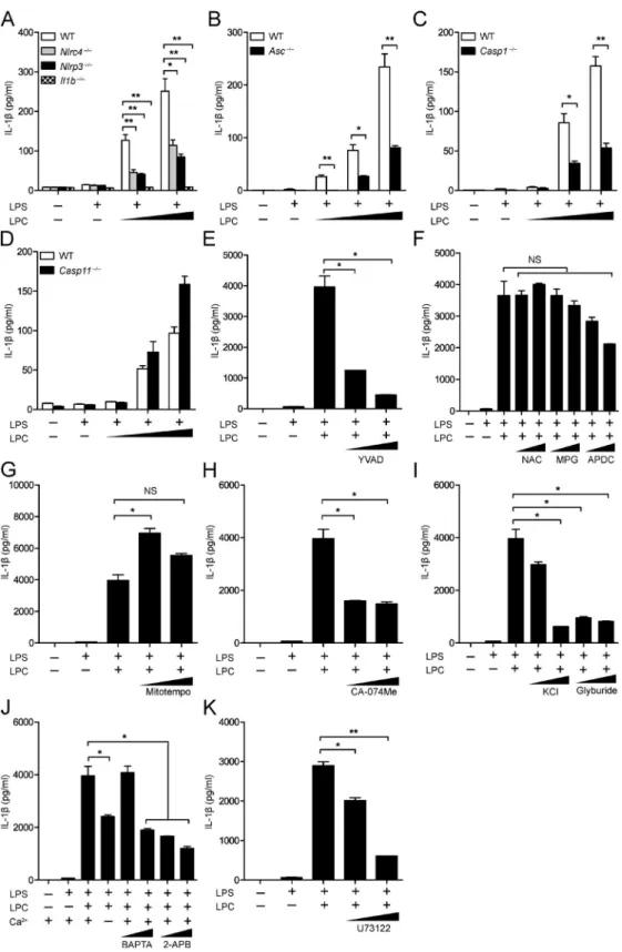
We assessed inflammasome activation by molecules associ-ated with CNS disease by analyzing LPC activity. To assess whether LPC could activate the inflammasome, we generated BMDMs from WT, Nlrp3−/−, Nlrc4−/−, Asc (apoptotic speck containing protein with a CARD)−/−, Casp1−/−, Il1b−/−, and Casp11−/− mice and primed these cells with LPS followed by LPC stimulation. Then, IL-1β release was quantified by ELI SA (Fig. 1, A–D) and immunoblotting (Fig. 2, A–D). In these experiments, cells were primed with LPS, which is a well-documented inducer of signal 1 of inflammasome ac-tivation, inducing the transcription and translation of in-flammasome components, such as pro–IL-1β, pro–caspase-1, and NLRP3. After LPS pretreatment, LPC was tested for signal 2 activity, which normally leads to caspase-1 and IL-1β maturation followed by secretion of IL-1β. A com-bination of LPS and LPC caused significant IL-1β release in WT BMDMs (Fig. 1, A–D; and Fig. 2, A–D). However, IL-1β secretion in Nlrp3−/−, Nlrc4−/−, Asc−/−, Casp1−/−, and Il1b−/− BMDMs was reduced compared with WT controls but not in Casp11−/− BMDMs (Fig. 1, A–D; and Fig. 2, A–D). These results indicate that inflammasome-associated proteins NLRC4, NLRP3, caspase-1, and ASC mediate LPC-induced IL-1β secretion in BMDMs, whereas the noncanonical in-flammasome-associated protein capase-11 is not involved. LPC-induced inflammasome activation was also inhibited by a caspase-1 inhibitor, YVAD (Z-Tyr-Val-Ala-Asp[OMe]-flu-oromethylketone; Fig. 1 E and Fig. 2 E).
To further explore how NLRP3 might mediate LPC-induced IL-1β secretion in BMDMs, we pretreated WT BMDMs with inhibitors of intracellular NLRP3 agonists be-fore LPC stimulation and measured IL-1β secretion by ELI SA (Fig. 1, F–K) and immunoblotting (Fig. 2, F–I). Inhibi-tors of reactive oxygen species (ROS; NAC [N-acetyl-l-cys-teine], MPG [N-(2-mercaptopropionyl)glycine], and APDC [(2R,4R)-4-aminopyrrolidine-2,4-dicarboxylic acid]) and mitochondrial ROS production (Mitotempo) did not signifi-cantly reduce LPC-induced inflammasome activation (Fig. 1,
F and G; and Fig. 2 F). Inhibitors of cathepsin B–mediated degradation (CA-074Me), K+ efflux (glyburide), and Ca2+
in-flux (BAP TA, 2-APB, and U73122) reduced LPC-induced IL-1β secretion in BMDMs (Fig. 1, H–K; and Fig. 2, G–I). This suggests that LPC induction of IL-1β secretion in BMDMs is dependent on cathepsin B, Ca2+ influx, and K+ efflux.
With the observation that LPC induces IL-1β secretion in WT BMDMs, we examined whether NLRP3 and NLRC4 are required for LPC-induced cell death. BMDMs were iso-lated from WT, inflammasome-associated gene–deficient mice, and noncanonical inflammasome-associated gene–defi-cient mice, LPS primed, and stimulated with increasing con-centrations of LPC. Supernatants were collected, and lactate dehydrogenase (LDH) was measured to assess the level of cell death (Fig. S1). Although there was a decrease in LDH levels in Nlrp3−/−, Nlrc4−/−, Asc−/−, and Casp1−/− BMDMs compared with WT BMDMs, the reduction was modest, suggesting that the inflammasome modestly impacted LPC-induced cell death. There was no difference in LDH levels in Casp11−/− BMDMs compared with WT BMDMs, excluding a role for the noncanonical inflammasome. LPC treatment of Il1b−/− and Il18−/− cells also showed no difference from controls.
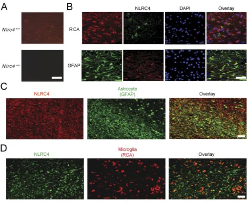
expression of nLrc4 is enhanced in the mouse cnS during neuroinflammation
The results in Figs. 1 and 2 revealed that NLRP3 and NLRC4 are important in LPC-mediated inflammasome ac-tivation. Although NLRP3 has been studied in several neu-rodegenerative conditions, the role of NLRC4 has not been extensively explored in the CNS. A previous study has shown that Nlrc4 RNA is expressed in mouse brain as well as in other tissues (Poyet et al., 2001). Because RNA expression does not always correlate with protein abundance, we assessed whether NLRC4 protein is found in the brain in the cu-prizone model during peak inflammatory cell accumulation, demyelination, and mature oligodendrocyte death (Hiremath et al., 1998; Arnett et al., 2001). Brains of C57BL/6 mice that had been treated with cuprizone for 4 wk were exam-ined for NLRC4 by immunohistochemistry (IHC) using an anti-NLRC4 antibody. As a specificity control, we showed that NLRC4 expression was detected in the brain of WT mice (Nlrc4+/+; 4-wk cuprizone treated) but not in
identi-cally treated Nlrc4−/− mice (Fig. 3 A). The protein was abun-dant in astrocytes (glial fibrillary acidic protein [GFAP]+)
and modestly abundant in microglia expressing the lectin Ricinus communis agglutinin 1 (RCA1+) at the corpus
cal-losum after 4 wk of cuprizone-induced demyelination in WT mice (Fig. 3 B). NLRC4 was not found at detectable levels by NeuN+ neurons and CNPase+ oligodendrocytes (Fig. S2).
nLrc4 is abundant in lesions and astrocyte-rich regions of human brain tissue from MS patients
Figure 1. the inflammasome mediates LPc-induced IL-1βsecretion in mouse BMdMs. (A–D) WT, Nlrc4−/−, Nlrp3−/−, Il1b−/−(A), Asc−/−(B), Casp1−/−(C),
and Casp11−/−(D) BMDMs were LPS primed (100 ng/ml) overnight before LPC stimulation at concentrations of 25, 50, and 100 µM for 1 h before IL-1β was
of California Los Angeles (UCLA) Human Brain and Spinal Fluid Resource Center (HBS FRC). The clinical synopsis of three controls and three MS samples used for this study is listed in Table 1. Three individuals were diagnosed with chronic ac-tive MS and exhibited demyelination, with two having gliosis. Normal control brain tissues were devoid of inflammatory lesions and did not display CNS neuropathology. The plaque location of samples taken from the MS brains is shown in Fig. S3. The three samples shown on the left were diagnosed with MS, whereas the samples on the right (labeled Normal) were not. NLRC4 expression was greatly elevated in the lesion associated with extensive astrogliosis as marked by GFAP as demonstrated in the overlay panel of anti-NLRC4 and anti- GFAP (Fig. 3 C). Microglial accumulation was also evident,
although fewer RCA+ microglia cells were detected
com-pared with astrocytes (compare Fig. 3, C and D, middle), and the overlay of anti-NLRC4 and anti-RCA staining showed few NLRC4+RCA+ cells compared with NLRC4+GFAP+
cells (Fig. 3, C and D, right). A summary of Image J (National Institutes of Health) quantification is shown in Tables 2 and 3. The MS samples showed greatly enhanced NLRC4 protein levels in plaque, white matter, and, to a lesser extent, gray matter, whereas normal controls did not express detectable NLRC4 protein. Astrocytes identified by GFAP staining ex-pressed the highest amount of NLRC4, whereas microglial cells identified by RCA staining also expressed the protein but to a lower extent. Neurons and oligodendrocytes did not express detectable levels. These results in human specimens
KCl (50 and 100 mM) and glyburide (100 and 200 µM; I); or the calcium inhibitors BAP TA (10 and 20 µM), 2-APB (20 and 100 µM; J), and U73122 (2 and 10 µM; K). Then, IL-1β was collected from supernatants for analysis by ELI SA. Each control and experimental condition was performed in triplicate. All data are representative of at least two independent experiments. Results are displayed as the mean ± SEM. *, P < 0.05; **, P < 0.01; unpaired Student's t test. Figure 2. the inflammasome mediates LPc-induced IL-1βmaturation in mouse BMdMs. (A–D) WT, Nlrp3−/−, Nlrc4−/−, and Il1b−/−(A), Asc−/−(B),
Casp1−/−(C), and Casp11−/−(D) BMDMs were LPS primed (100 ng/ml) overnight before LPC stimulation (25, 50, and 100 µM). Pro–IL-1β and IL-1β in the
agree with the findings observed in mice in the previous paragraph (Fig. 3, A and B; and Fig. S2).
Astrocytic nLrc4 and nLrP3 display the same specificity as their macrophage counterparts
The findings in the previous paragraph led us to compare LPC-induced inflammasome activation in macrophages and astrocytes. Astrocytes were isolated from day-0–2 postnatal pups, and purity was verified by immunocytochemistry (ICC) using the astrocytic marker GFAP for detection of desired as-trocytes versus the microglial marker Iba-1 for contaminating microglia (Fig. 4 A). In BMDMs, NLRC4 is known to respond to PAMPs such as bacterial flagellin and T3SS (Ma et al., 2000; Franchi et al., 2006, 2010; Miao et al., 2006, 2010), whereas NLRP3 reacts to a plethora of activators, including ATP (Mariathasan et al., 2006; Duncan et al., 2007). First, we con-firmed responses of Nlrc4−/− BMDMs to PAMP stimulation, finding that WT and Nlrc4−/− BMDMs produced similar levels of IL-1β in response to the known NLRP3 stimuli LPS + ATP, which should not be affected by the absence of NLRC4. As expected, IL-1β response to LPS and flagellin was ablated in the Nlrc4−/− BMDMs (Fig. 4 B). Likewise, Nlrp3−/− and WT BMDMs showed no statistical difference in IL-1β production in response to LPS and flagellin, but response to LPS + ATP was ablated in the absence of NLRP3 (Fig. 4 C). This confirmed the specificity of the NLRC4 and NLRP3 inflammasomes in BMDMs. Next, we assessed whether astrocytic NLRC4 dis-played the same specificity as BMDM NLRC4 using primary astrocytes isolated from Nlrc4−/− and WT newborn mice (Wu et al., 2010). Once astrocyte purity was confirmed, astrocytes
were pretreated with LPS and transfected with S. typhimurium flagellin, and IL-1β release was measured by ELI SA. Upon transfection of WT astrocytes with increasing concentrations of flagellin, IL-1β release increased in a dose-dependent fashion. In contrast, Nlrc4−/− astrocytes showed little or no increase in IL-1β release when transfected with increasing concentrations of flagellin (Fig. 4 D). As observed with Nlrp3−/− BMDMs (Fig. 4 C), when Nlrp3−/− astrocytes were transfected with in-creasing amounts of flagellin, there was no difference in IL-1β release compared with WT astrocytes (Fig. 4 E). Again, response to LPS + ATP was ablated in Nlrp3−/− but not in Nlrc4−/− as-trocytes (Fig. 4, D and E). These results show that, similar to BMDMs, NLRC4 in astrocytes is required for inflammasome activation by flagellin, whereas NLRP3 in astrocytes is required for the LPS + ATP response.
Both nLrc4 and nLrP3 are required for LPc-induced IL-1β
secretion from primary mouse astrocytes
To extend the previous findings of LPC activating the inflam-masome in BMDMs, we next examined whether LPC can also activate astrocyte-derived inflammasome activity. To establish inflammasome activation by LPC in astrocytes, we investi-gated whether LPC stimulation would result in the release of IL-1β in WT mouse astrocytes. A dose-dependent increase of released IL-1β resulted in WT astrocytes treated with in-creasing concentrations of LPC. Furthermore, when WT as-trocytes were stimulated with LPC for 4 h as compared with 2 h, the amount of IL-1β released was enhanced (Fig. 5 A). This suggests that LPC acted to stimulate IL-1β release in astrocytes in a concentration- and time-dependent manner.
Figure 3. nLrc4 is expressed in mouse cnS during neuroinflammation and in
human brain tissue from MS patients. (A)
WT mice brains were examined for NLRC4 ex-pression by IHC using a mouse NLRC4 antibody. NLRC4 expression (red) was detected after 4 wk of cuprizone-induced demyelination in WT (Nlrc4+/+) but not in Nlrc4−/−mice. (B) The
Next, we investigated whether NLRC4 mediated LPC stimulation of inflammasome activation. Upon 4 h of LPC stimulation, the IL-1β released from Nlrc4−/− astrocytes was attenuated compared with WT astrocytes (Fig. 5 B). Addition-ally, Nlrp3−/− astrocytes also showed reduced IL-1β release in response to LPC (Fig. 5 C). The adapter protein of NLRP3 and NLRC4 inflammasome ASC was also required for LPC-in-duced IL-1β production in mouse astrocytes (Fig. 5 D). These data agree with the data generated in BMDMs. This indi-cates that, in contrast to the flagellin-activated inflammasome, which only requires NLRC4, and the ATP-induced masome, which only requires NLRP3, LPC-induced inflam-masome requires both NLRC4 and NLRP3.
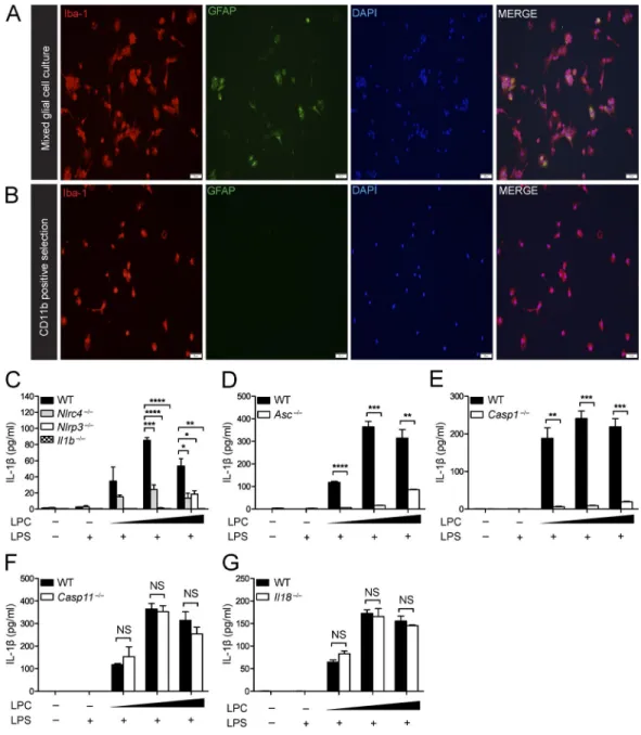
the inflammasome mediates LPc-induced IL-1β secretion in microglia
The NLRP3 inflammasome has been suggested to mediate IL-1β secretion in microglia in neurodegenerative diseases such as Alzheimer’s disease and Parkinson’s disease and in response to neuropathology-associated markers such as α-synuclein and β-amyloid (Rock et al., 2010; Codolo et al., 2013; Freeman and Ting, 2016). Next, we examined whether LPC induces IL-1β release in microglia and whether this process is dependent on the inflammasome. Microglia were isolated using CD11b-pos-itive selection. ICC (Fig. 6, A and B) was used to assess the separation of microglia (Iba-1+ cells) from GFAP+ astrocytes
before and after CD11b selection. Microglia were isolated from brains of WT, Nlrc4−/−, Nlrp3−/−, Il1b−/− (Fig. 6 C), Asc−/− (Fig. 6 D), Casp1−/− (Fig. 6 E), Casp11−/− (Fig. 6 F), and Il18−/−
(Fig. 6 G) neonatal mice (postnatal day 3–5). Once microglial purity was confirmed, microglial cells were LPS primed and stimulated with LPC at increasing concentrations (20, 25, and 50 µM). As expected, there was an increase in IL-1β secretion in response to LPC stimulation of WT microglia, but Nlrc4−/−, Nlrp3−/−, Il1b−/−, Asc−/−, and Casp1−/− microglia showed sig-nificantly attenuated IL-1β secretion compared with WT mi-croglia. Casp11−/− and Il18−/− samples did not differ from WT controls. These results suggest that NLRC4, NLRP3, caspase-1, and the adapter protein ASC play a critical role in mediating LPC-induced IL-1β secretion in microglia, whereas IL-18 and caspase-11 are not involved.
To determine whether the inflammasome mediates py-roptosis in microglia upon LPC treatment, we isolated mi-croglia from WT and inflammasome-deficient mice. These cells were LPS primed and stimulated with LPC, and su-pernatants were collected to measure LDH levels (Fig. S4). Although LDH levels were detectable in both WT and inflammasome-deficient microglia, there was no signifi-cant difference among any of the canonical or noncanon-ical inflammasome-deficient microglia (Nlrc4−/−, Nlrp3−/−, Asc−/−, Casp1−/−, Il1b−/−, Il18−/−, Casp1−/−, and Casp11−/−) compared with WT microglia, suggesting that the inflam-masome and noncanonical inflaminflam-masome do not enhance LPC-induced microglial death.
the inflammasome promotes microglial accumulation and astrogliosis
The in vitro system indicates that NLRC4 and NLRP3 in astrocytes and microglia mediate inflammasome activation by
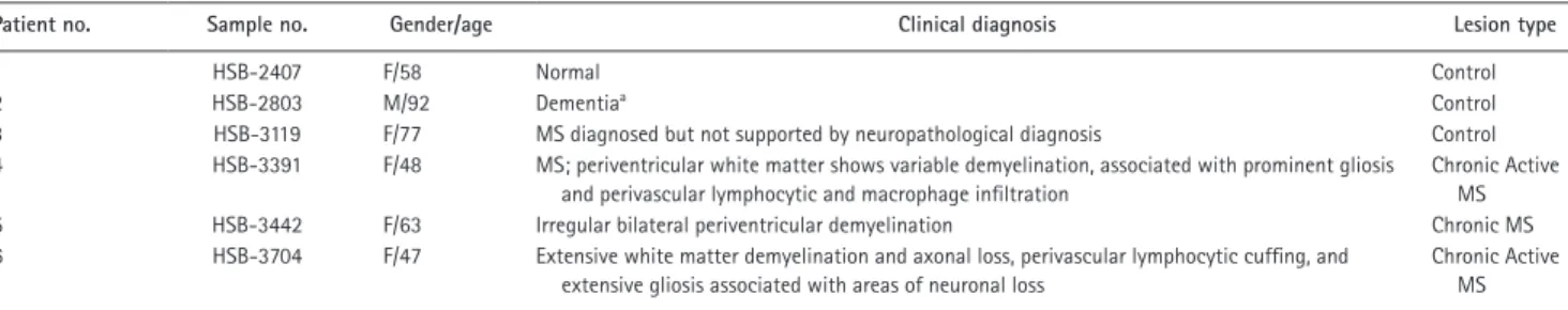
Table 1. MS patients: clinical synopsis
Patient no. Sample no. Gender/age Clinical diagnosis Lesion type
1 HSB-2407 F/58 Normal Control
2 HSB-2803 M/92 Dementiaa Control
3 HSB-3119 F/77 MS diagnosed but not supported by neuropathological diagnosis Control 4 HSB-3391 F/48 MS; periventricular white matter shows variable demyelination, associated with prominent gliosis
and perivascular lymphocytic and macrophage infiltration
Chronic Active MS 5 HSB-3442 F/63 Irregular bilateral periventricular demyelination Chronic MS 6 HSB-3704 F/47 Extensive white matter demyelination and axonal loss, perivascular lymphocytic cuffing, and
extensive gliosis associated with areas of neuronal loss
Chronic Active MS
The clinical pathology of MS and control brain tissues obtained from the UCLA HBS FRC is described. Each brain was evaluated by neuropathologists at UCLA. Brain tissue from three MS cases was examined along with three control brains from cases without MS. In all cases, the tissue was taken from the region around or at the corpus callosum, as shown in Fig. S2.
aDementia with no distinctive pathology.
Table 2. distribution of reactivity for nLrc4 in MS lesions
Ab MS brain Control brain
Plaque Adjacent
white matter Adjacent gray matter NAWM NAGM
NLRC4 39.585 28.714 1.511 0 0
Immunofluorescence indicating NLRC4 presence in tissue sections was quantified by National Institutes of Health ImageJ software (Schneider et al., 2012). The images were unstacked. Each image was split into channels, the threshold was set, and then the signal was measured. The numbers represent mean values from the output results. Abbreviations used: Ab, antibody; NAGM, normal-appearing gray matter; NAWM, normal-appearing white matter.
Table 3. cellular distribution of reactivity for nLrc4
Ab Cellular distribution
Astrocytes
(GFAP) Macrophages (RCA)Microglia/ Oligodendrocytes (O4) Neurons (NeuN)
NLRC4 23.085 12.1 0 0
LPC. In parallel with the in vitro studies, we also assessed whether NLRP3 and NLRC4 play a role in the cuprizone model of neuroinflammation. This model consists of feeding mice with chow supplemented with the neurotoxicant cupri-zone (0.2%), which results in a CNS disease model comprised of astrogliosis and microglial activation followed by oligoden-drocyte cell death and demyelination.
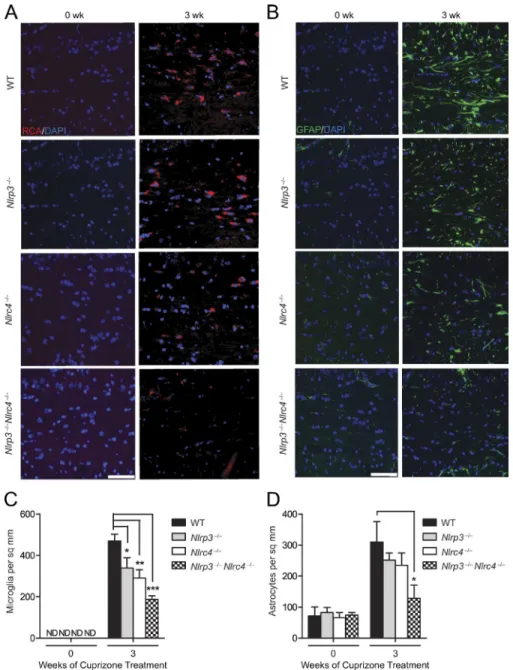
To explore whether the NLRC4 and NLRP3 in-flammasomes have a role during cuprizone-induced neu-roinflammation, we examined microglial accumulation in WT (control), Nlrc4−/−, Nlrp3−/−, and double KO (DKO) Nlrp3−/−Nlrc4−/− mice (Fig. 7 A). Microglial cells are res-ident immune cells of the CNS that release cytokines and chemokines and also phagocytose dead cells, cellular debris, and invading pathogens (Hanisch and Kettenmann, 2007;
Napoli and Neumann, 2009). The microglia populations at the corpus callosum were quantified by RCA-1 lectin staining (Fig. 7 C). Among the untreated groups (indi-cated as 0 wk), age-matched WT, Nlrc4−/−, Nlrp3−/−, and Nlrp3−/−Nlrc4−/− mice showed no difference in numbers of microglia at the corpus callosum, indicating that dele-tion of Nlrc4 and/or Nlrp3 did not affect microglial ac-cumulation in nondiseased mice at steady state (Fig. 7 C). After 3-wk cuprizone treatment, there was a significant re-duction in microglial accumulation in Nlrc4−/−, Nlrp3−/−, and Nlrp3−/−Nlrc4−/− mice compared with WT controls (Fig. 7 C), with the DKO showing the most significant re-duction. These results indicate that NLRC4 and NLRP3 contribute to microglia accumulation at the corpus callo-sum during demyelination.
Figure 4. Astrocytic nLrc4 and nLrP3 display the same inflammasome
specific-ity as macrophage nLrs. (A)
Immunocy-tochemical detection of GFAP (green)- but not Iba-1(red)–stained cells from a purified mouse astrocyte culture used in Fig. 4 (D and E). DAPI (blue) was used to label nuclei. Representative images from the experiment performed in triplicate are shown. Bar,16 µM. (B) WT and Nlrc4−/−BMDMs were LPS primed
(400 ng/ml) for 4 h and transfected with the transfecting reagent DOT AP and 25, 50, or 100 ng of S. typhimurium flagellin for 1 h before IL-1β was collected from supernatant. WT and Nlrc4−/− LPS-primed BMDMs were
also stimulated with 5 mM ATP, as a positive control for inflammasome activation. Cells were transfected with DOT AP alone or stim-ulated with flagellin alone without LPS as a negative control. Data are representative of two independent experiments. (C) WT and Nlrp3−/−BMDMs were similarly treated as in
B. Data are representative of two indepen-dent experiments. (D) WT and Nlrc4−/−primary
mouse astrocytes were LPS primed (400 ng/ml) for 4 h and transfected with 50, 100, or 200 ng flagellin for 1 h before IL-1β was collected from supernatant. LPS-primed primary mouse WT and Nlrc4−/−astrocytes were also
stimu-lated with 5 mM ATP, as a positive control, and transfected with DOT AP alone or stimulated with flagellin alone as a negative control. Data are representative of three independent ex-periments. (E) WT and Nlrp3−/−primary mouse
Next, we examined astrogliosis in WT, Nlrc4−/−, Nlrp3−/−, and Nlrp3−/−Nlrc4−/− mice (Fig. 7 B). The astro-cyte population at the corpus callosum was studied using GFAP staining as a marker of astrocyte activation. Among the control untreated groups (0 wk), age-matched WT, Nlrc4−/−, Nlrp3−/−, and Nlrp3−/−Nlrc4−/− mice showed no difference in the number of astrocytes at the corpus callosum (Fig. 7 D), indicating that the basal level of astrocytes is unaffected by deletion of Nlrc4 and/or Nlrp3. After 3 wk of cuprizone treatment, there was a reduction of astrocytes in Nlrc4−/− and Nlrp3−/− compared with WT mice, although the difference did not reach significance. In contrast, there was a signifi-cant reduction of astrocytes in Nlrp3−/−Nlrc4−/− DKO mice (Fig. 7 D). These results indicate that NLRC4 and NLRP3 exacerbate astrogliosis in the CNS during demyelination.
the inflammasome promotes demyelination in mice
In the cuprizone model, overt loss of myelination follows astro-gliosis and microglial accumulation (Matsushima and Morell, 2001). To assess whether NLRC4 and NLRP3 play a role in demyelination, WT, Nlrc4−/−, Nlrp3−/−, and Nlrp3−/−Nlrc4−/− mice were treated with cuprizone. Representative scoring of the extent of demyelination was measured by Luxol Fast blue (LFB) staining for myelin (Fig. 8 A). Slides were read by a reader blinded to the identity of tissues using a scale of 0 (no demyelination) to 3 (complete demyelination), and demye-lination was quantified (Fig. 8 B). Cuprizone-treated (3 wk) WT, Nlrc4−/−, Nlrp3−/−, and Nlrp3−/−Nlrc4−/− mice showed increased demyelination at the corpus callosum (denoted by the purple, bracketed area; Fig. 8 A) compared with untreated (0 wk) WT, Nlrc4−/−, Nlrp3−/−, and Nlrp3−/−Nlrc4−/− mice.
3-wk cuprizone–treated Nlrc4−/− and Nlrp3−/− mice showed demyelination scores lower than WT mice, although the dif-ference did not reach significance. In contrast, there was a significant reduction of demyelination in Nlrp3−/−Nlrc4−/− mice. This suggests that, in addition to promoting microglial accumulation and astrogliosis, NLRC4 and NLRP3 cooper-ate to enhance demyelination.
the inflammasome-intact mice have elevated G2A expression
The results thus far indicate that LPC has a proinflamma-tory effect on microglia, astrocytes, and BMDMs and that the inflammasome mediates LPC-induced IL-1β secretion in all three cell types (Fig. 1, Fig. 5, and Fig. 6, C–G). The re-sults also demonstrate that the NLRP3 and NLRC4 inflam-masomes exacerbate microglial accumulation and astrogliosis in vivo. Directly assessing LPC in the brain during cuprizone treatment proved to be technically difficult, with inconsistent results obtained. Alternately, we examined the expression of an LPC receptor, namely G2A, a G-protein–coupled receptor (GPR132) that is known to trigger release of cyclic adenosine monophosphate and intracellular Ca2+ to promote
apopto-sis and chemotaxis of macrophages, monocytes, and microg-lia upon binding to LPC (Rikitake et al., 2002; Lin and Ye, 2003). To determine whether G2A expression was detectable during the course of neuroinflammation caused by cupri-zone treatment, we examined brains from mice subjected to 4-wk cuprizone treatment. G2A expression was confirmed in cuprizone-treated WT brains (Fig. 9 A). Brain sections from WT, Nlrc4−/−, Nlrp3−/−, and Nlrp3−/−Nlrc4−/− DKO mice were stained for the presence of G2A (denoted by black arrows)
Figure 5. nLrc4 and nLrP3 mediate LPc-induced
IL-1β secretion from primary mouse astrocytes. (A) WT
astrocytes were LPS primed (1 µg/ml) for 3 h and incubated with 50 and 100 µM LPC for 2 or 4 h, as indicated, before IL-1β was collected from the supernatant for analysis by ELI SA. Data are representative of three independent experiments. (B) WT and Nlrc4−/−astrocytes were treated as described in
A and assayed for IL-1β secretion at 4 h. Data are represen-tative of three independent experiments. (C and D) WT and Nlrp3−/−astrocytes (C) or WT and Asc−/−astrocytes (D) were
with hematoxylin used as a counter stain (Fig. 9 B). Quanti-fication of G2A staining in untreated and cuprizone-treated brains was performed (Fig. 9 C). Untreated (0 wk) brains from WT, Nlrc4−/−, Nlrp3−/−, and Nlrp3−/−Nlrc4−/− mice showed low levels of G2A expression, with no statistical dif-ferences among these genotypes. Brains isolated from cupri-zone-treated Nlrp3−/− and Nlrc4−/− mice showed a reduction in G2A+ cells, but the difference did not reach significance
when compared with cuprizone-treated WT brains, whereas
G2A in the brains of cuprizone-treated Nlrp3−/−Nlrc4−/− DKO mice showed a significant reduction. These data suggest that both NLRP3 and NLRC4 are required for elevated G2A expression under a neuroinflammatory state.
As the cuprizone model represents a mouse model of neuroinflammation and demyelination, we assessed whether G2A expression was elevated in human MS tissue. The pres-ence of G2A in brain tissues from MS patients and patients without MS was analyzed (Fig. 9 D). In normal brain tissue,
Figure 6. the inflammasome mediates LPc-induced IL-1βsecretion in microglia. (A and B) ICC of Iba-1 and GFAP staining of mixed glial cell cultures (A) and CD11b-positive selected cells (B) isolated from postnatal day-3–5 WT mouse pups indicates the degree of microglial purity. Bars, 50 µM. (C–G) WT, Nlrc4−/−, Nlrp3−/−, Il1b−/−(C), Asc−/−(D), Casp-1−/−(E), Casp-11−/−(F), and Il18−/−(G) microglia were LPS primed (100 ng/ml) for 3 h and stimulated with
G2A expression was very low to barely detectable, whereas G2A expression was abundant in brain tissue from MS patients. These data suggest that G2A is elevated in neuroinflamma-tion-associated conditions in both human and mouse tissues.
dIScuSSIon
There is increasing evidence that inflammatory processes in the CNS play a large part in neurological disorders. As an understanding of how DAMPs activate neuroinflam-mation is fundamental to our understanding of CNS dis-ease etiology, this work focuses on LPC, which has been associated with neurodegenerative and demyelination dis-eases. The results show that LPC serves as a DAMP that induces an atypical pattern of inflammasome activation that is dependent on both NLRP3 and NLRC4, as well as
ASC and caspase-1. Conventionally, NLRP3 and NLRC4 mediate inflammasome complexes of distinct and nonover-lapping specificities (Strowig et al., 2012; Broderick et al., 2015; Guo et al., 2015; Broz and Dixit, 2016). Recent stud-ies with Salmonella infection indicate that both NLRP3 and NLRC4 are involved in inflammasome activation by this bacteria, which actually represents a complex patho-gen, unlike LPC (Qu et al., 2016). In addition to the in vitro study here, analysis of an animal model of neuroin-flammation and demyelination shows that both NLRP3 and NLRC4 exacerbate disease outcome, thus providing an in vivo correlate for in vitro characterization. Finally, analysis of purified astrocyte and microglial preparations show that both NLRP3 and NLRC4 are required for inflammasome activation by LPC in these cells.
Figure 7. nLrP3 and nLrc4 promote
neu-roinflammation. (A) Representative images of
mouse brain sections stained for the microg-lial marker, RCA. Samples were obtained from cuprizone-treated WT, Nlrc4−/−, Nlrp3−/−, and
Nlrc4−/−Nlrp3−/− mice. RCA+ (red) microglial
Although NLRP3 has been extensively studied, NLRC4 is primarily studied in the context of microbial infection. However, Nlrc4 mRNA is detected in the adult mouse brain (Poyet et al., 2001), is elevated in brain tissues from Alzhei-mer’s patients, and is shown to mediate palmitate-induced in-flammasome activation in astrocytic cultures (Liu and Chan, 2014). Additionally, the absence of NLRC4 reduced brain in-jury, although this process appears to be independent of IL-1
(Denes et al., 2015). Heightened expression of NLRC4 has been associated with inflammatory diseases such as Kawasaki disease (Ikeda et al., 2010) and atopic dermatitis (Macaluso et al., 2007). Using peripheral blood mononuclear cells from pa-tients, these authors demonstrated increased gene expression of NLRC4 in patients relative to healthy controls, leading the authors to suggest a role for NLRC4 in these inflammatory diseases, although the direct relevance of elevated NLRC4 in these diseases is undefined. A direct link between NLRC4 and inflammation has been determined for recurrent macro-phage activation syndrome, where this disorder was associated with an activating NLRC4 mutation (Canna et al., 2014).
Our previous study with Nlrp3−/−, Casp1−/−, Il1β−/−, and Il18−/− mice in the cuprizone model showed a NL-RP3-dependent mechanism, leading to demyelination and loss of mature oligodendrocytes (Jha et al., 2010). Moreover, our results in Nlrp3−/− mice showed that NLRP3 deficiency only partially delayed demyelination and neuroinflammation, uncovering the possible involvement of redundant NLR pro-teins during neuroinflammation and demyelination. This pre-vious work did not analyze the cell types that are involved in inflammasome activation.
The current work provides evidence that NLRC4 is strongly expressed in astrocytes and to a lesser extent in microglia during neuroinflammation. Expression of glial NLRC4 was enhanced in the mouse brain during chronic stages of neuroinflammation in the cuprizone model and in pathological brain sections from MS patients. As the role of the inflammasome in astrocytes is not well understood, the importance of this study lies in the functional delineation of LPC-induced NLRC4 and NLRP3 inflammasome activation in astrocytes. We also show that the canonical inflammasome
components NLRP3, NLRC4, ASC, caspase-1, and IL-1β
mediate LPC-induced IL-1β secretion in BMDMs and mi-croglia, whereas caspase-11, which represents the noncanoni-cal inflammasome pathway, is not required. These data suggest that inflammasome activation by LPC in macrophages and these two glial populations occurs via indistinguishable path-ways. The importance of NLRC4 and NLRP3 in the CNS is further supported by findings in the cuprizone model, where mice lacking individual NLR (Nlrc4−/− or Nlrp3−/−) exhib-ited modestly reduced astrogliosis, microglial accumulation, and expression of the LPC cognate receptor G2A, whereas Nlrp3−/−Nlrc4−/− DKO mice showed the most significant reduction when compared with WT mice. This suggests that NLRP3 and NLRC4 may function in a synergistic or coop-erative fashion during neuroinflammation.
This study also finds that astrocytes express a relatively high level of NLRC4. Astrocytes, the most abundant cell type in the CNS, are increasingly appreciated for their broad reg-ulatory impact on CNS homeostasis and function. Astrocytes perform multiple and diverse roles in the CNS, including the maintenance of extracellular ionic balance, promotion of neuronal survival, formation of synapses, and maintenance of the blood–brain barrier (Molofsky et al., 2012). They also
Figure 8. the inflammasome promotes demyelination. (A) Repre-sentative images of brain sections of untreated (0 wk) and 3-wk cupri-zone–treated WT, Nlrc4−/−, Nlrp3−/−, and Nlrc4−/−Nlrp3−/−mice stained
for the presence of myelin at the corpus callosum (purple dashed lines) with LFB (blue). Bar, 50 µM. (B) Quantification of LFB-stained WT, Nlrc4−/−,
Nlrp3−/−, and Nlrc4−/−Nlrp3−/−mice brain sections. Each circle represents
the mean observed LFB score for one mouse. WT, Nlrc4−/−, Nlrp3−/−, and
Nlrc4−/−Nlrp3−/−mice were scored by a blinded reader on a scale of 0 (no
provide nutrients such as glucose to neurons and play a key role in the repair and scarring process in the brain. Activated astrocytes are a major contributor to neuroinflammation and are important for synapse formation. With respect to the inflammasome genes, astrocytes express the MHC class II transactivator (CII TA; Collawn and Benveniste, 1999; Stüve et al., 2002), NOD1 and NOD2, and the inflammasome adapter protein ASC (de Rivero Vaccari et al., 2008). How-ever, inflammasome activity in astrocytes has not been widely studied, so questions of NLRC4 mediating inflammasome functions in astrocytes remain unanswered.
It is commonly understood in the field that different in-flammasome NLRs display distinct specificities for agonists/
ligands. However, we initially proposed the model wherein NLRs may be able to mix and match, thus expanding the repertoire of PAMP recognition (Ting and Davis, 2005). In-deed, a few pairs of NLRs have been found to display com-mon specificity. For example, the NAIP2–NLRC4 complex physically associates with a T3SS molecule, whereas NAIP5– NLRC4 associates with flagellin (Kofoed and Vance, 2011; Zhao et al., 2011; Halff et al., 2012). Although NLRP3 and NLRC4 display distinct specificities, they are both required for inflammasome activation by Salmonella (Broz et al., 2010), and endogenous NLRP3 and NLRC4 have been colocalized in cells upon Salmonella infection (Man et al., 2014). More recently, analysis of a mutant NLRC4 unexpectedly revealed
Figure 9. the inflammasome elevates
expres-sion of the LPc receptor G2A. (A) Brain
sec-tions from 4-wk cuprizone–treated WT mice were stained for the presence of G2A at the corpus callosum of the brain, as indicated by black ar-rowheads, with a hematoxylin counter stain. (Left) The negative control (no G2A antibody was used), which shows no brown staining. (Right) G2A-pos-itive cells (brown staining as indicated by black ar-rowheads). Bar, 20 µm. (B) Representative images of 0-wk and 4-wk cuprizone–treated brains from WT, Nlrc4−/−, Nlrp3−/−, and Nlrc4−/−Nlrp3−/−mice
are shown. G2A-positive cells (as indicated by black arrowheads) are detected within the corpus callosum. Bar, 20 µM. (C) G2A-positive cells within the corpus callosum of cuprizone-treated brains of WT, Nlrc4−/−, Nlrp3−/−, and Nlrc4−/−Nlrp3−/−mice
that this mutant protein can still recruit NLRP3, which then engages ASC to activate the inflammasome upon infection with S. typhimurium (Qu et al., 2016). It will be interesting to examine whether this mutant NLRC4 exhibits intact in-flammasome response to LPC.
In summary, this work provides direct evidence for the functional importance of NLRC4 and NLRP3 in astrocytic and microglial inflammasome activation and, furthermore, shows that NLRC4 and NLRP3 mediate inflammasome ac-tivity by a CNS-associated DAMP, namely LPC. Considering the expanding importance of astrocytes and microglia in neu-rological homeostasis and disease states, these findings have broad implications for understanding a plethora of neurolog-ical disorders where neuroinflammation is evident.
MAterIALS And MethodS Mice
Nlrc4−/− mice on the C57BL/6 background were provided by V. Dixit (Genentech, San Francisco, CA). Nlrp3−/− mice backcrossed on the C57BL/6 background have been previ-ously described (Jha et al., 2010). C57BL/6 mice were pur-chased from The Jackson Laboratory and were maintained at the University of North Carolina facility. Nlrp3−/− mice were crossed with Nlrc4−/− mice to generate Nlrp3−/−Nlrc4−/− mice. Asc−/−, Casp1−/− (Casp11Tg), and Casp11−/− mice were originally provided by V. Dixit. Il1b−/− mice, originally pro-vided by D. Chaplin (University of Alabama at Birmingham, Birmingham, AL), and Il18−/− mice (The Jackson Laboratory) were also used. All mice were 8–10 wk old before the start of treatment. All mice were allowed to acclimate to the facility for 1–2 wk before cuprizone treatment. C57BL/6, Nlrc4−/−, Nlrp3−/−, and Nlrp3−/−Nlrc4−/− mouse lines were bred and maintained in house. All experiments with animals were con-ducted under protocols approved by the Institutional Animal Care and Use Committee at University of North Carolina Chapel Hill and the National Institutes of Health Guidelines for Animal Care and Use. All animals were housed under spe-cific pathogen–free conditions in sterile isolated caging in Association for Assessment and Accreditation of Laboratory Animal Care–accredited facilities.
cuprizone treatment
8–10-wk-old male mice were fed 0.2% (wt/wt) cuprizone (oxalic bis [cyclohexylidenehydrazide]; C9012; Sigma-Al-drich) mixed into ground chow ad libitum for 3–4 wk to induce progressive demyelination (Arnett et al., 2002; Franco- Pons et al., 2007). Untreated control mice were maintained on a diet of normal pellet chow.
tissue preparation
Mice were anesthetized and perfused with PBS followed by 4% paraformaldehyde (PFA). Brains were removed, postfixed in PFA, and embedded in paraffin. 5-µm coronal sections were cut at the fornix region of the corpus callosum. For frozen sections, mice were perfused and postfixed as described
previously (Arnett et al., 2001). Brains were allowed to soak in 30% sucrose in PBS and snap frozen on dry ice in optimal cutting temperature medium. 5- and 20-µm coronal sections were cut at the fornix region of the corpus callosum for anal-ysis by IHC. All analyses were restricted to the midline corpus callosum as described previously (Plant et al., 2007).
human brain tissue samples
The MS and control tissues were obtained from the UCLA HBS FRC with written informed consent from all donors, and the use of human tissues was reviewed via expedited re-view procedures (45 CFR 46. 110 b [2]) and approved by the UCLA Institutional Review Board. The clinical pathol-ogy of patients is listed in Table 1, with material evaluated by neuropathologists at the UCLA HBS FRC. Brain tissues from three MS cases were examined along with three con-trol brains from cases without MS. Among MS brains, three different samples were obtained from each brain including normal-appearing white matter, normal-appearing gray mat-ter, and plaque. Considering the control brains, two samples were obtained from each brain including normal-appearing white matter and normal-appearing gray matter. In all cases, the tissues were taken from regions around or at the corpus callosum (Fig. S3). The brain samples were obtained in 4% PFA and subsequently embedded in paraffin and sectioned into 5-µm coronal sections.
histological staining
To examine demyelination, paraffin sections were rehydrated through a graded series of alcohol washes and stained with LFB (S3382; Sigma-Aldrich) as described previously (Arnett et al., 2002). Sections were read by a double-blinded reader and graded on a scale from 0 (no demyelination) to 3 (com-plete demyelination), with higher scores indicating increased pathology. For detection of microglia/macrophages, sections were rehydrated and permeabilized with 0.1% Triton/PBS for 20 min at room temperature (RT) and then incubated with RCA-1 lectin (1:500; L-1080; Vector Laboratories) at 37°C for 1 h. Only RCA-1+ cells with observable DAPI-stained
nuclei were included in the quantification.
Ihc
anti-body was detected by incubation with goat anti–rabbit Alexa Fluor 488–conjugated antibody (A11034; Molecular Probes/ Thermo Fisher Scientific) for 1 h at RT. Immunopositive cells with an observable DAPI-stained nucleus were counted twice with sample identities covered from reviewers. Cell counts were averaged for four to seven mice per time point.
G2A staining was performed on paraffin specimens using G2A (M-20) antibody (goat polyclonal; sc-9692; lot 2315; Santa Cruz Biotechnology, Inc.). Antigen retrieval was performed with EDTA-based buffer (950-500; Discovery CC1), pH 8.5, for 64 min at 100◦C and then blocked with
Rodent Decloaker (10X; RD913L; Biocare Medical) for 1 h at RT. The samples were given a hydrogen peroxidase block for 8 min at RT and then incubated in the primary antibody (1:50 dilution) for 2 h at RT in a Tris-based di-luent (760-212; Discovery PSS Didi-luent), followed by the secondary antibody (polyclonal rabbit anti–goat immuno-globulin/HRP; P0160; Dako) incubation for 1 h at RT. The samples were treated with diaminobenzidine and Hematox-ylin II (790-2208; Ventana) for 12 min, followed by Bluing Reagent (760-2037; Ventana) for 4 min. The slide staining was performed using a Discovery Ultra Automated IHC staining system (Ventana).
Icc
ICC was performed on glial cells plated on polylysine-coated coverslips. To detect the presence of astrocytes, coverslips were permeabilized with Triton X-100 for 20 min and washed with PBS containing 0.05% Tween 20 for 20 min. Covers-lips were blocked with 5% normal donkey serum (D9963; Sigma-Aldrich) in Triton X-100 for 30 min. Coverslips were incubated overnight with chicken primary GFAP antibody (ab1334436; Abcam) in 5% normal donkey serum (0.5:1,000) in Triton X-100 overnight. Coverslips were washed four times with PBS containing 0.05% Tween 20 and blocked with 5% normal donkey serum for 30 min. Coverslips were incubated with a donkey anti–chicken Alexa Fluor 488 anti-body (703-545-155; Jackson ImmunoResearch Laboratories, Inc.) at 1:500 for 1 h at RT.
To detect the presence of microglia, coverslips were per-meabilized with Triton X-100 for 20 min and washed with PBS containing 0.05% Tween 20 for 20 min. Coverslips were blocked with 5% normal goat serum (S-1000; Vector Labora-tories) in Triton X-100 for 30 min. Coverslips were incubated overnight with rabbit primary Iba-1 antibody (ab178847; Abcam) in 5% normal goat serum (1:100) in Triton X-100 overnight. Coverslips were washed four times with PBS con-taining 0.05% Tween 20 and blocked with 5% normal goat serum for 30 min. Coverslips were incubated with a bioti-nylated goat anti–rabbit antibody (BA-1000; Vector Labora-tories) in 5% normal goat serum at 1:100 for 1 h at RT. Then, coverslips were incubated with an avidin-conjugated Alexa Fluor 594 antibody (S32356; Invitrogen) at 1:500 for 1 h at RT. Coverslips were mounted with DAPI media (H-1200; Vector Laboratories) to detect the presence of nuclei.
Imaging
For imaging of IHC, microglial and astrocytic cell counts were taken from the midline of the corpus callosum, confined to images of a 0.033-mm2 area taken with a 50× oil
immer-sion objective, whereas LFB-stained images of the corpus cal-losum were taken at a magnification of 20. For ICC, images of microglia and astrocytes were taken at 20×. An upright mi-croscope (BX-40; Olympus) with a camera (Optronics) and Scion image acquisition software were used for taking images of the mouse brains at RT. The scale bar was added by Scion image acquisition software. An upright microscope (BX-61; Olympus) with bright-field, dark-field, differential interfer-ence contrast, and epifluorescinterfer-ence capability with Velocity software (PerkinElmer), an ORCA RC camera (Hamamatsu Photonics), and a color camera (RET IGA 4000R; QImag-ing) were used for imaging human brain tissue at RT. Images of a ruler were also taken at 50× by an upright microscope (BX-40) or at 20× by an upright microscope (BX-61), and the scale bars for Fig. 3 and Fig. S2 were generated based on ruler images. Immunofluorescence in tissue sections was quantified by the National Institutes of Health ImageJ soft-ware (Schneider et al., 2012).
For imaging of G2A-stained sections, G2A+ cell counts
were taken from the midline of the corpus callosum and taken with a 100× oil immersion objective. An inverted microscope (Eclipse Ts2r; Nikon) with a camera (DS-Ri2; Nikon) and NIS-Elements Advanced Research image acquisition software were used for taking images of G2A-stained mouse brains at RT. The scale bar was added by the image acquisition software.
Astroglial isolation and purification
Primary mouse glial cultures were generated from brains as described previously with slight modifications (McCarthy and de Vellis, 1980). Three to four brains from neonatal mice (1–2 d old) were used to seed each T75 flask. In brief, the tissue was triturated and processed with a papain dissociation sys-tem according to the manufacturer’s instructions (LK003150; Worthington Biochemical Corporation). Cells obtained as a result of tissue processing were resuspended in DMEM con-taining 10% FBS (Hyclone), 100 µg/ml streptomycin, and 50 U/ml penicillin and were plated in a T75 flask. These cultures were maintained in a 5%-CO2 humidified incubator at 37°C
Microglial isolation and purification
Brains were obtained from postnatal day 3–5 mouse pups and enzymatically treated with a tissue dissociation kit (LK003150). Microglial cells were isolated from mixed glial cell populations through CD11b-positive selection (130-093-634; magnetic-activated cell sorting; Miltenyi Biotec). Microglial purity was confirmed by staining for the presence of a microglial marker (Iba-1) and absence of an astrocyte marker (GFAP) in mixed glial cell cultures and cultures that were subjected to CD11b-positive selection.
Flagellin transfection of astrocytes
Primary glial cultures were harvested from a pool of 0–2-d-old neonatal mouse pup brains as previously described (Mc-Carthy and de Vellis, 1980). Then, astrocytes were replated in a 96-well plate at a density of 50,000 cells per well. As-trocytes were primed with 400 ng/ml LPS (tlrl-3pelps; In-vivoGen) in DMEM containing 10% FBS for 4 h. Before transfection, wells were rinsed two to three times with PBS to remove any serum. Then, astrocytes were DOT AP trans-fected with S. Typhimurium flagellin (tlrl-epstfla-5; Invivo-Gen) in serum-free DMEM media for 1 h at concentrations of 6.5–200 ng per well. Supernatants from each of the wells were collected and then measured for the mouse cytokine IL-1β using an ELI SA kit (OptEIA) according to the manu-facturer’s instructions (BD).
LPS and LPc stimulation of BMdMs
BMDMs were obtained by flushing femurs of WT, Nlrp3−/−, Nlrc4−/−, Nlrc4−/−Nlrp3−/−, Asc−/−, Casp1−/−, Casp11−/−, Il18−/−, and Il1b−/− mice with DMEM containing 10% FBS and penicillin/streptomycin. Cells were treated with ACK lysis buffer. Cells were cultured with DMEM, supplemented with 30% GM-CSF containing L929 media, 20% FBS, and penicil-lin/streptomycin, for 5–7 d to allow cells to differentiate into BMDMs. Cells were plated at a density of 100,000 cells per well in a 96-well plate or 1,000,000 cells per well in a 6-well plate. BMDMs were stimulated with DMEM containing 10% FBS and 100 ng/ml LPS overnight. After wells were rinsed twice with PBS, the cells were stimulated with serum free DMEM containing 25, 50, or 100 µM LPC for 1 h. Cells were pelleted by spinning at 400 g for 5 min, and the supernatant was col-lected and analyzed for IL-1β secretion by ELI SA and West-ern blotting or LDH release for cell toxicity (11644793001; Roche). With regard to inhibitor usage, the inhibitors were added after LPS priming and 30 min before 50-µM LPC stimulation and were maintained in cell culture during LPC stimulation. The caspase-1 inhibitor YVAD (FMK005) was purchased from R&D Systems. NAC (A9165) and MPG (M6635) were purchased from Sigma-Aldrich. APDC (ALX-550-178-M001), Mitotempo (ALX-430-150-M005), Ca-074 Me (BML-PI126-0001), glyburide (BML-KC120-0005),
BAP TA-AM (BML-CA411-0025), 2-APB
(ALX-400-045-M100), and U73122 (BML-ST391-0005) were pur-chased from Enzo Life Sciences.
LPS and LPc stimulation of astrocytes
Astrocytes were replated at a density of 50,000 cells per well in a 96-well dish plate. Then, astrocytes were primed with 1 µg/ml LPS in DMEM containing 10% FBS overnight. After rinsing wells with PBS two to three times, then, cells were stimulated with prewarmed serum-free DMEM (37°C) containing LPC concentrations of 50 and 100 µM for 2 and 4 h. Supernatants from each of the wells were collected, and IL-1β levels were measured.
LPS and LPc stimulation of microglia
Microglia were seeded at a density of 50,000 cells per well in a 96-well dish plate. First, microglia were primed with 100 ng/ml LPS in DMEM containing 10% FBS for 3 h before LPC stimulation. After rinsing cells with PBS, cells were then stimulated with prewarmed serum-free DMEM (37°C) containing LPC concentrations of 20, 25, and 50 µM for 1 h. Supernatants from each of the wells were collected and then assessed for mouse cytokine IL-1β or for cytotoxic-ity by LDH release assay.
Immunoblotting
Cells were resuspended in lysis buffer (50 mM Tris, 150 mM NaCl, 1% Triton X-100, protease inhibitor cocktail, and phosphatase inhibitor cocktail, pH 7.4). Cell lysates were in-cubated on ice for 30 min, and cell debris was pelleted by centrifugation at 16,000 g for 30 min at 4°C. Clear lysates and cell supernatants were separated by 4–16% Bis-Tris gels (NativePAGE Novex; Thermo Fisher Scientific) and then transferred onto polyvinylidene fluoride membranes (EMD Millipore). The membranes were blocked with 5% milk pro-tein before probing with primary antibodies. Primary anti-bodies included anti–IL-1β (AF-401-SP; R&D Systems), anti–caspase-1 (AG-20B-0044-C100; Adipogen), and anti– β-actin (sc-1615; HRP conjugate; Santa Cruz Biotechnology, Inc.). Appropriate HRP-conjugated secondary antibodies were used, and proteins were detected using Enhanced Che-miluminescent reagent (Thermo Fisher Scientific).
Statistical analysis
All values are shown as mean ± SEM. Two-tailed, unpaired Student’s t test was used to assess statistical significance in Prism (5.0; GraphPad Software). In all tests, p-values of <0.05 (*, P < 0.05; **, P < 0.01; ***, P < 0.001; ****, P < 0.0001) were considered statistically significant. All figures are repre-sentative of at least two independent experiments.
online supplemental material