Rachel L. Redler
A dissertation submitted to the faculty at the University of North Carolina at Chapel Hill in partial fulfillment of the requirements for the degree of Doctor of Philosophy in the Department
of Biochemistry and Biophysics in the School of Medicine.
Chapel Hill 2014
Approved by: Sharon Campbell
Michael Caplow
!
! ABSTRACT
Rachel L. Redler: The impact of post-translational modifications on aggregation of Cu, Zn superoxide dismutase in amyotrophic lateral sclerosis
(Under the direction of Nikolay V. Dokholyan)
Aberrant conformers of disease-linked proteins have been proposed as cytotoxic agents in several late-onset neurodegenerative disorders, including Alzheimer’s disease and amyotrophic lateral sclerosis (ALS). Mutations in the gene encoding Cu, Zn superoxide dismutase (SOD1) are present in a subset of familial ALS (FALS) cases; most of these mutations destabilize the
protein, although typically by a small margin relative to SOD1’s exceptionally high stability. Therefore, SOD1 with FALS-linked substitutions often misfolds and aggregates, adopting aberrant conformations that interact with numerous cellular components and disrupt their functioning, despite having a more stable folded state than would be expected for an
! further destabilizes SOD1 containing a FALS-linked substitution within the dimer interface
! ACKNOWLEDGEMENTS
This work was supported by the National Institutes of Health Predoctoral Fellowship F31NS073435 from the National Institute of Neurological Disorders and Stroke to R.L.R., as well as the National Institutes of Health grant R01GM080742 and the ARRA supplement 3R01GM080742-03S1 to N.V.D and National Institutes of Health Predoctoral Fellowship F31AG039266 from the National Institute on Aging to E.A.P. This research is based in part upon work conducted using the UNC Michael Hooker Proteomics Center, which is supported in part by the NIH-NCI Grant No. CA016086 to the Lineberger Comprehensive Cancer Center.
! PREFACE
Chapter 1 is reprinted from “Redler, R. L. and Dokholyan, N.V. (2012) The complex molecular biology of amyotrophic lateral sclerosis (ALS). Prog Mol Biol Transl Sci 107:215-262”, Copyright 2012, with permission from Elsevier.
! TABLE OF CONTENTS
LIST OF TABLES………..……....xi
LIST OF FIGURES………...xii
LIST OF ABBREVIATIONS………...xiv
CHAPTER 1: INTRODUCTION………..………..1
ALS is a deadly neurodegenerative disorder…………...………1
! Etiology of ALS……….………..3
! SOD1-related pathology as a general model for ALS………..………...4
! Misfolding and aggregation is the most likely source of SOD1 toxicity…………...……..5
! ! SOD1 aggregate structure……….………...8!
Mechanism of SOD1 aggregation……….………...10
Toxicity of SOD1 aggregates……….………12
References………..14
!
Introduction………26
Methods………..27
Expression and purification of SOD1 variants………..…27
Size exclusion chromatography……….28
Determination of dimer dissociation rate constants using surface plasmon resonance………30
Comparison of SOD1 monomer stability using thermal denaturation monitored by circular dichroism (CD) spectroscopy……….32
All-atom DMD simulations of glutathionylated SOD1 mutants………...33
Dimer interface contact maps………34
Calculation of dimer interface area………34
Results………35
SOD1 wild type and mutant dimers are destabilized by glutathionylation under physiological conditions………..35
Effects of glutathionylation on dimer dissociation kinetics………...39
Glutathionylation has little effect on SOD1 monomer stability………40
Structural effects of glutathionylation on SOD1 dimer interface………..41
Discussion………..45
! CHAPTER 3: NON-NATIVE SOLUBLE SOD1 OLIGOMERS CONTAIN A
CONFORMATIONAL EPITOPE LINKED TO CYTOTOXICITY IN ALS…..56 Introduction………56
Methods………..57
Cloning, expression and purification of recombinant SOD1 from S. cerevisiae...57
High resolution mass determination of intact recombinant SOD1……….58
Time-resolved analytical size exclusion chromatography (SEC)………..58
Estimation of molecular weight of oligomers using size exclusion chromatography combined with multi-angle light scattering (SEC-MALS)…….59 Measurement of C4F6 epitope exposure of isolated apo-SOD1 oligomer populations……….59 Effect of reducing agent treatment on apo-SOD1 oligomer stability……….60
Results………61
Formation of metastable soluble oligomers by apo-SOD1 with FALS-linked substitutions……….61
Glutathionylation at Cys-111 induces monomerization of apo-SOD1 and increases propensity to form non-native oligomers………63 Metastable oligomers show enhanced exposure of an epitope common to SOD1 found in ALS patients……….65 Cys-111 modulates soluble oligomer formation through mechanism(s)
independent of intermolecular disulfide bonding………..67
!
Identification of species with potential toxicity in ALS………70
Oxidative modification of Cys-111 induces conformational changes that promote oligomer assembly and exposure of the disease-linked C4F6 epitope....71
References………..74
CHAPTER 4: DISCUSSION AND FUTURE DIRECTIONS………..……78
Vulnerability of SOD1 to destabilizing post-translational modifications………..78
Relative cytotoxicities of misfolded/aggregated SOD1 species………79
Future directions………81
Effects of Cys-111 glutathionylation and Thr-2 phosphorylation on SOD1 dimer structure…..………..…81
Assessment of oligomer cytotoxicity…………...………..87
! LIST OF TABLES
Table 1.1. Genetic loci associated with ALS………...3
! LIST OF FIGURES
Figure 1.1. El Escorial criteria for diagnosis of ALS………..2
Figure 1.2. Location of FALS-causative mutations on the SOD1 structure………7
Figure 1.3. General mechanism of SOD1 aggregation………8
Figure 1.4. Diverse pathological processes in SOD1-related FALS are highly interrelated and many stem directly from SOD1 misfolding/aggregation
and cytosolic calcium overload………..13 Figure 2.1. Wild type SOD1 dimers are destabilized by Cys-111 glutathionylation………36
Figure 2.2. Effect of Cys-111 glutathionylation on Kd of selected FALS mutants………...38
Figure 2.3. Dimer dissociation rate constants for unmodified and glutathionylated SOD1……..39
Figure 2.4. Effect of glutathionylation on monomer thermal stability………..41
Figure 2.5. Effects of glutathionylation on the SOD1 dimer interface………..42
Figure 2.6. Comparison of Cα and Cβ dimer interface contacts……….44
Figure 2.7. Summary of effects of Cys-111 glutathionylation on the stabilities of WT SOD1 and the FALS mutants I112T and A4V………...45 Figure 3.1. Formation of metastable soluble non-native oligomers of metal-free SOD1………..61
Figure 3.2. Estimation of apoSOD1 oligomer size by SEC-MALS………....…..62
Figure 3.3. Cys-111 glutathionylation promotes the formation of non-native apo-SOD1 oligomers……….64
! Figure 3.5. Intermolecular disulfide bonding is not universally required for
the persistence of metastable non-native oligomers in vitro………..68
Figure 3.6. Model of early SOD1 oligomerization………71
Figure 4.1. Strategies for crystallization of post-translationally modified SOD1……….82
Figure 4.2. Crystal structure of SOD1-T2E………...83
Figure 4.3. Crystal structure of SOD1-T2D………...84
Figure 4.4. Effect of glutathionylation on SOD1-T2E dimer stability and interface composition ………..………….…….86
! LIST OF ABBREVIATIONS
ALS Amyotrophic lateral sclerosis
CNS Central nervous system
DMD Discrete molecular dynamics
EDTA Ethylenediaminetetraacetic acid
EMG Electromyography
FALS Familial amyotrophic lateral sclerosis
FTLD Frontotemporal lobar dementia
GSSG Oxidized glutathione
GS-SOD1 Glutathionylated SOD1
LMN Lower motor neuron
µ-ESI-FT-ICR-MS Microcapillary electrospray ionization Fourier transform ion cyclotron resonance mass spectrometry
MALS Multi-angle light scattering MS/MS Tandem mass spectrometry
p-SOD1 Phosphorylated SOD1
RNS Reactive nitrogen species
ROS Reactive oxygen species
! SEC Size exclusion chromatography
SOD1 Cu, Zn superoxide dismutase
SPR Surface plasmon resonance
TDP-43 43 kDa trans-activating response region DNA-binding protein
! CHAPTER ONE: INTRODUCTION
ALS is a deadly neurodegenerative disorder
Amyotrophic lateral sclerosis (ALS) was first described by the noted French neurologist Jean-Martin Charcot in 1869, who connected the progressive paralytic syndrome with lesions in both white and grey matter of the central nervous system (CNS) (1). Over 140 years later, ALS is the most common adult-onset motor neuron disorder, affecting approximately 1-2 per 100,000 people worldwide. Considering the short course of disease progression (death/tracheotomy typically within 2-5 years of diagnosis), 1 of every 800 individuals is expected to face ALS in his/her lifetime (2-4).
! of patients experience some cognitive change (such as loss in executive function) without
crossing the threshold required for a diagnosis of dementia (8).
Clinical presentation varies but most commonly consists of weakness, fasciculations (twitching muscles), and/or hyperreflexivity of facial muscles (bulbar onset) or limbs (spinal onset). Interestingly, initial symptoms usually appear at a focal site and later spread along contiguous anatomic paths (9). Diagnosis is achieved by a combination of clinical examination and electromyography (EMG), in which positive sharp waves and fibrillation potentials provide evidence for active denervation. The El Escorial criteria were developed in 1990 and are still utilized to diagnose and classify ALS cases as “possible,” “probable,” or “definite” (10) (Figure 1.1). Guidelines on implementation of the El Escorial criteria have been revised to place greater emphasis on electrophysiological abnormalities, which can be detected earlier and thus facilitate timely diagnosis (11).
! Etiology of ALS
The majority of ALS cases (≈ 82%) are sporadic (SALS) (9), having no apparent heritability. Up to 5% of SALS cases are caused by mutations in the 43 kDa trans-activating response region DNA-binding protein (TDP-43). TDP-43 mutations have also been linked to ≈ 3% of inherited, or “familial” ALS (FALS) (12). The most commonly-occurring mutations in FALS patients are found in the gene for Cu, Zn superoxide dismutase (SOD1) and account for approximately 20% of all FALS (13, 14). Most of these mutations are missense mutations that cause autosomal dominant ALS, except the D90A polymorphism, which can also behave as a recessive mutation (15). FALS-causative mutations have also been found in genetic loci corresponding to alsin, a guanine exchange factor for Rac1 that plays a role in cytoskeletal dynamics (16, 17); senataxin, a DNA/RNA helicase that may be involved in RNA processing (18, 19); vesicle-associated membrane protein-associated protein B (VAPB), which facilitates intracellular vesicular trafficking (20); and angiogenin (21-23) (Table 1.1). Some polymorphisms
found in ALS patients do not segregate completely with disease and may represent
genetic risk factors rather than causative mutations. !
! Mutations in the neurofilament-heavy subunit (24, 25), vascular endothelial growth factor
(VEGF) (26) and ciliary neurotrophic factor (CNTF) (27, 28) fall under this category. All genetic loci that have been reported as putative modifiers of ALS susceptibility are listed in the ALS Online Genetics Database (http://alsod.iop.kcl.ac.uk).
There is evidence to suggest that specific environmental factors play a prominent role in the etiology of some ALS cases. Geographically-limited populations with dramatically increased ALS incidence, such as inhabitants of the Kii peninsula in Japan (29), the Chamorro people of Guam, Gulf War veterans (30, 31), and Italian soccer players (32), certainly lead one to suspect the environment as a potential modifier of disease susceptibility. There also have been reports of ALS in individuals with intense exposure to particular stressors, such as harsh chemicals and heavy metals (33, 34), viral infection (35), electrical shock (36) and traumatic nerve injury (37). Most of these reports, however, involve a very small number of cases and do not permit rigorous evaluation of these stressors as potential risk factors for ALS.
SOD1-related pathology as a general model for ALS
! Misfolding and aggregation is the most likely source of SOD1 toxicity
SOD1 is a ubiquitous cytosolic enzyme whose primary function is the dismutation of the superoxide radical (O2¯•) to a less oxidizing species (H2O2) via a bound Cu2+ ligand. Although this enzyme plays an important role as a cellular antioxidant, the ability of SOD1 mutants to selectively kill motor neurons is not linked to a loss of dismutase function. Not only do many FALS mutants retain enzymatic activity at or near wild type levels (41-43), SOD null mice do not exhibit neurodegeneration (44). Furthermore, the toxicity of SOD1 mutants cannot be rescued by co-expression of wild type SOD1 (45). This evidence has lead to widespread acceptance of the hypothesis that SOD1 mutants acquire a novel toxic property independent of their enzymatic function.
Despite over 15 years of research, the mode(s) by which SOD1 mutants selectively kill motor neurons has not been clearly delineated. However, a large body of evidence implicates a common propensity to misfold and aggregate as the primary toxic gain of function.
! common feature of ALS, regardless of genotype. It thus appears that ALS is a protein
conformational disorder, akin to other neurodegenerative diseases such as Alzheimer’s, Parkinson’s and Huntington’s (2).
Though a primary role for SOD1 aggregation in FALS seems likely, deconstruction of the molecular determinants and mechanisms of this process is incomplete. SOD1 is an extremely stable enzyme in its fully mature, homodimeric form, remaining active in the presence of 6 M guanidinium or 8 M urea (59, 60). SOD1 owes its extraordinary stability largely to the
coordination of Zn2+, which constrains the relatively unstructured electrostatic and zinc-binding loops, “tethering” them together and protecting the protein core, an eight-stranded Greek key β -barrel (47, 61, 62) (Figure 1.2). The catalytic copper ligand and an intrasubunit disulfide bridge between Cys-57 and Cys-146 appear to contribute relatively little to monomer thermodynamic stability, but the latter modification constrains loop mobility and facilitates dimer formation (59, 61, 63). Metal-bound, disulfide-oxidized SOD1 forms an exceptionally stable homodimer, with low nanomolar binding affinity (64, 65). These maturation events are mutually interdependent— metal binding promotes disulfide bond formation, disulfide bond formation and metal binding promote dimerization, and dimeric SOD1 is more resistant to disulfide reduction/metal loss (61, 64, 66).
! disulfide reduced mutant SOD1 was shown directly by Tiwari et al. using
1-anilinonaphthalene-8-sulfonic acid (ANS), a fluorescent dye that binds to hydrophobic surfaces (73). Munch et al. obtained similar results using a different hydrophobic dye, Sypro Orange, and found that increased exposure of hydrophobic regions promotes aggregation (74). A general model of SOD1 aggregation in ALS has emerged in which dimer dissociation and subsequent metal loss (and/or disulfide reduction) induce structural distortions that favor assembly into non-native oligomers (oligomers other than the native homodimer) (Figure 1.3). FALS mutations promote aggregation by increasing the tendency of SOD1 to lose its stabilizing post-translational
modifications and/or by decreasing the intrinsic stability of the apo-monomer (46, 48-50, 61, 75-77). Substantial gaps remain in our understanding of the relation between SOD1 aggregation and ALS pathology. These include aggregate structure, mechanism of formation and toxicity.
SOD1 aggregate structure
No high-resolution structural information is available for misfolded monomeric SOD1 or non-native oligomers. The transient nature of many structurally-perturbed SOD1 species makes
! their isolation for study impractical. However, misfolded dimeric or monomeric SOD1 can be
detected using an antibody specific for residues 145-151, which are normally buried within the native dimer interface (78). SOD1 monomers with a more substantially disrupted fold can be tracked using an antibody recognizing the natively-buried residues of β-strand 4 (residues 42-48) (79). Chromatographic methods have also been utilized to isolate misfolded SOD1 using their affinity to hydrophobic resins (80). Continued study using these and similar methods will be useful in tracking the spatial and temporal distribution of misfolded SOD1 in cell culture, transgenic mouse models, and ALS patients, providing insight into the molecular determinants and cellular consequences of SOD1 destabilization.
Electron microscopic, immunohistological, and biochemical studies have shed some light on the structural properties of SOD1 aggregates. Both insoluble, detergent-resistant aggregates and soluble oligomers have been noted in cell culture, transgenic mice and in vitro (56, 57, 81-83). These species contain metal-free SOD1 that is full-length and usually lacks the native disulfide bridge (84). Aggregates formed in vitro under near-physiological conditions are often fibrillar and bind thioflavin T (ThT+, suggestive of amyloid character) (75, 85-87), while in vivo aggregates sometime appear amorphous or pore-shaped and do not always bind
amyloid-sensitive dyes (79, 82, 88-90). Soluble misfolded SOD1 populates a wide range of oligomeric states and also accumulates as non-native monomers, dimers or trimers (55, 80, 85). The
! Mechanism of SOD1 aggregation
The likelihood that misfolded SOD1 samples a multitude of conformational and energetic states also complicates detailed mechanistic study of oligomer formation. However, it is clear that post-translational modifications of the SOD1 polypeptide modulate oligomer formation to some extent. As discussed above, the native intramolecular disulfide bridge and metal binding both impart exceptional stability to SOD1 and, unsurprisingly, loss of these factors drives misfolding and aggregation. However, reduction of the native Cys57-Cys146 disulfide has been putatively linked to the initiation, but not elongation, of amyloid-like fibril formation in vitro (75, 86). Fully mature, but metal-free, SOD1 incubated at physiological pH and temperature can be induced to aggregate by disrupting noncovalent interactions with a chaotrope, but treatment with a reducing agent instead results in a 20-fold shorter lag period (86). Disulfide bond
reduction, while apparently dispensible for fibril formation in vitro, may specifically accelerate nucleation. Indeed, the presence of a small amount of disulfide-reduced wild type or mutant SOD1 appeared to “recruit” disulfide-intact wild type SOD1 into fibrils without need for additional reducing agent (86). The mechanism by which disulfide-reduced SOD1 facilitates fibril nucleation has not yet been demonstrated, although the requirement of cysteines 57 and 146 suggest that intermolecular cross-linking between these two residues may play a role (86). It is also unclear whether in vivo SOD1 aggregation, which is not always amyloid-like, proceeds by elongation of nuclei.
! oligomerization. However, more recent studies in mutant SOD1 transgenic mice show that
aberrant disulfide linkages are present only in large-scale aggregates appearing late in the disease (92, 93). A secondary role for intermolecular disulfide cross-linking in aggregation is
unsurprising given the reducing environment of the cytosol and may be due to kinetic “trapping” of SOD1 in a misfolded state after an initial destabilizing trigger, such as Zn2+ loss or altered conformational dynamics resulting from mutation (76, 77). Cell culture experiments reveal a key role for Cys-111 in the promotion of SOD1 oligomerization, as mutation of this residue, but not Cys-6, attenuated oligomer formation and protected cells from mutant SOD1-mediated toxicity (94).
An emerging question in the study of mutant-mediated SOD1 aggregation is the extent of involvement of wild type protein. Since most FALS patients with SOD1 mutations are
! overexpress the protein (38). However, mice overexpressing SOD1WT alone, while exhibiting
minor deficits in motor function, do not experience paralysis or die prematurely (96). Thus, FALS mutants clearly possess intrinsic pathogenicity independent of gene dosage. Mutant-wild type heterodimers and disulfide-linked aggregates containing both wild-type and mutant SOD1 have been observed (81, 97), suggesting that wild type SOD1 is “recruited” into non-native oligomers by pathogenic mutants, possibly under conditions of oxidative stress. These studies present an incomplete picture of the role of SOD1WT in aggregation but highlight the need for further scrutiny of the physiological relevance of commonly-used transgenic mouse models.
Toxicity of SOD1 aggregates
While misfolding and aggregation has been convincingly linked to ALS pathogenesis, the species responsible for motor neuron death has not been identified. Insoluble inclusion bodies appear in brain stem and spinal cord coincident with symptom onset and accumulate
progressively in the terminal stages (98-102), leading to an initial belief that large-scale aggregates are themselves toxic. However, the ability to detect soluble misfolded SOD1 led to the discovery that these non-native forms are present from birth (80, 103) and selectively enriched in motor neurons (78, 80) of FALS transgenic mice. It thus appears that small
misfolded SOD1 may be the actual toxic culprit(s), present throughout life but causing symptoms only when cells can no longer keep their deleterious effects in check. In such a scenario,
assembly of soluble misfolded SOD1 into relatively inert inclusions is expected to be
! toxicities of small soluble oligomers and large-scale aggregates of SOD1 remain to be directly
proven. Similarly, no consensus has yet been reached on the mode(s) by which non-native SOD1 kills cells. The evidence at present, though sometimes contradictory, identifies a diverse set of targeted organelles, signaling pathways, and other cellular processes (107). On a subcellular level, ALS pathology is staggeringly complex and includes abnormalities in nearly all cellular compartments (Figure 1.4). Many of these are undoubtedly secondary effects or compensatory mechanisms for an initial dysfunctional “trigger,” the identification of which has remained elusive despite nearly 20 years of research on the molecular bases of ALS.
! Figure 1.4. Diverse pathological processes in SOD1-related FALS are highly
interrelated and many stem directly from SOD1 misfolding/aggregation and cytosolic calcium overload. mutSOD1 = mutant SOD1; UTR = untranslated region; VDAC = voltage-dependent anion channel; ETC = electron transport chain; UPR = unfolded protein response; ROS/RNS = reactive oxygen/nitrogen species.
! REFERENCES
1. Goetz, C. G. (2000) Amyotrophic lateral sclerosis: early contributions of Jean-Martin Charcot. Muscle Nerve. 23, 336.
2. Bruijn, L. I., Miller, T. M. and Cleveland, D. W. (2004) Unraveling the mechanisms involved in motor neuron degeneration in ALS. Annu.Rev.Neurosci. 27, 723.
3. Cleveland, D. W. and Rothstein, J. D. (2001) From Charcot to Lou Gehrig: deciphering selective motor neuron death in ALS. Nat.Rev Neurosci. 2, 806.
4. Rothstein, J. D. (2009) Current hypotheses for the underlying biology of amyotrophic lateral sclerosis. Ann.Neurol. 65 Suppl 1, S3.
5. Chou, S. M. and Norris, F. H. (1993) Amyotrophic lateral sclerosis: lower motor neuron disease spreading to upper motor neurons. Muscle Nerve. 16, 864.
6. Eisen, A. and Weber, M. (2001) The motor cortex and amyotrophic lateral sclerosis. Muscle Nerve. 24, 564.
7. Mochizuki, Y., Mizutani, T. and Takasu, T. (1995) Amyotrophic lateral sclerosis with marked neurological asymmetry: clinicopathological study. Acta Neuropathol. 90, 44. 8. Lomen-Hoerth, C., Anderson, T. and Miller, B. (2002) The overlap of amyotrophic
lateral sclerosis and frontotemporal dementia. Neurology. 59, 1077.
9. Ravits, J. M. and La Spada, A. R. (2009) ALS motor phenotype heterogeneity, focality, and spread: deconstructing motor neuron degeneration. Neurology. 73, 805.
10. Brooks, B. R. (1994) El Escorial World Federation of Neurology criteria for the diagnosis of amyotrophic lateral sclerosis. Subcommittee on Motor Neuron Diseases/Amyotrophic Lateral Sclerosis of the World Federation of Neurology Research Group on
Neuromuscular Diseases and the El Escorial "Clinical limits of amyotrophic lateral sclerosis" workshop contributors. J Neurol Sci. 124 Suppl, 96-107.
11. de Carvalho, M., Dengler, R., Eisen, A., England, J. D., Kaji, R., Kimura, J., Mills, K., Mitsumoto, H., Nodera, H., Shefner, J. and Swash, M. (2008) Electrodiagnostic criteria for diagnosis of ALS. Clin Neurophysiol. 119, 497-503.
! 13. Deng, H. X., Hentati, A., Tainer, J. A., Iqbal, Z., Cayabyab, A., Hung, W. Y., Getzoff, E.
D., Hu, P., Herzfeldt, B. and Roos, R. P. (1993) Amyotrophic lateral sclerosis and structural defects in Cu,Zn superoxide dismutase. Science. 261, 1047.
14. Rosen, D. R., Siddique, T., Patterson, D., Figlewicz, D. A., Sapp, P., Hentati, A., Donaldson, D., Goto, J., O'Regan, J. P. and Deng, H. X. (1993) Mutations in Cu/Zn superoxide dismutase gene are associated with familial amyotrophic lateral sclerosis. Nature. 362, 59.
15. Al-Chalabi, A., Andersen, P. M., Chioza, B., Shaw, C., Sham, P. C., Robberecht, W., Matthijs, G., Camu, W., Marklund, S. L., Forsgren, L., Rouleau, G., Laing, N. G., Hurse, P. V., Siddique, T., Leigh, P. N. and Powell, J. F. (1998) Recessive amyotrophic lateral sclerosis families with the D90A SOD1 mutation share a common founder: evidence for a linked protective factor. Hum.Mol.Genet. 7, 2045.
16. Hadano, S., Hand, C. K., Osuga, H., Yanagisawa, Y., Otomo, A., Devon, R. S., Miyamoto, N., Showguchi-Miyata, J., Okada, Y., Singaraja, R., Figlewicz, D. A.,
Kwiatkowski, T., Hosler, B. A., Sagie, T., Skaug, J., Nasir, J., Brown, R. H., Jr., Scherer, S. W., Rouleau, G. A., Hayden, M. R. and Ikeda, J. E. (2001) A gene encoding a putative GTPase regulator is mutated in familial amyotrophic lateral sclerosis. Nat.Genet. 29, 166. 17. Yang, Y., Hentati, A., Deng, H. X., Dabbagh, O., Sasaki, T., Hirano, M., Hung, W. Y.,
Ouahchi, K., Yan, J., Azim, A. C., Cole, N., Gascon, G., Yagmour, A., Ben-Hamida, M., Pericak-Vance, M., Hentati, F. and Siddique, T. (2001) The gene encoding alsin, a protein with three guanine-nucleotide exchange factor domains, is mutated in a form of recessive amyotrophic lateral sclerosis. Nat.Genet. 29, 160.
18. Chance, P. F., Rabin, B. A., Ryan, S. G., Ding, Y., Scavina, M., Crain, B., Griffin, J. W. and Cornblath, D. R. (1998) Linkage of the gene for an autosomal dominant form of juvenile amyotrophic lateral sclerosis to chromosome 9q34. Am.J.Hum.Genet. 62, 633. 19. Chen, Y. Z., Bennett, C. L., Huynh, H. M., Blair, I. P., Puls, I., Irobi, J., Dierick, I., Abel,
A., Kennerson, M. L., Rabin, B. A., Nicholson, G. A., Auer-Grumbach, M., Wagner, K., De, J. P., Griffin, J. W., Fischbeck, K. H., Timmerman, V., Cornblath, D. R. and Chance, P. F. (2004) DNA/RNA helicase gene mutations in a form of juvenile amyotrophic lateral sclerosis (ALS4). Am.J.Hum.Genet. 74, 1128.
! 21. Greenway, M. J., Alexander, M. D., Ennis, S., Traynor, B. J., Corr, B., Frost, E., Green,
A. and Hardiman, O. (2004) A novel candidate region for ALS on chromosome 14q11.2. Neurology. 63, 1936.
22. Greenway, M. J., Andersen, P. M., Russ, C., Ennis, S., Cashman, S., Donaghy, C.,
Patterson, V., Swingler, R., Kieran, D., Prehn, J., Morrison, K. E., Green, A., Acharya, K. R., Brown, R. H., Jr. and Hardiman, O. (2006) ANG mutations segregate with familial and 'sporadic' amyotrophic lateral sclerosis. Nat.Genet. 38, 411.
23. Wu, D., Yu, W., Kishikawa, H., Folkerth, R. D., Iafrate, A. J., Shen, Y., Xin, W., Sims, K. and Hu, G. F. (2007) Angiogenin loss-of-function mutations in amyotrophic lateral sclerosis. Ann.Neurol. 62, 609.
24. Al-Chalabi, A., Andersen, P. M., Nilsson, P., Chioza, B., Andersson, J. L., Russ, C., Shaw, C. E., Powell, J. F. and Leigh, P. N. (1999) Deletions of the heavy neurofilament subunit tail in amyotrophic lateral sclerosis. Hum.Mol.Genet. 8, 157.
25. Figlewicz, D. A., Krizus, A., Martinoli, M. G., Meininger, V., Dib, M., Rouleau, G. A. and Julien, J. P. (1994) Variants of the heavy neurofilament subunit are associated with the development of amyotrophic lateral sclerosis. Hum.Mol.Genet. 3, 1757.
26. Lambrechts, D., Storkebaum, E., Morimoto, M., Del-Favero, J., Desmet, F., Marklund, S. L., Wyns, S., Thijs, V., Andersson, J., van, M. I., Al-Chalabi, A., Bornes, S., Musson, R., Hansen, V., Beckman, L., Adolfsson, R., Pall, H. S., Prats, H., Vermeire, S., Rutgeerts, P., Katayama, S., Awata, T., Leigh, N., Lang-Lazdunski, L., Dewerchin, M., Shaw, C., Moons, L., Vlietinck, R., Morrison, K. E., Robberecht, W., Van, B. C., Collen, D., Andersen, P. M. and Carmeliet, P. (2003) VEGF is a modifier of amyotrophic lateral sclerosis in mice and humans and protects motoneurons against ischemic death. Nat.Genet. 34, 383.
27. Al-Chalabi, A., Scheffler, M. D., Smith, B. N., Parton, M. J., Cudkowicz, M. E.,
Andersen, P. M., Hayden, D. L., Hansen, V. K., Turner, M. R., Shaw, C. E., Leigh, P. N. and Brown, R. H., Jr. (2003) Ciliary neurotrophic factor genotype does not influence clinical phenotype in amyotrophic lateral sclerosis. Ann.Neurol. 54, 130.
28. Giess, R., Goetz, R., Schrank, B., Ochs, G., Sendtner, M. and Toyka, K. (1998) Potential implications of a ciliary neurotrophic factor gene mutation in a German population of patients with motor neuron disease. Muscle Nerve. 21, 236.
29. Kokubo, Y., Kuzuhara, S. and Narita, Y. (2000) Geographical distribution of
! 30. Haley, R. W. (2003) Excess incidence of ALS in young Gulf War veterans. Neurology.
61, 750.
31. Horner, R. D., Kamins, K. G., Feussner, J. R., Grambow, S. C., Hoff-Lindquist, J., Harati, Y., Mitsumoto, H., Pascuzzi, R., Spencer, P. S., Tim, R., Howard, D., Smith, T. C., Ryan, M. A., Coffman, C. J. and Kasarskis, E. J. (2003) Occurrence of amyotrophic lateral sclerosis among Gulf War veterans. Neurology. 61, 742.
32. Chio, A., Benzi, G., Dossena, M., Mutani, R. and Mora, G. (2005) Severely increased risk of amyotrophic lateral sclerosis among Italian professional football players. Brain. 128, 472.
33. Sutedja, N. A., Fischer, K., Veldink, J. H., Van Der Heijden, G. J., Kromhout, H., Heederik, D., Huisman, M. H., Wokke, J. J. and Van den Berg, L. H. (2009) What we truly know about occupation as a risk factor for ALS: A critical and systematic review. Amyotroph.Lateral.Scler., 10, 295.
34. Sutedja, N. A., Veldink, J. H., Fischer, K., Kromhout, H., Heederik, D., Huisman, M. H., Wokke, J. H. and Van den Berg, L. H. (2009) Exposure to chemicals and metals and risk of amyotrophic lateral sclerosis: A systematic review. Amyotroph.Lateral.Scler., 10, 302. 35. Mattson, M. P. (2004) Infectious agents and age-related neurodegenerative disorders.
Ageing Res.Rev. 3, 105.
36. Jafari, H., Couratier, P. and Camu, W. (2001) Motor neuron disease after electric injury. J.Neurol.Neurosurg.Psychiatry. 71, 265.
37. Kurtzke, J. F. (1991) Risk factors in amyotrophic lateral sclerosis. Adv.Neurol. 56, 245. 38. Turner, B. J. and Talbot, K. (2008) Transgenics, toxicity and therapeutics in rodent
models of mutant SOD1-mediated familial ALS. Prog.Neurobiol. 85, 94.
39. Andersen, P. M., Nilsson, P., Keranen, M. L., Forsgren, L., Hagglund, J., Karlsborg, M., Ronnevi, L. O., Gredal, O. and Marklund, S. L. (1997) Phenotypic heterogeneity in motor neuron disease patients with CuZn-superoxide dismutase mutations in Scandinavia. Brain. 120 ( Pt 10), 1723.
40. Hand, C. K., Khoris, J., Salachas, F., Gros-Louis, F., Lopes, A. A., Mayeux-Portas, V., Brewer, C. G., Brown, R. H., Jr., Meininger, V., Camu, W. and Rouleau, G. A. (2002) A novel locus for familial amyotrophic lateral sclerosis, on chromosome 18q.
! 41. Bruijn, L. I., Becher, M. W., Lee, M. K., Anderson, K. L., Jenkins, N. A., Copeland, N.
G., Sisodia, S. S., Rothstein, J. D., Borchelt, D. R., Price, D. L. and Cleveland, D. W. (1997) ALS-linked SOD1 mutant G85R mediates damage to astrocytes and promotes rapidly progressive disease with SOD1-containing inclusions. Neuron. 18, 327.
42. Gurney, M. E., Pu, H., Chiu, A. Y., Dal Canto, M. C., Polchow, C. Y., Alexander, D. D., Caliendo, J., Hentati, A., Kwon, Y. W. and Deng, H. X. (1994) Motor neuron
degeneration in mice that express a human Cu,Zn superoxide dismutase mutation. Science. 264, 1772.
43. Wong, P. C., Pardo, C. A., Borchelt, D. R., Lee, M. K., Copeland, N. G., Jenkins, N. A., Sisodia, S. S., Cleveland, D. W. and Price, D. L. (1995) An adverse property of a familial ALS-linked SOD1 mutation causes motor neuron disease characterized by vacuolar degeneration of mitochondria. Neuron. 14, 1105.
44. Reaume, A. G., Elliott, J. L., Hoffman, E. K., Kowall, N. W., Ferrante, R. J., Siwek, D. F., Wilcox, H. M., Flood, D. G., Beal, M. F., Brown, R. H., Jr., Scott, R. W. and Snider, W. D. (1996) Motor neurons in Cu/Zn superoxide dismutase-deficient mice develop normally but exhibit enhanced cell death after axonal injury. Nat.Genet. 13, 43.
45. Bruijn, L. I., Houseweart, M. K., Kato, S., Anderson, K. L., Anderson, S. D., Ohama, E., Reaume, A. G., Scott, R. W. and Cleveland, D. W. (1998) Aggregation and motor neuron toxicity of an ALS-linked SOD1 mutant independent from wild-type SOD1. Science. 281, 1851.
46. Khare, S. D., Caplow, M. and Dokholyan, N. V. (2006) FALS mutations in Cu, Zn superoxide dismutase destabilize the dimer and increase dimer dissociation propensity: a large-scale thermodynamic analysis. Amyloid. 13, 226.
47. Furukawa, Y. and O'Halloran, T. V. (2005) Amyotrophic lateral sclerosis mutations have the greatest destabilizing effect on the apo- and reduced form of SOD1, leading to
unfolding and oxidative aggregation. J.Biol.Chem. 280, 17266.
48. Hough, M. A., Grossmann, J. G., Antonyuk, S. V., Strange, R. W., Doucette, P. A., Rodriguez, J. A., Whitson, L. J., Hart, P. J., Hayward, L. J., Valentine, J. S. and Hasnain, S. S. (2004) Dimer destabilization in superoxide dismutase may result in disease-causing properties: structures of motor neuron disease mutants. Proc Natl Acad Sci U S A. 101, 5976.
! 50. Shaw, B. F. and Valentine, J. S. (2007) How do ALS-associated mutations in superoxide
dismutase 1 promote aggregation of the protein? Trends Biochem Sci. 32, 78.
51. Radunovic, A. and Leigh, P. N. (1996) Cu/Zn superoxide dismutase gene mutations in amyotrophic lateral sclerosis: correlation between genotype and clinical features. J.Neurol.Neurosurg.Psychiatry. 61, 565.
52. Cudkowicz, M. E., McKenna-Yasek, D., Sapp, P. E., Chin, W., Geller, B., Hayden, D. L., Schoenfeld, D. A., Hosler, B. A., Horvitz, H. R. and Brown, R. H. (1997) Epidemiology of mutations in superoxide dismutase in amyotrophic lateral sclerosis. Ann.Neurol. 41, 210.
53. Bystrom, R., Andersen, P. M., Grobner, G. and Oliveberg, M. (2010) SOD1 mutations targeting surface hydrogen bonds promote amyotrophic lateral sclerosis without reducing apo-state stability. J.Biol.Chem. 285, 19544.
54. Wang, Q., Johnson, J. L., Agar, N. Y. and Agar, J. N. (2008) Protein aggregation and protein instability govern familial amyotrophic lateral sclerosis patient survival. PLoS Biol. 6, e170.
55. Gruzman, A., Wood, W. L., Alpert, E., Prasad, M. D., Miller, R. G., Rothstein, J. D., Bowser, R., Hamilton, R., Wood, T. D., Cleveland, D. W., Lingappa, V. R. and Liu, J. (2007) Common molecular signature in SOD1 for both sporadic and familial
amyotrophic lateral sclerosis. Proc.Natl.Acad.Sci.U.S.A. 104, 12524.
56. Shibata, N., Asayama, K., Hirano, A. and Kobayashi, M. (1996) Immunohistochemical study on superoxide dismutases in spinal cords from autopsied patients with amyotrophic lateral sclerosis. Dev Neurosci. 18, 492.
57. Shibata, N., Hirano, A., Kobayashi, M., Sasaki, S., Kato, T., Matsumoto, S., Shiozawa, Z., Komori, T., Ikemoto, A. and Umahara, T. (1994) Cu/Zn superoxide dismutase-like immunoreactivity in Lewy body-like inclusions of sporadic amyotrophic lateral sclerosis. Neurosci.Lett. 179, 149.
58. Matsumoto, S., Kusaka, H., Ito, H., Shibata, N., Asayama, T. and Imai, T. (1996) Sporadic amyotrophic lateral sclerosis with dementia and Cu/Zn superoxide dismutase-positive Lewy body-like inclusions. Clin.Neuropathol. 15, 41.
! 60. Forman, H. J. and Fridovich, I. (1973) On the stability of bovine superoxide dismutase.
The effects of metals. J.Biol.Chem. 248, 2645.
61. Ding, F. and Dokholyan, N. V. (2008) Dynamical roles of metal ions and the disulfide bond in Cu, Zn superoxide dismutase folding and aggregation. Proc.Natl.Acad.Sci.U.S.A. 105, 19696.
62. Tiwari, A. and Hayward, L. J. (2005) Mutant SOD1 instability: implications for toxicity in amyotrophic lateral sclerosis. Neurodegener.Dis. 2, 115.
63. Banci, L., Bertini, I., Cantini, F., D'Onofrio, M. and Viezzoli, M. S. (2002) Structure and dynamics of copper-free SOD: The protein before binding copper. Protein Sci. 11, 2479. 64. Doucette, P. A., Whitson, L. J., Cao, X., Schirf, V., Demeler, B., Valentine, J. S., Hansen,
J. C. and Hart, P. J. (2004) Dissociation of human copper-zinc superoxide dismutase dimers using chaotrope and reductant. Insights into the molecular basis for dimer stability. J Biol Chem. 279, 54558.
65. Khare, S. D., Caplow, M. and Dokholyan, N. V. (2004) The rate and equilibrium constants for a multistep reaction sequence for the aggregation of superoxide dismutase in amyotrophic lateral sclerosis. Proc Natl Acad Sci U S A. 101, 15094.
66. Arnesano, F., Banci, L., Bertini, I., Martinelli, M., Furukawa, Y. and O'Halloran, T. V. (2004) The unusually stable quaternary structure of human Cu,Zn-superoxide dismutase 1 is controlled by both metal occupancy and disulfide status. J.Biol.Chem. 279, 47998. 67. Rakhit, R., Crow, J. P., Lepock, J. R., Kondejewski, L. H., Cashman, N. R. and
Chakrabartty, A. (2004) Monomeric Cu,Zn-superoxide dismutase is a common misfolding intermediate in the oxidation models of sporadic and familial amyotrophic lateral sclerosis. J Biol Chem. 279, 15499.
68. Lindberg, M. J., Normark, J., Holmgren, A. and Oliveberg, M. (2004) Folding of human superoxide dismutase: disulfide reduction prevents dimerization and produces marginally stable monomers. Proc.Natl.Acad.Sci.U.S.A. 101, 15893.
69. Ray, S. S., Nowak, R. J., Strokovich, K., Brown, R. H., Jr., Walz, T. and Lansbury, P. T., Jr. (2004) An intersubunit disulfide bond prevents in vitro aggregation of a superoxide dismutase-1 mutant linked to familial amytrophic lateral sclerosis. Biochemistry. 43, 4899.
! lateral sclerosis-associated variants: destabilization of the copper/zinc superoxide
dismutase electrostatic loop. J.Biol.Chem. 284, 30965.
71. Durazo, A., Shaw, B. F., Chattopadhyay, M., Faull, K. F., Nersissian, A. M., Valentine, J. S. and Whitelegge, J. P. (2009) Metal-free superoxide dismutase-1 and three different amyotrophic lateral sclerosis variants share a similar partially unfolded beta-barrel at physiological temperature. J.Biol.Chem. 284, 34382.
72. Teilum, K., Smith, M. H., Schulz, E., Christensen, L. C., Solomentsev, G., Oliveberg, M. and Akke, M. (2009) Transient structural distortion of metal-free Cu/Zn superoxide dismutase triggers aberrant oligomerization. Proc Natl Acad Sci U S A. 106, 18273. 73. Tiwari, A., Liba, A., Sohn, S. H., Seetharaman, S. V., Bilsel, O., Matthews, C. R., Hart,
P. J., Valentine, J. S. and Hayward, L. J. (2009) Metal deficiency increases aberrant hydrophobicity of mutant superoxide dismutases that cause amyotrophic lateral sclerosis. J.Biol.Chem. 284, 27746.
74. Munch, C. and Bertolotti, A. (2010) Exposure of hydrophobic surfaces initiates
aggregation of diverse ALS-causing superoxide dismutase-1 mutants. J.Mol.Biol. 399, 512.
75. Furukawa, Y., Kaneko, K., Yamanaka, K., O'Halloran, T. V. and Nukina, N. (2008) Complete loss of post-translational modifications triggers fibrillar aggregation of SOD1 in the familial form of amyotrophic lateral sclerosis. J.Biol.Chem. 283, 24167.
76. Khare, S. D., Ding, F. and Dokholyan, N. V. (2003) Folding of Cu, Zn superoxide dismutase and familial amyotrophic lateral sclerosis. J.Mol.Biol. 334, 515.
77. Khare, S. D. and Dokholyan, N. V. (2006) Common dynamical signatures of familial amyotrophic lateral sclerosis-associated structurally diverse Cu, Zn superoxide dismutase mutants. Proc.Natl.Acad.Sci.U.S.A. 103, 3147.
78. Rakhit, R., Robertson, J., Vande, V. C., Horne, P., Ruth, D. M., Griffin, J., Cleveland, D. W., Cashman, N. R. and Chakrabartty, A. (2007) An immunological epitope selective for pathological monomer-misfolded SOD1 in ALS. Nat.Med. 13, 754.
! 80. Zetterstrom, P., Stewart, H. G., Bergemalm, D., Jonsson, P. A., Graffmo, K. S.,
Andersen, P. M., Brannstrom, T., Oliveberg, M. and Marklund, S. L. (2007) Soluble misfolded subfractions of mutant superoxide dismutase-1s are enriched in spinal cords throughout life in murine ALS models. Proc.Natl.Acad.Sci.U.S.A. 104, 14157.
81. Deng, H. X., Shi, Y., Furukawa, Y., Zhai, H., Fu, R., Liu, E., Gorrie, G. H., Khan, M. S., Hung, W. Y., Bigio, E. H., Lukas, T., Dal Canto, M. C., O'Halloran, T. V. and Siddique, T. (2006) Conversion to the amyotrophic lateral sclerosis phenotype is associated with intermolecular linked insoluble aggregates of SOD1 in mitochondria.
Proc.Natl.Acad.Sci.U.S.A. 103, 7142.
82. Johnston, J. A., Dalton, M. J., Gurney, M. E. and Kopito, R. R. (2000) Formation of high molecular weight complexes of mutant Cu, Zn-superoxide dismutase in a mouse model for familial amyotrophic lateral sclerosis. Proc Natl Acad Sci U S A. 97, 12571.
83. Shibata, N., Hirano, A., Kobayashi, M., Siddique, T., Deng, H. X., Hung, W. Y., Kato, T. and Asayama, K. (1996) Intense superoxide dismutase-1 immunoreactivity in
intracytoplasmic hyaline inclusions of familial amyotrophic lateral sclerosis with posterior column involvement. J Neuropathol Exp.Neurol. 55, 481.
84. Shaw, B. F., Lelie, H. L., Durazo, A., Nersissian, A. M., Xu, G., Chan, P. K., Gralla, E. B., Tiwari, A., Hayward, L. J., Borchelt, D. R., Valentine, J. S. and Whitelegge, J. P. (2008) Detergent-insoluble aggregates associated with amyotrophic lateral sclerosis in transgenic mice contain primarily full-length, unmodified superoxide dismutase-1. J.Biol.Chem. 283, 8340.
85. Banci, L., Bertini, I., Durazo, A., Girotto, S., Gralla, E. B., Martinelli, M., Valentine, J. S., Vieru, M. and Whitelegge, J. P. (2007) Metal-free superoxide dismutase forms soluble oligomers under physiological conditions: a possible general mechanism for familial ALS. Proc Natl Acad Sci U S A. 104, 11263.
86. Chattopadhyay, M., Durazo, A., Sohn, S. H., Strong, C. D., Gralla, E. B., Whitelegge, J. P. and Valentine, J. S. (2008) Initiation and elongation in fibrillation of ALS-linked superoxide dismutase. Proc.Natl.Acad.Sci.U.S.A. 105, 18663.
87. DiDonato, M., Craig, L., Huff, M. E., Thayer, M. M., Cardoso, R. M., Kassmann, C. J., Lo, T. P., Bruns, C. K., Powers, E. T., Kelly, J. W., Getzoff, E. D. and Tainer, J. A. (2003) ALS mutants of human superoxide dismutase form fibrous aggregates via framework destabilization. J Mol Biol. 332, 601.
88. Jonsson, P. A., Graffmo, K. S., Andersen, P. M., Brannstrom, T., Lindberg, M.,
! 89. Matsumoto, G., Kim, S. and Morimoto, R. I. (2006) Huntingtin and mutant SOD1 form
aggregate structures with distinct molecular properties in human cells. J.Biol.Chem. 281, 4477.
90. Matsumoto, G., Stojanovic, A., Holmberg, C. I., Kim, S. and Morimoto, R. I. (2005) Structural properties and neuronal toxicity of amyotrophic lateral sclerosis-associated Cu/Zn superoxide dismutase 1 aggregates. J Cell Biol. 171, 75.
91. Niwa, J., Yamada, S., Ishigaki, S., Sone, J., Takahashi, M., Katsuno, M., Tanaka, F., Doyu, M. and Sobue, G. (2007) Disulfide bond mediates aggregation, toxicity, and ubiquitylation of familial amyotrophic lateral sclerosis-linked mutant SOD1. J.Biol.Chem. 282, 28087.
92. Karch, C. M., Prudencio, M., Winkler, D. D., Hart, P. J. and Borchelt, D. R. (2009) Role of mutant SOD1 disulfide oxidation and aggregation in the pathogenesis of familial ALS. Proc.Natl.Acad.Sci.U.S.A. 106, 7774.
93. Karch, C. M. and Borchelt, D. R. (2008) A limited role for disulfide cross-linking in the aggregation of mutant SOD1 linked to familial amyotrophic lateral sclerosis.
J.Biol.Chem. 283, 13528.
94. Cozzolino, M., Amori, I., Pesaresi, M. G., Ferri, A., Nencini, M. and Carri, M. T. (2008) Cysteine 111 affects aggregation and cytotoxicity of mutant Cu,Zn-superoxide dismutase associated with familial amyotrophic lateral sclerosis. J.Biol.Chem. 283, 866.
95. Fukada, K., Nagano, S., Satoh, M., Tohyama, C., Nakanishi, T., Shimizu, A., Yanagihara, T. and Sakoda, S. (2001) Stabilization of mutant Cu/Zn superoxide dismutase (SOD1) protein by coexpressed wild SOD1 protein accelerates the disease progression in familial amyotrophic lateral sclerosis mice. Eur.J.Neurosci. 14, 2032. 96. Jaarsma, D., Haasdijk, E. D., Grashorn, J. A., Hawkins, R., van, D. W., Verspaget, H. W.,
London, J. and Holstege, J. C. (2000) Human Cu/Zn superoxide dismutase (SOD1) overexpression in mice causes mitochondrial vacuolization, axonal degeneration, and premature motoneuron death and accelerates motoneuron disease in mice expressing a familial amyotrophic lateral sclerosis mutant SOD1. Neurobiol.Dis. 7, 623.
97. Wang, L., Deng, H. X., Grisotti, G., Zhai, H., Siddique, T. and Roos, R. P. (2009) Wild-type SOD1 overexpression accelerates disease onset of a G85R SOD1 mouse.
! 98. Sasaki, S., Warita, H., Murakami, T., Shibata, N., Komori, T., Abe, K., Kobayashi, M.
and Iwata, M. (2005) Ultrastructural study of aggregates in the spinal cord of transgenic mice with a G93A mutant SOD1 gene. Acta Neuropathol. 109, 247.
99. Turner, B. J., Lopes, E. C. and Cheema, S. S. (2003) Neuromuscular accumulation of mutant superoxide dismutase 1 aggregates in a transgenic mouse model of familial amyotrophic lateral sclerosis. Neurosci.Lett. 350, 132.
100. Wang, J., Xu, G. and Borchelt, D. R. (2002) High molecular weight complexes of mutant superoxide dismutase 1: age-dependent and tissue-specific accumulation. Neurobiol.Dis. 9, 139.
101. Wang, J., Xu, G., Gonzales, V., Coonfield, M., Fromholt, D., Copeland, N. G., Jenkins, N. A. and Borchelt, D. R. (2002) Fibrillar inclusions and motor neuron degeneration in transgenic mice expressing superoxide dismutase 1 with a disrupted copper-binding site. Neurobiol.Dis. 10, 128.
102. Wang, J., Xu, G., Li, H., Gonzales, V., Fromholt, D., Karch, C., Copeland, N. G., Jenkins, N. A. and Borchelt, D. R. (2005) Somatodendritic accumulation of misfolded SOD1-L126Z in motor neurons mediates degeneration: alphaB-crystallin modulates aggregation. Hum.Mol.Genet. 14, 2335.
103. Jonsson, P. A., Ernhill, K., Andersen, P. M., Bergemalm, D., Brannstrom, T., Gredal, O., Nilsson, P. and Marklund, S. L. (2004) Minute quantities of misfolded mutant superoxide dismutase-1 cause amyotrophic lateral sclerosis. Brain. 127, 73.
104. Arrasate, M., Mitra, S., Schweitzer, E. S., Segal, M. R. and Finkbeiner, S. (2004) Inclusion body formation reduces levels of mutant huntingtin and the risk of neuronal death. Nature. 431, 805.
105. Caughey, B. and Lansbury, P. T. (2003) Protofibrils, pores, fibrils, and
neurodegeneration: separating the responsible protein aggregates from the innocent bystanders. Annu.Rev.Neurosci. 26, 267.
106. Kirkitadze, M. D., Bitan, G. and Teplow, D. B. (2002) Paradigm shifts in Alzheimer's disease and other neurodegenerative disorders: the emerging role of oligomeric assemblies. J.Neurosci.Res. 69, 567.
! 108. Andersen, P. M. (2006) Amyotrophic lateral sclerosis associated with mutations in the
CuZn superoxide dismutase gene. Curr.Neurol Neurosci.Rep. 6, 37.
109. Esteban, J., Rosen, D. R., Bowling, A. C., Sapp, P., Kenna-Yasek, D., O'Regan, J. P., Beal, M. F., Horvitz, H. R. and Brown, R. H., Jr. (1994) Identification of two novel mutations and a new polymorphism in the gene for Cu/Zn superoxide dismutase in patients with amyotrophic lateral sclerosis. Hum.Mol Genet. 3, 997.
110. Nogales-Gadea, G., Garcia-Arumi, E., Andreu, A. L., Cervera, C. and Gamez, J. (2004) A novel exon 5 mutation (N139H) in the SOD1 gene in a Spanish family associated with incomplete penetrance. J Neurol Sci. 219, 1-6.
! CHAPTER 2
GLUTATHIONYLATION AT CYS-111 INDUCES DISSOCIATION OF WILD TYPE AND FALS MUTANT SOD1 DIMERS
Introduction
Amyotrophic lateral sclerosis (ALS) is a late-onset neurodegenerative disorder for which effective treatment is extremely limited. The majority of ALS cases have no known genetic cause, but substantial insights into the disease have been gained by the study of Cu/Zn superoxide dismutase (SOD1), mutations of which are linked to inherited, or familial, ALS (FALS). Mutant SOD1 appears to play a prominent role in FALS pathology through an acquired propensity to misfold and aggregate (1-5). Notably, misfolded and/or aggregated wild type SOD1 has also been documented in patients with sporadic ALS (6, 7), suggesting that this enzyme can be induced to adopt toxic conformational states by non-genetic factors. Such a phenomenon was recently demonstrated for wild type SOD1: Bosco et al. found that oxidative modification of SOD1WT induced structural rearrangement and conformational similarity to the FALS-associated G93A mutant (6). Oxidative stress may thus represent a factor in the cellular environment that is capable of inducing SOD1 to adopt noxious misfolded conformations.
! as well as by reversibly modifying proteins to prevent permanent oxidation (10, 11). Mixed
protein-glutathione disulfides may later be removed by glutaredoxins, making protein
S-glutathionylation an important defense against irreversible oxidative damage to proteins (11). We recently found that SOD1 is heavily glutathionylated at cysteine-111 in human tissue, with the modified enzyme constituting nearly 50% of the pool of SOD1 in freshly-drawn erythrocytes (12).
SOD1 misfolding and aggregation is initiated by dissociation of the native homodimer, leaving monomers more prone to loss of the stabilizing zinc ion (13-15). As such, dimer dissociation is a critical first step in the pathway to SOD1 aggregate formation. In light of our finding that a major fraction of SOD1 in human cells is glutathionylated at cysteine-111, a residue proximal to the dimer interface (12), we considered it pertinent to evaluate the influence of this modification on SOD1 dimer stability. We find that cysteine-111 glutathionylation has a profound effect on the dimer stabilities of wild type SOD1 and the FALS mutant A4V, and that this effect is attributable to decreased association rate (kon). Using Discrete Molecular Dynamics (DMD) simulations, we show that glutathionylation affects specific interface contacts in the SOD1 dimer, offering structural insight into the experimental differences we observe in measurements of dimer stability.
Methods
Expression and purification of SOD1 variants
anion-! exchange chromatography using a MonoQ column (GE Healthcare), which resolves two
populations of SOD1 isolated from S. cerevisiae. µ-ESI-FT-ICR-MS analysis shows that SOD1 eluted at lower ionic strength is primarily unmodified, while the protein eluted at higher ionic strength is enriched >8-fold in the glutathione modification (12). The degree of enrichment is estimated by comparing mass spectrum intensities of modified and unmodified SOD1 in the high- and low-charge populations. By comparing the ratio of intensities for unmodified and modified SOD1, we control for any differences in ionization of these two species. Assuming equal ionization of SOD1 with and without glutathione, approximately 75% of SOD1 monomers in the high-charge population are glutathionylated, and over 90% of SOD1 dimers are expected to be modified on one or both subunits. The glutathione-enriched population eluted at high ionic strength is referred to as glutathionylated SOD1 (GS-SOD1).
Size exclusion chromatography
Purified samples of SOD1 at 88 or 8.8 µM in a buffer containing 20 mM Tris, 150 mM
NaCl, pH 7.8 were applied to a Superdex 200 PC 3.2/30 column (GE Healthcare) equilibrated in the sample buffer at 4 °C using a 20 µl sample loop. DTT treatment was administered by
dialyzing samples overnight against sample buffer containing 1 mM DTT.
! not self-associate, there is skewness toward the trailing edge of peaks that increases with the
quantity of protein loaded (17). To deconvolute peaks corresponding to monomeric and dimeric SOD1 while taking this skewness into account, we assume the A280 curve for unmodified wild type SOD1 as the peak shape for completely dimeric SOD1. This assumption is justified since SEC experiments are performed at an SOD1 concentration well above the previously reported value of Kd, which is 10 nM (15). This “standard” dimeric SOD1 curve is subtracted from each data set to be deconvoluted to yield the signal attributable to monomeric SOD1. To control for the band-broadening effects of increased sample load, we subtract the curve for dimeric SOD1 collected at equivalent total protein concentration. The concentrations of monomeric and dimeric SOD1 in each sample are then calculated using:
[M]= AM AM+AD
×[D]total×2
[D]= AD
AM+AD
×[D]total
where [M] and [D] are the concentrations of SOD1 monomer and dimer, respectively in the equilibrated sample; AM and AD are the areas under the curves corresponding to monomeric and dimeric SOD1, respectively; and [D]total is the starting concentration of dimeric SOD1 (88 or 8.8
µM). Kd is then calculated usingKd = [M]2
[D] . A Kd of 10 nM for unmodified SOD1 (15) corresponds to monomer concentrations of 0.93 and 0.29 µM at equilibrium when the initial
dimer concentrations are 88 and 8.8 µM, respectively, as calculated using:
0.01µM = [M] 2
! Hence, the maximum contribution of unmodified SOD1 dimer dissociation to the estimated
monomer population for GS-SOD1 is less than 5% (see Figure 2.1b).
Determination of dimer dissociation rate constants using surface plasmon resonance
Wild type and mutant SOD1 dimers were biotinylated on a single subunit as previously described (13); briefly, SOD1 was incubated with a primary amine-reactive biotinylating agent (EZ-Link NHS-LC-LC Biotin, Pierce) for 30 minutes at 25°C and brought to 20 mM Tris, 150 mM NaCl, pH 7.8 using a 1 ml Sephadex G-25 medium spin column. Dimer dissociation was monitored by surface plasmon resonance (SPR) using a Biacore 2000 instrument with
biotinylated SOD1 dimers immobilized on a streptavidin-coated flow cell (sensor chip SA or Biotin CAPture Kit, GE Healthcare). Biotinylated SOD1 at approximately 40 µM was loaded onto the surface at 5 µl/minute until achieving a signal gain of 1500 – 2500 response units (RU),
at which point the dissociation reaction was initiated by flowing SOD1-free buffer over the surface. Biotinylation and SPR measurements were conducted at 25 °C and biotinylated SOD1
was stored at 4 °C until use.
! was performed for the first 1000 s only. While SOD1 dimer dissociation is a first order process,
an additional, fast decay was present in all reactions, evidenced by the comparatively poor fit of a single exponential. We therefore fit SPR data to a double exponential decay, and the rate constant for dimer dissociation was taken to be that of the process that accounted for the majority of signal loss (>70%). The half time of the minor exponential function was invariably between 1 and 15 minutes, and accounted for approximately 5 - 30% of the signal loss during the reaction (Table 2.1). Due to the consistent presence of this process across all reactions, we conclude it to be an instrumental artifact, or perhaps the dissociation of transient noncovalent interactions
Average fast rate (s-1, n = 3) Average contribution of fast rate (%, n = 3)
WT 1.60 x 10⁻³ ± 3.7 x 10⁻⁴ 3.8 ± 0.7
GS-WT 2.23 x 10⁻³ ± 9.3 x 10⁻⁴ 4.6 ± 2.9
I112T 1.27 x 10⁻³ ± 2.2 x 10⁻⁴ 9.2 ± 2.9
GS-I112T 1.03 x 10⁻³ ± 0.7 x 10⁻⁴ 16.4 ± 1.3
A4V 1.10 x 10⁻² ± 2.1 x 10⁻³ 31.2 ± 3.7
GS-A4V 1.20 x 10⁻² ± 1.7 x 10⁻³ 15.8 ± 10.0 Table 2.1. Average values for the rate and contribution of the artificial fast decay obtained by double exponential fit to Guggenheim data. The derivative of raw SPR data was fit to a double exponential:
(1)
Average rates are listed ± S. D. The contribution of each rate is calculated using:
(2)
! between non-immobilized and immobilized SOD1 dimers following the transition from sample
loading to buffer flow. Such observations have been made previously concerning SPR measurements (20).
Comparison of SOD1 monomer stability using thermal denaturation monitored by circular dichroism (CD) spectroscopy
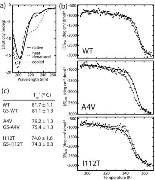
SOD1 variants with and without Cys-111 glutathionylation were analyzed using a Jasco J-815 CD spectrometer (Jasco Inc. Easton, MD). Yeast-expressed SOD1 mutants were dialyzed overnight against 10 mM phosphate buffer and diluted to 0.2 mg/ml for analysis. Sample spectra were taken at 20 °C and 96 °C and the major loss of signal occurred at 230 nm. Upon cooling to
20 °C, the decrease in ellipticity at 230 nm was reversible for all samples to within 65 – 85% of
the initial value (Figure 2.4a). All subsequent unfolding experiments were temperature ramps from 20 °C to 96 °C monitored at 230 nm by 1 °C increments with a 5 s dwell time at each.
Dialysis buffer was used as a blank. To obtain apparent melting temperature Tm (Tm*) values, blank-corrected thermal melting data were fit to a modified form of the van’t Hoff equation, as previously described in (21). This equation includes parameters for the melting transition as well as the baselines corresponding to the native and denatured states (22):
θ(T)=anT+bn+(adT+bd)K
1+K
where ΔGu is the difference in Gibbs free energy between the native and denatured states at a
given temperature T, and R is the universal gas constant. ΔGu was calculated according to the
Gibbs-Helmholtz equation (23):
ΔGu=ΔHu(1−
T Tm
)− ΔCu[(Tm−T)+Tln(
T Tm
)]
where Tm is the temperature at which ΔGu=0, and ΔHu and ΔCu are the changes in enthalpy and
heat capacity, respectively, associated with thermal denaturation. Data were fit with the
parameters an, ad, bn, bd, ΔHu, ΔCu, Tm using non-linear least-squares regression, and Tm values
were reported as apparent Tm (Tm*)due to the incomplete reversibility of the unfolding transition.
All-atom DMD simulations of glutathionylated SOD1 mutants
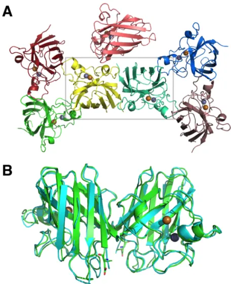
To obtain the structures of post-translationally modified mutant and wild type SOD1, we
use the known X-ray crystallographic structure of wild type SOD1 (PDBID: 1SPD) as a
reference structure, and constrain glutathione molecules to their respective SOD1 residues.
Mutations are made to these structures using the Eris suite (24), avoiding changes to residues
participating in the metal-binding, glutathionylation, or disulfide bond interactions. The overall
structure energy was minimized using an all-atom protein model with discrete molecular
! We perform equilibration and production simulations using DMD. DMD is a molecular
dynamics engine that uses discrete potentials in place of continuous potentials, which transforms the simulation into simple calculations of ballistic equations, increasing the speed and efficiency of the simulation and extending sampling of conformational space. Each system is equilibrated for 500 ps at 226 K with a heat exchange occurring every 5 fs. We conduct 50 ns equilibrium simulations of dimeric SOD1 277 K. We perform simulations for each case of mutant or wild type, both the glutathionylated and unmodified structures, resulting in 6 cases total (2
(glutathionylated or unmodified) × 3 (two mutants and wild type)).
Dimer interface contact maps
In our DMD simulations, we define two residues as being in contact in the dimer interface if two Cα atoms of opposing chains are within 10 Å of each other. At each simulation snapshot (5 picoseconds of simulation time), we evaluate the contacts present between the two monomers. We then normalize the count between every pair of residues over the entire
simulation.
Calculation of dimer interface area
! resulting in the total buried area of both monomers in the respective dimer structure. We divide
this resulting total area by two (since two monomers form the interface) to obtain the dimer interface area. All SASAs are calculated using the Gaia suite (28).
Results
SOD1 wild type and mutant dimers are destabilized by glutathionylation under physiological conditions
Size exclusion chromatography (SEC) analysis of GS-SOD1WT reveals the substantial destabilization of dimers by this physiologically prevalent modification (Figure 2.1). GS-SOD1 used in these assays was isolated from the endogenous pool of enzyme expressed in S. cerevisiae using ion-exchange chromatography, yielding a population that is heavily (~8-fold) enriched in glutathionylated protein (12). While some unmodified enzyme remains in this sample, we do not perform additional in vitro glutathionylation of SOD1, in order to avoid non-physiological modification of cysteine-6 (12). Hence, we report a lower limit for the destabilizing effect of cysteine-111 glutathionylation.
We examined the effect of cysteine-111 glutathionylation on the SOD1 monomer-dimer equilibrium by assaying the oligomeric state of unmodified and glutathionylated SOD1 at
physiological pH and concentration (estimated as 50 – 100 µM in neurons (29, 30)). Unmodified
wild type SOD1 is completely dimeric under these conditions (thick solid curve, Figure 2.1a), in agreement with the previously reported Kd of 10 nM for wild type SOD1 expressed in S.
cerevisiae (15). Glutathionylation of SOD1WT results in the appearance of a significant
! the overlapping peaks for dimeric and monomeric SOD1. We estimate the monomer contribution
Figure 2.1. Wild type SOD1 dimers are destabilized by Cys-111 glutathionylation. (a) Size exclusion chromatography at physiological [SOD1] (88µM) shows marked
! while accounting for peak skewness (see discussion in Materials and Methods) by assuming the
peak shape for unmodified SOD1 (solid curves, Figure 2.1b) to be characteristic of dimeric SOD1. By removing the contribution of this curve from the observed A280 (dashed curves, Figures 2.1b and 2.2b), we obtain the signal attributable to monomeric SOD1 (dotted curves, Figures 2.1b and 2.2b). We estimate the Kd of the GS-SOD1WT homodimer to be approximately 10-20 µM, which represents an increase of approximately 1000-fold over that of the extremely stable unmodified enzyme (previously reported as 10 nM (15)). SOD1A4V is also destabilized by glutathionylation, experiencing an approximately 30-fold increase in Kd (Figure 2.2). In contrast, SOD1I112T stability is unaffected by this modification, remaining dimeric when glutathionylated (Figure 2.2a). The mean elution volume for monomeric SOD1A4V is slightly lower (~1.84 ml) compared to that of the wild type (~1.89 ml). This mutation is reported to increase the radius of gyration of monomeric SOD1 (31), accounting for the decrease in mobility in SEC. The
differences in oligomeric state between unmodified and glutathionylated SOD1A4V and SOD1WT are observed in replicate experiments and are abrogated by treatment with DTT to remove the glutathione moiety (thin curves, Figures 2.1a and 2.2a), implying that the observed
!
Figure 2.2. Effect of Cys-111 glutathionylation on Kd of selected FALS mutants.
(a) Size exclusion chromatography at physiological [SOD1] (88µM) shows
destabilization of GS-SOD1A4V that is reversed by treatment with DTT to remove the glutathione moeity. The oligomeric state of SOD1I112T (bottom panel) is relatively unaffected by modification. Solid lines indicate unmodified SOD1 while dashed lines represent GS-SOD1. Broad and fine lines show these species before and after
! Effects of glutathionylation on dimer dissociation kinetics
To measure the effect of glutathionylation on the rate of SOD1 dimer dissociation, we use surface plasmon resonance (SPR) to monitor the dissociation of biotinylated SOD1 dimers immobilized to a streptavidin-coated sensor chip (13). We observe a clear distinction between the effects of the A4V and I112T mutations on dissociation kinetics. SOD1112T has an average half time of 1.10 hours, compared to 1.29 hours for the wild type, a difference that is within experimental error (Figure 2.3). SOD1A4V dimers, by contrast, dissociate significantly faster than the wild type, with an average half time of 4.51 minutes (Figure 2.3). This observation is in stark agreement with the common classification of A4V as a mutation that particularly affects dimer
! stability (32, 33), with the dimer dissociation rate constant koff for unmodified SOD1A4V nearly
20-fold greater than that of the unmodified wild type. Glutathionylation has a minimal effect on dissociation rate for all SOD1 variants studied: koff values for unmodified and glutathionylated dimers do not differ significantly for the wild type and the A4V mutant. SOD1I112T shows a significant, but small (20%), increase in dimer dissociation rate as a result of glutathionylation. The minimal effect of glutathionylation on the dissociation rate constants (koff) of SOD1WT and SOD1A4V dimers cannot account for the significant destabilization at equilibrium revealed by SEC (Figures 2.1 and 2.2); thus, the effects of this modification on Kd are attributable to decreases in the association rate constant (kon) of modified monomers.
Glutathionylation has little effect on SOD1 monomer stability
We assess the effect of mutations and glutathionylation on SOD1 monomer stability using thermal unfolding experiments monitored by circular dichroism (CD). Because CD primarily reflects protein secondary structure content, we expect that changes in signal upon thermal denaturation of SOD1 are mainly attributable to the loss of β-strand structure as
!
Structural effects of glutathionylation on SOD1 dimer interface
Using DMD simulations, we show that dimer interface contacts are changed in the glutathionylated versus the unmodified structures. SOD1WT and SOD1A4V exhibit a general loss
Figure 2.4. Effect of glutathionylation on monomer thermal stability. (a) Representative CD spectra of SOD1 before and after cooling shows reversible decrease in ellipticity at 230 nm. (b) Representative curves for thermally-induced unfolding of unmodified (closed symbols) and glutathionylated (open symbols) wild type and mutant SOD1 monitored by circular dichroism at 230 nm. (c) Apparent Tm (Tm*) values obtained by fitting
!
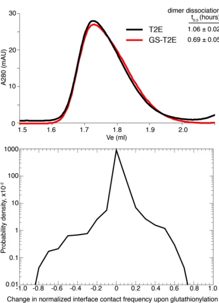
Figure 2.5. Effects of glutathionylation on the SOD1 dimer interface. (a) The top ten most frequently changed Cα interface contacts upon glutathionylation are highlighted with a three-dimensional rod representation for SOD1WT, SOD1A4V, and SOD1I112T. Rod thickness is proportional to the change in frequency of the interaction. Blue rods represent a loss in frequency of the interaction; red rods represent a gain in frequency of the interaction. (b) Distributions of changes in frequency of Cα interface contacts upon Cys-111
! in overall interface Cα contacts, while the I112T mutant experiences a shift in Cα dimer interface
contacts upon glutathionylation (Figures 2.5a-b). In SOD1I112T, residues that lose interface contacts are balanced by neighboring residues that gain contacts, resulting in an overall change in composition of the interface, without significantly changing the number of interface contacts. Interestingly, in wild type SOD1, we observe that, while the net number of Cα contacts decreases upon glutathionylation, the net number of Cβ contacts increases (Figure 2.6). This would indicate rearrangement of the side chains in the dimer interface in order to accommodate the glutathione moiety. We do not observe this effect in the A4V or I112T mutants, which have the same
qualitative distribution of losses and gains in contact frequency upon glutathionylation in Cα and Cβ contacts.
!
!
Figure 2.6. Comparison of Cα and Cβ dimer interface contacts. Distributions of changes in frequency of both Cα-Cα (backbone) and Cβ-Cβ (side-chain) dimer interface contacts upon
Cys-111 glutathionylation for (a) wild type, (b) A4V, and (c) I112T SOD1. Wild type SOD1 shows a reversal of behavior in the two types of contacts; the interface undergoes an overall loss of backbone contacts, but gains chain contacts, indicating the ability of the side-chains to rearrange upon separation of the monomer backbones. This behavior is not observed in A4V or I112T SOD1, whose distributions have similar qualitative behavior between
backbone and side-chain contacts.
![Figure 2.1. Wild type SOD1 dimers are destabilized by Cys-111 glutathionylation. (a) Size exclusion chromatography at physiological [SOD1] (88µM) shows marked](https://thumb-us.123doks.com/thumbv2/123dok_us/8282306.2193361/51.918.249.640.176.774/figure-dimers-destabilized-glutathionylation-exclusion-chromatography-physiological-marked.webp)