THE INFLUENCE OF LOWER EXTREMITY BIOMECHANICS ON BIOCHEMICAL MARKERS OF BONE TURNOVER DURING ARMY CADET BASIC TRAINING
Timothy C. Mauntel
A dissertation submitted to the faculty at the University of North Carolina at Chapel Hill in partial fulfillment of the requirements for the degree of Doctor of Philosophy in the Human Movement Science Curriculum in the Department of Allied Health Sciences in the School of
Medicine.
Chapel Hill 2016
iii ABSTRACT
Timothy C. Mauntel: The Influence of Lower Extremity Biomechanics on Biochemical Markers of Skeletal Stress During Army Cadet Basic Training
(Under the direction of Darin A. Padua)
Lower extremity stress fracture rates are high among military personnel, result in substantial lost duty time, and inhibit military readiness. Stress fracture risk factors include aberrant biomechanics, previous musculoskeletal injury, physical fitness, and anthropometric measurements. It is unknown how these risk factors influence bone formation and resorption (turnover) biomarkers. Elucidating the relationships between stress fracture risk factors and bone turnover biomarkers will provide insight into how these factors influence bone health. Our primary aim was to characterize the effects of stress fracture risk factors on bone turnover biomarkers. Our secondary aim was to validate an automated markerless motion capture system. We hypothesized the presence of stress fracture risk factors would result in bone biomarker profiles indicative of high turnover rates. We also hypothesized the markerless motion capture system would provide valid kinematic measurements.
iv stereophotogrammetric motion capture system.
Lower extremity stress fracture risk factors predicted post-CBT bone turnover
biomarkers. Overall movement quality was not predictive, but variables associated with sagittal plane displacement and foot position at initial ground contact did predict post-CBT bone
turnover biomarkers. Injury during CBT, physical fitness test performance, and mass also predicted post-CBT bone turnover biomarkers.
Moderate agreement was observed between the markerless and stereophotogrammetric motion capture systems. Better agreement was observed for sagittal than frontal plane joint angles and for maximum and displacement angles than initial ground contact joint angles.
v
TABLE OF CONTENTS
LIST OF TABLES ... viii
LIST OF FIGURES ... ix
LIST OF ABBREVIATIONS ... x
CHAPTER I ... 1
1.2 – Operational Definitions... 4
1.3 – Assumptions and Limitations ... 6
1.4 – Delimitations ... 7
1.5a – Independent (Predictor) Variables ... 7
1.5b – Dependent Variables ... 9
1.6 – Specific Aims and Research Hypotheses ... 9
1.6a – Specific Aim 1 ... 10
1.6b – Specific Aim 2 ... 11
1.6c – Specific Aim 3 ... 15
1.6d – Specific Aim 4 ... 17
1.7 – Significance ... 18
CHAPTER II ... 20
2.1 – General Information and Introduction ... 20
2.2 – Military Training Related Injuries ... 21
2.2a – Military Training Related Injuries: Military Training ... 21
2.2b – Military Training Related Injuries: Epidemiology ... 23
2.2c – Military Training Related Injuries: Risk Factors ... 25
2.3 – Bone Tissue ... 34
2.3a – Bone Tissue: Stress Fractures ... 35
vi
2.3c – Bone Tissue: Biochemical Makers of Bone Turnover – Response to
Physical Activity... 39
2.3d – Biochemical Markers of Bone Turnover: Data Collection Considerations ... 44
2.4 – Automated Markerless Motion Capture Systems ... 46
CHAPTER III ... 49
3.1 – Experimental Design Overview ... 49
3.2 – Participants ... 50
3.2a – Inclusion Criteria ... 51
3.2b – Exclusion Criteria... 51
3.3 – Data Collection Procedures ... 51
3.3a – Post-Cadet Basic Training Serum Samples ... 51
3.3b – Biomechanical Assessment ... 52
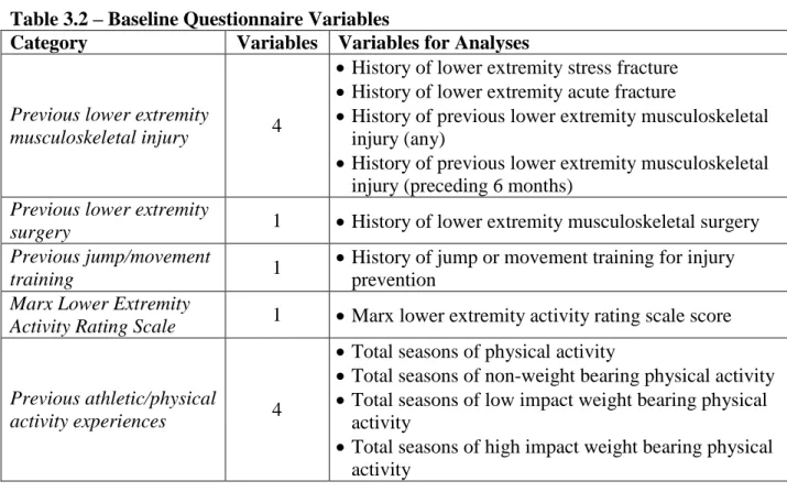
3.3c – Baseline Questionnaire (BLQ)... 56
3.3d – Prior Physical Activity ... 56
3.3e – Army Physical Fitness Test (APFT) ... 57
3.3f – Body Mass Index (BMI) ... 57
3.3g – Food Consumption Log ... 57
3.3h – Cadet Basic Training Injury Log ... 57
3.4 – Data Reduction and Statistical Plan ... 58
3.4a – Data Processing and Reduction ... 58
3.4b – Data Analyses ... 61
CHAPTER IV... 67
Manuscript 1: Trunk and Lower Extremity Movement Patterns and Stress Fracture Risk Factors Influence Biomarkers of Bone Turnover In Military Training ... 67
Manuscript 2: Trunk and Lower Extremity Kinematics and Stress Fracture Risk Factors Influence Biomarkers of Bone Turnover In Military Training ... 89
Manuscript 3: Validation of a Markerless Motion Capture System Trunk and Lower Extremity Joint Angles During a Jump-Landing Assessment ... 109
CHAPTER V ... 130
5.1 – Introduction ... 130
5.2 – Methods ... 130
vii
5.4 – Interpretation of Results ... 132
5.5 – Strengths and Limitations ... 135
5.6 – Conclusions ... 137
APPENDIX 3.1 – LESS OPERATIONAL DEFINITIONS ... 138
APPENDIX 3.2 – BASELINE QUESTIONNAIRE ... 141
APPENDIX 4.1 – MOVEMENT QUALITY AND BIOMARKERS OF BONE TURNOVER ... 147
APPENDIX 4.2 – STRESS FRACTURE RISK FACTORS AND BIOMARKERS OF BONE TURNOVER ... 150
viii
LIST OF TABLES
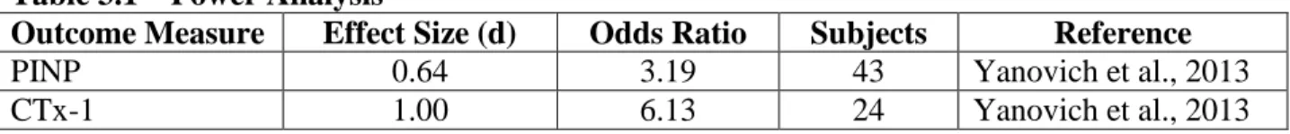
Table 3.1 – Power Analysis ... 50
Table 3.2 – Baseline Questionnaire Variables ... 61
Table 3.3 – Data Analyses Table ... 65
Table 4.1 – USMA Participant Demographics Presented as Means ± SD ... 70
Table 4.3 – Summary of Landing Error Scoring System (LESS) Items ... 86
Table 4.4 – Predictability of the Landing Error Scoring System on Biomarkers of Bone Turnover ... 87
Table 4.5 – Predictability of Stress Fracture Risk Factors and Movement Quality on Biomarkers of Bone Turnover ... 88
Table 4.6 – Summary of Trunk and Lower Extremity Kinematic Variables ... 105
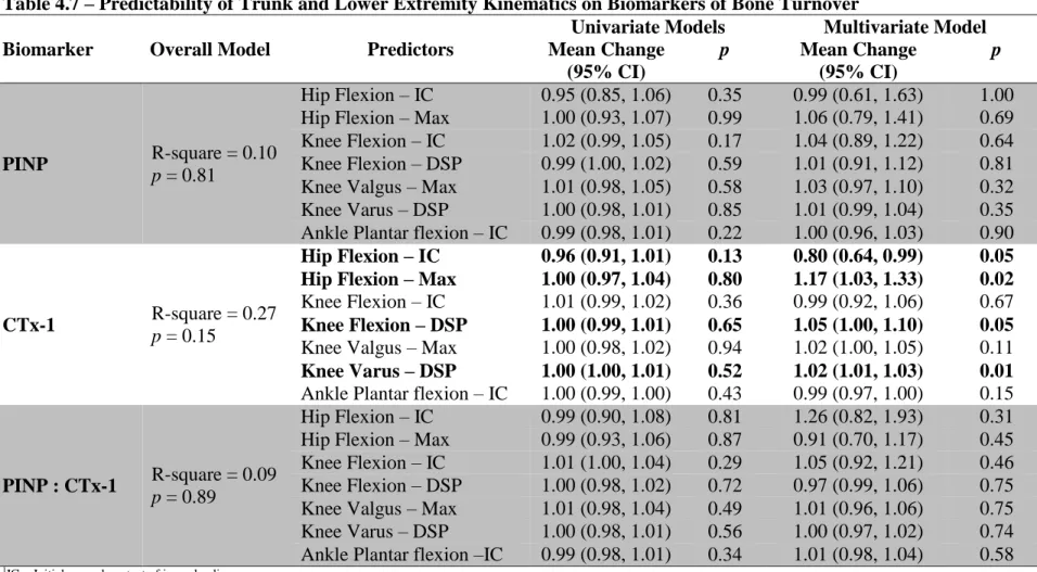
Table 4.7 – Predictability of Trunk and Lower Extremity Kinematics on Biomarkers of Bone Turnover ... 106
Table 4.8 – Predictability of Stress Fracture Risk Factors and Trunk and Lower Extremity Kinematics on Biomarkers of Bone Turnover ... 107
Table 4.9 – Markerless Motion Capture System Reliability Participant Demographics ... 110
Table 4.10 – Trunk and Ankle Joint Angle Means, 95% Confidence Intervals, Intraclass Correlation Coefficients, and Pearson Product-Moment Correlation Coefficients ... 121
Table 4.11 – Hip Joint Angle Means, 95% Confidence Intervals, Intraclass Correlation Coefficients, and Pearson Product-Moment Correlation Coefficients ... 122
Table 4.12 – Knee Joint Angle Means, 95% Confidence Intervals, Intraclass Correlation Coefficients, and Pearson Product-Moment Correlation Coefficients ... 124
ix
LIST OF FIGURES
x
LIST OF ABBREVIATIONS
APFT Army Physical Fitness Test BALP Bone-specific alkaline phosphate BLQ Baseline questionnaire
BMI Body mass index
CTx-1 Cross-linked collagen telopeptide DPD Deoxypyridinoline
ELISA Enzyme-linked immunosorbent assays LESS Landing Error Scoring System
PABAK Prevalence and Bias Adjusted Kappa statistic PICP Procollagen type I carboxy-terminal propeptide PINP Procollagen type I aminoterminal propeptide TRAP5b Tartrate resistant acid phosphate
1 CHAPTER I INTRODUCTION
1.1 – Background and Introduction
Musculoskeletal injuries affect 63% of non-deployed military personnel1 and are the most significant medical issue limiting military readiness.2 Lower extremity injuries account for 39% of non-deployed military personnel injuries, with 82% of these injuries resulting from overuse mechanisms.1 The direct and indirect costs associated with musculoskeletal injuries are estimated at $3.7 billion annually for the Department of Defense.3 One of the most common injuries affecting military personnel is lower extremity stress fractures,1,4 which affect nearly 1 in 3 male service members.5 These injuries result in significant lost duty time, medical costs, and attrition.4 Given the high prevalence of musculoskeletal injuries and their substantial physical and financial costs it is critical to understand the factors that contribute to individuals sustaining injury during military training.3
Military training is highly repetitive but also involves bouts of high intensity exercise, this training regimen results in high training loads that are associated with increased lower extremity injury rates.6-8 This is especially true for overuse bone injuries (e.g. stress fractures).1,4 Musculoskeletal stress occurring during military training may be amplified by aberrant
2
Aberrant biomechanical patterns can be easily identified with common clinical movement assessments (e.g. jump-landing tasks).11,12 The jump-landing task has been developed into a validated clinical movement assessment that is scored on visual observation of aberrant movement patterns (the Landing Error Scoring System or LESS). The LESS is capable of discriminating between individuals at increased lower extremity injury risk from those who are not.11-13 Individuals who score high on the LESS (>6) and individuals who score low on the LESS (≤4) display different three-dimensional lower extremity biomechanical patterns.11 The aberrant biomechanical patterns observed among individuals with high LESS scores have been associated with traumatic and overuse musculoskeletal injuries.4,9,10,12,13
The LESS is a movement assessment that meets many of the requirements put forth by a consortium of civilian and military experts on injury risks and prevention.11 Primarily, it is valid, reliable, and can be implemented quickly across a large number of individuals. However, the LESS does have its limitations.14 The LESS requires video replay and manual scoring of jump-landing trials, which is time consuming and therefore prohibitive for clinicians to implement.11,14 Thus, there has been a call for automated systems that accurately and quickly identify individuals at increased injury risk.14,15
3
capture system is required before wide-spread implementation can occur to aid clinicians in identifying lower extremity injury risks.
Biochemical markers (biomarkers) associated with musculoskeletal system stress may be useful in identifying individuals who are overstressing their musculoskeletal systems, prior to them sustaining an injury.5,17,18 Biomarkers indicative of skeletal stress (“bone turnover”) change with alterations in physical activity, thus they may be able to identify individuals prior to
injury.5,17,19-27 Biomarkers indicative of bone formation (procollagen type I aminoterminal propeptide [PINP]) and resorption (cross-linked collagen telopeptide [CTx-1]) (i.e. turnover) are altered by military training.5,17,25,27 Bone turnover biomarkers also increase following traumatic lower extremity joint injuries,28,29 which are common amongst military personnel.1-3 Examining serum biomarkers representative of bone turnover will provide insight into the extent to which lower extremity biomechanics influence skeletal stress during military training.
Bone turnover biomarkers may also be influenced by other known stress fracture risk factors. These factors include modifiable and non-modifiable factors. Modifiable risk factors include training load,6,30-32 aerobic and anaerobic fitness,14,33-40 physical activity preceding military training,33,35,38,41-43 body composition,17,33,44 and lifestyle choices.14,33,37,45 Non-modifiable risk factors include previous history of musculoskeletal injury,37,45 age,37,38 race,34,37,44 and sex.44,46 It is therefore important to consider the aforementioned factors when assessing bone turnover biomarkers.
4
turnover. Understanding the influence of biomechanics on bone turnover biomarkers will allow for the development of intervention strategies to reduce injury risk and optimize performance during military training.
1.2 – Operational Definitions
1) Cadet Basic Training: A 6-week course completed by new cadets at the United States
Military Academy (West Point) the summer prior to the start of their first academic year. The course is designed to improve physical fitness, teach basic military skills (e.g. marksman-ship, first aid, land navigation), and improve confidence.
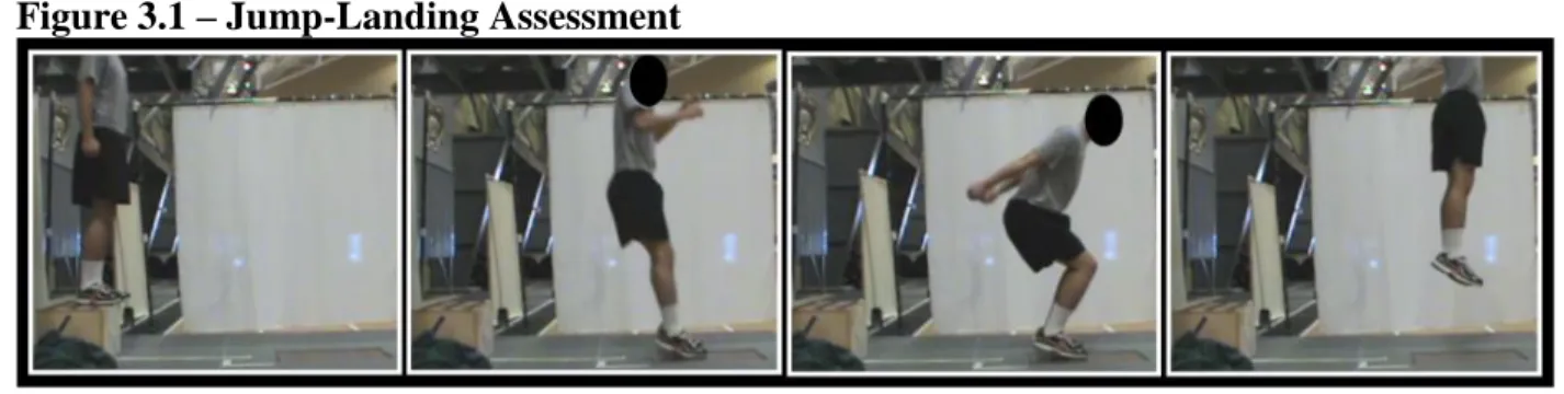
2) Jump-Landing Movement Assessment: A clinical movement assessment in which the study participant jumps from a 30cm tall box to a target area located a standardized 0.9m away from the front of the box. Participants complete a vertical jump for maximal height immediately following landing in the target area. Biomechanical patterns are identified during the landing phase of the initial jump (initial ground contact peak knee flexion).11
a. Initial Ground Contact: The video frame immediately preceding the video frame in which the entire foot is in contact in the ground, or when the ground reaction force is ≥10N.
b. Peak Knee Flexion: The maximum knee flexion angle the participant reaches following initial ground contact.
3) Landing Error Scoring System (LESS): A valid and reliable clinical movement assessment during which lower extremity movement patterns are visually observed during a jump-landing movement assessment.11
5
measured and evaluated as an indicator of normal or pathogenic biologic processes47 (skeletal response to stress induced by biomechanical patterns and military basic training), that is measured through blood serum.
a. Procollagen type I aminoterminal propeptide (PINP): A biochemical marker indicative of type I collagen neogenesis, representative of bone formation.
b. Cross-linked collagen telopeptide (CTx-1): A biochemical marker indicative of type I collagen breakdown, representative of bone resorption.
5) Biochemical Marker Turnover: The ratio between biochemical markers indicative of tissue neogenesis and tissue breakdown (type I collagen, bone).
6) Baseline Questionnaire (BLQ): A comprehensive questionnaire that is designed to assess previous and current physical activity levels, previous and current injury history, and overall current physical well-being
7) Army Physical Fitness Test (APFT): A test of physical fitness administered by the United States Army to determine the muscular strength, muscular endurance, and cardiorespiratory fitness of each cadet. The APFT includes 2 minutes of push-ups, 2 minutes of sit-ups, and a timed 2-mile run. The raw score and standardized score (0 – 100 points) for each event and a cumulative score (0 – 300 points) are recorded.
8) Previous Physical Activity: The physical activity the cadet participated in prior to beginning Cadet Basic Training.
a. Previous Physical Activity Level: The number of seasons (season = participation in a physical activity ≥3 times a week for ≥3 months) an individual completed structured physical activity.
6
activity multiplied by the average duration of physical activity.
c. Previous Physical Activity Type: Physical activity that either directly loads (weight bearing) or does not directly load the lower extremity completed prior to beginning Cadet Basic Training.
9) Body Mass Index (BMI): An index of mass-to-height used to classify individuals into categories of underweight, normal, overweight, and obese. This value is obtained with the following equation: BMI = mass (kg) / height (cm)2.48
1.3 – Assumptions and Limitations
The following assumptions and limitations will apply to this study:
1) The PhysiMaxTM LESS Scoring Platform is a valid measure of trunk and lower extremity movement patterns.
2) Participants will jump for maximal effort during the jump-landing assessments. 3) Participants will give maximal effort throughout Cadet Basic Training.
4) The enzyme-linked immunosorbent assay (ELISA) kits for measurement of bone turnover biomarkers will be reliable within <10% inter and intra-assay coefficients of variation. 5) Circulating serum concentrations of bone biomarkers (PINP and CTx-1) measured within 2
weeks of completing Cadet Basic Training accurately and reliably reflect bone turnover rates. 6) The rates of bone turnover of military cadets completing Cadet Basic Training at the United
7 1.4 – Delimitations
The following delimitations were made for this study.
1) 45 male cadets were recruited from the United States Military Academy (West Point). 2) All participants were injury-free at the time of the jump-landing movement assessment
testing.
3) All participants were healthy with no history of neurological or metabolic disorders. 4) All serum biomarker concentrations were measured using enzyme-linked immunosorbent
assays (ELISA) and spectrophotometry.
1.5a – Independent (Predictor) Variables 1) Lower Extremity Movement Quality
a. LESS total score b. LESS individual items
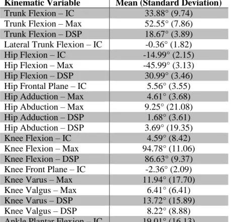
c. Average frontal and sagittal plane trunk, hip, knee, and ankle joint angles at initial ground contact, maximum values, and displacements
2) Previous Physical Activity Levels
a. Volume of physical activity prior to Cadet Basic Training b. Total number of previous physical activity seasons 3) Previous Physical Activity Type
8 e. History of jump/movement training 4) Physical Fitness Levels
a. APFT standardized composite score b. APFT individual event scores
i. Raw scores
ii. Standardized scores
5) Musculoskeletal Injury History (dichotomous)
a. Previous history of lower extremity stress fracture b. Previous history of lower extremity acute fracture
c. Previous history of lower extremity musculoskeletal injury (e.g. ligamentous sprain, meniscal injury)
i. Any history
ii. Injury within 6 months preceding Cadet Basic Training
iii. History of musculoskeletal injury during Cadet Basic Training a. Duration of time loss from Cadet Basic Training following
musculoskeletal injury d. Previous history of orthopaedic surgery 6) Body Compositions Measurements
a. Height
i. Pre-Cadet Basic Training ii. Post-Cadet Basic Training b. Mass
9 iv. Post-Cadet Basic Training
v. Change from Pre-to-Post-Cadet Basic Training measurements c. Body Mass Index (BMI)
vi. Pre-Cadet Basic Training vii. Post-Cadet Basic Training
viii. Change from Pre-to-Post-Cadet Basic Training measurements
7) Post-Cadet Basic Training blood draw preceding 12 hours physical activity and food consumption
a. Food Consumption i. Time
ii. Protein vs Non-Protein rich foods b. Exercise
i. Time
ii. Weight bearing vs Non-weight bearing 1.5b – Dependent Variables
1) Biomarkers Representative of Bone Turnover – Individual
a. Procollagen type I aminoterminal propeptide (PINP) at post-Cadet Basic Training b. Cross-linked collagen telopeptide (CTx-1) at post-Cadet Basic Training
2) Biomarkers Representative of Bone Turnover – Turnover Ratio a. PINP : CTx-1 at post-Cadet Basic Training
1.6 – Specific Aims and Research Hypotheses
10 1.6a – Specific Aim 1
Characterize the effects of lower extremity biomechanics on biomarker profiles representing bone turnover through predictive models incorporating serum biomarker measures collected following military basic training (post-Cadet Basic Training).
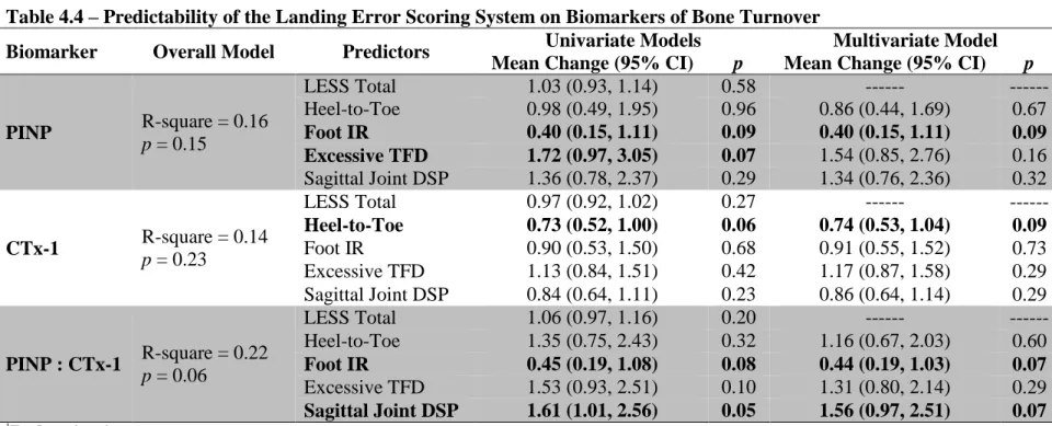
Hypothesis 1a: Qualitative measures of lower extremity movement quality (LESS total score and individual LESS items) will be predictive of post-Cadet Basic Training PINP, CTx-1, and PINP : CTx-1 serum concentration levels.
Hypothesis 1a.1: Higher LESS scores (poorer movement quality) will result in higher serum concentrations of PINP and CTx-1. Higher LESS scores will also result in smaller PINP : CTx-1 ratios.
Hypothesis 1a.2: Positive findings of sagittal plane LESS items (trunk, hip, knee, and ankle items at initial ground contact and displacements) will result in higher serum concentrations of PINP and CTx-1. Positive findings of sagittal plane LESS items will also result in smaller PINP : CTx-1 ratios.
Hypothesis 1a.3: Positive findings of frontal plane LESS items (hip and knee alignments at initial ground contact and displacement) will result in higher serum concentrations of PINP and CTx-1. Positive findings of frontal plane LESS items will also result in smaller PINP : CTx-1 ratios. Frontal plane trunk items will not be predictive of serum
concentrations of PINP, CTx-1, or PINP : CTx-1 ratios.
Hypothesis 1a.4: Positive findings of transverse plane LESS items (foot internal and external rotation) will not be predictive of serum concentrations of PINP, CTx-1, or PINP : CTx-1 ratios.
11
quality (average trunk, hip, knee, and ankle angles) will be predictive of PINP, CTx-1, and PINP : CTx-1 serum concentration levels.
Hypothesis 1b.1: Smaller trunk, hip, and knee sagittal plane joint angles at initial ground contact, maximum values, and displacements will result in higher serum concentrations of PINP and CTx-1. Smaller trunk, hip, and knee sagittal plane joint angles will also result in smaller PINP : CTx-1 ratios.
Hypothesis 1b.2: Larger hip and knee frontal plane joint angles at initial ground contact, maximum values, and displacements will result in higher serum concentrations of PINP and CTx-1. Larger hip and knee frontal plane joint angles will also result in smaller PINP : CTx-1 ratios. Frontal plane trunk angles will not be predictive of serum concentrations of PINP, CTx-1, or PINP : CTx-1 ratios.
1.6b – Specific Aim 2
Characterize the effects of known stress fracture risk factors on biomarker profiles representing bone turnover through predictive models incorporating serum biomarker measures collected following military basic training (post-Cadet Basic Training).
Hypothesis 2a: Previous physical activity volume will be predictive of PINP, CTx-1, and PINP :
CTx-1 serum concentration levels.
Hypothesis 2a.1: Smaller volumes of previous physical activity will result in higher serum concentrations of PINP and CTx-1. Smaller volumes of previous physical activity will also result in smaller PINP : CTx-1 ratios.
12
Hypothesis 2b: Previous physical activity type will be predictive of PINP, CTx-1, and PINP :
CTx-1 serum concentration levels.
Hypothesis 2b.1: Fewer seasons of previous weight bearing physical activity will result in higher serum concentrations of PINP and CTx-1. Fewer seasons of previous weight bearing physical activity will also result in smaller PINP : CTx-1 ratios.
Hypothesis 2b.2: Smaller Marx lower extremity activity rating scores will result in higher serum concentrations of PINP and CTx-1. Smaller Marx lower extremity activity rating scores will also result in smaller PINP : CTx-1 ratios.
Hypothesis 2b.3: Previous history of jump or movement training will result in smaller serum concentrations of CTx-1, but not PINP. Previous history of jump or movement training will also result in larger PINP : CTx-1 ratios.
Hypothesis 2c: Physical fitness levels will be predictive of PINP, CTx-1, and PINP : CTx-1
serum concentration levels.
Hypothesis 2c.1: Lower composite APFT scores will result in higher serum
concentrations of PINP and CTx-1. Lower composite APFT scores will also result in smaller PINP : CTx-1 ratios.
Hypothesis 2c.2: Lower APFT push-ups and sit-ups raw and standardized scores will result in higher serum concentrations of PINP and CTx-1. Lower APFT push-ups and sit-ups raw and standardized scores will also result in smaller PINP : CTx-1 ratios.
Hypothesis 2c.3: Higher APFT raw run time and lower standardized score will result in higher serum concentrations of PINP and CTx-1. Higher APFT raw run time and lower standardized score will also result in smaller PINP : CTx-1 ratios.
13 CTx-1 serum concentration levels.
Hypothesis 2d.1: History of lower extremity fracture (acute and stress) will result in higher serum concentrations of PINP and CTx-1. History of lower extremity fracture will also result in smaller PINP : CTx-1 ratios.
Hypothesis 2d.2: History of lower extremity musculoskeletal injury (any history and within 6 months preceding Cadet Basic Training) will result in higher serum
concentrations of CTx-1, but not PINP. History of lower extremity fracture (acute and stress) will also result in smaller PINP : CTx-1 ratios.
Hypothesis 2d.3: History of orthopaedic surgery will result in higher serum
concentrations of PINP and CTx-1. History of orthopaedic surgery will also result in smaller PINP : CTx-1 ratios.
Hypothesis 2d.4: History of musculoskeletal injury during Cadet Basic Training will result in higher serum concentrations of PINP and CTx-1. History of musculoskeletal injury during Cadet Basic Training will also result in smaller PINP : CTx-1 ratios. Hypothesis 2d.5: Longer duration of time loss from Cadet Basic Training training as the result of a musculoskeletal injury will result in higher serum concentrations of PINP and CTx-1. Longer duration of time loss from Cadet Basic Training training as the result of a musculoskeletal injury will also result in smaller PINP : CTx-1 ratios.
Hypothesis 2e: Anthropometric measurements will be predictive of PINP, CTx-1, and PINP :
CTx-1 serum concentration levels.
14
Hypothesis 2e.2: Lower Pre- and Post-Cadet Basic Training mass will result in higher serum concentrations of PINP and CTx-1. Lower Pre- and Post-Cadet Basic Training mass will also result in smaller PINP : CTx-1 ratios.
Hypothesis 2e.3: Greater Pre-to-Post-Cadet Basic Training changes in mass will result in higher serum concentrations of PINP and CTx-1. Greater Pre-to-Post-Cadet Basic
Training changes in mass will also result in smaller PINP : CTx-1 ratios.
Hypothesis 2e.4: Lower Pre- and Post-Cadet Basic Training BMI will result in higher serum concentrations of PINP and CTx-1. Lower Pre- and Post-Cadet Basic Training BMI will also result in smaller PINP : CTx-1 ratios.
Hypothesis 2e.5: Greater Pre-to-Post-Cadet Basic Training changes in BMI will result in higher serum concentrations of PINP and CTx-1. Greater Pre-to-Post-Cadet Basic Training changes in BMI will also result in smaller PINP : CTx-1 ratios.
Hypothesis 2f: Food consumption and physical activity within 12 hours preceding the post-Cadet
Basic Training blood draw will be predictive of CTx-1 and PINP : CTx-1 serum concentration levels, but not PINP.
Hypothesis 2f.1: Protein rich food consumption within 12 hours of the post-Cadet Basic Training blood draw will result in higher serum concentrations of CTx-1, but not PINP. Protein rich food consumption within 12 hours of the post-Cadet Basic Training blood draw will also result in smaller PINP : CTx-1 ratios.
15 1.6c – Specific Aim 3
Characterize how each significant predictor variable in specific aim 2 modifies the effects of lower extremity biomechanics on biomarker profiles representing bone turnover through predictive models incorporating serum biomarker measures collected following military basic training (post-Cadet Basic Training).
Hypothesis 3a: Previous physical activity exposure will interact with lower extremity
biomechanics and significantly alter the effects of lower extremity biomechanics on serum biomarker measures. Smaller volumes of previous physical activity and fewer seasons will exacerbate the effects of lower extremity biomechanics and result in higher PINP and CTx-1 serum concentrations and smaller PINP : CTx-1 ratios.
Hypothesis 3b: Previous physical activity type will interact with lower extremity biomechanics
and significantly alter the effects of lower extremity biomechanics on serum biomarker
measures. Fewer seasons of previous weight bearing physical activity and smaller Marx lower extremity activity rating scores will exacerbate the effects of lower extremity biomechanics and result in higher PINP and CTx-1 serum concentrations and smaller PINP : CTx-1 ratios. Previous history of jump or movement training will not significantly interact with lower extremity
biomechanics.
Hypothesis 3c: Physical fitness levels will interact with lower extremity biomechanics and
significantly alter the effects of lower extremity biomechanics on serum biomarker measures. Worse composite and individual APFT scores will exacerbate the effects of lower extremity biomechanics and result in higher PINP and CTx-1 serum concentrations and smaller PINP : CTx-1 ratios.
16
biomechanics and significantly alter the effects of lower extremity biomechanics on serum biomarker measures. A history of fracture or lower extremity musculoskeletal injury will exacerbate the effects of lower extremity biomechanics and result in higher PINP and CTx-1 serum concentrations and smaller PINP : CTx-1 ratios.
Hypothesis 3e: History of orthopaedic surgery will interact with lower extremity biomechanics
and significantly alter the effects of lower extremity biomechanics on serum biomarker measures. A history of orthopaedic surgery will exacerbate the effects of lower extremity biomechanics and result in higher PINP and CTx-1 serum concentrations and smaller PINP : CTx-1 ratios.
Hypothesis 3f: Sustaining a musculoskeletal injury during Cadet Basic Training will interact
with lower extremity biomechanics and significantly alter the effects of lower extremity
biomechanics on serum biomarker measures. Sustaining a musculoskeletal injury during Cadet Basic Training will exacerbate the effects of lower extremity biomechanics and result in higher PINP and CTx-1 serum concentrations and smaller PINP : CTx-1 ratios. The longer duration of time loss from Cadet Basic Training as a result of the musculoskeletal injury will result in higher PINP and CTx-1 serum concentrations and smaller PINP : CTx-1 ratios.
Hypothesis 3g: Pre- and Post-Cadet Basic Training mass will interact with lower extremity
biomechanics and significantly alter the effects of lower extremity biomechanics on serum biomarker measures. Lower Pre- and Post-Cadet Basic Training mass will exacerbate the effects of lower extremity biomechanics and result in higher PINP and CTx-1 serum concentrations and smaller PINP : CTx-1 ratios.
Hypothesis 3h: Pre-to-Post-Cadet Basic Training changes in mass will interact with lower
17
serum biomarker measures. Greater Pre-to-Post-Cadet Basic Training changes in mass will exacerbate the effects of lower extremity biomechanics and result in higher PINP and CTx-1 serum concentrations and smaller PINP : CTx-1 ratios.
Hypothesis 3i: Pre- and Post-Cadet Basic Training BMI will interact with lower extremity
biomechanics and significantly alter the effects of lower extremity biomechanics on serum biomarker measures. Lower Pre- and Post-Cadet Basic Training BMI will exacerbate the effects of lower extremity biomechanics and result in higher PINP and CTx-1 serum concentrations and smaller PINP : CTx-1 ratios.
Hypothesis 3j: Pre-to-Post-Cadet Basic Training changes in BMI will interact with lower
extremity biomechanics and significantly alter the effects of lower extremity biomechanics on serum biomarker measures. Greater Pre-to-Post-Cadet Basic Training changes in BMI will exacerbate the effects of lower extremity biomechanics and result in higher PINP and CTx-1 serum concentrations and smaller PINP : CTx-1 ratios.
Hypothesis 3k: Protein rich food consumption and weight bearing physical activity within 12
hours of the post-Cadet Basic Training blood draw will not significantly alter the effects of lower extremity biomechanics on PINP or CTx-1 serum biomarker concentrations or PINP : CTx-1 ratios.
1.6d – Specific Aim 4
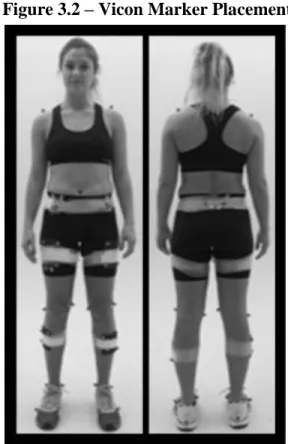
Validate the trunk and lower extremity angles calculated by the PhysiMaxTM markerless motion capture system against the current gold-standard (marker based stereophotogrammetry system [Vicon]) of motion capture systems.
Hypothesis 4a: Frontal and sagittal plane trunk angles calculated by the PhysiMaxTM markerless
18
gold-standard of motion capture systems. Maximum joint angles will demonstrate the best agreement between motion capture systems, followed by joint angle displacements, and then joint angles at initial ground contact.
Hypothesis 4b: Frontal and sagittal plane hip angles calculated by the PhysiMaxTM markerless
motion capture system will be valid measures of hip kinematics as compared to the current gold-standard of motion capture systems. Maximum joint angles will demonstrate the best agreement between motion capture systems, followed by joint angle displacements, and then joint angles at initial ground contact.
Hypothesis 4c: Frontal and sagittal plane knee angles calculated by the PhysiMaxTM markerless
motion capture system will be valid measures of knee kinematics as compared to the current gold-standard of motion capture systems. Maximum joint angles will demonstrate the best agreement between motion capture systems, followed by joint angle displacements, and then joint angles at initial ground contact.
Hypothesis 4d: Sagittal plane ankle angles calculated by the PhysiMaxTM markerless motion
capture system will be valid measures of ankle kinematics as compared to the current gold-standard of motion capture systems. Maximum joint angles will demonstrate the best agreement between motion capture systems, followed by joint angle displacements, and then joint angles at initial ground contact.
1.7 – Significance
19
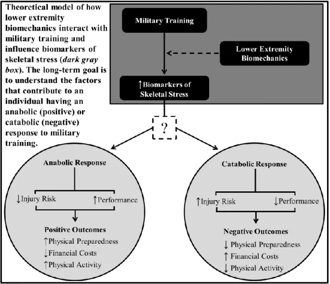
fracture risk factors influence bone turnover biomarkers during military training. This
contribution is significant because it is a major step towards understanding why some individuals have an anabolic (positive) response while others have a catabolic (negative) response to military training (Figure 1.1 – Theoretical Model). Understanding the influence of biomechanics on biomarkers of skeletal stress will allow for the development of intervention strategies to reduce injury risk and optimize performance during military training. These intervention strategies will positively impact the physical readiness of our military and reduce the enormous costs of musculoskeletal injuries.3,4
20 CHAPTER II
REVIEW OF THE LITERATURE 2.1 – General Information and Introduction
Lower extremity musculoskeletal injuries significantly affect military personnel.2,49 These injuries result in substantial medical costs, forced attrition from physical activity,3,50,51 and long-term physical52,53 and financial consequences.54 The direct and indirect costs associated with musculoskeletal injuries cost the Department of Defense $3.7 billion annually.3 Training related injuries not only affect non-deployed military personnel, but are among the top reasons why individuals are medically evacuated from war zones.55-57 Thus, they are a primary concern for military commanders58 and healthcare professionals.14 Of particular importance are lower extremity fractures that result in the greatest amount of lost duty time.34 Many lower extremity fractures result from overuse mechanisms, and thus are preventable.1,4,59 It is therefore essential to identify the factors that increase lower extremity stress fractures risk so that targeted injury intervention strategies may be implemented.58
Lower extremity non-contact injury risks are multifactorial in nature.14,33,35,37,38,40-43,45 1 primary predictor of non-contact injury is lower extremity biomechanical patterns.9,10,12,14,35,36 Laboratory based movement assessments effectively identify high-risk biomechanical
21
capture system; however, the kinematic measures calculated by this system have yet to be validated.16
Biochemical markers (biomarkers) of bone turnover may be beneficial in identifying individuals at high-risk of stress fracture, prior to them becoming injured.5,17,18 Bones are
dynamic tissues that are constantly remodeling.61 As boney tissue remodels, proteins are cleaved off the ends of procollagen and collagen fibers during the formation and resorption processes. These proteins can be measured in the blood.62-66 Previous research has found changes in bone turnover concentrations resulting from military training,5,17,27 and that aberrant lower extremity biomechanics can increase stress fracture risk.9 However, it is still unknown how lower
extremity biomechanics and other stress fracture risk factors influence bone turnover biomarker concentrations during military training.
2.2 – Military Training Related Injuries
2.2a – Military Training Related Injuries: Military Training
Military training is highly repetitive but also involves episodes of high intensity exercise. Military training results in high training loads8 that are associated with high lower extremity injury rates.1,4,6-8 Military training involves planned events designed to challagne the human body and improve aerobic and anaerobic fitness. In addition to the planned physical training events, military personnel further stress their bodies through running and marching between training events. This additional running can add an additional 18 miles a week of weight-bearing activity, further stressing the musculoskeletal system.39
22
physical activity 219.5 minutes/day, moderate physical activity for 74.5 minutes/day, moderately intense physical activity 97 minutes/day, and vigorous physical activity 22.1 minutes/day. These same recruits stood for 522 minutes/day, sat for 271 minutes/day, walked for 103.5 minutes/day, completed menial chores 119 minutes/day, participated in calisthenics for 44 minutes/day, and engaged in load carriage for 449 minutes/day.67 The cumulative effects of military training increase musculoskeletal injury risk.1,4,6-8
Military personnel are carrying heavier loads than they ever have historically.68 External load carriage increases stress on the musculoskeletal system. The increased stress occurs rapidly and the musculoskeletal system may not have sufficient time to adapt and withstand the greater loads. External loads change trunk and lower extremity biomechanical patterns that can increase injury risk.68 Brown et al.69 showed greater external loads negatively affect jump-landing
biomechanical patterns. Medium (20kg) and heavy (40kg) loads resulted in more stiff landings with less hip but not knee flexion, compared to a light (6kg) load. Normalized vertical ground reaction forces also increased with each increase in external load.69 Collectively the increased stress placed on the musculoskeletal system by external loads and the aforementioned changes in lower extremity and trunk biomechanics increase injury risk.68,69
23
physical fitness and potentially a loss of physical performance.3 2.2b – Military Training Related Injuries: Epidemiology
Musculoskeletal injury is the primary medical issue limiting military physical readiness.2 During Army basic training over 45% of male recruits sustain at least one musculoskeletal injuries of which nearly half are overuse lower extremity injuries.37 Similar injury rates (58.5%) are observed among British infantrymen, during pre-deployment training; 30-35% of these injuries are directly related to soldier specific physical training.38,72 Among the most commonly injured non-deployed military personnel are United States Army infantrymen.40 Injury rates among these individuals are as high as 1.42 injuries per infantryman.34 The peak incidence of musculoskeletal injuries occurs between weeks 4 and 6 of training.5,26
Lower extremity and low back injuries are the most commonly reported injuries amongst individuals completing military training.34,37,38,40,72 The most common lower extremity injury sites include the knee (18.5%),40,41,72 ankle (16%),41,72 and lower leg (8%).41,72 Lower extremity stress fractures, effect 2-32% of military trainees.4,5,17,41,55,59,73 Nearly half (46%) of these stress fractures occur within the first 4 weeks of training.5
24
training and sport related hospitalizations in the United States Army.76 The rates of ACL injuries among military personnel are nearly 10 times greater than general civilian population ACL injury rates.77,78
Musculoskeletal injuries are the primary medical issue limiting military physical
readiness.2,38 Musculoskeletal injuries largely contribute to the United States Army’s deployment readiness being at only 85%79 because they result in substantial lost duty time.3,34,38,41,76 Lost duty time is the total number of days a soldier is unable to perform regular duties; this is a
combination of days spent in the hospital, days on convalescent leave (time to recover), and days in a medical holding company.76 Musculoskeletal injuries result in total limited duty days
equivalent to 68,000 service members annually.3 Lost duty days reduce training and operational effectiveness and increase demands on medical care providers.38
Training and sport related injuries account for 11% of all military hospitalizations. Males miss 13 days per musculoskeletal injury requiring hospitalization76 and miss approximately 27 days per 100 person-weeks due to injury during United States Army basic training.41 Fractures account for the largest amount of lost duty days.34 Specifically, the average stress fracture rehabilitation requires 63 days to complete.59
There are long-term consequences associated with musculoskeletal injuries.
25 2.2c – Military Training Related Injuries: Risk Factors
A number of factors have been identified that increase military training related
musculoskeletal injury risk.14,33,35,37,38,40-43,45 Typically these factors are classified into those that are modifiable in nature, and those that are non-modifiable in nature. Modifiable risk factors include biomechanical patterns,9,11,35,36,45,81 training load,6,30-32 aerobic and anaerobic fitness, 14,33-40
physical activity preceding military training,33,35,38,41-43 body composition,17,33,44 and lifestyle choices.14,33,37,45 Non-modifiable risk factors include previous history of musculoskeletal injury,37,45 age,37,38 race,34,37,44 and sex.44,46
2.2c.1 – Military Training Related Injuries: Risk Factors – Biomechanical Patterns
Aberrant movement patterns are a primary predictor of acute and chronic lower extremity injuries in military and civilian populations.9,10,12,14,35,36 Aberrant biomechanical patterns can result from static skeletal malalignments61 but are more commonly the result of neuromuscular control deficiencies.10,11 Aberrant biomechanics increase the forces acting on normally aligned lower extremity segments or may cause normal forces to act on abnormally aligned lower extremity segments. Both of these examples can occur simultaneously, which results in further abnormal musculoskeletal loading and increased injury risk.61
Laboratory based9,10,60 and field-expedient11,12,14 movement assessments effectively identify aberrant, high-risk biomechanical patterns. Jump-landing,9-12 squatting,82,83 and lunge35,36,45,81 movement assessments are commonly employed to identify individuals at increased musculoskeletal injury risk.
jump-26
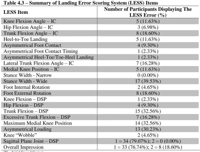
landing movement assessment while being videotaped from frontal and sagittal plane views. The videos are replayed and scored by trained raters using a standardized rubric to identify lower extremity and trunk movement errors. Items on the LESS are evaluated at initial ground contact, peak knee flexion, and the time between initial ground contact and peak knee flexion (landing phase). A larger LESS score is indicative of more aberrant biomechanical patterns than a smaller LESS score.11 The LESS is able to discriminate between individuals with high-risk (i.e. aberrant) biomechanics and individuals with low-risk biomechanics.11,12 Individuals who score ≥5 on the LESS have a greater risk of non-contact anterior cruciate ligament injury.12
The LESS has excellent intra-rater (ICC2,k=0.84, SEM=0.42) and good inter-rater reliability (ICC2,1=0.91, SEM=0.71).11 The originally validated LESS scoring rubric has been expanded from the original 17-item LESS rubric to a 22-item LESS rubric. The 5 additional LESS items include: further clarification of asymmetrical foot contact (timing and plantar flexion, 1 item each); excessive trunk flexion displacement; asymmetrical weight shift; and knee “wobble.”84
Females display significantly different trunk and hip biomechanical patterns during landing tasks, compared to males.11,85 Specifically, during jump-landing assessments, females have higher LESS scores, indicating greater injury risk. Females also display greater hip flexion and greater knee valgus at initial ground contact during stop-jump and drop-landing movement assessments. Females also have significantly more knee flexion at initial ground contact during the drop-landing, compared to males. 85 Because of these differences in biomechanical patterns during landing assessments, females were not included in this study.
27
assessments that examine upper extremity, trunk, and lower extremity movement quality. The FMS incorporates the deep squat and forward lunge movements to assess lower extremity and trunk biomechanics. Each test is visually sored real-time by a trained rater on a 4-level ordinal scale (0-3). Individual test scores are summed to provide a total score ranging from 0-21. Contrary to the LESS, lower FMS scores indicate poor movement quality while larger FMS scores indicate better movement quality.86,87 The FMS has similar inter-rater reliability (range=0.31-1.00; avg=0.74±0.18) as the LESS.88-90
The FMS can identify military personnel at increased musculoskeletal injury risk.35,36,81 Male United States Marine Corps officer candidates who score ≤14 on the FMS are at 2 times greater acute musculoskeletal injury risk during training than individuals who score >14. 45.8% of individuals with a cumulative FMS score ≤14 sustain an injury while only and 30.6% of individuals with FMS >14 sustain an injury. 36 Similar findings have been reported with similar cohorts of Marine Corps officer candidates and male United States Coast Guard cadets that go on to sustain a training related injury compared to those who do not (injured = ≤11, uninjured = ≥12).35,81
Finally, United Starts Army Rangers who have pain with an FMS clearing test are at greater risk of sustaining a musculoskeletal injury.45 FMS performance and subsequent
musculoskeletal injuries demonstrate how biomechanical factors influence injury risks. 2.2c.2 – Military Training Related Injuries: Risk Factors – Training Load
28
capable of attenuating forces and thus more force is transmitted to underlying bone, increasing skeletal stress and bone injury risk.61 Acute muscle fatigue alters lower extremity biomechanical patterns that can further increase injury risk.92-94 However, it is still unknown how repetitive, chronic bouts of fatiguing exercise alter biomechanics and effect injury risks.
Individuals entering the military likely have poor physical fitness levels prior to
beginning military training. Military personnel come from the general American population, of which only 22% adhere to the American College of Sports Medicine guidelines for physical activity.39 However, individuals entering military basic training self-report that they complete significant physical activity prior to training.37 14.9% of trainees report running 4+ days/week and 49.3% report running 1-3 days/week; 28.1% report participating in physical activity other than running 4+ days/week and 49.5% report participating in physical activity other than running 1-3 days/week. 60.6% of individuals report being more active than “average” and only 9% report being “inactive.”37
The potential exists that individuals are unaware of how physically active they actually are or how active they should be. Thus, they believe they are more physically active and fit than they actually are. Regardless, as individuals enter military basic training they have large increases in physical activity which increases injury risk.39
29
individuals train on their own they decrease their injury risk.
However, other studies show that when individuals have the option to engage in physical fitness outside of required military training they increase their injury risk.33,38,39 Individuals who run the most, additional to the running required by the military, have greater lower extremity injury risk, but no additional gains in physical fitness.33,39 Conversely, individuals who
minimally participate in physical fitness training in addition to what is required by the military are at greater injury risk.38 Collectively, these studies suggest that a minimum level of physical training is needed to stay fit and minimize the risk of injury, but if excessive physical training occurs there is increased injury risk, with no subsequent gains in physical performance.3
The type of physical training is also an important determinant of injury risk.7,30 Long-duration continual impact loading increases musculoskeletal injury risk. When the cumulative duration of training is reduced, and programs implementing variable training speeds and
durations are implemented, United States Marines have substantial reductions in musculoskeletal injuries and improvements in physical fitness.30 This is supported by Jones et al.37 who showed military units that complete the greatest amount of running have a greater incidence of lower extremity injury (41.8%) compared to units that complete the least amount of running (32.5%, rate ratio = 1.3). These units have no differences in physical fitness levels.37 Similar results are observed in civilian populations.7
2.2c.3 – Military Training Related Injuries: Risk Factors – Physical Fitness
30
times, an indicator of poor aerobic fitness, and a low cumulative FMS scores are 4.19 times as likely to sustain an injury as individuals who do not have poor aerobic fitness or movement quality.35
Performance on military standardized assessments of physical fitness is a key indicator of who goes on to sustain musculoskeletal injuries during military training and who does not. Overall low performance on standardized assessments of physical fitness increases injury risk.33,36 Low performing Marine Corp officer candidates (<280 points out of 300 available points) were 2.2 times more likely to sustain an injury as high performing candidates (≥280).36 Run assessment performance is most predictive of injury risks, especially lower extremity stress fractures.34,35,37,45,97 Non-deployed United States Army infantrymen in the slowest 2-mile run time quartile are 1.6 times more likely to be injured than those in the fastest quartile.34,37
Muscular strength also plays an important role in injury risks.4034,37,43,45,96,97 Military recruits who are ≥1 standard deviations below the mean for muscle strength, as measured by a 1-repetition max leg-press, are at greater lower extremity stress fracture risk.43 Multiple studies show that low performance on the sit-up component of standardized military physical fitness assessments also increases injury risk.34,37,45,97 United States infantrymen in the lowest quartile for number of sit-ups are 1.9 times more likely to be injured than those in the highest
quartile.34,37 Finally, upper extremity strength may be representative of total body strength as poor performance on push-up assessments is indicative of greater stress fracture risk.40,96
31
associations between physical fitness and lower extremity injury risk during military training and are not representative of the body of literature as a whole.
2.2c.4 – Military Training Related Injuries: Risk Factors – Previous Physical Activity Previous experience with weight-bearing physical activity is protective against lower extremity musculoskeletal injuries during military training.33,40,43,96 Male military recruits who are not physically active prior to starting military training have substantially more limited duty days, than recruits who participate in physical activity prior to training.43 Individuals with low prior running and exercise frequency are at the greatest risk of injury. Also, individuals who rate themselves as less physically active than average and exercise less are at increased injury risk.37 Finnish military conscripts who engage in brisk leisure time physical activity prior to military training experience fewer overuse musculoskeletal injuries during their initial military training than individuals who do not engage in weight-bearing physical activity.40 Finally, military personnel who perform resistance and agility training prior to military training also have lower injury risk.33
32
2.2c.5 – Military Training Related Injuries: Risk Factors – Demographic Measures
Height, mass, and body composition have all been identified as potential risk factors for musculoskeletal injury,33,40 and specifically stress fractures during military training.17,44,96,97 Individuals who are underweight,17,40,44 overweight,40 or obese33 based on their body mass index (BMI = mass [kg] / (height [cm]2)48 are at increased musculoskeletal injury risk during military training. Similar findings are observed when looking at stress fracture risk specifically.
Individuals with low body weight,44,97 shorter individuals,97 and taller individuals17 are also at greater stress fracture risk. However, these findings are not consistent.17,96
Interactions exist between measures of body composition and physical fitness. As previously described, poor physical fitness is a primary predictor of future musculoskeletal injury during military training. Individuals who are either underweight or overweight and have poor performance on a Cooper’s run test, a measure of aerobic physical fitness, are more likely to sustain an injury.40
Measures of skeletal length and width are also potential risk factors for lower extremity stress fractures among military personnel.96 Male, United States Naval recruits who go on to sustain stress fractures have significantly longer tibias and near significantly smaller thigh girth.96 The smaller thigh girth may be an indication of less muscle mass. Muscle mass is an important factor as muscles absorb forces and help to attenuate forces that would otherwise act through the bone.61 Similarly, United States Marines with smaller pelvic width are more likely to sustain a stress fracture than healthy controls.97 Lower total body bone mineral content also increases stress fracture risk during military training.96
2.2c.6 – Military Training Related Injuries: Risk Factors – Unmodifiable Risks
33
during military training. These factors include previous history of musculoskeletal injury,35,37,45 age,37,38 race,34,37,44 sex, 44,46 and history of smoking.33,37,45 It is important to consider and assess these risk factors when determining lower extremity injury risks.
Previous history of musculoskeletal injuries increase overuse37,45 and acute injury risk.98,99 Military cadets at the three largest United States military academies completing Cadet Basic Training are at increased risk for medically treated lower extremity injuries if they have a history of previous injury. Importantly, this increased injury risk was observed specifically for lower extremity stress fractures.99 Similarly, United States Army Rangers with a previous history of musculoskeletal surgery, history of recurrent musculoskeletal injury, or limited duty days in the preceding year as the result of injury are at increased risk of sustaining an overuse
musculoskeletal injury during training.45 Lisman et al.35 reported there is no increase in overuse or acute lower extremity injury risk among United States Marine Corps officer candidates with a previous history of injury, but individuals with a previous history of lower extremity injury are at an overall greater risk of future injury. Similar trends in increased injury risk following initial musculoskeletal injuries have been observed in civilian populations.98
A multitude of studies have identified age as an injury predictor.37,38,44,70 However, both younger38,70 and older37,44 age have been identified as risk factors. Civilian studies show
individuals younger than 30 years are at increased risk of sports-related musculoskeletal injuries; this is important to note because 70% of active duty military personnel are <30 years old.70 Younger British soldiers completing pre-deployment training were at increased risk of
34
risk of any musculoskeletal injury than individuals younger than 19 years.37 This is supported by Knapik et al.44 who report high rates of lower extremity stress fractures among older individuals completing military training. Other studies report no associations between injury risk and age.34,96 In studies that age is not a predictor of future injury, it is likely no difference was observed in injury rates between age groups because the study populations were very homogenous, with minimal differences in age between military trainees.96
An individual’s race44
and sex44,46 also influence lower extremity injury risk. United States military personnel who are black have decreased stress fracture risk, compared to all other races.44 However, this finding may only be relevant to stress fracture risk, and may not be
pertinent when any musculoskeletal injury risk is evaluated.37 Females, compared to males, are at increased risk of injury in both military41,44,46,100 and civilian51,101 populations. Because of the discrepancies in lower extremity injury rates between males and females, females were excluded from the study.
History of tobacco smoking is a strong predictor of musculoskeletal injury, especially stress fracture.20,23,26 Individuals with a history of smoking have a greater injury risk than individuals who do not have a history of smoking.20,23,26 Smoking impairs tissue healing102 and negatively affects bone mineral density.103 Collectively, impaired tissue healing and low bone mineral density increase stress fracture risk since the bones are weaker to begin with and require prolonged healing time as the result of smoking.
2.3 – Bone Tissue
non-35
collagenous proteins.62,66,104 Type I collagen is also found in skin, dentin, cornea, vessels, fibrocartilage, and tendons.62,63 Type I collagen is formed by osteoblasts in the form of pre-procollagen. Pre-collagen molecules contain amino-terminal (procollagen type I aminoterminal propeptide [PINP]) and carboxy-terminal propeptides (procollagen type I carboxyterminal propeptide [PICP]). These propeptides are cleaved off of the end of the pre-collagen molecules as new type I collagen is formed.66
Bone tissue remodels throughout life in response to physical load (e.g. ground reaction and muscular forces) and the metabolic environment.63,65,105 Bone remodeling helps maintain healthy bone density.66 Bone remodeling takes place on the surface of the bone and is regulated by osteoblasts (formation), osteoclasts (resorption), and osteocytes (maintenance);63,66 these cells all interact in tightly coupled processes.39,41 Bone remodeling strongly influences bone
properties, including collagen and bone-specific proteins.106
Bone remodeling is initiated by increased bone resorption.27 Generally, bone resorption takes 7-10 days while formation takes 2-3 months.39,41 The necessary substrates must be present for bone to remodel. If these substrates are not present it can result in bone resorption with limited bone formation, creating weakened bones.63,65 During normal bone growth, bone formation exceeds resorption and bone tissue is gained. This process can be inhibited in pathologic populations and more bone tissue is lost as resorption exceeds formation. If bone tissue is lost, bone mineral density drops, there is a loss in trabecular integrity, and increased fracture risk.63,65
2.3a – Bone Tissue: Stress Fractures
36
stress fractures result in significant lost duty time ranging from 13.1-23.6 weeks.107 Lost duty time negatively impacts the military’s readiness status.2
Lower extremity stress fracture risks are multifactorial in nature. These factors include bone composition, vascular supply, surrounding muscular attachments, systematic factors, and the type of physical activity an individual is engaged in.108
Bone remodeling is vital for bone health and maintaining “skeletal competence.” This is especially true for “targeted remodeling” that occurs in response to internal and external loading factors.61,63,65,105 Bone remodeling is dependent on a “feedforward” mechanism in which bone resorption precedes bone formation. This feedforward mechanism is largely controlled by the amount of bone deformation that occurs during weight-bearing activities.61,105 Factors that influence the amount of bony deformation include: the number of bone strain cycles, strain magnitude, and the strain rate 61,105
Strenuous exercise increases connective tissue matrix protein (e.g. collagen) turnover rate.105,109 Torsion and bending stresses are concentrated in the bone cortex.97 Repetitive
torsional and bending forces increase cyclic hydrostatic pressures which are sensed by osteocytes within the bone matrix. These mechanical pressures stimulate osteoclasts to begin resorbing cortical bone, and initiate the bone remodeling process.27 Initially osteoclastic activity outpaces osteoblast activity, resulting in greater bone resorption than formation44,61 causing “microfatigue damage.”27,109,110
Accelerated bone remodeling may compromise bone strength at fracture prone sites because mineralization of new bone is inhibited.111 This results in a vulnerable period when the bone is weakened and susceptible to stress fracture.44,61 Thus, bone stress injuries result from the bone not withstanding repetitive mechanical loading that results in structural fatigue.61
37
engage in repetitive weight-bearing activities and also may have low testosterone levels.108 Testosterone inhibits interleukin-6, which enhances osteoclast development. If interleukin-6 is not inhibited by testosterone it will enhance osteoclast development which will lead to increased bone resorption that may not be offset by bone formation.108
Stress fractures can occur on the compression (“low-risk”) or tension (“high-risk”) side of a bone’s bending axis.107
High-risk stress fractures require additional time to heal and are more likely to result in non-union and complete fractures, compared to low-risk stress fractures.107 It is important to consider the fracture location within a bone when developing a rehabilitation plan. 2.3b – Bone Tissue: Biochemical Makers of Bone Turnover – General Information
Type I collagen synthesizes or resorption releases biochemical markers (biomarkers) in the form of enzymes and proteins into the bloodstream which can then be measured via
laboratory analyses.63,65,66 Bone formation and resorption also releases these biomarkers.62,63 Biomarkers reflect the bone remodeling process and can reveal acute changes in bone turnover (formation vs resorption).66 Many biomarkers representative of bone formation and resorption can also be found in other tissues. However, non-skeletal tissues have slower turnover rates than bone and contribute very little to the circulating serum concentration levels.62,63 Biomarkers provide a more dynamic measure of bone turnover than more static measures including x-ray and dual-energy x-ray absorptiometry (DEXA).26,65,106 Therefore, biomarkers can effectively evaluate bone quality.18,64
38 18,62,63,106
Both PINP and PICP are specific products of proliferating osteoblasts and fibroblasts, and are cleaved off the ends of pre-collagen molecules as type I collagen is formed.66 As PINP is cleaved off the ends of the pre-collagen molecules it enters the blood stream and circulates as 2 fragments in the serum (100-kDa and 30-kDa fragments) that are detected by immunoassays.66 PINP and PICP concentrations are predominately associated with bone formation, but can also be released into the blood stream during other soft tissue formation, including skin.35,38,39,92
Serum concentrations of PINP and PICP can be effectively analyzed with commercially available enzyme-linked immunosorbent assays (ELISA). Assays evaluating PINP serum concentrations correlate better with bone formation and therefore have better diagnostic validity than assays evaluating PICP serum concentrations.38,39 Furthermore, PINP assays have good performance in clinical trials, are easily available, have relatively low variability, and good stability; therefore serum PINP is recommended by the International Osteoporosis Foundation as the biomarker of choice for assessing bone formation.18
Carboxy-terminal crosslinking telopeptide of type I collagen (CTx-1) is specific to type I collagen. CTx-1 is found in all tissues containing type I collagen, but has the highest percentage coming from bone.18,62,63,66 Free CTx-1 can be analyzed in either serum or urine, but similar to bone formation markers, serum concentrations appear to be more stable.18,62,63,66 However, because of the biological variability in CTx-1 measures, the differences between 2 measures must vary by a minimum 54% to be considered clinically meaningful.66
39
require radioimmunoassay. Therefore, it is recommended that CTx-1β be analyzed in conjunction with a biomarker of bone formation to assess bone turnover.63
CTx-1 is considered to be better than other biomarkers to assess bone resorption66 because of its performance in clinical trials, availability, relatively low variability, and good stability. Therefore, it has been recommended by the International Osteoporosis Foundation as the preeminent biomarker for assessing bone resorption.18
2.3c – Bone Tissue: Biochemical Makers of Bone Turnover – Response to Physical Activity
2.3c.1 – Bone Tissue: Biochemical Makers of Bone Turnover – General Physical Activity Bone remodeling is essential for maintaining healthy levels of bone tissue. Bone
remodeling is stimulated by weight-bearing activity.61,63,65,105 There is an initial increase in bone resorption, followed by bone formation. Changes in bone resorption and formation, in response to physical activity, can be detected by biomarkers indicative of the bone remodeling process. 19-24,112
A study of male high school students examined the effects of exercise on biomarkers of bone formation and resorption.24 The participants were randomized into exercise and control groups. Both groups completed 2 hours of “activity” each day for 4 weeks. Individuals in the exercise intervention group completed aerobic and weight training activities while the control group completed computer work. 24 No significant differences were observed between groups for any biomarker at baseline testing. However, significant increases in biomarkers of bone
40
In contrast to the aforementioned study, high intensity, repetitive (3 week) non-weight-bearing cycling exercise resulted in an overall slowdown in bone turnover rate.112 There were significant reductions in PINP, CTx-1, and NTx-1. These reductions were observed between day 1 and day 12 of the intervention, and PINP further decreased between day 12 and day 23.112 This study is important because it indicates that muscle contraction alone is not sufficient in
preserving bone strength. Muscle contraction must occur in conjunction with weight-bearing activity.112
Changes in bone biomarker concentrations have also been observed when physically active individuals stop participating in physical activity. Male professional soccer athletes were compared to healthy controls.19 Immediately following the competitive soccer season the soccer players had significantly greater CTx-1 concentrations compared to controls. Following the cessation of activity, CTx-1 increased while PICP decreased within 2 weeks. Indicating that there was more bone resorption than formation during this period.19 These same groups were also tracked as the soccer athletes returned to physical activity, and significate changes were observed after 10 days of increased activity. PICP significantly increased and CTx-1 decreased, suggesting that more bone formation was occurring in response to the physical activity.19
41
hours following activity.23 Male and female bone biomarker concentrations respond differently to similar bouts of physical activity; for this reason this study limited its analyses of biomarkers to male participants.
Long-term human studies examining skeletal biomarkers and bone density measurements are needed to establish the net effect of exercise on bone metabolism.23 This study is the first step in establishing such a long-term study with military personnel.
2.3c.2 – Bone Tissue: Biochemical Makers of Bone Turnover – Military Training
The high-intensity, repetitive nature of military training results in cyclic loading of the lower extremity.8 This cyclic loading results in changes in musculoskeletal tissues that increase injury risk.1,4,6-8 Many musculoskeletal tissue changes can be detected with biomarkers, prior to the onset of injury.5,17,25-27,113
Biomarkers of bone formation (PINP and bone-specific alkaline phosphate [BALP]) and resorption (CTx-1 and tartrate resistant acid phosphate [TRAP5b]) were tracked in male and female Israeli military trainees.27 All biomarker concentrations were significantly higher in males than females at baseline and throughout the entire course of training.27 Bone formation
biomarkers significantly increased over time for both sexes. BALP increased from months 0 to 2, then did not change from 2 to 4 months. Females demonstrated a greater percent increase in PINP than males from 0 to 2 months. Bone resorption biomarkers changed similarly for males and females. CTx-1 increased from 0 to 2 months, then returned to baseline levels by 4 months. TRAP5b increased from 0 to 2 months, then did not change.27