Multiple Transcripts Encode Full-Length Human Cytomegalovirus IE1
and IE2 Proteins during Lytic Infection
Kyle C. Arend, Benjamin Ziehr, Heather A. Vincent, Nathaniel J. Moorman
Department of Microbiology & Immunology, Lineberger Comprehensive Cancer Center, University of North Carolina at Chapel Hill, Chapel Hill, North Carolina, USA
ABSTRACT
Expression of the human cytomegalovirus (HCMV) IE1 and IE2 proteins is critical for the establishment of lytic infection and
reactivation from viral latency. Defining the mechanisms controlling IE1 and IE2 expression is therefore important for
under-standing how HCMV regulates its replicative cycle. Here we identify several novel transcripts encoding full-length IE1 and IE2
proteins during HCMV lytic replication. Two of the alternative major immediate early (MIE) transcripts initiate in the first
in-tron, intron A, of the previously defined MIE transcript, while others extend the 5
=
untranslated region. Each of the MIE
tran-scripts associates with polyribosomes in infected cells and therefore contributes to IE1 and IE2 protein levels. Surprisingly,
dele-tion of the core promoter region of the major immediate early promoter (MIEP) from a plasmid containing the MIE genomic
locus did not completely abrogate IE1 and IE2 expression. Instead, deletion of the MIEP core promoter resulted in increased
ex-pression of alternative MIE transcripts, suggesting that the MIEP suppresses the activity of the alternative MIE promoters. While
the canonical MIE mRNA was the most abundant transcript at immediate early times, the novel MIE transcripts accumulated to
levels equivalent to that of the known MIE transcript later in infection. Using two HCMV recombinants, we found that
se-quences in intron A of the previously defined MIE transcript are required for efficient IE1 and IE2 expression and viral
replica-tion. Together, our results identify new regulatory sequences controlling IE1 and IE2 expression and suggest that multiple
tran-scription units act in concert to regulate IE1 and IE2 expression during lytic infection.
IMPORTANCE
The HCMV IE1 and IE2 proteins are critical regulators of HCMV replication, both during primary infection and reactivation
from viral latency. This study expands our understanding of the sequences controlling IE1 and IE2 expression by defining novel
transcriptional units controlling the expression of full-length IE1 and IE2 proteins. Our results suggest that alternative
promot-ers may allow for IE1 and IE2 expression when MIEP activity is limiting, as occurs in latently infected cells.
T
he human cytomegalovirus (HCMV) IE1 and IE2 proteins are
critical regulators of the viral replicative cycle. Both proteins
are immediately expressed upon infection and together stimulate
the expression of host and viral genes necessary for virus
replica-tion (
1
). IE2 acts as a general transcription factor that broadly
transactivates host genes and viral early and late genes to facilitate
virus replication (
2–12
). IE1 promotes transcription from the
HCMV genome by inhibiting histone deactylases (HDACs) (
13–
15
), which otherwise limit virus transcription by forming
inhibi-tory chromatin structures on the viral genome. Reexpression of
IE1 and IE2 is also thought to be critical for the reactivation of
quiescent HCMV genomes from latent infection. Thus,
under-standing the regulatory mechanisms controlling IE1 and IE2
ex-pression is important to understand how HCMV regulates its
rep-licative cycle.
The mRNAs encoding the 72-kDa IE1 and 86-kDa IE2 proteins
are derived from a shared precursor RNA through alternative
splicing (
16
). The mature IE1 and IE2 transcripts share the same
first three exons (exons 1 through 3) but differ in their final exon.
Transcripts that include exon 4 encode the 72-kDa IE1 protein,
while transcripts that skip exon 4 and retain exon 5 encode the
86-kDa IE2 protein (
17
). Several IE1 and IE2 splice variants have
been identified. For example, IE1 transcript variants arise from
alternative splice donor and acceptor usage in exons 3 and 4,
re-spectively (
16–19
). Similarly, IE2 transcript variants arise from
alternative splicing within exon 5 (
17
). The proteins encoded by
these alternatively spliced transcripts differ in function from
full-length IE1 and IE2 proteins, highlighting the role of alternative
splicing in expanding the functions of proteins encoded by the
major immediate early (MIE) locus.
In addition to alternative splicing, multiple promoters direct
the transcription of several distinct RNAs from the MIE locus. The
most studied promoter in this region is the major immediate early
promoter (MIEP), whose activity is regulated by multiple cellular
and viral factors such as CREB/ATF, NF-
B, SP1, pp71, pp65, and
pTRS1 (
20–32
). In addition, the IE1 and IE2 proteins themselves
regulate MIEP activity. While IE1 stimulates MIEP activity, IE2
binds a wild-type
cis
repression sequence (crs) located adjacent to
the MIEP transcription start site, inhibiting transcription
initia-tion (
33–37
). Alternative promoters in the MIE locus also control
expression of IE1 and IE2 transcript variants in specific contexts. A
promoter proximal to the IE2-specific exon 5 drives the
expres-sion of several truncated IE2 isoforms during the late stage of
infection (
38–40
). An additional promoter located 5=
of the MIEP
Received20 April 2016Accepted18 July 2016
Accepted manuscript posted online27 July 2016
CitationArend KC, Ziehr B, Vincent HA, Moorman NJ. 2016. Multiple transcripts encode full-length human cytomegalovirus IE1 and IE2 proteins during lytic infection. J Virol 90:8855–8865.doi:10.1128/JVI.00741-16.
Editor:K. Frueh, Oregon Health & Science University
is active in latently infected human monocytes (
41
), suggesting
that MIE transcription may be driven by different promoters
dur-ing lytic and latent infections.
In this study, we identified several previously unrecognized
MIE transcripts encoding full-length IE1 and IE2 proteins that are
expressed during HCMV lytic infection. The novel IE1 and IE2
transcripts differed from the known MIE transcript in their
tran-scription start sites (TSSs), and thus their 5=
untranslated regions
(5=UTRs). Sequences surrounding each transcription start site
possessed promoter activity that increased during HCMV
infec-tion. Each transcript was associated with polyribosomes
(poly-somes) in infected cells, suggesting that each contributes to IE1
and IE2 protein expression during infection. While the canonical
MIE transcript is the most abundant transcript encoding IE1 or
IE2 during the immediate early stage of infection, the novel MIE
transcripts accumulated as infection progressed. Two of the newly
identified transcripts initiated in the first intron (intron A) of the
known MIE transcript. Recombinant viruses lacking intron A or
the region surrounding one of the “intronic” promoters expressed
lower levels of IE1 and IE2 mRNA and protein and replicated less
efficiently than the parental virus did. Together, our data show
that multiple transcripts encode full-length IE1 and IE2 proteins
during HCMV infection and suggest that multiple transcription
units act in concert to regulate IE1 and IE2 expression during lytic
replication.
MATERIALS AND METHODS
Cells and viruses.Primary human foreskin fibroblasts (HFFs), MRC-5 fibroblasts, and HeLa cells were grown in Dulbecco modified Eagle me-dium (DMEM) with 10% fetal bovine serum (FBS) and penicillin and streptomycin. Unless otherwise indicated, bacterial artificial chromosome (BAC)-derived HCMV AD169 strain containing a green fluorescent pro-tein (GFP) reporter driven by the simian virus 40 (SV40) promoter was used for all infections (42). The titers of cell-free virus were determined by the 50% tissue culture infective dose (TCID50) method on MRC-5 fibro-blasts.
Construction of recombinant viruses.Recombinant viruses were constructed in the AD169 genomic background by BAC-mediated recom-bineering as previously described (43–46). Briefly, the HCMV⌬UTR378 BAC was constructed by amplifying a kanamycin cassette flanked by FLP recombination target (FRT) sites with primers HCMV⌬ UTR378-FRTKanF (Kan stands for kanamycin, and F stands for forward) (5= -TGGTGACGATACTTTCCATTACTAATCCATAACATGGCTCTTT GCCACAAACCACGTCGTGGAATGCC-3=) and HCMV⌬ UTR378-FRTKanR (R stands for reverse) (5=-ATCGTGCTGTGCCTAAGTCT GGCCTCCACTGTTAGGAGCAAGGAGCTGCCTCCCATGTGCAG GTGCTG-3=), which contained 50-nucleotide homology arms flanking the desired insertion site in the HCMV genome. The PCR product was electro-porated into the recombination-competentE. coliEL250 strain, and recom-binants were selected by growth on LB containing kanamycin and chlor-amphenicol. The kanamycin cassette was removed by induction of Flp recombinase with arabinose, leaving a 34-bp FRT sequence in place of the 265 bp surrounding the UTR378 transcription start site (nucleotides 174085 to 174349 deleted, according to the numbering in reference genome FJ527563.1). The deletion of intron A was accomplished using a two-step recom-bination approach. In the first step, the KanSacB gene was amplified with primers HCMV⌬IntronA – KanSacBF (5=-GCGGCCGGGAACGGTGCATT GGAACGCGGATTCCCCGTGCCAAGAGTGACAATTCGAGCTCGG TACCCGG-3=) and HCMV⌬IntronA – KanSacBR (5=-AGGGTCCATCT TTCTCTTGGCAGAGGACTCCATCGTGTCAAGGACGGTGACATCC CGGGAAAAGTGCCACC-3=), which contained 50-nucleotide homol-ogy arms flanking intron A. Recombination with the AD169 genome re-sulted in the replacement of intron A with the KanSacB cassette.
Recom-binants were selected by growth on LB plates containing kanamycin and chloramphenicol. For the second step, the primers Exon1/2Fusion F and R ( F / R ) ( 5=- C G G C C G G G A A C G G T G C A T T G G A A C G C G G A TTCCCCGTGCCAAGAGTGACTCACCGTCCTTGACACGATGGAGT CCTCTGCCAAGAGAAAGATGGACCCT-3= and 5=-AGGGTCCATC TTTCTCTTGGCAGAGGACTCCATCGTGTCAAGGACGGTGAGTC ACTCTTGGCACGGGGAATCCGCGTTCCAATGCACCGTTCCCG GCCG-3=, respectively) were used to amplify a region spanning the exon 1/exon 2 splice junction using IE2 cDNA as a template. The KanSacB cassette was then removed by a second round of recombineering using the above PCR product. The recombinants were grown on LB agar with chlor-amphenicol and 6% sucrose to select against colonies that retained the SacB gene. Sucrose-resistant colonies were then screened for kanamycin sensitivity to ensure loss of the kanamycin cassette. The resulting recom-binant, HCMV⌬IntronA contained a seamless fusion of exons 1 and 2. (nucleotides 173738 to 174564 deleted, according to numbering in refer-ence genomeFJ527563.1). The absence of genomic rearrangements was confirmed by restriction digestion of the recombinant BAC DNAs at each step. No errors were detected when the 500 bp on either side of each mutation were sequenced. To further confirm the absence of additional errors, the entire genome of the parental strain and both recombinants was sequenced. Next-generation sequencing libraries were prepared from BAC DNA using the Nextera library preparation kit (Illumina), and the libraries were sequenced on an Illumina MiSeq instrument. No changes other than the intended mutations were detected in the recombinant ge-nomes.
Quantitative real-time PCR analysis.Total RNA was extracted using TRIzol according to the manufacturer’s directions as described previously (47,48). Briefly, cell pellets were resuspended in TRIzol and extracted with chloroform, and RNA was precipitated with isopropanol. The RNA was treated with Turbo DNase (Applied Biosystems) and quantified on a NanoDrop spectrophotometer. For quantitative reverse transcriptase PCR (qRT-PCR), cDNA was generated from 0.5g total RNA using the High Capacity cDNA reverse transcription kit (ThermoFisher) and ran-dom hexamer primers. The abundance of each transcript was determined using a real-time PCR machine (Bio-Rad) and SYBR Select master mix (ThermoFisher). The abundance of each product was determined by comparison to a standard curve generated from qPCR analysis of 10-fold serial dilutions of a DNA standard specific for each primer pair. For com-parative analysis of MIE transcript abundance, each of the UTR-specific primer pairs were confirmed to (i) amplify only the specific UTR se-quence, and not amplify the other MIE UTRs or HCMV BAC DNA (data not shown), (ii) have similar real-time PCR efficiencies (⬎95% efficiency for each primer pair), and (iii) have a similar linear range of detection of the appropriate DNA standard (linear between 108and 102copies;R2 ⬎0.97 for all experiments included in this work). Where indicated, phos-phonoacetic acid (PAA) (Sigma) was used at a final concentration of 200 g/ml. The primers used for qRT-PCR were IE1 qPCR F/R (5=-CAAGTG ACCGAGGATTGCAA-3= and 5=-CACCATGTCCACTCGAACCTT-3=, respectively), IE2 qPCR F/R (5=-TGACCGAGGATTGCAACGA-3=and 5=-CGGCATGATTGACAGCCTG-3=, respectively), GAPDH qPCR F/R (5=-CTGTTGCTGTAGCCAAATTCGT-3= and 5=-ACCCACTCCTCCA CCTTTGAC-3=, respectively), UTR70 qPCR (5=-TAGCTGACAGACTA ACAGAC-3=), exon2-3 qPCR (5= -GGTCACGGGTGTCTCGGGCCGT-3=), UTR136 qPCR (5=-TTGACCTCCATAGAAGACAC-3=), UTR378 qPCR (5=-CGCATTTGGAAGACTTAAGG-3=), UTR136/378 qPCR Re-verse (5=-CCTTGACACGATGGAGTCCT-3=), and UTR487 qPCR F/R (5=-GCATTATGCCCAGTACATGACC-3=and 5=-GAAATCCCCGTGA GTCAAACC-3=, respectively).
a 7-min extension at 72°C. The following PCR primers were used: exon 4 Out (5=-TGAATTTCTCTTCCGTCTGG-3=), exon 4 In (5=-AACCTTAA TCTGTTTGACGA-3=), exon 5 Out (5=-CTGCAAGAGTGGGTTGT CAG-3=), exon 5 In (5=-CTGGGCGAGGATGTCACCGA-3=), UTR70 Out (5=-ACAGACTAACAGACTGTTCC-3=), UTR70 In (5=-TCCATGG GTCTTTTCTGCAG-3=), UTR136 Out (5=-TGACCTCCATAGAAGACA CC-3=), UTR136 In (5=-TTCCCCGTGCCAAGAGTGAC-3=), UTR378 Out (5=-CGCTGACGCATTTGGAAGAC-3=), UTR378 In (5=-TGTTCT GATAAGAGTCAGAG-3=), UTR487 Out (5=-AAGTACGCCCCCTATT GACG-3=), and UTR487 In (5=-CCCCTATTGACGTCAATGAC-3=). PCR products were analyzed on agarose gels and visualized with ethidium bromide on an imager (Bio-Rad). PCR products were also sequenced to confirm the specificity of the PCR.
HCMV DNA was quantified essentially as described previously (49). Briefly, infected-cell pellets were lysed in DNA extraction buffer (400 mM NaCl, 10 mM Tris-HCl [pH 8.0], 10 mM EDTA) and digested with pro-teinase K (10 mg/ml) overnight at 37°C. The lysates were extracted with phenol-chloroform, digested with RNase A (10 mg/ml) for 1 h at 37°C, and again extracted with phenol-chloroform. DNA was precipitated with 100% ethanol and resuspended in 10 mM Tris-HCl (pH 8.0). The number of HCMV genomes was determined by comparing the threshold values to a series of HCMV BAC DNA standards containing from 108to 101HCMV genomes. Results were normalized to the total amount of DNA in the sample using qPCR primers specific for glyceraldehyde-3-phosphate dehydrogenase (GAPDH). The primers used were UL99 qPCR F/R (5= -GTGTCCCATTCCCGACTCG-3= and 5=-TTCACAACGTCCACCCAC C-3=, respectively) and GAPDH qPCR F/R (listed above).
Plasmid construction.To generate the promoter reporter constructs, the 500 nucleotides from⫺450 to⫹50 bp flanking each transcription start site were amplified by PCR and cloned into the HindIII and NotI sites of the pGL3-Basic vector (Promega) using Gibson cloning (NEB). The primers used were pGL3 Basic UTR70 F/R (5=-TCTTACGCGTGCTAGC CCGGCTATCGCCGATAGAGGCGACATCAAG-3=and 5=-TACCAA CAGTACCGGAATGCCAAGGCGGGCCATTTACCGTCATTG-3=, re-spectively), pGL3 Basic UTR136 F/R (5=-TCTACGCGTGCTAGCCCGG GCCGTATGTTCCCATAGTAACGCC-3=and 5=-TACCAACAGTACCG GAATGCCAGTGTCTTCTATGGAGGTCAAAACAG-3=, respectively), pGL3 Basic UTR378 F/R (5=-TCTTACGCGTGCTAGCCCGGGCATCG CCTGGAGACGCCATCCAC-3=and 5=-TACCAACAGTACGGAATGC CAATTCGCGTGGAGATCCCACGTTATG-3=, respectively), and pGL3 Basic UTR487 F/R (5=-TCTTACGCGTGCTAGCCCGGGCACCACCGT CCCCAGTGCCCGCAG-3=and 5=-TACCAACAGTACCGGAATGCCA CAGAAAAGACCCATGGAAAGGAAC-3=, respectively). The inserted region was sequenced to confirm the absence of mutations. To generate the plasmid pSVH⌬MIEP, the nucleotides⫺94 to⫹64 relative to the canonical transcription start site in the MIEP were deleted from the pSVH vector (50) (generously provided by Q. Tang) using PCR-mediated mutagenesis and Gibson cloning. Primers pSVH⌬MIEP F/R (5=-GGC ACCAAAATCAACGGGACTTTCCATAGAAGACACCGGGACCGA TCA-3= and 5=-GGAAAGTCCCGTTGATTTTGGTGCC-3=, respec-tively) were designed to (i) flank the region to be deleted and (ii) contain 20 nucleotides of sequence complementary to the other primer. The plas-mid was amplified by PCR, and the PCR product was circularized in a subsequent Gibson reaction. The clones were sequenced to confirm the absence of spurious mutations.
Luciferase assays.Luciferase assays were performed as described pre-viously (51). Briefly, 200,000 HeLa cells were transfected with the indi-cated plasmids using polyethylenimine (PEI) as the transfection reagent. Forty-eight hours after transfection, the cells were lysed in 150l of pas-sive lysis buffer (Promega). Twenty microliters of lysate was mixed with 100l of luciferase reagent (Promega), and luciferase was measured on a luminometer (Molecular Devices). The luciferase results were normalized to the amount of protein in each sample as determined by the Bradford protein assay kit (VWR). Luciferase reactions were performed in dupli-cate, and the graphs show the mean values of at least three biological
replicates performed on different days. In experiments using primary fi-broblasts, 4⫻106cells were electroporated with the reporter constructs and replated into six-well dishes 2 days prior to infection with HCMV. At the indicated time after infection, cells were harvested and analyzed as described above.
Western blotting.Western blotting was performed essentially as de-scribed previously (52). Briefly, cells were lysed in radioimmunoprecipi-tation assay (RIPA) buffer (50 mM Tris-HCl [pH 7.4], 150 mM NaCl, 1 mM EDTA, 1% NP-40, 1% sodium deoxycholate) with cOmplete pro-tease inhibitors (Roche) on ice for 15 min, and insoluble material was removed by centrifugation at 21,000⫻gfor 10 min. Protein concentra-tion was measured by the Bradford assay, and equal amounts of protein (20g) were loaded in each lane. Sample buffer (45 mM Tris-HCl [pH 6.8], 30% glycerol, 2% sodium dodecyl sulfate [SDS], 150 mM dithiothre-itol [DTT], and 180M bromophenol blue) was added to each sample prior to boiling at 95°C for 10 min. Insoluble material was removed by centrifugation for 5 min in a microcentrifuge at 21,000⫻g, and the samples were then resolved on SDS-polyacrylamide gels. Proteins were transferred to nitrocellulose membranes (Amersham) and blocked in Tris-buffered saline with Tween 20 (TBST) (20 mM Tris-HCl [pH 7.6], 140 mM NaCl, 0.1% Tween 20) with 5% nonfat milk for at least 1 h at room temperature. For monoclonal antibodies, the membranes were in-cubated with the indicated dilution of antibody in TBST with 1% bovine serum albumin (BSA) for 1 h at room temperature. For polyclonal anti-bodies, the membranes were incubated with the antibody in TBST with 5% BSA overnight at 4°C. Following incubation with primary antibody, the membranes were washed thrice with TBST and then incubated with horseradish peroxidase-coupled secondary antibodies for 1 h at room temperature in TBST with 1% BSA. The membranes were then incubated with WesternBright enhanced chemiluminescence (ECL) reagent (BioEx-press) and imaged on a chemiluminescence detection system (Bio-Rad). The following antibodies were used at the indicated dilution: ICP36 (UL44; Virusys), 1:1,000; tubulin (Sigma), 1:10,000; GFP (Roche), 1:2,000. The IE1 (53) (1:100), IE2 (54) (1:100), and pp28 (55) (1:100) antibodies were a generous gift from the Shenk lab.
5=rapid amplification of cDNA ends.5=rapid amplification of cDNA ends (5=RACE) was performed using the RLM-RACE (Ambion) kit ac-cording to the manufacturer’s protocol as previously described (47). Briefly, total or polysome-associated RNA was treated with calf intestinal phosphatase (CIP) to dephosphorylate uncapped or degraded RNAs. The 5=7-methylguanosine cap was then removed by incubation with tobacco acid pyrophosphatase (TAP). An RNA linker was ligated to the newly exposed phosphate on the 5=end of the RNA and was then reverse tran-scribed with random hexamer primers. Nested PCR was performed using primers specific for the linker sequence ligated to the mRNA 5=end and exon 2 of the MIE transcript. The primers used were 5=RACE Outer exon 2 (5=-TTCGGCCAACTCTGGAAACAGC-3=) and 5=RACE Inner exon 2 (5=-ATCAGGGTCCATCTTTCTCTTGG-3=). PCR products were visual-ized by ethidium bromide staining on agarose gels, shotgun cloned into the pCR-Blunt vector (Invitrogen), and sequenced by Sanger sequencing at the University of North Carolina (UNC) Genome Analysis Facility.
through a gradient fractionation system (Brandel) with continuous mon-itoring of the optical density at 254 nm (OD254) (UA-6; Isco). Gradient fractions containing polysomes were extracted with TRIzol and treated with Turbo DNase (Applied Biosystems), and the RNA was converted to cDNA as described above.
RESULTS
Multiple mRNAs with different transcription start sites encode
full-length IE1 and IE2 proteins.
In the course of identifying
splice junctions in HCMV transcripts using next-generation
se-quencing, we observed novel spliced transcripts in the major
im-mediate early (MIE) locus encoded on the same strand as the IE1
and IE2 reading frames (data not shown). These results suggested
the potential existence of undescribed IE1 and IE2 transcripts with
alternative 5=
termini. We therefore performed 5=
rapid
amplifi-cation of cDNA ends (5=
RACE) analysis to identify transcription
start sites (TSSs) for mRNAs containing exon 2 of the MIE locus.
To focus on transcripts that were translated during infection, we
used polysome-associated RNA in the 5=
RACE reactions.
Cyto-plasmic lysates of HCMV-infected cells were resolved through 10
to 50% linear sucrose gradients, and RNA was extracted from
fractions of the gradient containing polysomes. A representative
absorbance profile showing the location of polysomes in the
gra-dient is shown in
Fig. 1A
. For 5=
RACE analysis, we used the
RLM-RACE procedure, wherein the mRNA cap is cleaved from mature
transcripts by tobacco acid pyrophosphatase (TAP) and replaced
with an RNA oligonucleotide containing a known sequence.
Fol-lowing random-hexamer-primed reverse transcription, the cDNA
was amplified using primers specific to the RNA oligonucleotide
sequence and exon 2 of the MIE transcript. Multiple bands were
present when the PCR products were visualized on agarose gels
(
Fig. 1B
), demonstrating that transcripts containing exon 2 are
associated with multiple 5=
ends. No products were obtained in
the absence of TAP, demonstrating the specificity of the PCR for
mature, capped mRNAs.
To identify the 5=
ends of the exon 2-containing transcripts, we
cloned and sequenced the PCR products of the 5=
RACE reaction.
Herein we refer to each MIE transcript by the length of their 5=
untranslated region (UTR). In addition to the previously
identi-fied MIE TSS (referred to as UTR136) (
56
), multiple clones
con-taining additional 5=
ends were found (
Fig. 1C
). One of the novel
transcription start sites (UTR487) was located 351 nucleotides 5=
of the start site of UTR136. Several clones identified a TSS
previ-ously observed in latently infected monocytes isolated from
HCMV-infected individuals (UTR406) (
41
). Transcripts
initiat-ing at this site have a 270-nucleotide extension of the 5=
end of
exon 1 compared to the UTR136 start site. Two additional start
sites, UTR232 and UTR200, were located between the UTR406
TSS and the UTR136 TSS. Each of the above transcripts used the
previously described splice donor in exon 1 and splice acceptor in
exon 2 of the MIE transcript. The directionality of the splice donor
and acceptor sites confirmed that each transcript is encoded on
the same strand of the viral genome as the IE1 and IE2 open
read-ing frames (ORFs).
We also identified two transcripts that initiated in intron A of
the MIE locus. The first of these “intronic” transcripts, UTR378,
initiated in the middle of intron A, 350 nucleotides 3=
of the
ca-nonical exon 1 splice donor site. This transcript used a unique
splice donor site with a canonical splice donor sequence (
57
) and
was spliced to the known splice acceptor in MIE exon 2. We also
found several clones containing transcripts that initiated 54
nu-cleotides 5=
of the splice acceptor site of MIE exon 2 (UTR70).
These results show that multiple transcription start sites give rise
to a collection of transcripts containing MIE exon 2. The
associa-tion of each transcript with polysomes suggests that the transcripts
are both trafficked to the cytoplasm and translated into protein
during HCMV infection.
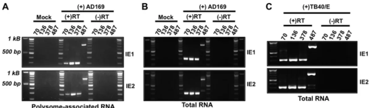
Each of the new MIE transcripts included exon 2, which
con-tains the initiator methionine for both the IE1 and IE2 proteins
(
16
). To determine whether the novel MIE transcripts encoded
full-length IE1 or IE2 protein, we used reverse transcriptase PCR
(RT-PCR) to determine whether IE1- or IE2-specific sequence
(exon 4 or exon 5, respectively) was found on mRNAs originating
from each of the newly identified TSSs. We focused our analysis on
four transcript isoforms: (i) the longest transcript (UTR487), (ii)
the previously described MIE transcript (UTR136), and (iii) the
two transcripts initiating in intron A (UTR378 and UTR70). Total
or polysome-associated RNA was extracted from HCMV-infected
cells and reverse transcribed using primers specific for either IE1
(exon 4) or IE2 (exon 5). The resulting cDNA was amplified using
primers specific to exon 4 or exon 5 together with a primer
recog-nizing the individual transcript isoforms. No PCR product was
obtained from cDNA from uninfected cells or in the absence of
reverse transcriptase, demonstrating the specificity of each
reac-tion (
Fig. 2A
and
B
). In the presence of reverse transcriptase, PCR
products were obtained from both polysome-associated (
Fig. 2A
)
and total (
Fig. 2B
) cDNA for each 5=
UTR linked to either IE1- or
IE2-specific exons. Each MIE transcript was also detected in total
RNA from cells infected with the HCMV TB40/E strain (
Fig. 2C
).
Sequence analysis of the amplicons confirmed that each transcript
contained exons 2 and 3 and was properly spliced to either exon 4
or exon 5 (data not shown). We conclude that transcripts arising
from each MIE transcription start site give rise to mature
cytoplas-mic transcripts encoding full-length IE1 or IE2 and that each
tran-script has the potential to contribute to IE1 and IE2 protein
ex-pression during infection.
To measure the temporal accumulation of each MIE
tran-script, we used quantitative reverse transcriptase PCR (qRT-PCR)
to measure the abundance of each transcript at different times
after infection. RNA was harvested at various times after infection,
and the abundance of each MIE transcript was determined by
comparison to a standard curve specific for each amplicon using
qRT-PCR (
Fig. 3A
). Each primer pair had a similar range of
de-tection and amplified its target with similar efficiency. The
abun-dance of the previously described MIE 5=
UTR (UTR136)
de-creased between 6 and 24 h after infection (
16
) (
Fig. 3A
). UTR136
abundance did not return to initial levels until 96 h after infection.
The abundance of the UTR378 transcript increased fivefold
be-tween 6 and 48 h after infection, with a maximal increase at 96 h.
The longest transcript, UTR487, displayed a similar trend, but it
was even more abundant at 96 h than at the 6-h time point.
Temporal regulation of abundance and promoter activity of
alternative MIE transcripts.
In comparing the abundance of the
different transcripts, the known MIE transcript (UTR136) was by
far the most abundant MIE transcript expressed 6 h after
infec-tion. However, by 72 h after infection, the UTR378 and UTR487
transcripts were expressed to at least the level of the UTR136
tran-script. Thus, the UTR136 transcript is the most abundant MIE
transcript at immediate early times after infection. However,
tran-scription of the novel MIE transcript isoforms increases as
infec-tion progresses, eventually rivaling expression of the previously
described MIE transcript, UTR136. Like the UTR136 transcript,
UTR378 transcript abundance was increased in the presence of
cycloheximide. In contrast, cycloheximide significantly reduced
UTR487 abundance (
Fig. 3B
). To measure the role of viral DNA
replication in the accumulation of each MIE transcript, we
mea-sured the effect of phosphonoacetic acid (PAA), an inhibitor of
viral DNA replication, on MIE transcript abundance. PAA
signif-icantly inhibited accumulation of the novel MIE transcripts, with
FIG 2Transcripts originating from each MIE transcription start site give rise to mature IE1 and IE2 mRNAs. Mock-infected (Mock) or HCMV-infected MRC-5 fibroblasts were harvested at 72 h after infection. (A) Polysome-associated RNA was extracted from AD169-infected cells and reverse transcribed using a primer specific for either exon 4 (IE1) or exon 5 (IE2). The resulting cDNA was amplified using a primer specific for either IE1 or IE2 together with a primer specific for each UTR (the primers are indicated by the length [in base pairs] of their their 5=UTR [UTR70, UTR136, UTR378, and UTR487]), and the PCR products were visualized on agarose gels. Reverse transcriptase was omitted in a set of samples [(-)RT] to ensure the absence of contaminating DNA. (B) Total RNA was extracted and analyzed as in panel A. (C) As in panel B, except cells were infected with the HCMV TB40/E strain (MOI of 3). The PCR products in panels A, B, and C were cloned and sequenced to ensure their specificity.
a more pronounced effect on accumulation of the UTR378 and
UTR487 mRNAs (
Fig. 3C
). These data show that the newly
iden-tified MIE transcripts have a complex expression profile, which
together with the UTR136 transcript allows for continued IE1/2
transcription throughout the HCMV lytic cycle.
Sequences surrounding transcription start sites act as
promot-ers that recruit RNA polymerase to initiate transcription. To
de-termine whether the sequences surrounding the novel MIE
tran-scription start sites possessed promoter activity, we cloned the
region from
⫺
450 to
⫹
50 bp flanking each transcription start site
into a promoterless luciferase reporter vector (
Fig. 4A
) and
mea-sured luciferase expression after transfection into HeLa cells (
Fig.
4B
) or primary human fibroblasts (
Fig. 4C
). Each promoter
re-gion increased the amount of luciferase activity in both cell types
compared to the empty luciferase vector lacking a promoter,
al-though to various degrees. The reporter containing the MIEP was
the most active promoter, increasing luciferase expression by
⬎
49,000-fold compared to the empty vector (
Fig. 4C
). The least
active promoter segment was the region surrounding the UTR378
TSS, which resulted in a modest but consistent
⬎
3-fold increase in
luciferase activity. The regions surrounding the UTR487 and
UTR70 transcription start sites had greater activity, increasing
lu-ciferase activity by 28- and 51-fold, respectively, compared to the
empty vector.
We next measured the effect of HCMV infection on the activity
of each promoter. Fibroblasts were electroporated with the
re-porter constructs and infected with HCMV, and luciferase activity
was measured over a time course of infection (
Fig. 4D
). Infection
had minimal effect on the activity of the UTR136 promoter,
in-creasing its activity by twofold throughout infection. Consistent
with our qRT-PCR analysis, each of the novel promoters was
in-duced by HCMV infection. Compared to uninfected cells, the
UTR70 promoter was induced 25-, 58-, and 50-fold at 24, 48, or 72
h after infection, respectively. At 24, 48, or 72 h after infection, the
UTR378 promoter was similarly induced 130-, 435-, and 470-fold,
respectively, and UTR487 was induced 5-, 20-, and 20-fold,
re-spectively (
Fig. 4D
). We conclude that the regions surrounding
the novel MIE transcription start sites have promoter activity that
is induced during HCMV infection.
The core promoter region of the MIEP is not necessary for
IE1 and IE2 expression outside the context of HCMV infection.
We next investigated whether the novel MIE promoters were
ac-tive outside of infection in the context of the MIE genomic locus.
The pSVH plasmid contains the region of the HCMV genome
from 840 nucleotides upstream of the MIEP transcription start
site to 1,413 nucleotides downstream of the polyadenylation
sig-nal following exon 5 (
17
). 5=
RACE analysis of
polysome-associ-ated RNA from pSVH-transfected HeLa cells using a gene-specific
primer recognizing MIE exon 2 produced a single prominent
band (
Fig. 5D
). When the PCR product was sequenced, it was
found to correspond to the previously defined MIEP transcription
start site, UTR136. Thus, the UTR136 transcript is the
predomi-nant MIE transcript expressed from the MIE locus in uninfected
cells.
Previous studies found that the highly active MIEP can
sup-press the activity of weaker promoters by competing for RNA
polymerase II recruitment (
58
,
59
). To determine whether
pro-moter competition might explain our failure to observe
tran-scripts arising from the alternative promoters in
pSVH-trans-fected cells, we generated a mutated version of the pSVH plasmid
(pSVH
⌬
MIEP) in which the core promoter region of the MIEP
(nucleotides
⫺
94 to
⫹
64 bp surrounding the transcription start
site) was deleted (
Fig. 5A
). Both IE1 and IE2 protein and mRNA
were expressed following pSVH
⌬
MIEP transfection, albeit at
sig-nificantly reduced levels compared to the level in
pSVH-trans-fected cells (
Fig. 5B
and
C
, respectively). This could reflect
in-creased alternative promoter activity driven by a change in
proximity to the MIE enhancer. In any case, these data show that
the core promoter region of the MIEP is not required for IE1 and
IE2 expression from the MIE genomic locus outside the context of
infection.
5=
RACE analysis of polysome-associated RNAs from
pSVH
⌬
MIEP-transfected cells generated two PCR products,
nei-ther of which matched the size of the UTR136 5=
UTR found in
cells transfected with wild-type pSVH. When the PCR products
were cloned and sequenced, they were found to correspond to the
UTR378 and UTR70 transcription start sites in intron A (
Fig. 5D
).
Thus, the regions flanking the alternative transcription start sites
in intron A have promoter activity that is sufficient to drive IE1
and IE2 expression in the context of the MIE genomic locus in the
absence of the MIEP. These data also suggest that promoter
sup-pression by the MIEP limits the activity of the intronic MIE
pro-moters.
Intron A of the MIE locus is important for efficient IE1 and
IE2 expression and HCMV replication.
Our results suggested
that sequences in intron A contributed to the expression of IE1
and IE2. We therefore constructed an HCMV recombinant,
HCMV
⌬
IntronA, wherein the entire intron A was deleted, leaving
a seamless fusion of exon 1 to exon 2 of the canonical MIE
tran-script. We also constructed a second recombinant, HCMV
⌬
UTR378, lacking the 250 nucleotides surrounding the
transcrip-tion start site for the UTR378 transcript (
Fig. 6A
). The entire
genome of each recombinant and the parental virus was
rese-FIG 5The core promoter region of the MIEP is not necessary for IE1 and IE2 expression outside the context of HCMV infection. (A) Cartoon showing a portion of the MIE locus in pSVH. The numbers show the locations of the MIE transcription start sites for the indicated MIE transcripts shown inFig. 1. pSVH⌬MIEP was created by removing a 158-bp region containing the MIEP core promoter (⫺94 to⫹64 relative to the transcription start site). (B) HeLa cells were left untransfected {negative control [(-) control]} or transfected with pSVH or pSVH⌬MIEP (⌬MIEP). IE1 and IE2 protein levels were measured by Western blotting at 24 h after transfection. (C) HeLa cells were transfected and harvested as in panel B. The relative abundance of the IE1 and IE2 mRNAs in pSVH⌬MIEP-transfected cells compared to pSVH-transfected cells was deter-mined by qRT-PCR using the⌬⌬CTmethod. IE1 and IE2 abundance in
pSVH-transfected cells is set at one. (D) HeLa cells were transfected as in panel B. 5=RACE analysis of polysome-associated RNA was performed using gene-specific primers located in exon 2. 5=RACE PCR products were visualized on agarose gels and subsequently cloned and sequenced. No PCR products were obtained in cells where tobacco acid pyrophosphatase (TAP) was omitted (⫺), demonstrating that the PCR products were derived from mRNAs containing a 5=m7G cap.
FIG 6Removal of MIE intron A delays HCMV replication. (A) Diagram depicting the genomic regions removed from each recombinant HCMV BAC. (B) MRC-5 fibroblasts were infected with wild-type (WT) HCMV, HCMV⌬Intron A (⌬Intron A), or HCMV⌬UTR378 (⌬UTR378) (MOI of 0.5). Cell-free virus was quantified at the indicated times after infection by the TCID50assay. (C) Fibroblasts were infected with HCMV (MOI of 3), and cell-free virus was quantified as
quenced to confirm the absence of spurious mutations. Infectious
virus was recovered following electroporation of the recombinant
genomes into MRC-5 fibroblasts, demonstrating that neither
in-tron A nor the sequences surrounding the UTR378 promoter are
essential for virus replication (
Fig. 6B
).
To measure the effect of each mutation on virus replication, we
compared the replication kinetics of the wild-type and
recombi-nant viruses. Cells were infected at a multiplicity of infection
(MOI) of 0.5 or 3, and the accumulation of cell-free virus was
measured over a single round of virus replication. We observed
reduced accumulation of cell-free virus with both mutants at 72
and 96 h after infection at either multiplicity of infection, though
the effect was more pronounced at the lower multiplicity (
Fig. 6B
and
C
). In addition, both recombinant viruses had a modest, but
reproducible, reduction in the accumulation of viral DNA (
Fig.
6D
). Thus, deletion of intron A or the region surrounding the
UTR378 promoter resulted in delayed replication kinetics
com-pared to wild-type virus.
To better define the defect in viral replication after
low-multi-plicity infection, we measured the accumulation of representative
immediate early, early, and late proteins after infection with the
recombinant viruses. Both recombinants expressed decreased
lev-els of the IE1 and E2 proteins compared to the wild-type virus. In
addition, the levels of the early protein pUL44 and the late protein
pp28 were expressed with delayed kinetics and to reduced levels
(
Fig. 7A
). The delayed replication of the HCMV
⌬
Intron A and
HCMV
⌬
UTR378 viruses thus correlated with decreased IE1 and
IE2 protein expression.
To determine whether the reduction in IE1 and IE2 protein
levels were the result of decreased IE1 and IE2 transcript levels, we
measured the accumulation of IE1 and IE2 transcripts after
infec-tion with each recombinant virus. Despite the presence of
equiv-alent numbers of viral genomes (
Fig. 7B
), IE1 and IE2 mRNA
levels were reduced in cells infected with the recombinant viruses
than in cells infected with the wild-type virus at early times of
infection (
Fig. 7C
). At late times after infection, both
recombi-nants expressed reduced levels of IE2 mRNA, but IE1 mRNA
lev-els were similar to those in cells infected with the wild-type virus
(
Fig. 7C
). Expression of the UTR136 transcript closely mirrored
IE1 expression; less UTR136 mRNA was present early after
infec-tion with either mutant, but its expression reached the same level
as in cells infected with wild-type virus by 72 h. Thus, the defect in
IE1 and IE2 mRNA levels correlated with a decrease in IE1 and IE2
protein expression. We conclude that sequences in intron A, and
the sequences surrounding the UTR378 TSS in MIE intron A, are
important for maximal expression of IE1 and IE2 and efficient
HCMV replication.
DISCUSSION
Proper regulation of IE1 and IE2 expression is critical for both
lytic and latent HCMV infection. In this study, we describe novel
IE1 and IE2 transcript isoforms arising from alternative
transcrip-tion start site usage. The novel transcripts encode full-length IE1
and IE2 proteins, but they differ from the previously described
MIE transcript in their 5=
UTRs (
Fig. 2
). Several of the new
tran-script isoforms extended exon 1 of the previously described MIE
transcript, while two of the novel transcripts initiated within
in-tron A (
Fig. 1
). The DNA sequences surrounding the new
tran-scription start sites had promoter activity (
Fig. 4B
and
C
) that was
induced upon HCMV infection (
Fig. 4D
), consistent with the
cumulation of the novel transcript isoforms as infection
pro-gressed (
Fig. 3A
). Deletion of MIE intron A or the region
sur-rounding the UTR378 TSS in intron A delayed the production of
cell-free virus (
Fig. 6B
and
C
) and decreased IE1 and IE2 mRNA
and protein accumulation (
Fig. 7
). Together, these results suggest
that multiple transcription units within the MIE region control
the expression of full-length IE1 and IE2 proteins during lytic
virus replication.
Several spliced and alternatively spliced transcripts are
tran-scribed from the MIE locus (
16–19
). Many of the alternative IE1
and IE2 mRNAs encode truncated versions of the IE1 and IE2
proteins that have distinct roles in regulating HCMV gene
expres-sion (
18
,
19
). Our results suggest that in addition, the MIE locus
gives rise to multiple transcripts encoding full-length IE1 and IE2
proteins. RT-PCR analysis of infected-cell RNA demonstrated
that each of the MIE transcription start sites was linked to both
exons 2 and 3, as well as the IE1- or IE2-specific exons (exon 4 or
5, respectively;
Fig. 2A
). The alternative transcripts were expressed
from both laboratory (AD169) and clinical (TB40/E) HCMV
strains (
Fig. 2B
and
C
) and thus likely are not the result of
adap-tation of the virus to
in vitro
passage. Each MIE transcript was
associated with polysomes in HCMV-infected cells, suggesting
that these transcripts contribute to IE1 and IE2 protein expression
(
Fig. 2A
). In addition, our results show that the upstream MIE
transcription start site previously found in latently infected
mono-cytes (UTR406 [
41
]) is also utilized during lytic infection (
Fig. 1
).
Thus, multiple transcription units contribute to the expression of
full-length IE1 and IE2 during lytic HCMV infection.
We also found that deleting either intron A of the MIE locus or
a portion of intron A containing one of the novel MIE promoters
decreased IE1 and IE2 mRNA and protein levels (
Fig. 7
) and
de-layed virus replication (
Fig. 6B
and
C
) without affecting viral entry
(
Fig. 7B
). These results are consistent with a role for the MIE
transcripts that initiate in intron A in the expression of IE1 and IE2
during infection. We obtained similar results with the smaller
de-letion of sequences surrounding the UTR378 start site.
Interest-ingly, while IE1 and UTR136 mRNA levels were similar at late
times after infection with the wild-type and deletion viruses, IE2
expression remained reduced at all time points (
Fig. 7
). This may
suggest that the alternative MIE transcripts are more important
for IE2 expression, particularly during the late stage of infection.
However, it is important to note that other undefined regulatory
elements could also be affected by these mutations and thus could
contribute to the observed decrease in IE1 and IE2 expression, as
suggested by the decreased expression of the UTR136 transcript at
early times after infection when intron A was deleted. Detailed
mapping of transcription factor binding sites and regulatory
se-quences controlling the intronic promoters together with a series
of subtle viral mutants will be needed to better define the factors
that regulate intronic promoter activity.
Our results suggest that the transcriptional regulation of
full-length IE1 and IE2 is more complex than previously appreciated.
We found that the MIE locus contains multiple promoters,
in-cluding two promoters located in intron A of the previously
de-scribed MIE transcript. This could explain results showing that the
presence of intron A modestly enhances the expression of
down-stream reporter genes (
60–62
). Perhaps this reflects a contribution
of the UTR378 and UTR70 promoters to transgene expression.
Our data also suggest that the coordinated regulation of the MIE
promoters allows for appropriate levels of IE1 and IE2 expression
throughout infection. The MIEP suppressed the weaker
alterna-tive MIE promoters in the context of the MIE locus (
Fig. 5
), which
correlates with the accumulation of the alternative MIE
tran-scripts later in infection when increased IE2 protein levels should
limit MIEP activity (
32
,
35
,
37
,
63
). We also find that the UTR378
transcript can be transcribed in the presence of cycloheximide,
suggesting a potential role in the immediate early expression of
IE1 and IE2 as well (
Fig. 3B
). However, the contribution of this
transcript to IE1 and IE2 expression at IE times is likely to be
minor, as it is approximately 10-fold less abundant than the
UTR136 transcript at this time.
We also found that maximal accumulation of the alternative
MIE transcripts required viral DNA replication (
Fig. 3C
). The
promoters of herpesvirus late genes contain TATT motifs, which
are recognized by a complex of viral proteins that facilitate RNA
polymerase II recruitment (
64
,
65
). Interestingly, TATT elements
are found approximately 30 nucleotides upstream of several of the
alternative MIE transcription start sites (UTR487, UTR406, and
UTR378; our unpublished observations), suggesting a role for
HCMV proteins in regulating the accumulation of the alternative
MIE transcripts. Thus, the specific sequence elements in the
alter-native MIE promoters may allow for the temporal or
context-specific regulation of IE1 and IE2 transcription.
Together, our data suggest a model for the regulation of IE1
and IE2 expression during HCMV infection. During the
immedi-ate early stage of infection, IE1 and IE2 transcription is controlled
primarily by the MIEP. As IE2 accumulates, it binds the
crs
se-quence in the MIEP and represses MIEP promoter activity.
Sup-pression of the MIEP by IE2 allows the weaker promoters
control-ling expression of the UTR70 and UTR378 transcripts to become
active, which together account for the majority of the IE1 and IE2
transcripts made during the later stages of infection. The
com-bined action of the multiple MIE promoters thus allows for
con-tinued transcription of IE1 and IE2 despite the accumulation of
IE2 protein as infection progresses.
ACKNOWLEDGMENTS
We thank members of the Moorman, Heise, de Silva, and de Paris labs as well as the members of the UNC Virology community for helpful discus-sions and input. We also thank Westefer Sanders for help sequencing the recombinant HCMV strain and Qi Tang (University of Puerto Rico) for providing the pSVH plasmid.
This work was supported by NIH grants AI03311 and AI123811 to N.J.M., the North Carolina University Cancer Research Fund, and awards from the UNC Virology Training grant (T32 AI07419 to B.Z. and K.C.A.) and National Science Foundation Graduate Research Fellowship grant (DGE-1144081) to K.C.A.
FUNDING INFORMATION
This work, including the efforts of Nathaniel John Moorman, was funded by HHS | NIH | National Institute of Allergy and Infectious Diseases (NIAID) (AI03311). This work, including the efforts of Nathaniel John Moorman, was funded by HHS | NIH | National Institute of Allergy and Infectious Diseases (NIAID) (AI123811). This work, including the efforts of Kyle C Arend and Benjamin Ziehr, was funded by HHS | NIH | National Institute of Allergy and Infectious Diseases (NIAID) (T32 AI07419). This work, including the efforts of Kyle C Arend, was funded by National Science Foundation (NSF) (DGE-1144081).
REFERENCES
1.Stinski MF, Thomsen DR, Stenberg RM, Goldstein LC.1983. Organi-zation and expression of the immediate early genes of human cytomega-lovirus. J Virol46:1–14.
2.Hermiston TW, Malone CL, Witte PR, Stinski MF.1987. Identification and characterization of the human cytomegalovirus immediate-early re-gion 2 gene that stimulates gene expression from an inducible promoter. J Virol61:3214 –3221.
3.Iwamoto GK, Monick MM, Clark BD, Auron PE, Stinski MF, Hun-ninghake GW.1990. Modulation of interleukin 1 beta gene expression by the immediate early genes of human cytomegalovirus. J Clin Invest85:
1853–1857.http://dx.doi.org/10.1172/JCI114645.
4.Geist LJ, Monick MM, Stinski MF, Hunninghake GW.1991. The im-mediate early genes of human cytomegalovirus upregulate expression of the interleukin-2 and interleukin-2 receptor genes. Am J Respir Cell Mol Biol5:292–296.http://dx.doi.org/10.1165/ajrcmb/5.3.292.
5.Crump JW, Geist LJ, Auron PE, Webb AC, Stinski MF, Hunninghake GW.1992. The immediate early genes of human cytomegalovirus require only proximal promoter elements to upregulate expression of interleu-kin-1 beta. Am J Respir Cell Mol Biol6:674 – 677.http://dx.doi.org/10 .1165/ajrcmb/6.6.674.
6.Monick MM, Geist LJ, Stinski MF, Hunninghake GW.1992. The im-mediate early genes of human cytomegalovirus upregulate expression of the cellular genes myc and fos. Am J Respir Cell Mol Biol7:251–256.http: //dx.doi.org/10.1165/ajrcmb/7.3.251.
7.Geist LJ, Monick MM, Stinski MF, Hunninghake GW.1994. The im-mediate early genes of human cytomegalovirus upregulate tumor necrosis factor-alpha gene expression. J Clin Invest93:474 – 478.http://dx.doi.org /10.1172/JCI116995.
8.Caswell R, Bryant L, Sinclair J.1996. Human cytomegalovirus immedi-ate-early 2 (IE2) protein can transactivate the human hsp70 promoter by alleviation of Dr1-mediated repression. J Virol70:4028 – 4037. 9.Yurochko AD, Huong SM, Huang ES.1999. Identification of human
cytomegalovirus target sequences in the human immunodeficiency virus long terminal repeat. Potential role of IE2-86 binding to sequences be-tween⫺120 and⫺20 in promoter transactivation. J Hum Virol2:81–90. 10. Lukac DM, Harel NY, Tanese N, Alwine JC.1997. TAF-like functions of human cytomegalovirus immediate-early proteins. J Virol71:7227–7239. 11. Petrik DT, Schmitt KP, Stinski MF.2007. The autoregulatory and trans-activating functions of the human cytomegalovirus IE86 protein use in-dependent mechanisms for promoter binding. J Virol81:5807–5818.http: //dx.doi.org/10.1128/JVI.02437-06.
12. Hagemeier C, Walker SM, Sissons PJ, Sinclair JH.1992. The 72K IE1 and 80K IE2 proteins of human cytomegalovirus independently trans-activate the c-fos, c-myc and hsp70 promoters via basal promoter ele-ments. J Gen Virol73(Part 9):2385–2393.http://dx.doi.org/10.1099/0022 -1317-73-9-2385.
13. Nevels M, Paulus C, Shenk T.2004. Human cytomegalovirus immedi-ate-early 1 protein facilitates viral replication by antagonizing histone deacetylation. Proc Natl Acad Sci U S A101:17234 –17239.http://dx.doi .org/10.1073/pnas.0407933101.
14. Saffert RT, Penkert RR, Kalejta RF.2010. Cellular and viral control over the initial events of human cytomegalovirus experimental latency in CD34⫹cells. J Virol84:5594 –5604.http://dx.doi.org/10.1128/JVI.00348-10.
15. Zalckvar E, Paulus C, Tillo D, Asbach-Nitzsche A, Lubling Y, Winterling C, Strieder N, Mucke K, Goodrum F, Segal E, Nevels M.2013. Nucleosome maps of the human cytomegalovirus genome reveal a temporal switch in chromatin organization linked to a major IE protein. Proc Natl Acad Sci U S A110:13126 –13131.http://dx.doi.org/10.1073/pnas.1305548110. 16. Stenberg RM, Witte PR, Stinski MF. 1985. Multiple spliced and
un-spliced transcripts from human cytomegalovirus immediate-early region 2 and evidence for a common initiation site within immediate-early re-gion 1. J Virol56:665– 675.
17. Stenberg RM, Depto AS, Fortney J, Nelson JA.1989. Regulated expres-sion of early and late RNAs and proteins from the human cytomegalovirus immediate-early gene region. J Virol63:2699 –2708.
18. Awasthi S, Isler JA, Alwine JC.2004. Analysis of splice variants of the immediate-early 1 region of human cytomegalovirus. J Virol78:8191– 8200.http://dx.doi.org/10.1128/JVI.78.15.8191-8200.2004.
19. Shirakata M, Terauchi M, Ablikim M, Imadome K, Hirai K, Aso T, Yamanashi Y.2002. Novel immediate-early protein IE19 of human cyto-megalovirus activates the origin recognition complex I promoter in a co-operative manner with IE72. J Virol76:3158 –3167.http://dx.doi.org/10 .1128/JVI.76.7.3158-3167.2002.
20. Bresnahan WA, Shenk TE.2000. UL82 virion protein activates expres-sion of immediate early viral genes in human cytomegalovirus-infected cells. Proc Natl Acad Sci U S A97:14506 –14511.http://dx.doi.org/10.1073 /pnas.97.26.14506.
21. Cantrell SR, Bresnahan WA.2005. Interaction between the human cyto-megalovirus UL82 gene product (pp71) and hDaxx regulates immediate-early gene expression and viral replication. J Virol79:7792–7802.http://dx .doi.org/10.1128/JVI.79.12.7792-7802.2005.
22. Cantrell SR, Bresnahan WA.2006. Human cytomegalovirus (HCMV) UL82 gene product (pp71) relieves hDaxx-mediated repression of HCMV replication. J Virol 80:6188 – 6191. http://dx.doi.org/10.1128/JVI .02676-05.
23. Cristea IM, Moorman NJ, Terhune SS, Cuevas CD, O’Keefe ES, Rout MP, Chait BT, Shenk T.2010. Human cytomegalovirus pUL83 stimu-lates activity of the viral immediate-early promoter through its interaction with the cellular IFI16 protein. J Virol84:7803–7814.http://dx.doi.org/10 .1128/JVI.00139-10.
24. Meier JL, Keller MJ, McCoy JJ.2002. Requirement of multiple wild-type cis-acting elements in the human cytomegalovirus major immediate-early distal enhancer for viral gene expression and replication. J Virol76:313– 326.http://dx.doi.org/10.1128/JVI.76.1.313-326.2002.
25. Hunninghake GW, Monick MM, Liu B, Stinski MF.1989. The promot-er-regulatory region of the major immediate-early gene of human cyto-megalovirus responds to T-lymphocyte stimulation and contains func-tional cyclic AMP-response elements. J Virol63:3026 –3033.
26. Penkert RR, Kalejta RF.2012. Tale of a tegument transactivator: the past, present and future of human CMV pp71. Future Virol7:855– 869.http: //dx.doi.org/10.2217/fvl.12.86.
27. Saffert RT, Kalejta RF.2006. Inactivating a cellular intrinsic immune defense mediated by Daxx is the mechanism through which the human cytomegalo-virus pp71 protein stimulates viral immediate-early gene expression. J Virol
80:3863–3871.http://dx.doi.org/10.1128/JVI.80.8.3863-3871.2006. 28. Romanowski MJ, Garrido-Guerrero E, Shenk T.1997. pIRS1 and pTRS1
are present in human cytomegalovirus virions. J Virol71:5703–5705. 29. Romanowski MJ, Shenk T.1997. Characterization of the human
cyto-megalovirus irs1 and trs1 genes: a second immediate-early transcription unit within irs1 whose product antagonizes transcriptional activation. J Virol71:1485–1496.
30. Macias MP, Huang L, Lashmit PE, Stinski MF.1996. Cellular or viral protein binding to a cytomegalovirus promoter transcription initiation site: effects on transcription. J Virol70:3628 –3635.
31. Isomura H, Tsurumi T, Stinski MF.2004. Role of the proximal enhancer of the major immediate-early promoter in human cytomegalovirus replication. J Virol 78:12788 –12799. http://dx.doi.org/10.1128/JVI.78.23.12788-12799 .2004.
repression sequence adjacent to the transcription start site of the human cytomegalovirus US3 gene is required to down regulate gene expression at early and late times after infection. J Virol72:9575–9584.
33. Liu B, Hermiston TW, Stinski MF.1991. A wild-type cis-acting element in the major immediate-early (IE) promoter of human cytomegalovirus is required for negative regulation by IE2. J Virol65:897–903.
34. Pizzorno MC, Hayward GS.1990. The IE2 gene products of human cytomegalovirus specifically down-regulate expression from the major immediate-early promoter through a target sequence located near the cap site. J Virol64:6154 – 6165.
35. Cherrington JM, Khoury EL, Mocarski ES.1991. Human cytomegalo-virus ie2 negatively regulates alpha gene expression via a short target se-quence near the transcription start site. J Virol65:887– 896.
36. Macias MP, Stinski MF.1993. An in vitro system for human cytomega-lovirus immediate early 2 protein (IE2)-mediated site-dependent repres-sion of transcription and direct binding of IE2 to the major immediate early promoter. Proc Natl Acad Sci U S A90:707–711.http://dx.doi.org /10.1073/pnas.90.2.707.
37. Reeves M, Murphy J, Greaves R, Fairley J, Brehm A, Sinclair J.2006. Autorepression of the human cytomegalovirus major immediate-early promoter/enhancer at late times of infection is mediated by the recruit-ment of chromatin remodeling enzymes by IE86. J Virol80:9998 –10009. http://dx.doi.org/10.1128/JVI.01297-06.
38. Sanders RL, Del Rosario CJ, White EA, Spector DH.2008. Internal dele-tions of IE2 86 and loss of the late IE2 60 and IE2 40 proteins encoded by human cytomegalovirus affect the levels of UL84 protein but not the amount of UL84 mRNA or the loading and distribution of the mRNA on polysomes. J Virol82:11383–11397.http://dx.doi.org/10.1128/JVI.01293-08.
39. White EA, Del Rosario CJ, Sanders RL, Spector DH.2007. The IE2 60-kilodalton and 40-kilodalton proteins are dispensable for human cy-tomegalovirus replication but are required for efficient delayed early and late gene expression and production of infectious virus. J Virol81:2573– 2583.http://dx.doi.org/10.1128/JVI.02454-06.
40. Sanchez V, Clark CL, Yen JY, Dwarakanath R, Spector DH.2002. Viable human cytomegalovirus recombinant virus with an internal deletion of the IE2 86 gene affects late stages of viral replication. J Virol76:2973–2989. http://dx.doi.org/10.1128/JVI.76.6.2973-2989.2002.
41. Kondo K, Xu J, Mocarski ES.1996. Human cytomegalovirus latent gene expression in granulocyte-macrophage progenitors in culture and in se-ropositive individuals. Proc Natl Acad Sci U S A93:11137–11142.http: //dx.doi.org/10.1073/pnas.93.20.11137.
42. Wang D, Bresnahan W, Shenk T.2004. Human cytomegalovirus en-codes a highly specific RANTES decoy receptor. Proc Natl Acad Sci U S A
101:16642–16647.http://dx.doi.org/10.1073/pnas.0407233101. 43. Yu D, Ellis HM, Lee EC, Jenkins NA, Copeland NG, Court DL.2000. An
efficient recombination system for chromosome engineering in Esche-richia coli. Proc Natl Acad Sci U S A97:5978 –5983.http://dx.doi.org/10 .1073/pnas.100127597.
44. Yu D, Silva MC, Shenk T.2003. Functional map of human cytomegalo-virus AD169 defined by global mutational analysis. Proc Natl Acad Sci U S A100:12396 –12401.http://dx.doi.org/10.1073/pnas.1635160100. 45. Terhune S, Torigoi E, Moorman N, Silva M, Qian Z, Shenk T, Yu D.
2007. Human cytomegalovirus UL38 protein blocks apoptosis. J Virol
81:3109 –3123.http://dx.doi.org/10.1128/JVI.02124-06.
46. Terhune SS, Moorman NJ, Cristea IM, Savaryn JP, Cuevas-Bennett C, Rout MP, Chait BT, Shenk T.2010. Human cytomegalovirus UL29/28 protein interacts with components of the NuRD complex which promote accumulation of immediate-early RNA. PLoS Pathog6:e1000965.http: //dx.doi.org/10.1371/journal.ppat.1000965.
47. Lenarcic EM, Ziehr B, De Leon G, Mitchell D, Moorman NJ.2014. Differential role for host translation factors in host and viral protein syn-thesis during human cytomegalovirus infection. J Virol88:1473–1483. http://dx.doi.org/10.1128/JVI.02321-13.
48. Ziehr B, Lenarcic E, Cecil C, Moorman NJ.2016. The eIF4AIII RNA helicase is a critical determinant of human cytomegalovirus replication. Virology489:194 –201.http://dx.doi.org/10.1016/j.virol.2015.12.009. 49. Moorman NJ, Shenk T. 2010. Rapamycin-resistant mTORC1 kinase
activity is required for herpesvirus replication. J Virol84:5260 –5269.http: //dx.doi.org/10.1128/JVI.02733-09.
50. Stenberg RM, Fortney J, Barlow SW, Magrane BP, Nelson JA, Ghazal P.
1990. Promoter-specific trans activation and repression by human cyto-megalovirus immediate-early proteins involves common and unique pro-tein domains. J Virol64:1556 –1565.
51. Ziehr B, Lenarcic E, Vincent HA, Cecil C, Garcia B, Shenk T, Moorman NJ.2015. Human cytomegalovirus TRS1 protein associates with the 7-methylguanosine mRNA cap and facilitates translation. Proteomics15:
1983–1994.http://dx.doi.org/10.1002/pmic.201400616.
52. Ziehr B, Vincent HA, Moorman NJ. 2016. Human cytomegalovirus pTRS1 and pIRS1 antagonize protein kinase R to facilitate virus replica-tion. J Virol90:3839 –3848.http://dx.doi.org/10.1128/JVI.02714-15. 53. Zhu H, Shen Y, Shenk T.1995. Human cytomegalovirus IE1 and IE2
proteins block apoptosis. J Virol69:7960 –7970.
54. Cuevas-Bennett C, Shenk T.2008. Dynamic histone H3 acetylation and methylation at human cytomegalovirus promoters during replication in fi-broblasts. J Virol82:9525–9536.http://dx.doi.org/10.1128/JVI.00946-08. 55. Silva MC, Yu QC, Enquist L, Shenk T.2003. Human cytomegalovirus
UL99-encoded pp28 is required for the cytoplasmic envelopment of teg-ument-associated capsids. J Virol77:10594 –10605.http://dx.doi.org/10 .1128/JVI.77.19.10594-10605.2003.
56. Stenberg RM, Thomsen DR, Stinski MF.1984. Structural analysis of the major immediate early gene of human cytomegalovirus. J Virol
49:190 –199.
57. Sharp PA.1988. RNA splicing and genes. JAMA260:3035–3041.http://dx .doi.org/10.1001/jama.1988.03410200091032.
58. Lee T, Bradley ME, Walowitz JL.1998. Influence of promoter potency on the transcriptional effects of YY1, SRF and Msx-1 in transient transfection analysis. Nucleic Acids Res26:3215–3220.http://dx.doi.org/10.1093/nar /26.13.3215.
59. Reddy JC, Hosono S, Licht JD.1995. The transcriptional effect of WT1 is modulated by choice of expression vector. J Biol Chem270:29976 –29982. http://dx.doi.org/10.1074/jbc.270.50.29976.
60. Chapman BS, Thayer RM, Vincent KA, Haigwood NL.1991. Effect of intron A from human cytomegalovirus (Towne) immediate-early gene on heterologous expression in mammalian cells. Nucleic Acids Res19:3979 – 3986.http://dx.doi.org/10.1093/nar/19.14.3979.
61. Hennighausen L, Fleckenstein B.1986. Nuclear factor 1 interacts with five DNA elements in the promoter region of the human cytomegalovirus major immediate early gene. EMBO J5:1367–1371.
62. Jeang KT, Rawlins DR, Rosenfeld PJ, Shero JH, Kelly TJ, Hayward GS.
1987. Multiple tandemly repeated binding sites for cellular nuclear factor 1 that surround the major immediate-early promoters of simian and hu-man cytomegalovirus. J Virol61:1559 –1570.
63. Lang D, Stamminger T.1993. The 86-kilodalton IE-2 protein of human cytomegalovirus is a sequence-specific DNA-binding protein that inter-acts directly with the negative autoregulatory response element located near the cap site of the IE-1/2 enhancer-promoter. J Virol67:323–331. 64. Perng YC, Qian Z, Fehr AR, Xuan B, Yu D.2011. The human
cytomeg-alovirus gene UL79 is required for the accumulation of late viral tran-scripts. J Virol85:4841– 4852.http://dx.doi.org/10.1128/JVI.02344-10. 65. Wong-Ho E, Wu TT, Davis ZH, Zhang B, Huang J, Gong H, Deng H,
Liu F, Glaunsinger B, Sun R.2014. Unconventional sequence require-ment for viral late gene core promoters of murine gammaherpesvirus 68. J Virol88:3411–3422.http://dx.doi.org/10.1128/JVI.01374-13. 66. Moser JM, Farrell ML, Krug LT, Upton JW, Speck SH.2006. A
gam-maherpesvirus 68 gene 50 null mutant establishes long-term latency in the lung but fails to vaccinate against a wild-type virus challenge. J Virol80:
1592–1598.http://dx.doi.org/10.1128/JVI.80.3.1592-1598.2006. 67. Pavlova IV, Virgin HW, IV, Speck SH.2003. Disruption of
gamma-herpesvirus 68 gene 50 demonstrates that Rta is essential for virus replication. J Virol77:5731–5739.http://dx.doi.org/10.1128/JVI.77.10 .5731-5739.2003.
68. Wu TT, Tong L, Rickabaugh T, Speck S, Sun R.2001. Function of Rta is essential for lytic replication of murine gammaherpesvirus 68. J Virol
75:9262–9273.http://dx.doi.org/10.1128/JVI.75.19.9262-9273.2001. 69. Wakeman BS, Johnson LS, Paden CR, Gray KS, Virgin HW, Speck SH.
2014. Identification of alternative transcripts encoding the essential mu-rine gammaherpesvirus lytic transactivator RTA. J Virol88:5474 –5490. http://dx.doi.org/10.1128/JVI.03110-13.
70. Goodwin MM, Canny S, Steed A, Virgin HW.2010. Murine gamma-herpesvirus 68 has evolved gamma interferon and stat1-repressible pro-moters for the lytic switch gene 50. J Virol84:3711–3717.http://dx.doi.org /10.1128/JVI.02099-09.