Identification and characterization of the Drosophila melanogaster meiotic MCM complex
Kathryn Patricia Kohl
A dissertation submitted to the faculty of the University of North Carolina at Chapel Hill in partial fulfillment of the requirements for the degree of Doctor of Philosophy in the
Curriculum in Genetics and Molecular Biology.
Chapel Hill 2013
ii ©2013
iii ABSTRACT
KATHRYN PATRICIA KOHL: Identification and characterization of the Drosophila melanogaster meiotic MCM complex
(Under the direction of Jeff Sekelsky)
iv
v
TABLE OF CONTENTS
LIST OF TABLES ... ix
LIST OF FIGURES ...x
LIST OF ABBREVIATIONS ... xiii
CHAPTER 1. INTRODUCTION ...1
COs and NCOs in meiotic recombination models ...2
Rise of the two-pathway paradigm ...5
Anti-CO and pro-CO activities of Sgs1 ...8
Extending the two-pathway paradigm ...11
Meiotic and mitotic DSB repair in meiosis ...12
Drosophila melanogaster as a meiotic model system ...16
Mini-chromosome maintenance proteins ...17
Investigations into the role of the Drosophila precondition proteins ...19
CHAPTER 2. EVOLUTION OF AN MCM COMPLEX IN FLIES THAT PROMOTES MEIOTIC CROSSOVERS BY BLOCKING BLM HELICASE...21
vi
Drosophila stocks and genetics ... 36
Identification of orthologs... 37
Sequence alignments and phylogenetic analysis ... 38
Molecular evolutionary analysis ... 38
Immunoprecipitation ... 40
Yeast two-hybrid assay ... 41
CHAPTER 3. MEI-MCMS AFFECT MEIOTIC CROSSOVER DISTRIBUTION...42
Introduction ...42
Results ...44
REC ATPase mutants differentially affect NDJ and COs ... 44
Hyperrecombination in a MEI-218 truncation mutant... 50
Discussion ...54
Materials and Methods ...60
Protein sequence alignments ... 60
Generating mei-MCM transgenic flies ... 60
Non-disjunction assay ... 61
vii
CHAPTER 4. ADDITIONAL GENETIC AND MOLECULAR
INVESTIGATIONS OF rec ...63
Introduction ...63
Results and Discussion ...66
Msh6 rec gene conversion assay ... 66
MCM yeast two-hybrid assay ... 71
Immunoprecipitation of REC ... 73
Immunofluorescence of REC ... 77
MCM RNA in situ hybridization ... 79
Materials and Methods ...82
Msh6 rec gene conversion assay ... 82
Yeast two-hybrid assay ... 83
Western blotting ... 84
FLAGREC transgene ... 84
Non-disjunction... 84
Immunofluorescence ... 85
RNA in situ hybridization ... 86
viii
Highlighted Findings ...89
Future Directions ...91
ix
LIST OF TABLES
Table 1. Percentage of crossovers attributed to each pathway
in the early two-pathway paradigm... 6
Table 2. Crossover distribution ... 29
Table 3. Models for the evolution of MCM8/rec in Diptera ... 34
Table 4. rec transgene recombination frequency ... 48
Table 5. rec transgene crossover number ... 49
Table 6. mini-MEI transgene crossover number ... 53
Table 7. mini-MEI transgene recombination frequency ... 53
Table 8. mei-MCM transgene phenotypes ... 55
Table 9. Intragenic recombination at the rosy locus ... 69
x
LIST OF FIGURES
Figure 1. Models of meiotic double-strand break repair ... 4
Figure 2. Two meiotic crossover pathways ... 14
Figure 3. Drosophila and Glossina uniquely lack MSH4, MSH5, and MCM9 ... 22
Figure 4. Drosophila and Glossina uniquely lack MSH4, MSH5, and MCM9 ... 23
Figure 5. Domains identified by a search of the Conserved Domain Database in Drosophilamelanogaster REC, MEI-217, MEI-218 and human MEI-218 ... 25
Figure 6. mei-MCM complex ... 26
Figure 7. mei-MCMs antagonize DmBLM ... 27
Figure 8. Crossovers in rec and mus309 mutants ... 28
Figure 9. Possible evolutionary scenarios for the loss of Msh4–Msh5 in Drosophila and the evolution of mei-MCM complex ... 30
Figure 10. Divergence of MCM8 in Drosophila ... 31
Figure 11. Rates of evolution of REC/MCM8 in Dipteran insects and select outgroups ... 33
Figure 12. Conservation of MEI-217 and MEI-218 as separate polypeptides throughout Drosophila ... 35
Figure 13. Structural changes in MEI-218 ... 36
xi
Figure 15. rec transgene non-disjunction... 46
Figure 16. rec transgene crossover distribution ... 48
Figure 17. MEI-218 protein alignment ... 51
Figure 18. mini-MEI transgene non-disjunction ... 52
Figure 19. mini-MEI transgene crossover distribution ... 52
Figure 20. rec ATPase mutant model ... 57
Figure 21. Double-strand break repair model. ... 64
Figure 22. rosy gene conversion assay ... 67
Figure 23. Self-virgining cross scheme for Msh6 rec gene conversion assay ... 68
Figure 24. Non-crossover events in Msh6 rec mutants... 70
Figure 25. MCM yeast two-hybrid assay ... 72
Figure 26. REC antibodies ... 75
Figure 27. P{FLAGREC} non-disjunction ... 76
Figure 28. C(3)G staining ... 78
Figure 29. RNA in situ hybridization ... 80
Figure 30. rec RNA in situ hybridization ... 81
xii
xiii
LIST OF ABBREVIATIONS
AAA – ATPases associated with a variety of cellular activities CO – crossover
DCO – double crossover dHJ – double Holliday junction DSB – double-strand break
DSBR – double-strand break repair hDNA – heteroduplex DNA
HJ – Holliday junction JM – joint molecule
MCM – mini-chromosome maintenance NCO – non-crossover
NDJ – non-disjunction
pre-RC – pre-replicative complex SCO – single crossover
CHAPTER 1
INTRODUCTION
2
segregation of homologous chromosomes and to increase genetic diversity. Third, the preferred repair template was changed from the sister chromatid in mitotic cells to the homologous chromosome in meiotic cells, since only COs between homologs give the aforementioned benefits. Finally, exquisite CO control mechanisms arose to ensure the optimal number and distribution of COs across the genome and relative to one another. In particular, every chromosome pair receives at least one CO and if additional COs occur, they tend not to occur near one another, a phenomenon called crossover interference (reviewed in Berchowitz and Copenhaver, 2010).
A complication obscuring the relationship between the mitotic and meiotic recombination pathways has been the apparent existence of two meiotic CO pathways – one pathway that produces COs subject to interference and another that produces non-interfering COs. Recent studies suggest that the non-interfering CO pathway fits the scenario described above - i.e., it is a derivative of the mitotic DSB repair pathway that contains numerous meiosis-specific embellishments. The non-interfering pathway, however, shares striking similarities to mitotic DSB repair in its original form. Additional discoveries reveal functions that are essential for generating meiotic COs can be carried out by different proteins in different species. These findings provide a new framework through which meiotic recombination pathways can be viewed and allow organisms previously thought to use disparate CO pathways to be brought under the same umbrella.
COs and NCOs in meiotic recombination models
3
intermediate in his model is a structure in which strands from two homologous duplexes swap pairing partners across a short region, yielding a four-stranded intermediate now known as the Holliday junction (HJ). Holliday proposed that these junctions are cleaved by DNA repair enzymes, now known as resolvases, to reestablish two separate duplexes. Depending on which strands are nicked, this process, now called resolution, could result in CO or NCO products. The equally likely outcomes of resolution fit with fungal recombination studies that suggested that COs and NCOs occur in equal numbers.
4
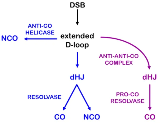
Figure 1. Models of meiotic double-strand break repair. (A) In the Szostak et al. (Szostak et al., 1983) model recombination initiates with a double-strand break (DSB) that is processed into an extended displacement loop (D-loop) and then a double Holliday junction (dHJ) structure. The dHJ is resolved into either a crossover (CO) or non-crossover (NCO) with equal probability. (B) In the revised model of Allers and Lichten (Allers and Lichten, 2001), some extended D-loops are unwound by an anti-CO helicase to produce NCOs, and dHJs are resolved by a pro-CO resolvase into COs.
5
mitotic DSB repair (reviewed in Pâques and Haber, 1999). In SDSA, after the invading strand is extended by DNA synthesis, helicases can disrupt the D-loop, freeing the nascent strand to anneal to the other end of the DSB.
Allers and Lichten noted another departure from the original DSBR model: Most JMs are processed into COs (Allers and Lichten, 2001) (Figure 1B). Although this discovery opposes the notion that resolution of a dHJ can produce a CO or a NCO with equal probability, it more readily accommodates the finding that NCOs outnumber COs, sometimes by a factor of ten or more (reviewed in Cole et al., 2012). Thus, in the revised model of Allers and Lichten, the backone of the original DSBR model is intact, but dHJs are now preferentially repaired as COs, and NCOs arise via SDSA instead of dHJ resolution. In this revised model, helicases that promote SDSA act as anti-CO factors and HJ resolvases become pro-CO factors rather than proteins that produce both COs and NCOs.
Rise of the two-pathway paradigm
6
two different pathways for meiotic CO formation (Table 1). The first pathway, which requires Msh4–Msh5, is responsible for a majority of COs in S. cerevisiae and all COs in C. elegans; the second, independent of Msh4–Msh5, produces the remaining COs in S. cerevisiae msh4 and msh5 mutants.
Class Type of CO Defining Proteins Sc Sp Percentage of COs Ce At Dm I interfering Msh4–Msh5 50-70 0 100 75-85 0 II interfering non- Mus81–Mms4 30-50 100 0 9-12 <10
Table 1. Percentage of crossovers attributed to each pathway in the early two-pathway paradigm. Organism abbreviations: Sc, Saccharomyces cerevisiae; Sp, Schizosaccharomyces pombe; Ce, Caenorhabditis elegans; At, Arabidopsis thaliana; Dm, Drosophila melanogaster. See text for references.
7
double mutants that lack both the Msh4–Msh5 and Mus81–Mms4 complexes have more severely reduced CO levels than mutants lacking either one, strongly supporting the existence of two pathways – one dependent on Msh4–Msh5 (Class I) and another on Mus81–Mms4 (Class II) (Berchowitz et al., 2007; de los Santos et al., 2003).
The nature of the COs produced by the two pathways was also in question – i.e., if there are two meiotic CO pathways, do the COs produced by them have different properties? A clue to the answer came from mathematical modeling of crossover interference. Copenhaver et al. (Copenhaver et al., 2002) were able to fit Arabidopsis data to a counting model for interference (Foss et al., 1993) if they assumed two types of COs, some that participate in interference and some that do not. Consistent with this prediction, experimental studies demonstrated that the residual COs in Arabidopsis and budding yeast msh4 and msh5 mutants do not display interference (Argueso et al., 2004; Higgins et al., 2004; Lu et al., 2008; Novak et al., 2001). Conversely, Mus81–Mms4-independent COs in S. cerevisiae and Arabidopsis do exhibit interference (Berchowitz et al., 2007; de los Santos et al., 2003). These results suggested that the Msh4–Msh5-dependent pathway produces COs subject to interference, whereas the Mus81–Mms4-dependent pathway produces non-interfering COs (Table 1). This formulation explains the finding that COs are non-interfering in S. pombe (Munz, 1994), as these COs are produced from the Mus81–Mms4 pathway, and the strong interference of all COs in C. elegans (Meneely et al., 2002), as these are all produced by the Msh4–Msh5 pathway.
8
still have some residual COs (de los Santos et al., 2003). In addition, though mathematical models of recombination in Drosophila fit best if most or all COs are interfering (Copenhaver et al., 2002), the Drosophila genome lacks Msh4 and Msh5 (Sekelsky et al., 2000), suggesting that another pathway produces interfering COs in this species.
Another shortcoming of the two-pathway paradigm is that the proteins used to define these pathways have very different functions: Mus81–Mms4 is an HJ resolvase whose activity presumably directly produces CO products (Boddy et al., 2001) (i.e., it is a pro-CO resolvase). Msh4–Msh5, however, does not directly produce COs, but instead blocks anti-CO helicases (i.e., it is an anti-anti-CO complex; see discussion below). Notably, the pro-CO resolvase that acts in the Msh4–Msh5-dependent pathway was unknown. Furthermore, the relationship between these pathways and the revised model for meiotic COs was unclear. Does the model fit both Class I and Class II pathways, with different proteins used for each, or is a second model necessary? These apparent weaknesses in the two-pathway paradigm for meiotic COs have largely been solved in the past year, as studies in a number of laboratories using different model organisms have clarified the roles and identities of pro-CO resolvases, anti-CO helicases, and anti-anti-CO complexes.
Anti-CO and pro-CO activities of Sgs1
9
preferentially repaired into NCOs, largely through the action of anti-CO helicases. One key anti-CO protein is the Bloom syndrome helicase BLM (reviewed in Andersen and Sekelsky, 2010). Although BLM likely has many anti-CO functions, two activities are relevant to DSB repair. First, studies in Drosophila suggested that BLM promotes SDSA, probably by disrupting D-loops after repair DNA synthesis (Adams et al., 2003; McVey et al., 2004). Second, in vitro studies demonstrated that BLM, together with topoisomerase IIIα and other proteins, can catalyze dHJ dissolution, a process in which
the two HJs are migrated toward one another and then decatenated (Wu and Hickson, 2003). Unlike resolution of dHJs, dissolution generates only NCOs.
Genetic studies suggested a similar anti-CO role for the S. cerevisiae BLM ortholog Sgs1 in meiosis. COs are reduced in mutants lacking ZMM proteins, including Msh4–Msh5, but, remarkably, COs are restored in double mutants that also lack Sgs1 (Jessop et al., 2006; Oh et al., 2007). An attractive interpretation of these results is that one function of ZMMs is to antagonize the anti-CO activity of Sgs1. Thus, Msh4–Msh5 is an anti-anti-CO protein.
10
suggests that when Sgs1 is absent, dHJs are resolved into COs and NCOs, as in the original DSBR model.
Additional insights came from physical studies of recombination in mutants lacking the known HJ resolvases. Three proteins, Mus81–Mms4, Yen1, and Slx1–Slx4, possess resolvase activity in vitro (Boddy et al., 2001; Fekairi et al., 2009; Ip et al., 2008). Mus81–Mms4 was shown to be important in generating mitotic crossovers, with Yen1 playing a compensatory or partially redundant role (Ho et al., 2010). Experiments by De Muyt et al. (De Muyt et al., 2012) and Zakharyevich et al. (Zakharyevich et al., 2012) found that single mutants lacking any one of these enzymes were still able to resolve most JMs and produce approximately normal numbers of COs. Even triple mutants lacking all three resolvases showed only a modest reduction in JM resolution and CO formation. These results suggest that the known resolvases collectively process only a small fraction of JMs. If these are JMs from the Class II pathway, then most JMs must be generated in the Class I pathway and be resolved by an unidentified resolvase.
11
What is the identity of the pro-CO resolvase that functions in the Class I pathway? It had previously been suggested that the mismatch repair proteins Mlh1–Mlh3 (MutLγ complex) and Exo1 might act in dHJ resolution (Nishant et al., 2008; Zakharyevich et al., 2010). COs are reduced in mlh3 mutants, but removal of Sgs1 restores COs, suggesting that Mlh3, like ZMMs, functions in the Class I pathway (Oh et al., 2007). Consistent with this hypothesis, Zakharyevich et al. (Zakharyevich et al., 2012) found that when all three known resolvases were removed, eliminating Mlh3 resulted in a similar reduction in COs as eliminating Sgs1. A parallel set of experiments suggested that Exo1 functions in a different pathway than Mus81–Mms4, putting Exo1 also in the Class I pathway.
These results are consistent with Sgs1 having the expected anti-CO functions: It promotes SDSA (in wild-type cells) and dHJ dissolution (when the three known resolvases and the putative pro-CO resolvase are all missing). Unexpectedly, the results also reveal a pro-CO role of Sgs1. This pro-CO role may be in influencing pathway choice: In the presence of Sgs1, the ZMM-dependent Class I CO pathway can be used, but in the absence of Sgs1, the alternative Class II pathway gives rise to both COs and NCOs from dHJ resolution.
Extending the two-pathway paradigm
12
exposure to ionizing radiation or by removal of RTEL-1 (yet another anti-CO helicase), the additional COs require MUS-81 but not the ZMM protein ZHP-3 (Youds et al., 2010). Crossover interference is reduced, consistent with these extra COs being formed through the Class II pathway. These findings expose the availability of the Class II pathway in C. elegans, even though it is normally not used to generate meiotic COs.
Additionally, Crismani et al. (Crismani et al., 2012) found that Arabidopsis mutants lacking the FANCM helicase had elevated COs. The additional COs were interference-insensitive, arose from a ZMM-independent pathway, and relied on Mus81 for formation. These findings parallel results seen in S. cerevisiae, where the absence of Sgs1 leads to COs being formed in an alternative, Mus81-dependent pathway. Furthermore, loss of FANCM rescues the meiotic defects of Arabidopsis zmm mutants, just like the removal of Sgs1 in S. cerevisiae zmm mutants (Crismani et al., 2012; Knoll et al., 2012). These findings strongly suggest that Arabidopsis FANCM functions as a meiotic anti-CO protein in a role that is antagonized by the ZMM proteins, similar to the role of Sgs1 in budding yeast. Thus, it appears that organisms can exchange proteins that occupy the same functional niche (in this case, swapping two anti-CO helicases) and still follow the framework of the two-pathway paradigm.
Meiotic and mitotic DSB repair in meiosis
13
unified model appears to be applicable to a more diverse set of model organisms than previously recognized. Furthermore, it is now apparent that the Class II pathway is strikingly similar to mitotic DSB repair in many respects. First, NCOs – not COs – are the predominant product. This outcome is achieved through SDSA, mediated by one or more anti-CO helicases. In instances where SDSA does not occur and a dHJ is generated, this intermediate can be resolved in an unbiased manner by “mitotic” resolvases to give either an NCO or a CO, but these COs are non-interfering. Despite these similarities, it should be noted that there are features of the Class II pathway that are unique to meiosis. For example, DSBs are generated by meiosis-specific Spo11 complexes, and engagement of DNA strands from the broken chromosome to the homologous chromosome is mediated in most species by meiosis-specific strand exchange proteins like Dmc1 (reviewed in Neale and Keeney, 2006). These events, however, may occur prior to the split between the Class I and Class II pathways (Figure 2).
14
Figure 2. Two meiotic crossover pathways. In this unified model, a DSB is processed into an extended D-loop. In the “mitotic-like” pathway (blue, Class II), the extended D-loop can be unwound by an anti-CO helicase to produce NCOs. In some cases a dHJ is generated and then resolved by an unbiased resolvase into either a CO or NCO product. In the meiosis-specific CO pathway (purple, Class I), an anti-anti-CO complex blocks the action of anti-CO helicases to promote formation of a dHJ intermediate, which is then acted upon by a pro-CO resolvase to form exclusively CO products. A dHJ is presented as a key intermediate to fit the original models and the detection of joint molecules (JMs) with properties of dHJs in physical assays. However, there are other models which posit additional/alternative intermediates, including single HJs and multi-chromatid JMs. Variations on the two-pathway model can accommodate these other intermediates and less-common fates of DSBs.
15
Over time, the Class I pathway evolved to place additional constraints on meiotic recombination to promote the optimal placement of COs. To ensure CO formation, additional regulation of anti-CO helicases active during mitotic DSB repair was developed. This functional niche was filled by meiosis-specific anti-anti-CO proteins like Msh4–Msh5. The Class I pathway also evolved so that dHJs are resolved in a biased way to produce COs but not NCOs. The Class II pathway remained available, perhaps as a failsafe to ensure that all DSBs are repaired.
16
the meiotic recombination field. The unified view of recombination pathways depicted in Figure 2 may help to guide some of these studies.
Drosophila melanogaster as a meiotic model system
The Drosophila model system is excellent for studying meiotic recombination. The long history of Drosophila meiotic research (Hawley, 1993), coupled with the absence of meiotic recombination in males (Morgan, 1912) (thus allowing the study of recombination events derived from a single parent only), the ability of Drosophila to tolerate high (0.5%) heterology (to aid in mapping recombination events) (Hilliker et al., 1991), and the increased likelihood of recovering non-disjunction (NDJ) progeny (some aneuplodies involving both the first and fourth chromosomes are viable) (Ashburner, 1989), all make Drosophila an ideal system for studying meiotic recombination.
17
The exchange class of meiotic recombination mutants contains four proteins hypothesized to form a complex necessary for HJ resolution: MEI-9, ERCC1, MUS312 and HDM (Joyce et al., 2009; Radford et al., 2005; Sekelsky et al., 1995; Yildiz et al., 2002). The precondition class genes include: mei-217, mei-218, recombination defective (rec) and Mcm5 (Baker and Carpenter, 1972; Blanton et al., 2005; Lake et al., 2007; Liu et al., 2000). Single mutants with null mutations in mei-217, mei-218 or rec exhibit identical phenotypes – a severe reduction in crossing over with residual COs showing the abnormal precondition distribution, high NDJ, normal synaptonemal complex formation, and no hypersensitivity to DNA damaging agents (Baker et al., 1976; Baker and Carpenter, 1972; Blanton et al., 2005; Carpenter, 1979; Grell, 1984; Liu et al., 2000). Epistasis experiments suggest that these precondition proteins function at an intermediate step in the meiotic recombination pathway (Blanton et al., 2005; Liu et al., 2000; Sekelsky et al., 1995). Likewise, a separation-of-function allele in Mcm5, called Mcm5A7, shows these same precondition mutant phenotypes (Lake et al., 2007).
Mini-chromosome maintenance proteins
-18
phosphates of ATP (Lindegren, 1955), while residues in the Walker B motif bind Mg2+ necessary for ATP catalysis (Schulz, 1992). MCM proteins are members of the AAA+ (ATPases Associated with a variety of cellular Activities) ATPase family (Koonin, 1993; Wu et al., 2007), which typically form toroidal, hexameric complexes (Hanson and Whiteheart, 2005).
19
While MCM2-7 are found in all eukaryotes, MCM8 and MCM9 are absent in some lineages (Blanton et al., 2005), and less is known about their cellular function. Early experiments with MCM8 produced contradictory results, as one report found that MCM8 interacted with the MCM4,6,7 complex (Johnson et al., 2003), while three other groups failed to show this interaction (Gozuacik et al., 2003; Maiorano et al., 2005; Volkening and Hoffmann, 2005). Furthermore, while one group provided evidence that MCM8 was required for loading the pre-RC (Volkening and Hoffmann, 2005), another group suggested MCM8 was required for DNA synthesis (Maiorano et al., 2005). More recently, data from two studies has shown that MCM8 and MCM9 form a complex involved in homologous recombination-mediated DNA repair (Lutzmann et al., 2012; Nishimura et al., 2012). In particular, Lutzmann et al. found that male and female MCM8-/- mice and MCM9-/- female mice are sterile, showing a role for the MCM8-MCM9 complex in gametogenesis (Lutzmann et al., 2012). Finally, the Drosophila ortholog of MCM8, REC, is required for normal meiotic CO formation (Blanton et al., 2005). Interestingly, whereas all other organisms possessing MCM8 also have MCM9, the Drosophila genome only encodes MCM8/REC (Blanton et al., 2005).
Investigations into the role of the Drosophila precondition proteins
20
CHAPTER 2
EVOLUTION OF AN MCM COMPLEX IN FLIES THAT PROMOTES MEIOTIC CROSSOVERS BY BLOCKING BLM HELICASE1
Crossovers (COs) between homologous chromosomes can be beneficial or detrimental, depending on their context (Andersen and Sekelsky, 2010). Meiotic COs increase genetic diversity and promote accurate chromosome segregation, whereas mitotic COs can lead to loss of heterozygosity, potentially triggering tumorigenesis. Mitotic COs are prevented by “anti-CO” proteins. A key anti-CO protein is the Bloom syndrome helicase BLM, which generates non-CO products by unwinding recombination intermediates that might otherwise be processed into COs (Chu and Hickson, 2009). In meiosis, CO formation is encouraged through inhibition of anti-CO proteins. The budding yeast Msh4-Msh5 heterodimer antagonizes the BLM ortholog Sgs1 (Jessop et al., 2006). Msh4 and Msh5 are found in all metazoans for which sequence is available, except Drosophila species and their fellow schizophoran Glossina morsitans, the tsetse fly (Figures 3 and 4). The lack of recognizable orthologs of these proteins suggests that these species evolved another protein or complex to block the anti-CO activity of BLM.
22
23
Figure 4. Drosophila and Glossina uniquely lack MSH4, MSH5, and MCM9. This figure is similar to Figure 3, but includes only arthropods. See Figure 3 for details.
24
MCM8 (Blanton et al., 2005); MCMs have properties reminiscent of Msh4-Msh5. MCM2 through MCM7, which are essential for replication in eukaryotes, form a heterohexamer that encircles DNA (Remus et al., 2009). Similarly, Msh4-Msh5 is thought to encircle recombination intermediates (Snowden et al., 2004). In both cases, this activity is regulated by adenosine triphosphate (ATP) binding and hydrolysis (Remus et al., 2009; Snowden et al., 2004).
25
Figure 5. Domains identified by a search of the Conserved Domain Database in Drosophila melanogaster REC, MEI-217, MEI-218 and human MEI-218. (A) In database descriptions COG1241 is “MCM2”, while smart00350 and pfam00493 are both listed as “MCM”. To the right of each domain is the E value, percent identity/percent similarity between the domain and input sequences, and the percentage of the domain definition that the alignment spans. For REC only the top three domain hits are shown. (B) Alignment of the Walker A and B motifs in human and Drosophila melanogaster MCM5, MCM8/REC, and MEI-218. These motifs are involved in ATP binding and hydrolysis. Identical or conserved residues have a black background. Numbers in parentheses denote number of amino acids between motifs. The consensus sequences are given below the alignment. The changes in both Drosophila and human MEI-218 suggest that this protein does not bind or hydrolyze ATP.
26
MEI-218 and REC (Figure 6B and C), which suggests that the mei-MCMs form a complex. This complex likely also contains one or more replicative MCMs. A meiosis-specific mutation in Mcm5 causes the same phenotypes as mei-MCM mutants (Lake et al., 2007), making MCM5 a strong candidate to be a component of the complex.
Figure 6. mei-MCM complex. (A) Structural domains identified through PHYRE. “MCM N-terminal domain” corresponds to Protein Data Bank fold 3f9v and “AAA ATPase domain” to fold
ID 3f8t. The “x” on Drosophila and human (Hs) MEI-218 symbolizes changes in the ATP
binding and hydrolysis motifs predicted to abolish ATPase activity (Figure 5). Red arrows on MEI-218 indicate segments used in yeast two-hybrid analysis. (B) Yeast two-hybrid interactions between MEI-217 and MEI-218. Cells expressing the indicated fusions to the GAL4 DNA binding domain (BD) or activating domain (AD) were streaked onto selective media. Growth on –trp –leu –his indicates an interaction. (C) Co-immunoprecipitation of REC and MEI-217. Epitope-tagged mei-MCMs were co-expressed in insect cells, immunoprecipitated with antibodies to the indicated epitope tags, blotted and probed with antibodies to REC and to the hemagglutinin (HA) tag.
C B
MCM N-terminal domain AAA ATPase domain
MCM5 29 298 338 643 733
A
REC 135 406 440 788 885
MEI-217 41 278279
MEI-218 850 1116 1186
HsMEI-218 5 275 311 618 681
27
Noting the genetic and biochemical similarities between mei-MCMs and Msh4-Msh5, we hypothesized that the mei-MCMs antagonize Drosophilamelanogaster BLM (DmBLM) in lieu of Msh4-Msh5. This hypothesis predicts that removing DmBLM should compensate for mei-MCM mutations; in budding yeast, the CO defect in msh4 mutants is suppressed by removing Sgs1 (Jessop et al., 2006). Few COs were made in rec and mei-218 single mutants, resulting in high nondisjunction (NDJ) of meiotic chromosomes (Figure 7A). In contrast, mutations in mus309, which encodes DmBLM, caused only a mild reduction in COs and correspondingly low levels of NDJ. Strikingly, mus309 mutations suppressed the high-NDJ phenotype of rec and mei-218 mutants (Figure 7A). Furthermore, the low CO rate in rec mutants returned to an approximately wild-type rate in mus309 rec double mutants (Figures 7B and 8, and Table 2); this finding indicates that mei-MCMs are not essential for generating meiotic COs if DmBLM is absent, thereby supporting our hypothesis that mei-MCMs oppose the known anti-CO activities of DmBLM (Adams et al., 2003; McVey et al., 2007).
28
Figure 8. Crossovers in rec and mus309 mutants. Crossover distribution across five intervals on chromosome 2L and proximal 2R is shown. The markers used in mapping are shown above
the graph. Hash marks between pr and cn indicate the position of the centromere. Solid lines
29
Table 2. Crossover distribution. Each row lists the number of non-crossover (NCO), single crossover (SCO), double crossover (DCO), and triple crossover (TCO) progeny for each indicated genotype. For NCO and SCO, the + symbol indicates wild-type for a marker, while the gene name indicates mutant for a marker, in the order along the chromosome (net ho dp b pr cn). Intervals for DCOs and TCOs are given in the columns on the right, with interval I being net to ho, etc. Numbers in parentheses denote number of times that particular TCO combination was observed in that genotype.
mei-MCMs appear to functionally replace Msh4-Msh5 in Schizophora, and presumably evolved to do so in response to natural selection. Several evolutionary scenarios could lead to this result (Figure 9), but most predict that there would be evidence of adaptive divergence of mei-MCM genes in Schizophora. REC was previously noted to be highly diverged in Drosophila (Blanton et al., 2005; Liu et al., 2009); we found that Glossina MCM8/REC is similarly divergent (Figure 10). The presence or absence of MCM8 correlates with that of its functional partner MCM9 throughout eukaryotes, except in Drosophila and Glossina, which retained MCM8/REC while losing MCM9 (Figures 3 and 4). The loss of MCM9 suggests that MCM8 evolved a novel function in an ancestor to Schizophora.
Genotype net ho
dp b pr
cn
+ + + + + + + ho
dp b pr
cn
ne
t + + + + +
+
+
dp b pr
cn
ne
t h
o + + + +
+ + + b
p r c n ne t ho dp + + +
+ + + + p
r c
n
ne
t ho
dp b +
+
+ + + + + c
n
ne
t ho
dp b pr
+ I a nd I I I a nd III I a nd I V I a nd V II a nd III II a nd IV I I a nd V III a nd IV III a nd V I V a nd V
wild-type 44 1 2320 0 7 3 2 5 2 5 9 10 1
rec 0 0 2036 0 0 0 0 0 0 0 0 0 0
mus309 33 0 1176 1 4 2 1 1 2 2 11 7 2
mus309 rec 80 10 1181 0 11 5 2 5 7 2 19 21 8
III,IV,V
I,III,V (3)
II,IV,V (2); I,III,IV (2)
I,II,IV (3)
705 31 29 136 103 87
13 25
22 89 104 61
1923
844 1023 14 51
1323 106 163 602 65 16 Tr
ipl e C ros sov er s ( TC O s)
I II III IV V
non-CO (NCO)
Single Crossovers (SCOs)
DC O flie s TC O flie s To ta l f lie s
30
31
gradual diminishing of Msh4–Msh5 activity. Dollo’s law of irreversibility suggests that once a trait is lost, it never re-evolves exactly as before (Collin and Cipriani, 2003; Collin and Miglietta, 2008). It may have been evolutionarily simpler to evolve the mei-MCMs than to restore Msh4– Msh5 activity (and potentially face the same evolutionary pressure that drove this activity down). (D) Positive selection for increased CO activity or antagonism of BLM drives evolution of mei-MCMs. CO rates are genetically labile and increased recombination rate is thought to facilitate adaptation (Adams et al., 2000). Schizophora represents a recent rapid radiation of lineages that may have diversified along with flowering plants in an anciently tropical world (Grimaldi and Engel, 2005; Wiegmann et al., 2011). Selection for increased crossover rate may have resulted in the repurposing of REC and its elevated rate of evolution. Once these new niches were filled, the need for elevated crossing over may have reduced. As Blanton et al. (Blanton et al., 2005) suggested, REC may promote repair synthesis to generate more stable intermediates that are refractory to unwinding by DmBLM. Thus, in this reduced CO scenario selection would act to preserve REC at the cost of Msh4–Msh5.
It is likely that the actual events encompassed elements of one or more of these scenarios. In three of these scenarios, the strong pressure to find a means to segregate chromosomes accurately may have driven the development of recombination-independent segregation in male Drosophila and Glossina (Gooding and Rolseth, 1995; Morgan, 1912). Consistent with this speculative hypothesis, male recombination is retained in mosquitoes (Gilchrist and Haldane, 1947; McClelland, 1966; Zheng et al., 1996), which also have conserved Msh4–Msh5 (Figures 3 and 4).
32
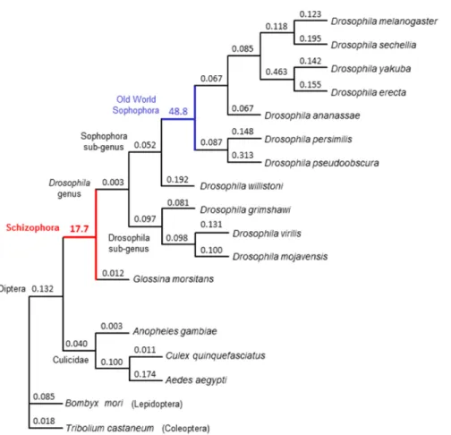
Divergence in rec and loss of MCM9 occurred after the split between mosquitos and higher flies 200 to 250 million years ago, but prior to the emergence of the Schizophora 65 million years ago. To test whether patterns of sequence evolution were consistent with positive selection leading to the divergence of rec, we estimated the ratio between the rate of base pair substitutions at nonsynonymous sites (dN) and the rate at synonymous sites (dS) among dipterans in MCM8/rec. We compared 15 evolutionary models, ranging from conservation of dN/dS ratios across all taxa surveyed to allowing free evolution of dN/dS ratios along all branches, and including models testing specific hypotheses about the evolution of rec along different branches of the insect phylogeny. The best-fitting model (P = 0.0002 versus the next best model) supports the hypothesis that rapid protein-coding divergence was driven by positive selection prior to the split of tsetse flies from fruit flies (Figures 10 and 11, and Table 3). Thus, we infer that natural selection likely drove the repurposing of REC into its new role as an antagonist of DmBLM. Recent evolution of rec shows much lower levels of nonsynonymous changes, suggesting subsequent functional constraint (Figure 11). MEI-217 and MEI-218 have also diverged substantially from the ancestral MCM structure: They split into two polypeptides, and MEI-218 acquired an N-terminal extension (Figures 12 and 13).
33
in Drosophila by Morgan 100 years ago (Figure 9) (Morgan, 1912). Resolving the conundrum of why the mei-MCMs supplanted Msh4-Msh5 will require a deeper understanding of both the evolutionary origins of the mei-MCMs and the functional differences between mei-MCMs and Msh4-Msh5.
Figure 11. Rates of evolution of REC/MCM8 in Dipteran insects and select outgroups. The tree represents a topology of phylogenetic relationships among the insect species shown. Branch lengths are proportional for ease of reading and do not reflect divergence, which is shown in Figure 10. dN/dS ratios along each branch for the best fitting model (Model 15) are shown. Values less than one suggest purifying selection, whereas values greater than one suggest positive selection along that branch. Consistent with our prediction of rapid evolution of REC before the
emergence of Schizophora, the branch leading to Glossina and Drosophila has a high positive
34
Model Type1
Para-meters Likelihood Log- Description
1 0 33 -29353.66955 One dN/dS rate for all taxa
2 2 34 -29353.13881 One rate for D. melanogaster;
one rate for all other taxa
3 2 34 -29352.42575 One rate for mosquitoes;
one rate for all other taxa
4 2 34 -29348.27369 One rate for Old World Sophophora;
one rate for all other taxa
5 2 34 -29340.13854 One rate for Sophophora;
one rate for all other taxa
6 2 34 -29322.82126 One rate for higher flies
one rate for all other taxa
7 2 34 -29313.19077 One rate for Drosophila;
one rate for all other taxa
8 2 35 -29341.74626 One rate for Drosophila;
one rate for Glossina; one rate for all other taxa
9 2 35 -29313.18287 One rate for ancestor to higher flies;
one rate for Drosophila; one rate for all other taxa
10 2 35 -29313.15944 One rate for ancestor to higher flies;
one rate for Sophophora; one rate for all other taxa
11 2 36 -29313.15172 One rate for ancestor to higher flies;
one rate for Drosophila; one rate for Glossina; one rate for all other taxa
12 2 36 -29310.98226 One rate for ancestor to higher flies;
one rate for Drosophila; one rate for Sophophora; one rate for all other taxa
13 2 37 -29310.95229 One rate for ancestor to higher flies;
one rate for Drosophila; one rate for Sophophora; one rate for Glossina; one rate for all other taxa
14 2 51 -29300.10477 dN/dS rate varies for all higher flies
15 1 63 -29280.42216 dN/dS rate varies for all taxa
Table 3. Models for the evolution of MCM8/rec in Diptera. 1Model-type: 0, common rate all lineages; 1, lineage-specific rates; 2, two or more rates assigned to different lineages (detailed in description).
35
Figure 12. Conservation of MEI-217 and MEI-218 as separate polypeptides throughout
36
Figure 13. Structural changes in MEI-218. These schematics illustrate the structures of MEI-218 orthologs from humans and several insects. Purple cylinder represents the N-terminal MCM domain, orange the C-terminal AAA ATPase-like domain. The human and Bombyx mori orthologs are similar in structure (as are other vertebrates and arthropods other than Drosophila and Glossina) to canonical MCMs, whereas Drosophila and Glossina are substantially longer. In Drosophila the two domains are found on separate polypeptides.
Materials and Methods
Drosophila stocks and genetics
Flies were maintained on standard medium at 25˚C. All experimental flies were
37
meiotic null. This genotype was used in all assays with mus309 mutants. Nondisjunction was scored by crossing mutant females to y cv v f / T(1:Y)BS males. The number of exceptional progeny indicative of nondisjunction (Bar-eyed females and wild-type-eyed males) was multiplied by two to account for triplo-X and nullo-X progeny, which do not survive to adulthood. This number was divided by the total number of progeny and expressed as a percentage. Crossovers were scored by crossing net dppd-ho dp b pr cn /+ virgin females of various genetic backgrounds to net dppd-ho dp b pr cn males.
Identification of orthologs
Orthologs were found through searches of public databases. The amino acid sequence of the human protein was used in BLASTP searches of refseq_protein at NCBI (http://blast.ncbi.nlm.nih.gov/Blast.cgi), using the default parameters. In cases where orthologs were not immediately identified, searches were repeated using sequence from a more closely related species and searching the nr database. If this was unsuccessful, TBLASTN was done using the nr database. Finally, species-specific databases were searched, either from the NCBI genomic BLAST page (http://www.ncbi.nlm.nih.gov/mapview/) or from species-specific websites. Additionally, a sequence for rec from D. pseudoobscura was obtained from (McGaugh and Noor, 2012).
38
TBLASTN searches. Apparent conservation of MEI-217 allowed high-confidence CDS (coding sequence) predictions, but divergence of MEI-218 made predictions for this gene less reliable.
Sequence alignments and phylogenetic analysis
Protein sequences were aligned in MEGA5 (Tamura et al., 2011) using the MUSCLE algorithm (Edgar, 2004). Maximum-likelihood trees were generated in MEGA5 using the method based on the Whelan and Goldman model (Whelan and Goldman, 2001). Initial trees for the heuristic search were obtained automatically. When the number of common sites was <100 or less than one fourth of the total number of sites, the maximum parsimony method was used; otherwise BIONJ method with MCL distance matrix was used. A discrete Gamma distribution was used to model evolutionary rate differences among sites (5 categories (+G, parameter = 2.8099)). The rate variation model allowed for some sites to be evolutionarily invariable ([+I], 0.0000% sites).
Molecular evolutionary analysis
39
40
etc.). All fit the data better than Models 1-3 and generally improve the fit as their complexity grows. However, Model 15, which allows for dN/dS ratio to vary freely among all branches, has the best fit to the data and is a significantly better than any alternative. This model suggests strong adaptive evolution prior to the divergence of the Schizophora and prior to the radiation of the Old World Sophophora. Simpler models (e.g., 4-14) similarly support that the dN/dS rate of rec in higher flies is different compared to the other Dipteran and our outgroups.
Immunoprecipitation
rec was cloned into pFastBac with an N-terminal FLAG tag. mei-217 and mei-218 were cloned into pFastBacDual with N-terminal HA and Strep-II tags, respectively. As per the Bac-to-Bac Baculovirus System (Invitrogen, Carlsbad, CA) protocol, constructs were transformed into DH10Bac cells. Sf9 cells were transfected with bacmid DNA extracted from transformed DH10Bac cells. Following two rounds of viral amplification in Sf9 cells, High Five cells were infected with either a single virus or with both viruses. Cells were harvested 2.5 days after infection and were stored at -80˚C until needed. Pellets were resuspended in lysis buffer (50mM Tris-HCl pH 7.5, 150mM NaCl, 1mM EDTA, 1% Triton X-100), sonicated using a Bioruptor (Diagenode, Denville, NJ)
on highest setting with 30s on/30s off cycles for 20min at 4˚C. Cell suspension was
41
immunoprecipitation was carried out. Beads were washed in a cellulose acetate filter spin column (Pierce, Rockford, IL) with either TBS (FLAG purification) or PBS (HA purification) prior to boiling in SDS-PAGE sample buffer. Samples were run on a 4-15% SDS-PAGE gel, transferred to PVDF membrane, and probed with appropriate antibodies: anti-HA (Sigma) 1:20,000; anti-REC-C (raised to amino acids 875-885; Pacific Immunology, San Diego, CA) 1:20,000; HRP-conjugated anti-rabbit secondary (Santa Cruz Biotechnology, Santa Cruz, CA) 1:20,000. SuperSignal West Pico Chemiluminescent Substrate (Pierce) was used to detect proteins.
Yeast two-hybrid assay
mei-217 was cloned into pGBD-DEST, a Gateway-compatible derivative of pGBD-C1 (James et al., 1996) constructed with the Gateway Vector Conversion System (Life Technologies, Carlsbad, CA). Full-length or truncated mei-218 was cloned into pACT2.2gtwy (Addgene plasmid 11346 deposited by Guy Caldwell), a Gateway-compatible derivative of pACT2.2. Constructs were transformed into Saccharomyces cerevisiae strain PJ69-4A (James et al., 1996). Co-transformants were selected on plates of SD minimal medium containing dropout supplements lacking leucine (-leu) and tryptophan (-trp) for 3 days at 30˚C. Single colonies were streaked onto fresh –trp –leu
plates and grown for 3 days at 30˚. Colonies were then streaked onto –trp –leu –histidine
CHAPTER 3
MEI-MCMS AFFECT MEIOTIC CROSSOVER DISTRIBUTION
Introduction
Meiotic recombination initiates via a programmed double-strand break (DSB) created by the archaeal topoisomerase VI-like protein Spo11 in all organisms thus far examined (Keeney et al., 1997). This step appears to be a hallmark of meiosis, and as such, the presence of a Spo11 homolog within a genome is used as part of a “meiosis detection toolkit” to identify organisms that undergo meiosis (Malik et al., 2008). Not all breaks are created equal, however, as some DSBs are repaired into crossovers (COs), while others will form non-crossovers (NCOs) instead.
43
and telomeres (Hassold and Hunt, 2007). Also, when more than one CO occurs on a chromosome, they tend to be more widely-spaced than expected by random chance, a phenomenon called CO interference (Berchowitz and Copenhaver, 2010).
Interference was first described in Drosophila melanogaster (Sturtevant, 1913), and has since been found to operate in many model systems including Arabidopsis thaliana, Caenorhabditis elegans and Saccharomyces cerevisiae (Copenhaver et al., 2002; Fogel and Hurst, 1967; Meneely et al. 2002). Notably, COs in the fission yeast Schizosaccharomyces pombe do not exhibit interference (Munz, 1994). Although it is unknown how interference works, several models hypothesize how CO interference operates. In the “polymerization model”, a CO produces some signal that is perpetuated along a chromosome, thus discouraging the formation of COs nearby (King and Mortimer, 1990). The “stress relief model” invokes mechanical stress along chromosomes that is alleviated at CO sites, thus inhibiting nearby COs (Borner et al., 2004). Finally, a “counting model” suggests that an organismal-specific number of NCOs must separate COs (Foss et al., 1993).
44
2009). These results led to the suggestion that the mei-MCMs have an early role in committing some DSBs to the CO pathway (Joyce and McKim, 2009). However, other epistasis experiments have placed RECOMBINATION DEFECTIVE (REC) at an intermediate step in the meiotic recombination pathway, downstream of strand invasion but upstream of exchange/CO formation (Blanton et al., 2005).
To investigate and tease apart the potential early and late roles of the mei-MCMs in CO formation, I created separation-of-function alleles of rec. I used a non-disjunction (NDJ) assay to test the ability of these alleles to segregate chromosomes and a CO assay to study their effect on CO number and CO distribution. I created a truncation mutant of another mei-MCM, MEI-218, to investigate the function of this protein’s conserved C-terminus. Similar to the rec separation-of-function alleles, I assayed this “mini-MEI” allele for NDJ, CO number and CO distribution.
Results
REC ATPase mutants differentially affect NDJ and COs
45
Figure 14. MCM Walker A/B alignment. Protein sequence alignment of D. melanogaster MCM2-MCM8 (REC). Positions that have identical or conserved residues in all proteins have a black background. Positions with identical or conserved residues in six of the seven proteins have a dark gray background. Positions with identical or conserved residues in four or five of the seven proteins have a light gray background. Walker A and Walker B motifs are indicated. Asterisks mark residues mutated in {UAS-rec*} transgenes.
46
and Luo, 1999). Since meiotic COs are important for the proper segregation of homologous chromosomes, transgenes were scored for NDJ as a readout of CO formation (Figure 15).
Figure 15. rec transgene non-disjunction. Percent non-disjunction (NDJ) was scored in the indicated genotypes. “KA” and “DA” superscripts denote the Walker A and Walker B mutant
alleles of rec, respectively, while “WT” superscript denotes the wild-type rec gene. “nos” and
“tub” indicate the rec transgene was under expression of the nanos-GAL4 or tubulin-GAL4 driver, respectively. Over 1000 individuals were scored for each genotype. ***p<0.0001, as compared to wild-type. n.s., not statistically significant compared to either wild-type (blue lettering) or compared between paired genotypes.
47
wild-type. This result suggests that allowing REC to bind, but not hydrolyze, ATP prevents proper meiotic chromosome segregation.
48
2324 flies scored) is statistically significant (p<0.0001), further supporting the idea that the Walker B mutant does not have a precondition CO distribution.
Figure 16. rec transgene crossover distribution. Crossover (CO) distribution across five intervals on chromosome 2L and proximal 2R is shown. The markers used in mapping are shown above the graph. Hash marks between pr and cn indicate the position of the centromere. Solid lines depict the number of map units (m.u.) per megabase pair (Mb) in each interval for more than 2000 individuals for each genotype. Dashed lines indicate the mean CO rate across the entire region. {UASp-rec*} transgenes were driven using the nanos-GAL4 driver.
Table 4. rec transgene recombination frequency. Recombination frequency is expressed as map units across the intervals shown. Numbers in parentheses denote the percentage of wild-type
recombination frequency. *The ratio of the percentage of wild-type recombination frequency
49
As expected based on their CO distribution pattern, the wild-type rec and Walker A mutant rec transgenics had ratios close to one, signifying that the change in CO frequency in the centromere-proximal region was similar to the change across the chromosome arm (Table 4). Interestingly, however, it appeared that most of these intervals had increased recombination frequencies in the Walker A mutant compared to wild-type, even though the CO distribution appeared normal. To investigate this possibility further, I calculated the number of multiple COs observed in these mutants (Table 5). I found that the {UASp-recWT} transgene did not have a significantly different number of single (SCO), double (DCO), or triple (TCO) COs as compared to the wild-type control. In contrast, the {UASp-recKA} transgene had significantly more DCOs than wild-type. The {UASp-recKA} transgene also had significantly more DCOs than the {UASp-recWT} transgene (p=0.0135). Therefore, the difference in COs between the Walker A transgene and wild-type flies is not a consequence of rec being expressed off a transgene and instead is a result of the point mutation within the Walker A motif.
Table 5. rec transgene crossover number. Number of single crossover (SCO), double crossover (DCO), and triple crossover (TCO) progeny observed in each genotype out of the total number of progeny (N) scored. {UASp-rec*} transgenes were driven using the nanos-GAL4
driver. #p=0.0002, as compared to wild-type. *p<0.0001, as compared to wild-type. NSnot
statistically significant, as compared to wild-type.
N
SCO
DCO
TCO
wild-type
2320
952
44
1
rec
2036
113
*0
*0
NSUAS-REC
WT3134
1399
NS80
NS1
NSUAS-REC
KA2281
949
NS86
#5
NS50
Hyperrecombination in a MEI-218 truncation mutant
While the C-terminus of MEI-218 contains a relatively well-conserved AAA ATPase domain, the protein’s N-terminus is disordered and much more divergent (Kohl et al., 2012). In particular, the N-terminus is of variable lengths in the various Drosophilidsfor which genome sequence is available (Kohl et al., 2012), and this region can only be poorly aligned even in closely related Drosophilids (Figure 17). Furthermore, it is through the conserved C-terminus, not the N-terminus, that MEI-218 interacts with MEI-217 (Kohl et al., 2012). To investigate whether this divergent N-terminal sequence was necessary for MEI-218’s meiotic function, I created a {UASp} transgenic truncation mutant of MEI-218 that eliminates the first 526 amino acids (44%) of the protein, called the “mini-MEI” allele (Figure 17). This allele was placed into a mei-218 null mutant background, and was driven using the same nanos-GAL4 driver as the rec transgenics.
51
Fi
gur
e
17
. M
EI -218 p ro te in al ign m en t. D . m el ano gas te r and D . ps eud oobs cu ra M EI -218 pr ot ei n s eq ue nc e a lig nm ent
. P
os iti ons tha t hav e i de nt ica l o r co ns er ved re si du es in b ot h p ro tei ns hav
e a b
52
Figure 18. mini-MEI transgene non-disjunction. Percent non-disjunction (NDJ) was scored in the indicated genotypes. Over 1200 individuals were scored for each genotype. Color of
asterisks indicates genotypes statistically compared. ***p<0.0001, *p=0.0202.
Figure 19. mini-MEI transgene crossover distribution. Crossover (CO) distribution across five intervals on chromosome 2L and proximal 2R is shown. The markers used in mapping are shown above the graph. Hash marks between pr and cn indicate the position of the centromere. Solid lines depict the number of map units (m.u.) per megabase pair (Mb) in each interval for
53
increase in SCOs, multiple COs, or both (Table 6). I found that the number of SCOs in the mini-MEI allele was not statistically different from wild-type, but that this allele did have a highly significant increase in DCOs compared to wild-type. To determine if the increased CO rate was obscuring an altered CO distribution, I calculated the ratio of crossing-over in the centromere-proximal region to crossing-over across the entire chromosome arm (Table 7). Interestingly, the mini-MEI allele showed a ratio greater than one (3.8). Using the definition of precondition proteins of Blanton et al. (Blanton et al., 2005), this results suggests that like the mei-218 null, the mini-MEI allele exhibited a precondition distribution of COs. However, the title “precondition mutant” has historically described flies which have a reduction in COs (Carpenter and Sandler, 1974; Sandler et al., 1968), which is clearly not the case for the mini-MEI allele.
Table 6. mini-MEI transgene crossover number. Number of single crossover (SCO), double crossover (DCO), and triple crossover (TCO) progeny observed in each genotype out of the total
number of progeny (N) scored. *p<0.0001, as compared to wild-type. NSnot statistically
significant, as compared to wild-type.
Table 7. mini-MEI transgene recombination frequency. Recombination frequency is expressed as map units across the intervals shown. Numbers in parentheses denote the
percentage of wild-type recombination frequency. *The ratio of the percentage of wild-type
recombination frequency across the centromere-proximal interval (pr-cn) compared to the percentage of wild-type frequency across the entire chromosome arm.
N
SCO
DCO
TCO
wild-type
2320
952
44
1
mei-218
1744
51
*0
*0
NSmini-MEI
1819
731
NS142
*4
NSnet-dppd-ho dppd-ho-dp dp-b b-pr pr-cn total net-cn ratio pr-cn /
total*
wild-type 5.09 7.54 27.33 3.49 1.51 44.96 1.0
54 Discussion
The results from the Walker A and Walker B mutant rec alleles (Table 8) are best analyzed in light of other MCM ATPase mutant analyses conducted previously. Firstly, the oligomeric nature of MCM proteins is crucial to the complex’s ability to hydrolyze ATP since the active sites are formed in trans using the Walker A and Walker B motifs of one subunit and the conserved arginine finger from a neighboring subunit (Davey et al., 2003). Of the several models for AAA+ protein ATP hydrolysis (“probabilistic” (Martin et al., 2005), “sequential” (Stitt and Xu, 1998), and “concerted” (Gai et al., 2004)), only one has been suggested for an MCM protein. A study of the archaeal Sulfolobus solfataricus MCM complex found that helicase activity was maintained even when multiple ATP binding sites were inactive, suggesting that MCM ATP hydrolysis is “probabilistic” (Moreau et al., 2007). Unlike archaeal MCM complexes, which are most often homomultimers, the replicative MCM2-7 complex is comprised of six non-redundant subunits. It has been shown that the ATP active sites in the MCM2-7 complex are functionally distinct and that mutations within the different subunits’ Walker A and Walker B motifs have varying effects on viability and ATPase activity (Bochman et al., 2008; Gomez et al., 2002).
55
Table 8. mei-MCM transgene phenotypes. Phenotypes for the various mei-MCM transgenes and mei-MCM (rec and mei-218) null mutants are summarized. For non-disjunction (NDJ) and
crossover (CO) number, “↑” indicates increased levels as compared to wild-type, “↓” indicates
decreased levels as compared to wild-type, and “–” indicates a level comparable to wild-type. For CO distribution, “precondition” denotes a precondition mutant distribution of COs, whereas “normal” denotes a type distribution of COs and “~normal” denotes an approximately wild-type distribution of COs.
complex, since MEI-217 and MEI-218 lack a Walker A motif and therefore would have been unable to provide this compensation (Kohl et al., 2012). A separation-of-function allele in Mcm5 shares the same meiotic phenotypes of the mei-MCMs (Lake et al., 2007), suggesting MCM5 is an additional member of the complex. It is unknown whether additional replicative MCMs are components of the mei-MCM complex, since four of the six DmMCM2-7 have been genetically characterized, and null mutations in each are pupal lethal (Feger et al., 1995; Lake et al., 2007; Schwed et al., 2002; Treisman et al., 1995). It is likely that all MCM2-7 are essential for viability in Drosophila, as they are in yeast (Tye, 1999), and thus without separation-of-functional alleles, like the Mcm5A7 allele, we cannot ascertain the proteins’ roles in meiosis.
NDJ
CO
number
CO
distribution
rec
or
mei-218
↑
↓
precondition
UAS-REC
WT−
−
normal
UAS-REC
KA−
↑
normal
UAS-REC
DA↑
↓
~normal
57
Figure 20. rec ATPase mutant model. (A) In wild-type, double-strand breaks (DSBs) are formed across a chromosome. The mei-MCM complex is recruited to a DSB site, thereby designating that DSB as a future crossover (CO) in an ATP hydrolysis-independent reaction. The chosen DSB occupies a central location on the chromosome arm. During DSB repair, the mei-MCM complex hydrolyzes ATP to facilitate CO formation. Once all DSBs are repaired, a CO checkpoint verifies that at least one CO was formed per chromosome. (B) In the rec Walker A mutant background, DSB formation, CO designation and DSB repair occur as in wild-type, resulting in CO formation. The CO checkpoint does not recognize the CO, however, triggering the formation of additional DSBs. (C) In the rec Walker B mutant, DSB formation and CO designation occur as in wild-type. The mei-MCM complex is unable to hydrolyze ATP during DSB repair, however, preventing CO formation. The CO checkpoint senses the lack of COs and triggers the formation of additional DSBs. This model depicts one potential explanation for the phenotypes observed, although other scenarios are possible (see Discussion). Relative timing of these events may also vary.
58
By using the rec Walker B mutant, I was able to uncouple the early and late roles for REC in meiotic recombination. Standard null alleles of rec result in ~95% reduction in COs and an abnormal, precondition distribution of residual COs (Blanton et al., 2005). This suggests that REC is required to make a majority of meiotic COs and that REC has a role in CO distribution. In a NDJ assay, the Walker B rec mutant also behaves like these null alleles of rec – both exhibit very high NDJ. Interestingly, while this phenotype appears to result from a strong reduction in crossing over in both cases, the Walker B mutant has a different effect on CO distribution than other rec null alleles. rec mutants show a polar reduction in crossing over but the Walker B mutant did not have this precondition distribution. This suggests that when REC was prevented from hydrolyzing bound ATP because of a mutated Walker B motif, REC was able to localize to sites of future COs properly but was unable to complete some function – one that presumably requires ATP hydrolysis – to physically form COs (Figure 20). This hypothesis that the mei-MCMs function as a pre-CO complex to mark sites of future COs is reminiscent of the pre-RC role of the replicative MCMs.
59
Like the rec Walker A mutant, the mini-MEI mutant had a hyperrecombination phenotype with increased DCOs as compared to wild-type. As discussed above for the rec Walker A mutant, this phenotype could arise from an increase in DSBs, a change in defining the sites of future COs, a change in the CO-sensing pathway, or a combination of these possibilities. It is noteworthy that I have identified mutant versions of two mei-MCM proteins that show a significant increase in the number of DCOs but retain normal meiotic chromosome disjunction. It should be noted that we have also observed the hyperrecombination phenotype previously, in the meiotic mus309 rec double mutants (Kohl et al., 2012). In both the mini-MEI and mus309 rec mutants, the distribution of COs did not follow a wild-type pattern, but instead showed a more even distribution across the chromosome, reminiscent of the mus309 and rec single mutant distributions. The rec Walker A mutant, on the other hand, showed a wild-type distribution of COs. Despite these differences, the shared hyperrecombination phenotypes in these three cases suggests that the mei-MCM complex is necessary for some mechanism of CO regulation – whether this mechanism involves designating future COs, sensing COs, or interfering with neighboring COs remains to be seen.
60 Materials and Methods
Protein sequence alignments
Protein sequences were aligned using the “Complete Alignment” setting on ClustalX (Larkin et al., 2007), using all default settings. Alignments were then viewed using the GeneDoc program.
Generating mei-MCM transgenic flies
The rec cDNA was cloned into the p{attBUASpW} vector downstream of the UASp germline expression promoter (Rørth, 1998) using Gateway technology (Invitrogen, Carlsbad, CA). The vector also contains a mini-white gene and PhiC31 attB site. In addition to the wild-type (unchanged) version of rec, two additional vectors were created using QuikChange (Agilent Technologies, Inc., Santa Clara, CA) to insert the Walker A mutation (K479A) and Walker B mutation (D537A). Each transgene was injected using standard PhiC31 transformation procedures (Best Gene Inc., Chino Hills, CA) into the 99F8 genomic location, creating {UASp-recWT}, {UASp-recKA}, and {UASp-recDA}, respectively.