DOI: 10.2478/v10004-007-0033-3 Original Scientific Paper
DYNAMICS OF THE LUNG FUNCTION IN ASBESTOS
PLEURAL DISEASE
Irena PERI], Dragan ARAR, Igor BARI[I], Ivana GOI]-BARI[I], Neven PAVLOV,
and Jadranka TOCILJ
1University Hospital Split, Split, Croatia
Received in February 2007 Accepted in October 2007
As a rule, asbestosis is a disease of workers who are occupationally exposed to inhalation of asbestos dust, leaving permanent alterations on the lung parenchyma or pleura. In our ten-year study, we investigated 318 workers with pleural asbestosis from whom we took medical history which included occupational exposure to asbestos, radiological examinations and lung function, which is mandatory for the diagnosis and the follow up of the disease. We analysed functional parameters such as forced vital capacity (FVC) and forced expiratory volume in the first second (FEV1) and intermediate forced expiratory flow at 25 % to 75 % (FEF25 %-75 %). In addition, we investigated the predicted values of functional parameters according to smoking and non-smoking habits. We found a significant reduction in vital capacity, particularly in smokers after 25 years of exposure to asbestos. During the first 15 years, values of vital capacity on the group basis remained inside the 80 % of the normal values and were not significant for assessing the dynamics of the lung function. To better assess the effects of occupational asbestos exposure, it is necessary to interpret lung function data not only on the group basis, but also for each subject individually.
KEY WORDS: individual monitoring, occupational exposure, pleural asbestosis, smoking, vital capacity
Asbestosis is a chronic disease of the lungs caused by exposure to asbestos fibres. Asbestos is a silicate of fabric structure, chemically built of bound and free silicon dioxide, calcium, nitric, magnesium, iron and water. In traces, it also contains nickel, manganese, chromium and cobalt (1). It is a cheap raw material, very resistant to heat and friction and largely used in industry (2).
Most of the inhaled asbestos fibres are mechanically cleared from the respiratory system and only particles of aerodynamic diameter of 7 µm to 10 µm can reach alveoli. Particles that have not been cleared from the respiratory system or completely phagocyted cause permanent irritation that can lead to changes in the lung parenchyma or pleura (3-6). Morgan and Holmes have proved that the critical length of asbestos fibre is 17 µm (6). Only short fibres of the chrysotile type of asbestos that follow air flow, the long, rigid crocidolite
type, as well as other amphibole types can penetrate into alveoli and induce pathological changes in the lung.
Lung asbestosis is a chronic progressive disease with fibrosis of the lung parenchyma. Inhaled asbestos fibres induce chronic inflammatory processes in the interstice, disturbing thus the fine structure of alveolar-capillary membrane. First accumulate inflamed cells that eject a range of mediators, causing inflammation which can also affect pleura. This induces fibroblast growth and an irreversible fibrosis of alveolar walls (7). In the end, diffuse or local plaques develop on parietal pleura that gradually become calcified (8, 9). Its diagnosis is based on data about exposure to asbestos dust and on radiological, functional and histopathological findings (10). Today, radiology is done and interpreted according to the 1980 recommendations of the International Labour
Organization (ILO) (11). The basic radiological examination includes posterior and anterior, profile and inclined x-ray of the thoracic organs, shot by “hard” technique, and then analysed and coded using the ILO classification. Every x-ray analysis is done separately by two radiologists, whose results are then coded using the standard ILO classification.
Asbestos diagnosis has been improved thanks to computer tomography (CT) of the thoracic organs and to high resolution computer tomography (HRCT) (12, 13).
Lung function tests show the type and the extent of the functional lesion of the lung. This lesion occurs at the level of lung ventilation and gas diffusion, that is, at the level of alveolar-capillary membrane. Functional parameters of the lung are essential in order to follow up persons occupationally exposed to asbestos. Earlier, most studies used classical methods to measure lung ventilation parameters (14-18).
Asbestos is no longer in use in most developed countries of the world. It has been banned in the EU with enforcement deadline by the end of 2005. In 2006, Croatia also adopted the ban. In spite of these regulatory measures, exposure to asbestos has not completely been eliminated due to earlier massive use and its resistance to biodegradation. It is therefore appropriate to continue with the surveillance of possible effects of asbestos, including the evaluation of adverse health effects which could be caused by earlier exposure to asbestos, particularly in the working environment.
The goal of our study was to establish dynamic changes in lung ventilation parameters, especially in the vital capacity, forced expiratory volume in the first second, and medium expiratory flow in subjects who had verified diagnosis of pleural asbestosis in relation to the time of exposure to asbestos dust through inhalation. We also studied how smoking affected the dynamics of changes in vital lung capacity.
SUBJECTS AND METHODS
The study included workers who had at least five years of working experience in the factory of asbestos and cement products Salonit in Vranjic (126 subjects or 40 %), factory of asbestos products Plobest Plo~e (139 examinees or 43 %), and in the local shipbuilder Brodogra|evna industrija Split (BIS, 53 subjects or 17 %), all in Croatia. Of 318 subjects, 243 were men
(76.4 %) with the average age of 53 years and 76 women (23.7 %) with the average age of 49 years (Table 1).
Table 1 Subject distribution by sex and age
Sex No. of subjects Age / years Average Range Men 243 (76.3 %) 53 35-74 Women 76 (23.7 %) 49 31-62 Table 2 Subject distribution by the duration of asbestos exposure
Factory Exposure / years
Average Range
Salonit 19.6 5-25
Plobest 21.3 5-30
BIS 17.8 7-23
All 19.6 5-30
Using medical history data, we established the duration of the inhalation of asbestos dust and the average duration of exposure for each group of workers shown in Table 2.
According to the smoking habit, we divided the subjects in smokers, non-smokers and ex-smokers.
All subjects underwent posterior and anterior, profile and inclined x-ray of the thoracic organs. The shots were done using the “hard” technique, and then analyzed and coded according to the ILO classification. Each x-ray was analysed independently by two radiologists and both coded the results according to standard ILO radiographs. Our study included only patients with the verified diagnosis of pleural asbestosis.
Ventilatory lung function was measured using a Vitalograph (Minhard), England. Before the examination, each subject was clinically checked up in order to exclude acute diseases of the respiratory tract that might have affected the results. At least one hour before the examination, the subjects did not smoke or take any alcohol or medicine. Before the examination, we recorded their body weight, height and age. The test was performed in the upright position with a clip on their nose. Measurements included forced vital capacity (FVC), forced expiratory volume in the first second (FEV1), the ratio between FEV1 and FVC (FEV1/FVC) and the average expiratory flow (FEV25 %-75 %). Three measurements were taken, and if they did not differ for more than 5 %, the highest was considered relevant for analysis. The obtained results were compared with the predicted values of the lung function (14-18).
Additionally, the dynamics of the vital capacity in smokers and non-smokers was analysed regarding the duration of exposure to asbestos dust. The obtained results were analysed using single- and multi-variant data analysis.
RESULTS
Table 1 shows their age distribution by sex. Subjects had been exposed to asbestos at workplace for 5 to 30 years, with average exposure of 19.6 years. Average exposure to asbestos dust in subjects who worked in the factory Salonit was 20 years, in Plobest 21.3 years, and in the BIS shipyard 17.8 years. Table 2 shows exposure time distribution by workplace. Pleural asbestosis was diagnosed based on the analysis of occupational asbestos exposure history and radiological examination of the thoracic organs. Subjects with the established diagnosis of
asbestos disease included 170 smokers (54 %), 110 non-smokers (35 %), and 38 (11 %) ex-smokers. Smokers were considered those who smoked at least five cigarettes a day. In the average they smoked from 20 to 30 cigarettes a day. Ex-smokers were considered subjects who quit smoking at least a month before the study started and who earlier smoked more than five cigarettes a day.
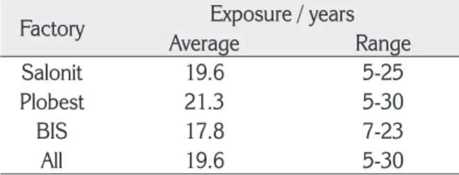
Figure 1 shows variations in lung function parameters in relation to asbestos exposure. Adjusted for age and sex, FVC shows a drop from 99 % to 90 % after the first ten years of exposure, reaching a statistically significant fall to 79 % with exposure of over 20 years (t=5.86; p<0.001). A drop in forced expiratory volume in the first second (FEV1) from 105 % to 97 % was observed after the first ten years of exposure, and it dropped significantly to 89 % in the next ten years of exposure (t=3.40; p<0.01). Forced expiratory flow (FEF25 %-75 %) dropped from 100 % to 90 % after the first ten years of exposure only to return Figure 2 Changes in vital capacity in smokers and non-smokers
to 100 % of the predicted values in the next ten years of exposure (t=2.01; p=0.13).
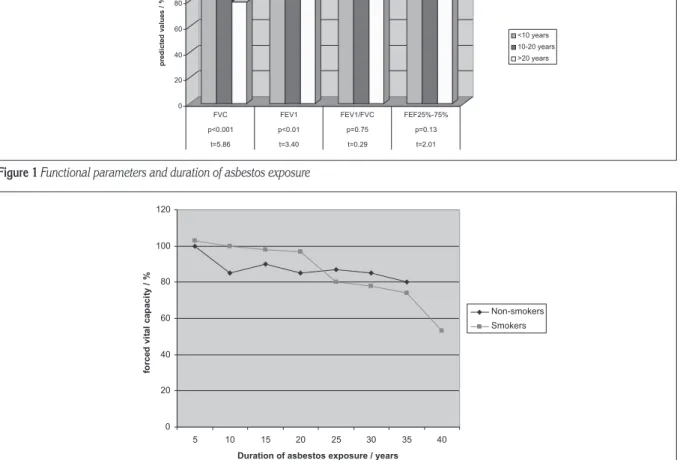
Additionally we analysed the dynamics of vital capacity among smokers and non-smokers in respect to exposure duration to asbestos dust. The results are shown in Figure 2.
At the beginning of exposure, median FVC in non-smokers was 110 % and in smokers 100 % of the predicted value. Vital capacity in non-smokers dropped to 90 % after the first ten years of exposure (t=9.12; p<0.001), remaining unchanged until after 20 years of exposure when it dropped further to 80 % of the predicted values (t=9.63; p<0.0001).
In smokers, FVC did not show significant difference from the predicted average after the first ten years of exposure to asbestos dust (t=0.29; p=0.74). After 20 years it came to a notable fall to under 80 % (t=5.80; p<0.001). After 35 years of exposure it fell to 50 % of the predicted values (t=12.06; p<0.000001).
DISCUSSION
Our results are in accordance with those of Wagner et al., whose study showed a drop in FVC, FEV1, and FEF25 %-75 % in 208 women occupationally exposed to asbestos (17). Wang et al. also found a significant drop in FVC and carbon monoxide (CO) transfer in 119 subjects exposed to asbestos. They believe that these two parameters are the most sensitive of early changes caused by asbestos (18). Yates et al. also got lower FVC and FEV1, while other parameters were also lower, but not at the same rate (19). In a study of functional parameters in subjects with different types of asbestos disease, Pa`anin found lower FVC, FVC and FEV1 in subjects with pleural and/or parenchymal changes, while with the parenchymal type of asbestosis only FVC dropped significantly (20). Murlidhar found that in 62 % of workers exposed to asbestos FVC dropped to less than 80 % of the normal values (21). Available bibliographical data agree with the conclusion that restrictive obstructions in ventilation are typical for asbestos disease. During a five year follow up, Wagner found a significant fall in FVC and DLCO, which was also corroborated by Yates in his ten-year follow up (17, 19). Recent studies confirm that restrictive obstructions in ventilation are typical of asbestos disease (21). Restrictive obstructions decreased linearly with the duration of asbestos exposure.
It seems that FVC tends to decrease minimally over a longer period. This is why we would like to
point out the need for individual follow up of every worker. In young people, spirometric values may vary from 20 % to 30 % above the predicted values. Only an individual follow up over a longer period could show a significant fall in functional parameters for a specific person. This is crucial for the assessment when such a person should be spared from further exposure to asbestos.
Analyzing the fluctuation of FVC in smokers and non-smokers, we established that both groups started with FVC around 100 % of the predicted values, and dropped in non-smokers to around 90 % until 15 years of exposure, and to 80 % in exposures longer than 15 years. Smokers showed a significant fall in FVC after 20 years of exposure to asbestos, which later fell to under 50 % of the predicted values. A study of 1298 subjects occupationally exposed to asbestos showed lower forced expiratory flow (FEF25 %-75 %) and forced expiratory volume in the first second (FEV1) in non-smokers who had pleural asbestosis, while FVC was 94 %, which was explained by the obstructive disturbance of ventilation without restriction. Similar was found for the parenchymal type of the disease (22). In that research smokers had lower FVC and FEV1 than non-smokers, which was not found in our subjects. A large study conducted in the USA on 8720 subjects confirmed that smokers had significantly reduced FEF25 %-75 %,FEV1 and FEF75 %-85 %, especially after 20 years of exposure to asbestos dust (23, 24).
CONCLUSIONS
The fall in vital capacity under 80 % of the predicted values could be expected only after 20 years of exposure to asbestos dust. This fall is more distinctive in smokers than in non-smokers. Smoking more significantly contributes to the decrease in the vital capacity of the lung than pleural asbestosis.
To better assess the effects of occupational asbestos exposure, it is necessary to interpret lung function data not only on the group basis, but also for each subject individually. In other words, subjects should be their own controls.
REFERENCES
1. Brandli BR, Gunter ME. A review of scientific literature examining the mining history, geology, mineralogy, and amphibole asbestos health effects of the Rainy Creek
igneous complex, Libby, Montana, USA. Inhal Toxicol 2006;18:949-62.
2. Kova~ S. Benign asbestos pleural effusion. Arh Hig Rada Toksikol 1985;36:33-42.
3. O’Reilly KM, Mclaughlin AM, Beckett WS, Sime PJ. Asbestos-related lung disease. Am Fam Physician 2007;75:683-8.
4. Morgan A, Black A, Evans N, Holmes A, Pritchard JN. Deposition of sized glass fibres in the respiratory tract of the rat. Ann Occup Hyg 1980;23:353-66.
5. Timbrell V. Deposition and retention of fibres in the lung. Ann Occup Hyg 1982;26:347-69.
6. Morgan A, Holmes A. Concentrations and dimensions of coated and uncoated asbestos fibres in human lung. Br J Ind Med 1980;37:25-31.
7. Ahn CS, Kim SJ, Oh SJ, Park KJ, Kim HJ, Ahn CM, Kim HK, Shin DH, Cho SH, Yang KM. Pulmonary asbestosis: radiologic-pathologic brief report. Yonsei Med J 1997;38:323-6.
8. Miller A, Miller JA. Diffuse thickening superimposed on circumscribed pleural thickening related to asbestos exposure. Am J Ind Med 1993;23:859-71.
9. Rui F, De Zotti R, Negro C, Bovenzi M. A follow-up study of lung function among ex-asbestos workers with and without pleural plaques. Med Lav 2004;95:171-9. 10. Broderick A, Fuortes LJ, Merchant JA, Galvin JR,
Schwartz DA. Pleural determinants of restrictive lung function and symptoms in an asbestos-exposed population. Chest 1992;101:684-91.
11. International Labour Office (ILO). International classification of radiographs of the pneumoconiosis. Occupational safety and health series XX. Geneva: International Labour Office, 1980.
12. Begin R, Ostiguy G, Filion R, Colman N, Bertrand P. Computed tomography in the early detection of asbestosis. Br J Ind Med 1993;50:689-98.
13. Oksa P, Suoranta H, Koskinen H, Zitting A, Nordman H. Hihg-resolution computed tomography in the early detection of asbestosis. Arch Occup Environ Health
1994;65:299-304.
14. Cristaudo A, Foddis R, Buselli R, Gattini V, Di Palma N, Guglielmi G. Medical surveillance of workers previously exposed to asbestos. Med Lav 2006;97:475-81. 15. Mustajbegovi} J, Kern J, Schachter EN, Zuskin E,
Pavi~i} F, Teufen. Ventilatory functions in Croatian population in comparison with European reference values. Croat Med J 2003;44:614-7.
16. Gibson GJ. Standardised lung function test. Eur Respir J 1993;6:155-71.
17. Wagner X, Yano E, Nonaka K, Wang M, Wang Z. Respiratory function of non-smoking female asbestos workers without radiographic signs of asbestosis. Arch Environ Health 1998;53:292-8.
18. Wang X, Yano E, Novak K, Wang M, Wang Z. Respiratory impairments due to dust exposure: a comparative study among workers exposed to silica, asbestos, and coalmine dust. Am J Ind Med 1997;31:495-502. 19. International Standards Organization (ISO). Proposed
analytical method for determination of asbestos fibers in air. Geneva: International Standard Organization, 1984.
20. Yates DH, Browne K, Stidolph PN, Neville E. Asbestos-related bilateral diffuse pleural thickening: natural history of radiographic and lung function abnormalities. Am J Res Crit Care Med 1996;253:301-6.
21. Pa`anin S, Mustajbegovi} J. Effect of age on asbestosis of the lung and/or pleura. Arh Hig Rada Toksikol 2003;54:5-10.
22. Murlidhar V, Kanhere V. Asbestosis in an asbestos composite mill at Mumbai: A prevalence study. Environ Health 2005;4:24.
23. Kilburn KH, Warshaw RH. Abnormal lung function associated with asbestos disease of the pleura, the lung, and both: a comparative analysis. Thorax 1991;46:33-8.
24. Kilburn KH, Warshaw RH. Airways obstruction from asbestos exposure. Effects of asbestosis and smoking. Chest 1994;106:1061-70.
Sa`etak
DINAMIKA FUNKCIJE PLU]A KOD AZBESTNE BOLESTI
Azbestoza je bolest izazvana udisanjem azbestnih ~estica koje ostavljaju trajne promjene na parenhimu plu}a i/ili pleuri. Dijagnoza se postavlja na osnovi anamnesti~kih podataka, uvidom u profesionalnu izlo`enost azbestu i radiolo{kom obradom te patohistolo{kom potvrdom promjena na plu}ima i/ili pleuri. Funkcionalna obrada plu}a obavezna je u postavljanju dijagnoze i pra}enju bolesti. Tijekom desetogodi{njeg istra`ivanja funkcionalno smo obradili 318 osoba profesionalno izlo`enih azbestu s dokazanom azbestozom pleure. Analizirane su vrijednosti funkcionalnih parametara, i to forsiranoga vitalnog kapaciteta (FVC), forsiranoga ekspiracijskog volumena u prvoj sekundi (FEV1) i srednjega ekspiracijskog protoka (FEF25 %-75 %). Dokazan je statisti~ki signifikantan pad vrijednosti FVC i FEV1. Dodatno smo istra`ili vrijednosti funkcionalnih parametara kod na{ih ispitanika s navikom pu{enja i nepu{a~a. U obje skupine prisutno je zna~ajno sni`enje vrijednosti vitalnog kapaciteta tijekom istra`ivanja, s tim da nakon 25 godina izlo`enosti azbestu kod pu{a~a dolazi do naglog pada vrijednosti vitalnog kapaciteta u odnosu na nepu{a~e. Bitno je uo~iti da tijekom prvih 15 godina vrijednosti vitalnog kapaciteta ostaju unutar 80 % normalnih vrijednosti te nemaju zna~enja za pra}enje dinamike funkcije plu}a kod azbestne bolesti. Individualnim pra}enjem profesionalno izlo`enih radnika ostvaruje se bolji uvid u dinamiku funkcije plu}a kod azbestne bolesti.
KLJU^NE RIJE^I: forsirani vitalni kapacitet, individualno pra}enje, pleuralna azbestoza, profesionalna izlo`enost, pu{enje
CORRESPONDING AUTHOR:
Irena Peri}
M. Krle`e 26, HR-21000 Split E-mail: irena.peric1ºst.t-com.hr