Hypokalemia and hyperkalemia are common electrolyte disorders caused by changes in potassium intake, altered excretion, or transcellular shifts. Diuretic use and gastrointestinal losses are common causes of hypokalemia, whereas kidney disease, hyperglycemia, and medication use are common causes of hyperkalemia. When severe, potassium disorders can lead to life-threatening cardiac conduction disturbances and neuromuscular dysfunction. Therefore, a first priority is determining the need for urgent treatment through a combination of history, physical examination, laboratory, and electrocardiography findings. Indications for urgent treatment include severe or symptomatic hypo-kalemia or hyperhypo-kalemia; abrupt changes in potassium levels; electrocardiography changes; or the presence of certain comorbid conditions. Hypokalemia is treated with oral or intravenous potassium. To prevent cardiac conduction dis-turbances, intravenous calcium is administered to patients with hyperkalemic electrocardiography changes. Insulin, usually with concomitant glucose, and albuterol are preferred to lower serum potassium levels in the acute setting; sodium polystyrene sulfonate is reserved for subacute treatment. For both disorders, it is important to consider poten-tial causes of transcellular shifts because patients are at increased risk of rebound potassium disturbances. (Am Fam Physician. 2015;92(6):487-495. Copyright © 2015 American Academy of Family Physicians.)
Potassium Disorders:
Hypokalemia and Hyperkalemia
ANTHONY J. VIERA, MD, MPH, and NOAH WOUK, MD, University of North Carolina at Chapel Hill School of Medicine, Chapel Hill, North CarolinaP
otassium disorders are common. Hypokalemia (serum potassium level less than 3.6 mEq per L [3.6 mmol per L]) occurs in up to 21% of hospitalized patients and 2% to 3% of outpatients.1-3 Hyperkalemia (serumpotas-sium level more than 5 mEq per L [5 mmol per L] in adults, more than 5.5 mEq per L [5.5 mmol per L] in children, and more than 6 mEq per L [6 mmol per L] in neonates) occurs in up to 10% of hospitalized patients and approximately 1% of outpatients.4,5 The
body’s plasma potassium concentration is closely regulated by a variety of mechanisms. Causes of Hypokalemia
Hypokalemia results from abnormal losses, transcellular shifts, or insufficient intake (Table 1).6-8 Abnormal losses are most
com-mon.9 Because the kidney can significantly
lower potassium excretion in response to decreased intake, insufficient intake is rarely the sole cause of hypokalemia, but it often contrib-utes to hypokalemia in hospitalized patients.9
RENAL LOSSES
Diuretic use is a common cause of renally mediated hypokalemia.10 When given in the
same dosage, chlorthalidone is more likely to induce hypokalemia than hydrochlorothia-zide, which is more often implicated because of its widespread use.11,12 Diuretic-induced
hypokalemia is dose-dependent and tends to be mild (3 to 3.5 mEq per L [3 to 3.5 mmol per L]), although it can be more severe when accompanied by other causes (e.g., gastroin-testinal [GI] losses).13
GI LOSSES
GI losses are another common cause of hypokalemia, particularly among hospital-ized patients.9 The mechanism by which
upper GI losses induce hypokalemia is indi-rect and stems from the kidney’s response to the associated alkalosis. As a portion of daily potassium is excreted in the colon, lower GI losses in the form of persistent diarrhea can also result in hypokalemia and may be accompanied by hyperchloremic acidosis.6
Evaluation and Management of Hypokalemia
GENERAL PRINCIPLES
Hypokalemia is often asymptomatic. Evalu-ation begins with a search for warning signs or symptoms warranting urgent treatment
More online at http://www. aafp.org/afp. CME This clinical content conforms to AAFP criteria for continuing medical education (CME). See CME Quiz Questions on page 441.
Author disclosure: No rel-evant financial affiliations. Patient information: A handout on this topic, written by the authors of this article, is available at http://www.aafp.org/ afp/2015/0915/p487-s1. html.
(Figure 1).7,14 These include weakness or
pal-pitations, changes on electrocardiography (ECG), severe hypokalemia (less than 2.5 mEq per L [2.5 mmol per L]), rapid-onset hypokalemia, or underlying heart disease or cirrhosis.7,15 Most cases of
hypokalemia-induced rhythm disturbances occur in individuals with underlying heart disease.10
Early identification of transcellular shifts is important because management may differ. Identification and treatment of concurrent hypomagnesemia are also important because magnesium depletion impedes potassium repletion and can exacerbate hypokalemia-induced rhythm disturbances.16,17
HISTORY AND PHYSICAL EXAMINATION A focused history includes evaluation for possible GI losses, review of medications, and assessment for underlying cardiac comorbidities. A history of paralysis, hyper-thyroidism, or use of insulin or beta agonists suggests possible transcellular shifts leading to redistributive hypokalemia. The physical examination should focus on identifying cardiac arrhythmias and neurologic mani-festations, which range from generalized weakness to ascending paralysis.
LABORATORY ANALYSIS AND ECG
The diagnosis should be confirmed with a repeat serum potassium measurement. Other laboratory tests include serum glucose and magnesium levels, urine electrolyte and creatinine levels, and acid-base balance. The most accurate method for evaluating urinary potassium excre-tion is a 24-hour timed urine potassium collecexcre-tion; nor-mal kidneys excrete no more than 15 to 30 mEq per L (15 to 30 mmol per L) of potassium per day in response to hypokalemia. A more practical approach is calculation of the urine potassium-to-creatinine ratio from a spot urine specimen; a ratio greater than 1.5 mEq per mmol (13 mEq per g) is indicative of renal potassium wasting.18 If no
cause is identified with the initial workup, assessment of thyroid and adrenal function should be considered.
Typically, the first ECG manifestation of hypokale-mia is decreased T-wave amplitude. Further progression can lead to ST-interval depression, T-wave inversions, PR-interval prolongation, and U waves. Arrhythmias associated with hypokalemia include sinus bradycar-dia, ventricular tachycardia or fibrillation, and torsade de pointes.19 Although the risk of ECG changes and
arrhythmias increases as serum potassium concentra-tion decreases, these findings are not reliable because some patients with severe hypokalemia do not have ECG changes.20
Treatment of Hypokalemia
The immediate goal of treatment is the prevention of potentially life-threatening cardiac conduction distur-bances and neuromuscular dysfunction by raising serum potassium to a safe level. Further replenishment can pro-ceed more slowly, and attention can turn to the diagnosis and management of the underlying disorder.15 Patients
with a history of congestive heart failure or myocardial infarction should maintain a serum potassium concen-tration of at least 4 mEq per L (4 mmol per L), based on expert opinion.15
Careful monitoring during treatment is essential because supplemental potassium is a common cause of hyperkalemia in hospitalized patients.21 The risk of
rebound hyperkalemia is higher when treating redis-tributive hypokalemia. Because serum potassium con-centration drops approximately 0.3 mEq per L (0.3 mmol per L) for every 100-mEq (100-mmol) reduction in total body potassium, the approximate potassium
Table 1. Causes of Hypokalemia
Abnormal losses
Medications Diuretics
Laxatives and enemas Corticosteroids Gastrointestinal losses Renal losses
Osmotic diuresis Mineralocorticoid excess Types I and II renal tubular acidosis Polydipsia
Intrinsic renal transport defects Hypomagnesemia Dialysis/plasmapheresis Transcellular shifts Medications Insulin overdose Beta2 sympathomimetics Decongestants Xanthines Amphotericin B Verapamil intoxication
Chloroquine (Aralen) intoxication Barium intoxication
Cesium intoxication
NOTE: Listed in approximate order of frequency. Information from references 6 through 8.
Transcellular shifts
(continued)
Alkalosis
Refeeding syndrome Increased beta2 adrenergic
stimulation Delirium tremens Head injury Myocardial ischemia Thyrotoxicosis
Familial hypokalemic periodic paralysis Hypothermia Inadequate intake Anorexia Dementia Starvation
Total parenteral nutrition
Pseudohypokalemia
Delayed sample analysis Significant leukocytosis
(> 75,000 cells per mm3 [75.0 × 10⁹ per L])
deficit can be estimated in patients with abnormal losses and decreased intake. For example, a decline in serum potassium from 3.8 to 2.9 mEq per L (3.8 to 2.9 mmol per L) roughly corresponds to a 300-mEq (300-mmol) reduc-tion in total body potassium. Addireduc-tional potassium will be required if losses are ongoing. Concomitant hypomag-nesemia should be treated concurrently.
For hypokalemia associated with diuretic use, stopping the diuretic or reducing its dosage may be effective.15 Another strategy,
if otherwise indicated to treat a comor-bid condition, is use of an angiotensin-converting enzyme (ACE) inhibitor, angiotensin receptor blocker (ARB), beta blocker, or potassium-sparing diuretic because each of these drugs is associated with an elevation in serum potassium.
It is appropriate to increase dietary po tassium in patients with low-normal and mild hypokalemia, particularly in those with a history of hypertension or heart disease.15 The effectiveness of increased
dietary potassium is limited, however, because most of the potassium contained in foods is coupled with phosphate, whereas most cases of hypokalemia involve chloride depletion and respond best to supplemen-tal potassium chloride.6,15
Because use of intravenous potassium increases the risk of hyperkalemia and can cause pain and phlebitis, intravenous potassium should be reserved for patients with severe hypokalemia, hypokalemic ECG changes, or physical signs or symp-toms of hypokalemia, or for those unable to tolerate the oral form. Rapid correction is possible with oral potassium; the fastest results are likely best achieved by combin-ing oral (e.g., 20 to 40 mmol) and intrave-nous administration.22
When intravenous potassium is used, standard administration is 20 to 40 mmol of potassium in 1 L of normal saline. Correction typically should not exceed 20 mmol per hour, although higher rates using central venous catheters have been successful in emergency situations.22
Con-tinuous cardiac monitoring is indicated if the rate exceeds 10 mmol per hour. In chil-dren, dosing is 0.5 to 1.0 mmol per L per kg over one hour (maximum of 40 mmol).23
Potassium should not be given in dextrose-containing solutions because dextrose-stimulated insulin secretion can exacerbate hypokalemia.
Nonurgent hypokalemia is treated with 40 to 100 mmol of oral potassium per day over days to weeks. For the pre-vention of hypokalemia in patients with persistent losses,
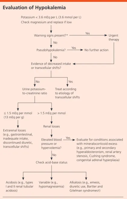
Evaluation of Hypokalemia
Figure 1. Suggested algorithm for the evaluation of hypokalemia.
Information from references 7 and 14.
*—Symptoms of hypokalemia, changes on electrocardiography, severe hypokalemia (less than 2.5 mEq per L [2.5 mmol per L]), rapid-onset hypokalemia, or underlying heart dis-ease or cirrhosis.
†—Autosomal recessive disorders of renal tubular transport. Potassium < 3.6 mEq per L (3.6 mmol per L) Check magnesium and replace if low
Warning signs present?* No Yes Pseudohypokalemia? Urgent therapy No Evidence of decreased intake or transcellular shifts? No further action No Yes Urine potassium-to-creatinine ratio Treat according to etiology of transcellular shifts
≤ 1.5 mEq per mmol (13 mEq per g)
> 1.5 mEq per mmol
Extrarenal losses (e.g., gastrointestinal, inadequate intake, discontinued diuretic, transcellular shifts) Renal losses Elevated blood pressure or hypervolemia?
Check acid-base status
Evaluate for conditions associated with mineralocorticoid excess (e.g., primary and secondary hyperaldosteronism, renal artery stenosis, Cushing syndrome, congenital adrenal hyperplasia)
Acidosis (e.g., types I and II renal tubular acidosis)
Variable (e.g., hypomagnesemia)
Alkalosis (e.g., emesis, diuretic use, Bartter and Gitelman syndromes†)
Yes
Yes
as with ongoing diuretic therapy or hyper-aldosteronism, 20 mmol per day is usually sufficient.15
Causes of Hyperkalemia
Hyperkalemia is caused by excess potassium intake, impaired potassium excretion, or transcellular shifts (Table 2).8,24 The etiology
of hyperkalemia is often multifactorial, with impaired renal function, medication use, and hyperglycemia as the most common contributors.25 Because healthy individuals
can adapt to excess potassium consumption by increasing excretion, increased potas-sium intake is rarely the sole cause of hyper-kalemia, and underlying renal dysfunction is common.24
IMPAIRED POTASSIUM EXCRETION
Renally mediated hyperkalemia results from derangement of one or more of the following processes: rate of flow in the distal nephron, aldosterone secretion and its effects, and functioning potassium secretory pathways. Hyperkalemia secondary to decreased distal delivery of sodium and water occurs with congestive heart failure, cirrhosis, acute kid-ney injury, and advanced chronic kidkid-ney disease. Conditions that cause hypoaldoste-ronism, such as adrenal insufficiency and hyporeninemic hypoaldosteronism (a com-mon complication of diabetic nephropathy and tubulointerstitial diseases), can lead to hyperkalemia.
TRANSCELLULAR SHIFTS
Various mechanisms promote the exit of potassium from cells or impede its entrance, thereby raising the plasma potassium concentration (redistributive hyper-kalemia). Increased plasma osmolality, such as with uncontrolled diabetes mellitus, establishes a concentra-tion gradient wherein potassium follows water out of cells. Relative insulin deficiency or insulin resistance, which also occurs in persons with diabetes, prevents potassium from entering cells. In response to acidosis, extracellular hydrogen is exchanged for intracellular potassium, although the net result is highly variable and depends in part on the type of acidosis; metabolic acidosis produces the greatest effect.26 Because 98% of
total body potassium is intracellular, any process that increases cell turnover, such as rhabdomyolysis, tumor
lysis syndrome, or red blood cell transfusions, can result in hyperkalemia.
MEDICATION-INDUCED HYPERKALEMIA
Medication use is a common cause of hyperkalemia, particularly in patients with baseline renal dysfunction or hypoaldosteronism.27 Medication-induced
hyperkale-mia is most often a result of the medication interfering with potassium excretion. Also, the administration of potassium to treat or prevent hypokalemia can inadver-tently cause hyperkalemia.19
ACE inhibitors contributed to one-half of all cases of drug-induced hyperkalemia in one sample, and approxi-mately 10% of outpatients who start an ACE inhibitor or an ARB will develop hyperkalemia within one year.23,28
Table 2. Causes of Hyperkalemia
Impaired excretion
Acute kidney injury/chronic kidney disease
Medications
Angiotensin-converting enzyme inhibitors and angiotensin receptor blockers Nonsteroidal anti-inflammatory drugs Potassium-sparing diuretics Trimethoprim Heparin Lithium Calcineurin inhibitors Decreased distal renal flow
Acute kidney injury/chronic kidney disease
Congestive heart failure Cirrhosis Hypoaldosteronism Hyporeninemic hypoaldosteronism Adrenal insufficiency Adrenocorticotropic hormone deficiency Primary hyporeninemia Primary renal tubular defects
Sickle cell disease
Systemic lupus erythematosus Obstructive uropathy Hereditary tubular defects Amyloidosis
NOTE: Listed in approximate order of frequency.
*—Dietary-induced hyperkalemia usually involves concurrent renal insufficiency. Information from references 8 and 24.
Transcellular shifts Insulin deficiency/resistance Acidosis Hypertonicity Hyperglycemia Mannitol Medications Beta blockers Digoxin toxicity Somatostatin Succinylcholine (Anectine) Cell breakdown/leakage Hyperkalemic periodic paralysis
Increased intake
Potassium supplementation Red blood cell transfusion Foods high in potassium*
Potassium-containing salt substitutes Protein calorie supplements Penicillin G potassium Certain forms of pica
Pseudohyperkalemia
Hemolysis Tourniquet use Fist clenching Blood sample cooling
Intravenous fluids with potassium Cell hyperplasia
Significant leukocytosis (> 75,000 cells per mm3 [75.0 × 10⁹ per L]) Erythrocytosis
Thrombocytosis
The incidence of hyperkalemia associated with use of potassium-sparing diuretics has risen since adding spi-ronolactone to standard therapy was shown to reduce morbidity and mortality in patients with congestive heart failure.29 Dual treatment with an ACE inhibitor
and an ARB increases the risk of harmful adverse effects, including hyperkalemia, and should be avoided.11 Other
commonly used medications known to cause hyperka-lemia include trimethoprim, heparin, beta blockers, digoxin, and nonsteroidal anti-inflammatory drugs.3
Evaluation and Management of Hyperkalemia GENERAL PRINCIPLES
As with hypokalemia, the immediate danger of hyper-kalemia is its effect on cardiac conduction and muscle strength, and initial efforts should focus on determin-ing the need for urgent intervention (Figure 2).14,30
The absence of symptoms does not exclude severe hyper-kalemia, because hyperkalemia is often asymptomatic. Because of their increased risk of developing hyperka-lemia, patients with underlying renal dysfunction merit special attention.22
HISTORY AND PHYSICAL EXAMINATION
Severe hyperkalemia (more than 6.5 mEq per L [6.5 mmol per L]) can cause muscle weakness, ascending paralysis, heart palpitations, and paresthesias. Chronic kidney dis-ease, diabetes, heart failure, and liver disease all increase the risk of hyperkalemia. Clinicians should review patients’ medications to identify those known to cause hyperkalemia, and ask patients about the use of salt sub-stitutes that contain potassium. The physical examina-tion should include assessment of blood pressure and intravascular volume status to identify potential causes
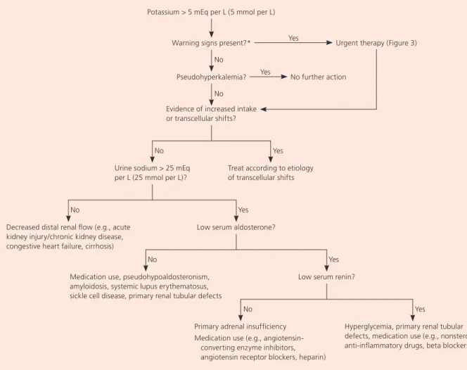
Evaluation of Hyperkalemia
Figure 2. Suggested algorithm for the evaluation of hyperkalemia.
Information from references 14 and 30.
*—Symptoms of hyperkalemia, changes on electrocardiography, severe hyperkalemia (greater than 6.5 mEq per L [6.5 mmol per L]), rapid-onset hyperkalemia, or underlying heart disease, cirrhosis, or kidney disease.
Potassium > 5 mEq per L (5 mmol per L)
Warning signs present?* No
Yes
Pseudohyperkalemia?
Urgent therapy (Figure 3)
No Evidence of increased intake or transcellular shifts?
No further action
No Yes
Urine sodium > 25 mEq per L (25 mmol per L)?
Treat according to etiology of transcellular shifts
Yes
No Yes
Decreased distal renal flow (e.g., acute kidney injury/chronic kidney disease, congestive heart failure, cirrhosis)
Low serum aldosterone?
No Yes
Medication use, pseudohypoaldosteronism, amyloidosis, systemic lupus erythematosus, sickle cell disease, primary renal tubular defects
Low serum renin?
No Yes
Primary adrenal insufficiency Medication use (e.g.,
angiotensin-converting enzyme inhibitors, angiotensin receptor blockers, heparin)
Hyperglycemia, primary renal tubular defects, medication use (e.g., nonsteroidal anti-inflammatory drugs, beta blockers)
of kidney hypoperfusion, which can lead to hyperkale-mia. Neurologic signs of hypokalemia include general-ized weakness and decreased deep tendon reflexes.11
LABORATORY ANALYSIS AND ECG
Repeat measurement of serum potassium can help iden-tify pseudohyperkalemia, which is common and typi-cally results from potassium moving out of cells during or after sample collection.31 Other laboratory studies
include measurement of serum blood urea nitrogen and creatinine, measure-ment of urine electrolytes and creatinine, and assessment of acid-base status. Fur-ther evaluation may include measurement of serum glucose to evaluate for hypergly-cemia, and measurement of serum renin, aldosterone, and cortisol to further inves-tigate kidney and adrenal function.
ECG should be considered if the potas-sium level is greater than 6 mEq per L; if there are symptoms of hyperkalemia; if there is suspicion of rapid-onset hyperka-lemia; or among patients with underlying kidney disease, heart disease, or cirrhosis who have a new case of hyperkalemia. Find-ings on ECG are neither sensitive nor spe-cific for hyperkalemia. Therefore, although ECG changes should trigger urgent treat-ment, treatment decisions should not be based solely on the presence or absence of ECG changes.32
Peaked T waves are the prototypical, and generally the earliest, ECG sign of hyperkalemia. Other ECG changes include P-wave flattening, PR-interval prolon-gation, widening of the QRS complex, and sine waves.19 Hyperkalemia-induced
arrhythmias include sinus bradycardia, sinus arrest, ventricular tachycardia, ven-tricular fibrillation, and asystole.19
Treatment of Hyperkalemia GENERAL PRINCIPLES
The goals of acute treatment are to pre-vent potentially life-threatening cardiac conduction and neuromuscular dis-turbances, shift potassium into cells, eliminate excess potassium, and resolve the underlying disturbance. Patients with chronic hyperkalemia should be counseled to reduce dietary potassium. Although redistributive hyperkalemia is uncommon, a cautious approach is warranted because treatment may not involve attempts to eliminate potassium, and cor-rection of the underlying problem can provoke rebound hypokalemia. Indications for prompt intervention are symptoms of hyperkalemia, changes on ECG, severe hyperkalemia (greater than 6.5 mEq per L), rapid-onset hyperkalemia, or underlying heart disease, cir-rhosis, or kidney disease.24,30,33-35 Potassium should be Management of Hyperkalemia
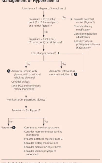
Figure 3. Suggested algorithm for the management of hyperkalemia.
(ECG = electrocardiography.)
NOTE: See Table 3 for a summary of medication therapy for hyperkalemia.
*—Symptoms of hyperkalemia, rapid-onset hyperkalemia, or underlying heart disease, cirrhosis, or kidney disease.
†—Avoid in patients with or at risk of developing abnormal bowel function. Potassium > 5 mEq per L (5 mmol per L)
Yes Potassium 5 to 5.9 mEq per L (5 to 5.9 mmol per L) and no risk factors?*
No Potassium > 6 mEq per L (6 mmol per L) or risk factors?*
Evaluate potential causes (Figure 2) Consider dietary modification Consider medication adjustments Consider sodium polystyrene sulfonate (Kayexalate)†
ECG changes present?
Administer intravenous calcium in addition to A
Administer insulin with glucose, with or without nebulized albuterol Consider dialysis Serial ECG and continuous
cardiac monitoring
Monitor serum potassium, glucose
Potassium < 6 mEq per L?
Continue to monitor potassium Consider more continuous cardiac
monitoring
Evaluate potential causes (Figure 2) Consider dietary modifications Consider medication adjustments Consider sodium polystyrene
sulfonate†
No Yes
A
No Yes
monitored often because patients are at risk of rede-veloping hyperkalemia until the underlying disorder is corrected and excess potassium is eliminated. Figure 3 is an algorithm for the management of hyperkalemia, and Table 3 22,30,36 summarizes medications used in the
treat-ment of the condition. URGENT TREATMENT
Intravenous Calcium. Intravenous calcium, which helps prevent life-threatening conduction disturbances by stabilizing the cardiac muscle cell membrane, should be administered if ECG changes are present.24,25,35
Intra-venous calcium has no effect on plasma potassium concentration. If after five minutes, follow-up ECG con-tinues to show signs of hyperkalemia, the dose should
be repeated.37 Clinicians should be aware that
intrave-nous calcium has a short duration, ranging from 30 to 60 minutes.
Insulin and Glucose. The most reliable method for shifting potassium intracellularly is administration of glucose and insulin. Typically, 10 units of insulin are administered, followed by 25 g of glucose to prevent hypoglycemia.37 Because hypoglycemia is a common
adverse effect even with the provision of glucose, serum glucose levels should be monitored regularly. Patients with a serum glucose level of more than 250 mg per dL (13.9 mmol per L) typically do not require coadministra-tion of glucose.
Inhaled Beta Agonists. Albuterol, a beta2 agonist,
is an underutilized adjuvant for shifting potassium
Table 3. Medications for the Treatment of Hyperkalemia
Medication Dosage Onset Duration
Approximate
potassium-lowering effect Mechanism Cautions
Acute treatment
Calcium Calcium chloride, 10 mL of 10% solution IV over 5 to 10 minutes, or calcium gluconate, 30 mL of 10% solution IV over 5 to 10 minutes Immediate 30 to 60 minutes — Stabilizes cardiac muscle cell membrane; no effect on serum potassium or total body potassium
May potentiate digoxin toxicity; calcium chloride can cause phlebitis and tissue necrosis
Insulin Regular insulin, 10 units IV followed immediately by 50 mL of 50% glucose (25 g) IV
15 minutes ≥ 2 hours 0.7 to 1 mEq per L (0.7 to 1 mmol per L) Shifts potassium into cells; no effect on total body potassium
May cause hypoglycemia; glucose is unnecessary if serum glucose level is > 250 mg per dL (13.9 mmol per L); additive effect when combined with albuterol Albuterol 10 to 20 mg
nebulized
30 minutes ≥ 2 hours 0.5 to 1 mEq per L (0.5 to 1 mmol per L) Shifts potassium into cells; no effect on total body potassium
Can cause tachycardia and thus should be used with caution in patients with underlying heart disease; potassium-lowering effect not reliable in all patients; additive effect when combined with insulin Subacute treatment Sodium polystyrene sulfonate (Kayexalate) Oral: 15 g, 1 to 4 times daily Rectal: 30 to 50 g every 6 hours in a retention enema
2 to 24 hours Variable Variable Binds potassium in exchange for sodium; lowers total body potassium Association with gastrointestinal complications, particularly when combined with sorbitol; should be avoided in patients at risk of abnormal bowel function
IV = intravenously.
intracellularly.24,37 All forms of administration (i.e.,
inhaled, nebulized, and intravenous where available) are effective. It should be noted that the recommended dose of nebulized albuterol (10 to 20 mg) is four to eight times greater than the typical respiratory dose. There is an additive effect when albuterol is combined with insu-lin.38 Albuterol’s potassium-lowering effect is mitigated
in some patients, particularly those with end-stage kid-ney disease; therefore, albuterol should not be used as monotherapy.30
Sodium Bicarbonate. Although sodium bicarbonate is often used to treat hyperkalemia, the evidence to sup-port this use is equivocal, showing minimal to no ben-efit.39 Therefore, sodium bicarbonate should not be used
as monotherapy. It may have a role as adjuvant therapy, particularly among patients with concurrent metabolic acidosis.24,39,40
LOWERING TOTAL BODY POTASSIUM
Potassium can be removed via the GI tract or the kid-neys, or directly from the blood with dialysis. Dialysis should be considered in patients with kidney failure or life-threatening hyperkalemia, or when other treatment strategies fail.23,37 Other modalities are not rapid enough
for urgent treatment of hyperkalemia.39
Currently available cation exchange resins, typi-cally sodium polystyrene sulfonate (Kayexalate) in the United States, are not beneficial for the acute treatment of hyperkalemia but may be effective in lowering total body potassium in the subacute setting.25,39 Because
sodium polystyrene sulfonate can be constipating, many formulations include sorbitol for its laxative effects. However, case reports linking the concomitant use of
sodium polystyrene sulfonate and sorbitol to GI injury prompted a U.S. Food and Drug Administration boxed warning.41,42 More recent reports implicate sodium
poly-styrene sulfonate alone.43 Therefore, use of the drug with
or without sorbitol should be avoided in patients with or at risk of abnormal bowel function, such as postopera-tive patients and those with constipation or inflamma-tory bowel disease.42
There is no evidence supporting the use of diuret-ics for the acute treatment of hyperkalemia. However, diuretics, particularly loop diuretics, may play a role in the treatment of some forms of chronic hyperkalemia, such as that caused by hyporeninemic hypoaldosteron-ism.39,44 Fludrocortisone is an option for hyperkalemia
associated with mineralocorticoid deficiency, including hyporeninemic hypoaldosteronism.29
Strategies to prevent chronic hyperkalemia include instructing patients to eat a low-potassium diet, discon-tinuing or adjusting medications, avoiding nonsteroidal anti-inflammatory drugs, and adding a diuretic if the patient has sufficient renal function.
Data Sources: An Essential Evidence search was conducted. Searches of PubMed, the Cochrane Database of Systematic Reviews, and the National Guideline Clearinghouse were completed using the key terms hypokalemia and hyperkalemia. The search included meta-analyses, randomized controlled trials, clinical trials, and reviews. Search dates: February, September, and December 2014.
The Authors
ANTHONY J. VIERA, MD, MPH, is an associate professor in the Depart-ment of Family Medicine at the University of North Carolina at Chapel Hill School of Medicine.
NOAH WOUK, MD, is a resident in the Department of Family Medicine at the University of North Carolina at Chapel Hill School of Medicine.
SORT: KEY RECOMMENDATIONS FOR PRACTICE
Clinical recommendation
Evidence
rating References
Patients with a history of congestive heart failure or myocardial infarction should maintain a serum potassium concentration of at least 4 mEq per L (4 mmol per L).
C 15
Intravenous potassium should be reserved for patients with severe hypokalemia (serum potassium < 2.5 mEq per L [2.5 mmol per L]), hypokalemic ECG changes, or physical signs or symptoms of hypokalemia, or for those unable to tolerate the oral form.
C 22
Prompt intervention and possible ECG monitoring are indicated for patients with severe hypokalemia (serum potassium < 2.5 mEq per L) or severe hyperkalemia (serum potassium > 6.5 mEq per L [6.5 mmol per L]); ECG changes; physical signs or symptoms; possible rapid-onset hyperkalemia; or underlying kidney disease, heart disease, or cirrhosis.
C 7, 15, 24, 30, 33-35
Intravenous calcium should be administered if hyperkalemic ECG changes are present. C 24, 25, 35 Intravenous insulin and glucose, inhaled beta agonists, and dialysis are effective in the acute
treatment of hyperkalemia.
B 39
Sodium polystyrene sulfonate (Kayexalate) may be effective in lowering total body potassium in the subacute setting.
C 25
ECG = electrocardiography.
A = consistent, good-quality patient-oriented evidence; B = inconsistent or limited-quality patient-oriented evidence; C = consensus, disease-oriented evidence, usual practice, expert opinion, or case series. For information about the SORT evidence rating system, go to http://www.aafp. org/afpsort.
Address correspondence to Anthony J. Viera, MD, MPH, University of North Carolina at Chapel Hill School of Medicine, 590 Manning Dr., CB 7595, Chapel Hill, NC 27599 (e-mail: [email protected]). Reprints are not available from the authors.
REFERENCES
1. Paice BJ, Paterson KR, Onyanga-Omara F, Donnelly T, Gray JM, Law-son DH. Record linkage study of hypokalaemia in hospitalized patients. Postgrad Med J. 1986;62(725):187-191.
2. Lippi G, Favaloro EJ, Montagnana M, Guidi GC. Prevalence of hypo-kalaemia: the experience of a large academic hospital. Intern Med J. 2010;40(4):315-316.
3. Liamis G, Rodenburg EM, Hofman A, Zietse R, Stricker BH, Hoorn EJ. Electrolyte disorders in community subjects: prevalence and risk factors. Am J Med. 2013;126(3):256-263.
4. Shemer J, Modan M, Ezra D, Cabili S. Incidence of hyperkalemia in hos-pitalized patients. Isr J Med Sci. 1983;19(7):659-661.
5. Paice B, Gray JM, McBride D, Donnelly T, Lawson DH. Hyperkalaemia in patients in hospital. Br Med J (Clin Res Ed). 1983;286(6372):1189-1192. 6. Gennari FJ. Hypokalemia. N Engl J Med. 1998;339(7):451-458. 7. Weiner ID, Wingo CS. Hypokalemia—consequences, causes, and
cor-rection. J Am Soc Nephrol. 1997;8(7):1179-1188.
8. Gennari FJ. Disorders of potassium homeostasis. Hypokalemia and hyperkalemia. Crit Care Clin. 2002;18(2):273-288.
9. Reid A, Jones G, Isles C. Hypokalaemia: common things occur com-monly - a retrospective survey. JRSM Short Rep. 2012;3(11):80. 10. Schulman M, Narins RG. Hypokalemia and cardiovascular disease. Am J
Cardiol. 1990;65(10):4E-9E.
11. Greenberg A. Diuretic complications. Am J Med Sci. 2000;319(1):10-24. 12. Dhalla IA, Gomes T, Yao Z, et al. Chlorthalidone versus hydrochlorothia-zide for the treatment of hypertension in older adults: a population-based cohort study. Ann Intern Med. 2013;158(6):447-455.
13. Morgan DB, Davidson C. Hypokalaemia and diuretics: an analysis of publications. Br Med J. 1980;280(6218):905-908.
14. Mount DB, Zandi-Nejad K. Disorders of potassium balance. In: Taal MW, Chertow GM, Marsden PA, Brenner BM, Rector FC, eds. Brenner and Rector’s The Kidney. Philadelphia, Pa.: Elsevier/Saunders; 2012. 15. Macdonald JE, Struthers AD. What is the optimal serum potassium level
in cardiovascular patients? J Am Coll Cardiol. 2004;43(2):155-161. 16. Whang R, Whang DD, Ryan MP. Refractory potassium repletion. A
conse-quence of magnesium deficiency. Arch Intern Med. 1992;152(1):40-45. 17. Millane TA, Ward DE, Camm AJ. Is hypomagnesemia arrhythmogenic?
Clin Cardiol. 1992;15(2):103-108.
18. Kamel KS, Ethier JH, Richardson RM, Bear RA, Halperin ML. Urine elec-trolytes and osmolality: when and how to use them. Am J Nephrol. 1990;10(2):89-102.
19. Diercks DB, Shumaik GM, Harrigan RA, Brady WJ, Chan TC. Electro-cardiographic manifestations: electrolyte abnormalities. J Emerg Med. 2004;27(2):153-160.
20. Weaver WF, Burchell HB. Serum potassium and the electrocardiogram in hypokalemia. Circulation. 1960;21:505-521.
21. Crop MJ, Hoorn EJ, Lindemans J, Zietse R. Hypokalaemia and subse-quent hyperkalaemia in hospitalized patients. Nephrol Dial Transplant. 2007;22(12):3471-3477.
22. Kim GH, Han JS. Therapeutic approach to hypokalemia. Nephron. 2002;92(suppl 1):28-32.
23. Ingram TC, Olsson JM. In brief: hypokalemia. Pediatr Rev. 2008; 29(9):e50-e51.
24. Evans KJ, Greenberg A. Hyperkalemia: a review. J Intensive Care Med. 2005;20(5):272-290.
25. Fordjour KN, Walton T, Doran JJ. Management of hyperkalemia in hos-pitalized patients. Am J Med Sci. 2014;347(2):93-100.
26. Aronson PS, Giebisch G. Effects of pH on potassium: new explanations for old observations. J Am Soc Nephrol. 2011;22(11):1981-1989. 27. Perazella MA. Drug-induced hyperkalemia: old culprits and new
offend-ers. Am J Med. 2000;109(4):307-314.
28. Raebel MA. Hyperkalemia associated with use of angiotensin-converting enzyme inhibitors and angiotensin receptor blockers. Car-diovasc Ther. 2012;30(3):e156-e166.
29. Gross P, Pistrosch F. Hyperkalaemia: again. Nephrol Dial Transplant. 2004;19(9):2163-2166.
30. Alfonzo A, Soar J, MacTier R, et al. Treatment of acute hyperka-laemia in adults. March 1, 2014. http://www.renal.org/guidelines/ joint-guidelines/treatment-of-acute-hyperkalaemia-in-adults#sthash. o9MgdJbw.dpbs. Accessed September 1, 2014.
31. Smellie WS. Spurious hyperkalaemia. BMJ. 2007;334(7595):693-695. 32. Montague BT, Ouellette JR, Buller GK. Retrospective review of the
frequency of ECG changes in hyperkalemia. Clin J Am Soc Nephrol. 2008;3(2):324-330.
33. Maxwell AP, Linden K, O’Donnell S, Hamilton PK, McVeigh GE. Manage-ment of hyperkalaemia. J R Coll Physicians Edinb. 2013;43(3):246-251. 34. Charytan D, Goldfarb DS. Indications for hospitalization of patients with
hyperkalemia. Arch Intern Med. 2000;160(11):1605-1611.
35. Soar J, Perkins GD, Abbas G, et al. European Resuscitation Council guidelines for resuscitation 2010 section 8. Cardiac arrest in special cir-cumstances: electrolyte abnormalities, poisoning, drowning, acciden-tal hypothermia, hyperthermia, asthma, anaphylaxis, cardiac surgery, trauma, pregnancy, electrocution. Resuscitation. 2010;81(10):1400-1433. 36. Lexicomp online. https://online.lexi.com/crlsql/servlet/crlonline
[sub-scription required]. Accessed September 23, 2014.
37. Weisberg LS. Management of severe hyperkalemia. Crit Care Med. 2008;36(12):3246-3251.
38. Lens XM, Montoliu J, Cases A, Campistol JM, Revert L. Treatment of hyperkalaemia in renal failure: salbutamol v. insulin. Nephrol Dial Trans-plant. 1989;4(3):228-232.
39. Mahoney BA, Smith WA, Lo DS, Tsoi K, Tonelli M, Clase CM. Emer-gency interventions for hyperkalaemia. Cochrane Database Syst Rev. 2005;(2):CD003235.
40. Allon M, Shanklin N. Effect of bicarbonate administration on plasma potassium in dialysis patients: interactions with insulin and albuterol. Am J Kidney Dis. 1996;28(4):508-514.
41. Lillemoe KD, Romolo JL, Hamilton SR, Pennington LR, Burdick JF, Wil-liams GM. Intestinal necrosis due to sodium polystyrene (Kayexalate) in sorbitol enemas: clinical and experimental support for the hypothesis. Surgery. 1987;101(3):267-272.
42. U.S. Food and Drug Administration. MedWatch. Kayexalate (sodium polystyrene sulfonate) powder. January 2011. http://www.fda.gov/ Safety/MedWatch/SafetyInformation/ucm186845.htm. Accessed Sep-tember 23, 2014.
43. Harel Z, Harel S, Shah PS, Wald R, Perl J, Bell CM. Gastrointestinal adverse events with sodium polystyrene sulfonate (Kayexalate) use: a systematic review. Am J Med. 2013;126(3):264.e9-e24.
44. Sebastian A, Schambelan M, Sutton JM. Amelioration of hyper-chloremic acidosis with furosemide therapy in patients with chronic renal insufficiency and type 4 renal tubular acidosis. Am J Nephrol. 1984;4(5):287-300.