Training Group
Preclinical Molecular Imaging
Preface . . . . 02
Laboratory for Preclinical Imaging and Imaging Technology of the Werner Siemens-Foundation . . . . 04
Members of the laboratory . . . . 04
Performance and infrastructure . . . . 06
Current scientific projects . . . . 10
Cooperation partners . . . . 16
Scientific publications . . . . 17
Graduate Program . . . . 18
Scientific focus . . . . 18
Curriculum . . . . 20
Conferences and workshops . . . . 21
Awards . . . . 21
Research budget . . . . 21
Fellowships and financial budget . . . . 22
What is special about our program? . . . . 23
Scientific Fields of the Program . . . . 24
Program partners . . . . 24
Coordinator . . . . 25
PhD Program Application Process . . . . 26
Prerequisites . . . . 26 Selection procedure . . . . 27 How to apply? . . . . 27 Location . . . . 28 Scientific environment . . . . 28 Cultural environment . . . . 30
Preface
Molecular and in particular small animal imaging is an emerging tool impacting on various biomedical research areas such as neurology, oncology, cardiology or immunology . Furthermore, the pharmaceutical industry recognizes the exceptional potential of molecular imaging for drug research .
The Laboratory for Preclinical Imaging and Imaging Technology was founded in 2005 and has been supported by the Werner Siemens-Foundation through an independent professorship since January 2008 . The laboratory maintains close scientific col-laborations with Siemens Medical Solutions (Knoxville, USA and Erlangen, Germany), Bruker Biospin MRI (Ettlingen, Germany) and major European pharmaceutical companies .
Our Objective…
…is to establish a PhD Research Training Group at the University of Tübingen, with a focus on Preclinical Molecular Imaging .
Our Vision…
… is to set up a novel graduate school without conventional rigid “student-like” structures . Instead, our focus will be on providing PhD candidates a research environment with the support nee-ded whilst allowing the individuals as much freedom as possible . The Werner Siemens-Foundation provides funding to establish this graduate school at the Medical Faculty of the University of Tübingen, to encourage graduate students to take up training in the very exciting, innovative and interdisciplinary research field of “preclinical molecular imaging”
Prof . Bernd Engler
President of the University Prof . Ingo AutenriethDean of the Medical Faculty Prof . Bernd PichlerProgram Director
Our Curriculum…
…will not be based on educational classes but rather candidates will be actively encouraged to attend international meetings in order to present their work in front of a specialized audiences, and network with top international researchers . In addition, scientific off-site meetings, journal clubs and seminars with talks from colleagues and first-class guest speakers from internationally reputable laborato-ries will build and support an excellent learning environment . Further unique features of our program include an optional research stay abroad in a collaborating laboratory in the US . All PhD students manage their own annual research budget to have a certain financial independence to pursue new ideas without the requirement of the supervisor’s permission . This not only educates the candidates scientifically, but also enables them to develop into mature researchers . The continuous performance control and final rating of the PhD work is not primarily based on the written thesis but on scientific output such as peer-reviewed publications, conference con-tributions, overall research goals achieved, awards aquired, and in the ideal case third-party grant funding obtained . We believe that this PhD research training group program will attract highly motivated junior researchers to develop their research career in a scientific stimulating environment as opposed to conventional training and research structures .
Laboratory for Preclinical Imaging
and Imaging Technology of the
Werner Siemens-Foundation
Members of the laboratory
The team of the Laboratory for Preclinical Imaging and Imaging Technology of the Werner Siemens-Foundation is an interdiscip-linary group, consisting of PhD and master students, postdocs, technicians and engineers as well as administrative officers . The members come from different scientific backgrounds such as physics, biology, medicine, biochemistry, chemistry, computer science, and engineering . Prof . Pichler also heads the Division of Radiopharmacy with ten highly qualified radiopharmacists, che-mists and engineers .
Daniel Bukala
Damaris Kukuk Uta Paulsen
Kristina Fischer Bernd Pichler
Manfred Kneilling
Julia Mannheim Markus Kühs
Martin Judenhofer
Nadine Kemmler Julian Schwab
Vesna Sossi Matthias Hofmann
Kerstin Fuchs Armin Kolb Christoph Grießinger Florian Maier Tanja Walsh llja Bezrukov Stefan Wiehr Hans Wehrl Andreas Schmid Konrad Lankes
Performance and infrastructure
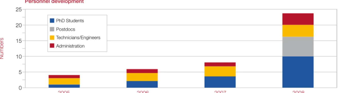
PerformanceProf . Bernd Pichler has been the head of the laboratory since 2005 and over the last 3 years the laboratory has developed from a small laboratory into a state-of-the-art facility for preclinical ima-ging . Our success is demonstrated by the continuous increase in the number of publications and the quality of the journals in which publications are being placed, as well as the amount of funds raised and the growth and development of personnel (Fi-gure 1-4) .
Number of journal publications
2005 2006 2007 2008 0 2 4 6 8 10 12 14 16 18 20 Number
Figure 1: Number of journal publications by the working group per year .
Figure 2: Accumulated impact points by all publications of the working group per year .
Impact factor 0 40 20 80 60 120 100 160 140
Impact factor (total)
2005 2006 2007 2008
Figure 3: Available funds raised per year, calculated from funds raised between 2005 and 2008 . Third party grant funding (status October 2008)
0 200 400 600 800 1 .000 1 .200 1 .400 Eur os (thousand) 2005 2006 2007 2008 2009 2010 First/last author Co-author
Figure 4: Personnel development and structure between 2005 and 2008 Personnel development 0 5 10 15 20 25 Numbers 2005 2006 2007 2008 PhD Students Postdocs Technicians/Engineers Administration
Infrastructure
The laboratory is equipped with latest small animal imaging technology such as two Inveon microPETs, an Inveon microCT, a com-bined SPECT/CT system, a PET/CT, 7 T MRI, optical imaging, 16 MeV cyclotron, radiochemistry laboratories, GMP laboratory for tra-cer production, and clinical as well as preclinical PET/MRI . The imaging modalities are complemented by molecular biology methods (rtPCR, autoradiography, ELISA etc .) and a state of the art cell culture laboratory .
The group currently spreads over 400 m2 of laboratory and 200 m2 of office space .
7T animal MRT Optical imaging Cell culture laboratory
Molecular biology laboratory
Inveon microPET scanner site High resolution microCT Radiopharmacy
PET Detector Laboratory MRI operator room
Current scientific projects
Current projects of the laboratory address two main research areas, biomedicine as well as physics including PET detector re-search . Thus, the laboratory provides an ideal environment for biologists, biochemists, veterinarians, chemists, physician, engi-neers and physicists .
Imaging of T-cell-trafficking in an animal model for contact hypersensitivity reaction
The goal of the study is to visualize the migration pro-perties of T-cells in animal models of inflammation . Ad-ditionally, the establishment of new labeling methods for the non-invasive tracking of T-cells by in vivo imaging is a focus of this work .
Non-invasive detection of amyloid plaques using combined functional and morphological imaging
This project involves the es-tablishment and evaluation of imaging techniques in transgenic mouse models of Alzheimers Disease, using positron emission tomo-graphy (PET) and magne-tic resonance tomography (MRT) .
Zoological studies: Comparative morphology of vertebrates
The projects deal with the compa-rative morphology of fish and the ear region of mammalians using microCT .
In vivo sizing of Echinococcus multilocularis metacestodes via
magnetic resonance tomography
This project comprises in vivo analysis of the growth of Echino-coccus multilocularis in the peritoneal cavity of infected jirds (Me-riones unguiculatus) using magnetic resonance tomography .
PET-imaging with highly specific ra-diolabeled monoclonal PSMA antibo-dies for targeting prostate cancer
The objective of this project is to in-vestigate the in vivo behavior and tu-mor uptake of radiolabeled anti-PS-MA specific monoclonal antibodies and to evaluate their potential as a diagnostic tracer for positron emissi-on tomography .
Investigation of novel PET Tracer in hormone-inde-pendent and hormone-dehormone-inde-pendent prostate cancer in xenograft mouse models
The object of this project is to evaluate PET biomar-kers for in vivo delineation of human prostate tumors in xenograft mouse models . Studies comprise the different pharmacodynamics and uptake charac-teristics of [18F]FLT, [18F]FDG, [11C]Choline and [18F]
FECh in the two hormone-independent cell lines PC-3 and DU145 and the two hormone-dependent tumor models CWR22 and PAC120 using small ani-mal PET imaging .
Pancreas tumor magnetic resonance imaging in the RIP1-Tag2 mouse model
The scope of this project is to reliably identify the pan-creas in a mouse, using magnetic resonance imaging and to investigate the minimum size of tumors that can be distinguished from normal pancreas tissue using magnetic resonance techniques .
Combined PET and MRI
This comprehensive project aims to develop and evaluate combined clinical and de-dicated animal PET/MRI systems . The project focuses on the development of MRI compatible PET detectors based on avalanche photo diodes (APDs) or Geiger-mode APDs (G-APDs) .
Other subprojects focus on novel PET/MR imaging applications which can be pursu-ed with such a system in the fields of oncology, neurology and cardiology .
The world-wide first PET/MRI clinical brain scanner, located in Tübingen, is being evaluated and the issue of MR-based PET attenuation correction, which is of major concern in human applications, is being investigated .
Simultaneous PET/MRI in oncology
The aim of this project is to assess the potential of simultaneous PET/MR imaging in oncological stu-dies . Advantages of combined PET/MR in imaging different cancer models will be evaluated . Strate-gies will be developed for integrated cancer ima-ging to yield a maximum of metabolical and ana-tomical information, therefore supporting treatment decisions and monitoring .
Simultaneous functional PET/ MRI in neurology
The goal of this project is to eva-luate the opportunities and limi-tations of simultaneous multi- functional PET/MRI imaging in neurology . Perfusion mea-surements obtained with PET and MRI will be compared and cross calibrated . In addition, the metabolic basics of the blood oxygen level dependent (BOLD) effect in functional MRI will be evaluated .
Simultaneous PET/MRI of an infarcted mouse myocardium
An attractive aspect of the combined PET/MRI system is the possi-bility to acquire both multi-functional and morphological data simul-taneously . Thus, multimodality imaging of a beating heart is feasible with very high registration accuracy . In this project mice with myo-cardial infarction are investigated, using [18F]FDG and MRI .
Next Generation PET/MRI scanner
Within this project we will develop the world first PET in-sert for small animal MRI scanners which enables the simulta-neous acquisition of whole body mouse
PET and MRI images . For optimal performance the in-sert must fit between the standard gradient coil and a rat whole body coil of the MRI . The planned FOV in axial direction is 72 mm, this is enough for whole body mouse or multi-bed rat scans .
G-APDs for PET/MRI detectors
This project focuses on investigating the suitability of Geiger-mode APDs (G-APDs) for usage in high resolution PET applica-tions . We are developing a novel detector which has the capacity to work in high magnetic fields of combined PET/MRI providing depth of interaction
information for opti-mal spatial resolution . Furthermore, fast rea-dout electronics, spe-cifically for G-APDs, will be developed .
Evaluation of clinical PET/MRI
In this work, the influence of the PET insert on the MR image quality of the world first clinical PET/MRI system will be analyzed . Furthermore, measurements to evaluate the PET performance of the MR compatible scanner will be performed .
In vivo PET-imaging of inflammation and hypoxia in a small
animal model of rheumatoid arthritis
This project focuses on the in vivo investigation of the role of hypoxia-induced angiogenesis during the development of rheumatoid arthritis (RA), using newly designed hypoxia-mar-kers to evaluate their clinical relevance and applicability . For this and most of the other projects we use in vitro methods like rtPCR, Western-Blot, ELISA or immunohistology to com-plement the in vivo imaging data .
PET in vivo quantification of the dopaminergic
system of rats and mice in an animal model of Parkinson’s Disease - Limits and possibilities
The objective of the study is to evaluate quantifi-cation limits of neurodegeneration in rats and mice using small animal PET and 7 Tesla MRI . As PET probes we use dopamine transporter and dopami-ne receptor ligands . Kidopami-netic modelling allows us to assess parameter like receptor density or KD .
MR-based Attenuation Correc-tion for PET/MR
In current PET/MR setups at-tenuation correction needs to be performed based on the MR image . This project uses machine learning methods, at-las registration and novel MR sequences to determine the attenuation map from the MR image .
Radiopharmacy and in vivo
hypoxia imaging in mice
In this project, our radiochemists work on the design and synthesis of novel PET markers for hypoxia
imaging . Performance evaluation of the Siemens Inveon small
animal PET scanner
Small animal PET scanners are used increasingly in bio-medical research . This stu-dy evaluates the relevant performance parameters of the Siemens Inveon PET, which is a state of the art small animal PET scanner .
Cooperation partners
International and national academic partners
> University of California, Davis, USA
> University of California, San Francisco, USA
> Universität Freiburg, Germany
> Universität Innsbruck, Austria
> Universität Münster, Germany
> University of Gießen, Germany
> Technische Universität München, Germany
> Washington University, St . Louis, USA
> Lawrence Berkeley National Laboratory, Berkeley, USA
> University of Queensland, Australia
Industry partners
> Astra Zeneca, UK
> Bayer Schering Pharma, Germany
> Boehringer-Ingelheim, Biberach, Germany
> Hamamatsu Photonics, Herrsching, Germany
> Merck, Germany
> Oncodesign, Dijon, France
> Siemens Medical Solutions, Erlangen, Germany
> Siemens Preclinical Solutions, Knoxville, TN, USA
> Synovo, Tübingen, Germany
Academic partners at Tübingen
> Clinic of Cardiology
> Clinic of Dentistry
> Clinic of Dermatology
> Clinic of Internal Medicine
> Clinic of Neurology
> Clinic of Radiology
> Institute of Anatomy
> Institute of Biochemistry
> Institute for Human Genetics
> Institute of Immunology
> Institute for Medical Microbiology and Hygiene
> Institute of Microbiology
> Institute of Molecular Biology
> Institute of Pathology
> Institute of Zoology
> Max Planck Institute for Biological Cybernetics
> University Eye Hospital
Scientific publications
The following list shows a selection of the most important recent scientific manuscripts:
M . Hofmann, F . Steinke, V . Scheel, G . Charpiat, J . Farquhar, P . Aschoff, M . Brady, B . Schölkopf, and B .J . Pichler: MR-based attenuation correction for PET/MR: A Novel Approach Combining Pattern Recognition and Atlas Registration . J Nucl Med . 49(11):1875-1883, 2008
H-P . Schlemmer, B .J . Pichler, M . Schmand, Z . Burbar, C . Michel, R . Ladebeck, K . Jattke, D . Townsend, C . Nahmias, P .K . Jacob, W .-D . Heiss, C .D . Claussen: Simultaneous MR/PET Imaging of the Human Brain: Feasibility Study . Radiology 248(3):1028-1035, 2008
B .J . Pichler, H .F . Wehrl, M .S . Judenhofer: Latest advances in molecular imaging instrumentation . J Nucl Med . 49 Suppl 2: 5S-23S ., 2008
H .F . Langer, R . Haubner, B .J . Pichler, M . Gawaz: Radionuclide imaging: A molecular key to the atherosclerotic plaque . J Am Coll Cardiol . 52(1):1-12, 2008
N . Müller-Hermelink*, H . Braumüller*, B . Pichler*, T . Wieder, R . Mailhammer, K . Schaak, K . Ghoreschi, A . Yazdi, R . Haubner, C .A . Sander, R . Mocikat, M . Schwai-ger, I . Förster, R . Huss, W .A . Weber, M . Kneilling, M . Röcken: TNFR1 signaling and IFN-gamma signaling determine whether T cells induce tumor dormancy or promote multistage carcinogenesis . Cancer Cell . 13(6):507-18, 2008 (*contributed equally) M . Föller, S . Feil, K . Ghoreschi, S . Koka, A . Gerling, M . Thunemann, F . Hofmann, B . Schuler, J . Vogel, B . Pichler, R .S . Kasinathan, J .P . Nicolay, S .M . Huber, F . Lang, R . Feil: Anemia and splenomegaly in cGKI-deficient mice . Proc Natl Acad Sci U S A . 105(18):6771-6, 2008
M .S . Judenhofer, H .F . Wehrl, D .F . Newport, C . Catana, S .B . Siegel, M . Becker, A . Thielscher, M . Kneilling, M . Lichy, M . Eichner, K . Klingel, G . Reischl, S . Widmaier, M . Röcken, R .E . Nutt, H .-J . Machulla, K . Uludag, S .R . Cherry, C .D . Claussen, B .J . Pichler: Simultaneous PET/MRI: A new approach for functional and morphological imaging . Nat Med . 14(4):459-65, 2008
C . Catana, D . Procissi, Y . Wu, M .S . Judenhofer, J . Qi, B .J . Pichler, R .E . Jacobs, S .R . Cherry: Simultaneous in vivo positron emission tomography and magnetic re-sonance imaging . Proc Natl Acad Sci U S A ., 11;105(10):3705-10, 2008
M . Angstenberger, J .W . Wegerner, B .J . Pichler, M .S . Judenhofer, S . Feil, S . Alberti, R . Feil, A . Nordheim: Severe intestinal obstruction upon induced smooth muscle-specific ablation of the transcription factor SRF in adult mice . Gastroenterology, 133(6):1948-59, 2007
Judenhofer, C . Catana, B .K . Swann, S .B . Siegel, W .-I . Jung, R .E . Nutt, S .R . Cherry, C .D . Claussen, B .J . Pichler: Simultaneous PET/MR images, acquired with a com-pact MRI compatible PET detector in a 7 Tesla magnet . Radiology, 244(3):807-14, 2007
M . Kneilling*, L . Hültner*, B .J . Pichler*, R . Mailhammer, L . Morawietz, S . Solomon, M . Eichner, J . Sabatino, T . Biedermann, V . Krenn, W . A . Weber, H . Ilges, R . Haubner, M . Röcken: Targeted mast cell cell–silencing prevents joint destruction and angio-genesis in experimental arthritis . Arth Rheum, 56(6):1806-1816, 2007 (*contributed equally)
A complete list of all publications of the laboratory and further information concer-ning the projects can be found at: www .preclinicalimaging .org
Graduate Program
Scientific focus
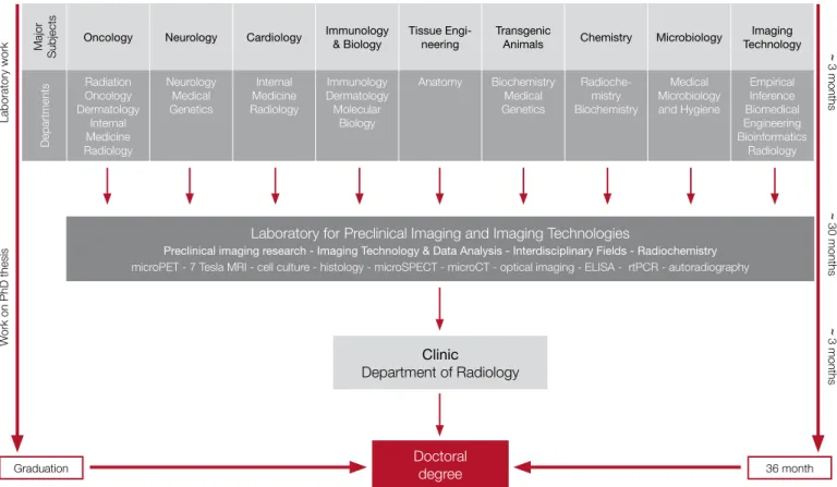
PhD candidates enter the program by selecting a major subject and gain their first laboratory experience in a laboratory focusing on their chosen subject in the first 3 months . This includes labo-ratories located at the University or at the Max Planck Institutes in Tübingen . PhD candidates can choose their major topic from a list of available projects, ranging from detector-physics, image reconstruction to basic biomedical research in the fields of on-cology, neurology, cardiology, immunology or infection biology (Figure 5) .
Subsequently, graduate training group members enter the small animal imaging program, working on the topic and project selec-ted as the major subject for their PhD thesis for about 30 months . During this time, the participants of the graduate program are located at the Laboratory for Preclinical Imaging and Imaging Technology . A close link to the initial training laboratory is manda-tory . The final 3 months are spent in the imaging department of the Clinic of Radiology to give PhD candidates an impression of clinical imaging applications (Figure 5) .
Participants of the graduate program gain theoretical and practi-cal training, and conduct a research project under supervision of an designated experienced senior scientist and are monitored by the thesis advisory committee . PhD candidates submit a written
Doctoral degree Qualified applicants
Dr . med .1 Physicians
Dr . sc . hum .2 University degree, Bachelor degree
Dr . rer . nat . 2 Physicist
Dr . rer . nat . 2 Biologist
MD/PhD 3 Physicians
PhD 3 University degree
1Dr . med is equivalent to an MD title
2Dr . rer . nat . and Dr . sc . hum . is equivalent to a PhD title 3planned new doctoral degrees at the University of Tübingen
Major
Subjects
Oncology Neurology Cardiology Immunology & Biology Tissue Engi-neering Transgenic Animals Chemistry Microbiology TechnologyImaging
Departments Radiation Oncology Dermatology Internal Medicine Radiology Neurology Medical Genetics Internal Medicine Radiology Immunology Dermatology Molecular Biology Anatomy Biochemistry Medical Genetics Radioche-mistry Biochemistry Medical Microbiology and Hygiene Empirical Inference Biomedical Engineering Bioinformatics Radiology
Laboratory for Preclinical Imaging and Imaging Technologies
Preclinical imaging research - Imaging Technology & Data Analysis - Interdisciplinary Fields - Radiochemistry
microPET - 7 Tesla MRI - cell culture - histology - microSPECT - microCT - optical imaging - ELISA - rtPCR - autoradiography
Clinic Department of Radiology Doctoral degree Graduation 36 month Laboratory work ~ 3 months ~ 30 months ~ 3 months W ork on PhD thesis
Figure 5: Organization of the graduate program . thesis on completion of their research project . Following the
ap-proval of their thesis by the committee and successful completion of an oral examination, the candidates receive a doctoral degree from the University of Tübingen .
Curriculum
A rich program of courses, lectures, workshops, off-site meetings and conferences is offered to the students . As part of their edu-cation, PhD candidates are required to participate in specified activities and fulfill given requirements as part of their PhD pro-gram . One focus is the development of soft skills (e .g . how to write a paper, how to prepare and give a talk, how to write a grant application) which is reflected in the course curriculum and assessment criteria .
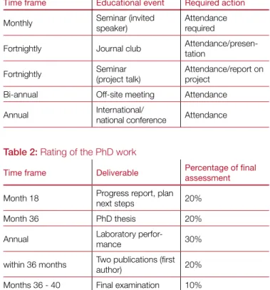
PhD candidates attend monthly seminars with a guest speaker and a fortnightly journal club . Every second week a participant talks about his project in a seminar . After 18 months, PhD candi-dates deliver a progress report and a plan on the next steps which makes up 20% of their final score . The PhD thesis in English will be handed in after 36 months and will be rated with 20% of the final score . Furthermore, the laboratory performance will be sub-ject to an annual evaluation (accounts for 30% of the final score) . Two publications as first author within three years (accounts for 20% of final score) are part of the PhD program . The final exami-nation will make up 10% of the total score (Table 1) .
To develop skills in writing grant applications, participants of the graduate program are required to write a grant application during the 3 year period either individually or as a group task . The inter-mediate progress report has to be written according to the NIH or DFG grant guidelines .
Time frame Educational event Required action
Monthly Seminar (invited speaker) Attendance required Fortnightly Journal club
Attendance/presen-tation Fortnightly Seminar
(project talk)
Attendance/report on project
Bi-annual Off-site meeting Attendance Annual International/ national conference Attendance
Table 2: Rating of the PhD work
Time frame Deliverable Percentage of final assessment
Month 18 Progress report, plan next steps 20%
Month 36 PhD thesis 20%
Annual Laboratory perfor-mance 30% within 36 months Two publications (first author) 20% Months 36 - 40 Final examination 10%
Table 1: Educational program Conferences and workshops
Participants of the program attend one international and one na-tional conference per year, to present their current results by a poster or an oral presentation . This will help students develop the skills needed in a scientific career, like confidence and pro-fessionalism while giving a lecture and presenting their own work to other scientists . Also, a scientific exchange with experienced researchers from other groups initiates new ideas and is stimu-lating for the PhD candidates . It is important for junior scientists to compare their work with the presentations from other groups . Additionally, off-site workshops with the whole work group are pl-anned twice a year, to foster exchange between group members and to stimulate the group interaction . These enhanced skills will enable students to present themselves in an optimal way for fu-ture job opportunities .
Awards
A committee of the research training group announces an annual publication award for the best paper and the best poster pub-lished . All PhD candidates of the laboratory are eligible for this award . Winners receive an official certificate and 1 .000 Euro to reward their success in the presentation of their scientific work .
Research budget
Students will receive their own research budget . The budget will be controlled and managed exclusively by the student and is fle-xibly applicable . Thereby students learn to take responsibility for their own budget, gain independence and flexibility within their project and are able to demonstrate their ability to handle a fixed budget . An additional budget can be awarded for successful grant applications, gained individually or as a group . These skills help students to show their ability to manage a fixed budget and will train them to raise additional money .
Currently 20 people work in the lab with different back-grounds from physics, biology, medicine, biochemistry,
Fellowships and financial budget
Initially is planned to fund five PhD fellowships for three years . The monthly net salary will be approximately 1000 Euro . Furthermo-re students Furthermo-receive an annual budget for their Furthermo-research project which is completely under their own control .
Every student should actively participate in one international and one national conference per year where an abstract as first author has to be accepted . Two off-site workshops per year with the whole work group are also planned .
Individuals or groups can strive for an additional flexible budget through a grant application annually . Furthermore performance-related internal grants for further project costs will be made availa-ble for the students . This will result in an additional flexiavaila-ble budget for all students per year .
Mentoring
The PhD candidates will be supervised and mentored by experi-enced senior researchers and have the support of administrative staff .
What is special about our program?
> First in Europe, focusing on preclinical molecular imaging and imaging technology, a field in biomedical research with great future prospects
> Unique program options for the students
– Up to two conferences (1 international, 1 national) per year · Networkt with other scientists
· Gain new ideas · Present the own work
– Two off-site workshops per year for focused research discussions and as a team building event to foster interactions between group members
– Invite individual experts – Interdisciplinary education
– Own research budget which is managed by the PhD candidate – Option for a research stay in the USA
– Practical orientated training and laboratory-focused education combined with theory, through seminars, conferences and workshops
– Innovative field of biomedical research
– Close contacts to large international pharmaceutical and bioengineering companies – A strong focus on the development of soft skills
· How to write a paper · How to apply for a grant · How to give a talk · Group interaction The laboratory organizes an annual imaging
Scientific Fields of the Program
The University of Tübingen plans to offer the interdisci-plinary preclinical molecular imaging PhD training pro-gram in the fields of:> biochemistry > cardiology > chemistry > imaging technology/physics > immunology > neurology > oncology > pharmacy > radiochemistry > tissue engineering > transgenic animals > toxicology > microbiology > infectious diseases > pharmacology
Program partners
The laboratory currently cooperates with the following institutions listed in Table 3 . Students can pick one of these departments to gain first experi-ence in laboratory work and seek insight in their chosen major subject .
Scientific field Name Department Organisation
Clinical microbiology, environmental and hos-pital hygiene, Infectious diseases
Prof . Autenrieth Medical Microbiology and Hygiene Tübingen University Medical physics, biomedical physics, experimental radiation oncology Radiation biology Prof . Bamberg
Radiation Oncology Tübingen University Prof . Rodemann
Signal transduction and transgenic disease models
Prof . Feil Biochemistry Tübingen University Neurodegeneration Prof . Gasser Neurology Tübingen University Cardiology and
cardio-vascular diseases Prof . Gawaz Internal Medicine III Tübingen University Hematology, oncology,
Iimmunology,
rheuma-tology and pneumology Prof . Kanz Internal Medicine II Tübingen University proteomics, functional
genomics, mouse genetics
Prof . Nordheim Molecular Biology Tübingen University
Scientific field Name Department Organisation
Immunobiology
(MHC functions) Prof . Rammensee Immunology Tübingen University Human genetics,
gene-tic mediated
neurode-generative diseases Prof . Rieß Medical Genetics Tübingen University Oncology and
au-toimmune disorders, immunology
Prof . Röcken Dermatology Tübingen University Theory and applications
of statistical learning
theory Prof . Schölkopf Empirical Inference
Max Planck Institute, Tübingen Clinical anatomy and
tissue engineering Prof . Skutella Anatomy Tübingen University Cardiomyopathy Prof . Kandolf Pathology Tübingen University Diabetis Prof . Häring Internal Medicine Tübingen University
Coordinator
Prof . Dr . rer . nat . Bernd Pichler
Laboratory for Preclinical Imaging and Imaging Technology of the Werner Siemens-Foundation Department of Radiology Eberhard-Karls-University Tübingen Röntgenweg 13 72076 Tübingen Germany www .preclinicalimaging .org
E-mail: bernd .pichler@med .uni-tuebingen .de Phone: +49-7071-29-83427
Fax: +49-7071-29-4451
Administration
Uta Paulsen, Dipl .-Biol .
Laboratory for Preclinical Imaging and Imaging Technology of the Werner Siemens-Foundation Department of Radiology
Eberhard-Karls-University Tübingen Röntgenweg 13
72076 Tübingen Germany
E-mail: uta .paulsen@med .uni-tuebingen .de Phone: +49-7071-29-83450
Fax: +49-7071-29-4451
PhD Program Application Process
Prerequisites
To be accepted as a graduate student at the research training group “Preclinical Molecular Imaging” at the Medical Faculty of the University of Tübingen, applicants must hold, or anticipate re-ceiving a university degree equivalent to a master of science that would formally allow them to enter a PhD or equivalent program in their home country, prior to enrollment .
Graduate university students shall fulfill the following conditions:
> college score better than 80% or
> college score better than 70% and a record of at least one peer-reviewed publication and an oral or poster presentation
Bachelors or FH students shall fulfill the following conditions: > College score better than 90%
or
> college score better than 80% and at least one publication in a peer reviewed journal and one oral or poster presentation
The objective is to find motivated students for scientific research through specific selection interviews . Students should have a background in at least one of the following fields:
> biochemistry > biology > biophysics > chemistry > computer science > medicine > physics
Selection procedure
The application deadline for the first round is October 1st, 2009 .
Shortlisted candidates will be invited for interviews 6 weeks after the application deadline . The final decision whether an applicant is accepted will be made by a committee, consisting of four se-nior advisors from Table 3 . In case less than five candidates were selected in the first round, a second round of recruitment will be carried out to assign the remaining fellowships .The deadline for the second round of recruitement will be May 1st, 2010 . Figure 6
shows the time table for the two rounds of recruitment .
How to apply?
Please visit our web-page www .preclinicalimaging .org to down-load the application forms .
Advertisement (Flyer, online)
Deadline Applications
Evaluation and compilation of shortlist
Invitations for interviews Selection of candidates 30 min interviews with the PhD committee
Start of program April - September December - April October 1st May 1st October 15th May 15th October 16th May 16th November June March September
Figure 6: Time table two rounds of recruitment
We have unique program options for the students
One focus of the PhD program is on practical laboratory education
Location
Tübingen is a traditional historic university town situated on the river Neckar, 40 km southwest of Stuttgart on the fringes of the Swabian mountains . The city officially first appears in records in 1191, however the local castle has records back to the year 1078 . The Eberhard Karls University is one of Germany‘s oldest universities, internationally recognized for medicine, natural sci-ences and the humanities . It was founded in 1477 by Count Eberhard V .
Due to the close proximity to Stuttgart, Tübingen is easily reached by airplane . Stuttgart has a modern international airport with non-stop flights to and from major international destinations . You can reach Tübingen from the airport in about 20 min by car . Public transport by bus or train is also available .
Scientific environment
Besides the university with its 14 faculties, Tübingen also has 17 hospitals affiliated with the University‘s Faculty of Medicine . In terms of third-party funds acquired, the number of Collaborati-ve Research Centres, Graduate Programmes, Research Groups, plus its involvement in national and international collaborations, the Faculty of Medicine in Tübingen is rated one of the top-ten German Faculties of Medicine in all accepted ranking lists .
Additionally, several close collaborations exist between the Uni-versity and the Max Planck Institutes which are also located in Tübingen:
> Max Planck Institute for Biological Cybernetics
> Max Planck Institute for Developmental Biology
> Friedrich Miescher Laboratory
Since October 2007, six faculties, the Max Planck Institute of Bio-logical Cybernetics, the Collaborative Research Centre 550, the Hertie Institute for Clinical Brain Research, the Graduate School of Neural and Behavioural Sciences, and numerous internal and external partners have participated in the Excellence Cluster „In-terdisciplinary Centre for Integrative Neuroscience (CIN) . It is fun-ded by the „Deutsche Forschungsgemeinschaft DFG“ (German Research Foundation), being one of the 20 Excellence Clusters within Germany .
The Laboratory for Preclinical Imaging and Imaging Technology of the Werner Siemens-Foundation is also a member of the CIN and is involved in the SFB 773 (Collaborative Research Center) “Understanding and Overcoming Therapy Resistance of Solid Tu-mors” with a central project . Furthermore, it participates in large national research associations funded by the Federal Ministry of Education and Research like “FORSYS Cooperation”, “Compe-tence Network Dementias – Neurodegeneration” and MoBiMed – Molecular Imaging in Medicine” .
The main research focus at the faculty of medicine is currently in following areas:
> Imaging and medical technology
> Infection biology
> Oncology and immunology
> Neuroscience
> Vascular medicine and diabetes
Tübingen offers an unique scientific environment, hosting institu-tions like the Hertie Institute for Clinical Brain Research („HIH“), which was established with promotional funds from the charitable Hertie Foundation in Tübingen . As a result of its close integrati-on with the Department of Neurology and hence with the Centre for Neurology it enables optimal co-ordination between basic re-search and medical applications . Another institution is the Center for Interdisciplinary Clinical Research, dealing with Cell Biology in
Diagnosis and Therapy of Organ System Diseases . LinksHomepage Laboratory for Preclinical Imaging
www .preclinicalimaging .org Homepage Clinical Center www .med .uni-tuebingen .de Homepage University www .uni-tuebingen .de
Homepage Max Planck Institutes, Tübingen www .tuebingen .mpg .de/
Hertie Institute for Clinical Brain Research
Max Planck Institute for Biological Cybernetics, Tübingen
Quelle: Max-Planck-Institut für biologische Kyber
Cultural environment
TübingenTübingen is a small town with 85 .000 inhabitants and 22 .000 students, making Tübingen the city with the youngest average population in Germany . Life in the city is dominated by its many students, it combines the flair of a lovingly restored medieval cen-ter of town with the colorful bustle and typical atmosphere of a young and cosmopolitan students‘ town . The cultural program comprises events, museums and collections as well as festivals, concerts, stage plays and readings with poets of international re-putation .
Official website Tübingen: www .tuebingen .de
Neckar front of Tübingen
Marketplace of Tübingen
Tübingen is situated in the southwest of Germany, on the Neckar river and at the foothills of the Swabian Alb and the Black Forest . In the old center of Tübingen, an unique ensemble of historic half timber buildings dating from the late 15th to the early 16th century
attract many visitors every year . Tübingen also offers a rich array of cultural activities year-round .
Tübingen
Swabian Alb Black Forest Karlsruhe Singen Bodensee München Würzburg/ HeilbronnStuttgart
The closest major city to Tübingen is Stuttgart, located 40 km northeast from Tübingen . Stuttgart, the capital of Baden-Würt-temberg, provides all the shopping possibilities and cultural life-style of a large city . It comprises a big range of cultural offerings including several museums, theaters and an opera .
Official website Stuttgart: www .stuttgart-tourist .de
Black Forest (Schwarzwald)
The Black Forest starts about 40 km west from Tübingen . It of-fers several opportunities for sporting activities such as hiking and cycling as well as great nature and cities that are worth a visit . With mountains up to 1493 m (Feldberg) it is also a popular skiing region in winter .
Official Black Forest page: www .blackforest-tourism .com
Further Information and Contact
Please visit our website www .preclinicalimaging .org to down-load a digital version of this brochure and the application forms!
Prof. Dr. Bernd Pichler Department of Radiology
Laboratory for Preclinical Imaging and Imaging Technology of the Werner Siemens-Foundation
Roentgenweg 13 72076 Tuebingen Germany
E-mail: bernd .pichler@med .uni-tuebingen .de Phone: +49-7071-29-83427
Fax: +49-7071-29-4451
Swabian mountains (Schwäbische Alb)
Situated close to Tübingen are the Swabian mountains, a high plateau with the highest mountain (Lemberg) reaching up to 1015 m . The spectacular landscape and magnificent nature make it attractive for hiking and cycling . Also worth a visit are the nu-merous castles, churches and monasteries as well as caves and places of discovery of important fossils and historical findings . Official site Tourist Organization: http://en .s-alb .de
Castle Hohenzollern near Hechingen
Aerial photo: “Guetenbach” near Freiburg
Winter landscape near Schluchsee Wuerttemberg State Opera
in Stuttgart
Albtrauf near Moessingen The New Castle in the centre
of the city