Available
online
at
www.sciencedirect.com
ScienceDirect
j ou rn a l h o m epa ge : h t t p : / / w w w . e l s e v i e r . c o m / l o c a t e / r p o r
Original
research
article
Dose
distribution
verification
for
GYN
brachytherapy
using
EBT
Gafchromic
film
and
TG-43
calculation
Somayeh
Gholami
a,b,
Hamid
Reza
Mirzaei
c,∗,
Ali
Jabbary
Arfaee
c,
Ramin
Jaberi
b,
Hassan
Ali
Nedaie
b,
Seied
Rabi
Mahdavi
d,
Eftekhar
Rajab
Bolookat
c,
Ali
S.
Meigooni
eaDepartmentofMedicalPhysicsandBiomedicalEngineering,FacultyofMedicine,TehranUniversityofMedical
Sciences,Tehran,Iran
bRadiationOncologyDepartment,CancerInstitute,TehranUniversityofMedicalSciences,Tehran,Iran
cRadiationOncologyDepartment,ShohadaeTajrishHospital,CancerResearchCenter,ShahidBeheshtiUniversity
ofMedicalSciences,Tehran,Iran
dDepartmentofMedicalPhysics,IranUniversityofMedicalSciences,Tehran,Iran eComprehensiveCancerCentersofNevada,LasVegas,NV,UnitedStates
a
r
t
i
c
l
e
i
n
f
o
Articlehistory:
Received23October2015 Receivedinrevisedform 20January2016 Accepted26June2016 Availableonline18July2016
Keywords: QAbrachytherapyphantom Filmdosimetry GZP6HDRsystem SelectronLDRsystem TG-43
a
b
s
t
r
a
c
t
Aim:Verificationofdosedistributionsforgynecological(GYN)brachytherapyimplantsusing EBTGafchromicfilm.
Background: Onemajorchallengeinbrachytherapyistoverifytheaccuracyofdose distri-butionscalculatedbyatreatmentplanningsystem.
Materialsand methods: A new phantomwas designed and fabricated using 90 slabsof 18cm×16cm×0.2cmPerspextoaccommodateatandemandOvoidassembly,whichis normallyusedforGYNbrachytherapytreatment.Thisphantomdesignallowstheuseof EBTGafchromicfilmsfordosimetricverificationofGYNimplantswithacobalt-60HDR sys-temoraLDRCs-137system.Gafchromicfilmswereexposedusingaplanthatwasdesigned todeliver1.5Gyofdoseto0.5cmdistancefromthelateralsurfaceofovoidsfromapair ofovoidassemblythatwasusedfortreatmentvaginalcuff.Foraquantitativeanalysisof theresultsforbothLDRandHDRsystems,themeasureddosevaluesatseveralpointsof interestswerecomparedwiththecalculateddatafromacommerciallyavailabletreatment planningsystem.ThisplanningsystemwasutilizingtheTG-43formalismandparameters forcalculationofdosedistributionsaroundabrachytherapyimplant.
Results:Theresultsoftheseinvestigationsindicatedthatthedifferencesbetweenthe cal-culatedandmeasureddataatdifferentpointswererangingfrom2.4%to3.8%fortheLDR Cs-137andHDRCo-60systems,respectively.
∗ Correspondingauthorat:RadiationOncologyDepartment,ShohadaeTajrishHospital,CancerResearchCenter,ShahidBeheshti Uni-versityofMedicalSciences,Tehran,Iran.Tel.:+982161192500;fax:+982161192501.
E-mailaddress:[email protected](H.R.Mirzaei).
http://dx.doi.org/10.1016/j.rpor.2016.06.005
Conclusion: TheEBTGafchromicfilmscombinedwiththenewlydesignedphantomcould beutilizedforverificationofthedosedistributionsarounddifferentGYNimplantstreated witheitherLDRorHDRbrachytherapyprocedures.
©2016GreaterPolandCancerCentre.PublishedbyElsevierSp.zo.o.Allrightsreserved.
1.
Background
Asradiationtherapytechniquesbecomemorecomplex,the quality assurance (QA) techniques have to be changed to provideasoundandpracticalmethodofverificationof treat-ment delivery in order to reduce errors during radiation therapy.1 A QA progressneeds to be adopted forcomplex
brachytherapy treatment techniques, particularly when it involvestreatmentwithacomplexgynecologic(GYN)system. GYNbrachytherapytreatmentsforthecervix,vaginaland endometrialcancershavebeencommonlyusedforover100 years.2Accuracyofdosecalculationplaysavitalroleinthe
brachytherapytreatmentplanning.Experimentalverification ofdoseaccuracyisoneofpossibleQAproceduresfor determi-nationofover-doseorunder-doseareainthebrachytherapy planningvolume.However,thistechniquefacesseveral chal-lengesrelatedtothemeasurementsofdosedistributionsina high-gradientregion.Oneoftheimportantcriteriaforthese experimentalsetupsistohave adosimeterwithhigh spa-tialresolutionsuchasGafchromicfilms.Gafchromicfilmsare beingusedas2Ddosimetersbyseveralinvestigatorsinvarious typesofapplications.3
Different investigators had demonstrated the useful-nessofthe EBT Gafchromicfilmsforbrachytherapy source dosimetry.4 Thesefilmsrequirenochemicalprocessingand
theyareinsensitivetoambientlight.Therefore,theycanbe cuttotheshapeoftheexperimentalgeometryforthebest representationofadosimetricsetup.
2.
Aim
Verification of dose distributions for gynecological (GYN) brachytherapyimplantsusingEBTGafchromicfilm.
3.
Materials
and
methods
EBTGafchoromicfilmdosimetryfortwodifferent
brachyther-apy systems for GYN treatment were considered in this
study:theGZP6highdoserate(HDR)60Cobrachytherapyunit thatwasintroducedbyNuclearPowerInstituteofChinafor brachytherapyprocedures,5andtheSelectronlow-dose-rate
(LDR)137Csremoteafterloadingsystemdistributedby
Nucle-tron(NucletronBV,Veenendaal,TheNetherlands).6
3.1. GZP6HDRsystem
GZP6afterloadingunit(NuclearPowerInstituteofChina)with HDR 60Co sources has one stepping source and five
non-steppingsource-braids.7TheGZP6treatmentplanningsystem
isabletoproducedosedistributionsinthetransverseand lon-gitudinalplanes.Itcalculatesdose usingthe superposition
dose calculation technique. This system could be used
for intracavitary treatment of cervical, vaginal, endome-trial, rectal, esophageal and nasopharyngeal malignancies. The sources in this system were consisted of 60Co active
pellets (3.5mm long and 1.5mm diameter) sealed by tita-nium capsulesandsphericalstainlesssteelinactive pellets (1.5mmdiameter)whichwerecoveredbyasteelspring.8
TG-43 recommended dosimetric characteristics of the sources in this system have been evaluated by several different investigators.7–9
Inthisstudychannels3and4ofGZP6HDRsystemwere utilizedtodeliver1.5Gydoseto0.5cmdistancefromthe lat-eralsurfaceofovoidswithin4.62min.Eachofthechannels 3and4containsonestationaryactivepellet(theyarenearly identicalintheirsourcestrengthandgeometry)andtheycan beusedintheovoids.
3.2. SelectronLDRsystem
SelectronCs-137LDRsystemhassomeactivepelletspherical sources(suppliedbyAmershamCorporation,Louisville,CO) andinactiveordummypelletsinanapplicatorset.10
Differ-entcombinationsofactiveandinactivepelletsareusedfor GYNcancer.TheNucletronPLATOtreatmentplanningsystem (TPS)calculatesthedosedeliveredbytheunitatthepointof interest.10TheTPSdeterminesthedosedistributionaround
differentcombinationsofsourcesandspacers byassuming eachactivepelletasapointsource,usingthesuperposition dosecalculationtechnique.
The Selectron unit consists of spherical Cs-137 pellets composedof1.5mmactivesourcecoreofceramic, encapsu-lated in0.5mmsteel,withatotaldiameterof2.5mm.11 In
addition, thissystemcontainssomenon-activepelletsasa
dummy,withthesame dimensionsandchemical
composi-tionastheactivepellets.6Liuetal.havepublishedtheTG-43
recommendeddosimetriccharacteristicsofthesourcesinthis system.12
Inthisstudy,fromasetof8pellets,numbers2–7are con-sidered active,andthe remaindersare non-active,inorder todeliver1.5Gyofdoseto0.5cmdistancefromthe lateral surfaceofovoidswithin1.01h.
3.3. EBTGafchromicfilmdosimetry
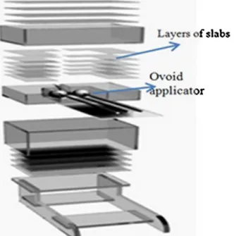
Thenewphantomwasdesignedandfabricatedfrom90slabs of18cm×16cm×0.2cmPerspextoaccommodateGYN appli-cators(tandem andovoid).ThecompositionofthePerspex takentobeH,8%;C,60%;O,32%,withadensityof1.18g/cm3.13
Thisconfigurationenablesustoverifythedosedistributions around the applicatorwitha high spatialresolution. Fig.1
showstheschematicdiagramofthisphantomwithitslayers andtheovoidapplicatorwhichisplacedinthephantom.
Fig.1–Schematicsofdifferentlayersofthephantomand Oviodapplicator.
The thin layer of the slabs was carefully machined to accommodatelayersofGafchromicfilmsinbetweentheslabs forradiation dosimetry.Theirradiatedfilms were cutwith scissorsinvarioussizesandshapetomatchthecurvatureand locationoftheapplicator.Largesize(3cmdiameter)ovoids wereselectedforbothGZP6andSelectronmachines.Withthis assembly,EBTGafchromicfilms14wereexposedusingaplan
with1.5Gydosedeliveriesto0.5cmdistancefromthelateral surfaceoftheovoidsforbothunits.
Foraquantitativeevaluation,onlyapairoftheovoidshas beenusedinorder tobeable toreproducethe experimen-talsetupandgeometryoftheassemblyforrepetitionofthe measurement,andalsofordifferentsourcetypes.
Fig.2showstheschematicdiagramofthepointsofinterest andtheapplicator.Thesepointsofinterestinclude,point“D” representingvaginalmucosa,locatedat0.5cmdistancefrom thelateralsurfaceoftheovoidand“F”thatwaslocatedat1cm distancefromthelateralsurfaceoftheovoid.
Thedifferencesbetweentheplannedandmeasureddata demonstratethe accuracyofthe planned dosedistribution
Fig.2–SchematicdiagramoftheOvoidapplicatorsand calculatedpointsrelativetotheapplicator.DandFshow pointsofinterestfordosecalculation(graphsarenottothe scale).
around applicators.These measurements were repeatedat least3timestoimprovethestatisticalfluctuationofthefinal data.
SamplesofEBTGafchromicfilmswerecalibratedto deter-minethesensitivitycurve(i.e.responsevs.dose).Acalibrated teletherapy cobalt-60 system was chosen for calibration process of the Gafchromic films. For 137Cs dose
calcula-tion because of energy dependence of Gafchromic films,15 response curve and the energy dependence ofGafchromic filmfordifferentradiationenergiesfromChiu-Tsaostudywas considered.16Calibrationcurveanditsrelativeenergy
depend-encecoefficientwereusedfortheanalysisoftheirradiated filmsaroundtheGYNapplicatorfor60Coand137Cssources
withinthenewphantomdesign.130piecesofEBTfilmswith size of 2cm×1.5cm were selected for calibrating samples usingateletherapycobalt-60systemintherangeof25–800cGy dosewith25cGyinterval.Thesefilmswereexposedat1cm depth ina30cm×30cm×10cmslabPerspexlayerswitha 20cm×20cm radiationfield size.After24hfromexposure, filmswerescannedusingaflatbedMicrotekscanner(model: ScanMaker9800XL,MicrotekLaboratories,Inc.,Dayton,OH). Sincethepeakabsorptionoftheradiochromicfilmisinthe redregionofthevisiblespectrum(636nm),extractionofthe redchannelfromtheRGBimagecanimprovescanner sensitiv-ity.AssuggestedintheguidelineoftheGafchromicfilms,the irradiatedfilmsforbothcalibrationandmeasurementprocess werescannedinthelandscapeorientation.Forfilmcalibration andbothUnitexperimentalmeasurements,filmswereused fromthesamebatch.
Inaddition,wecompliedwiththerecommendationon pro-jectingthefilmorientationonsmallpiecesthatwereusedfor radiationdosimetry.Bothcalibrationsandexperimentalfilm scanningwereperformedusing100dpiresolutions.
ThepixelvalueswereobtainedfromIMAGEJsoftware (Java-basedimageprocessingprogram,NationalInstituteofHealth). Thescannersallowtheacquisitionoftransmissionscansin upto16bitspercolor.InEq.(1)netopticaldensity(NOD)was calculatedfromsubtractionofODirrandODunirrwhich repre-sented the opticaldensitiesofun-irradiated andirradiated film,respectively:
NOD=ODirr−ODunirr=
−log10 (PVirr−PVdark) (PVblank−PVdark)
−
−log10(PVunirr−PVdark) (PVblank−PVdark)
(1)
wherethePVirr,PVunirr,PVdarkandPVblankrepresentthepixel values measured inthe irradiated film,un-irradiated film, zero-lighttransmittedimagesoftheopaquesheetscanand blank,respectively.
Theselectedregionofinterestwasa2mm×2mminthe centerofthefilmpiecetoavoidopticaldensity(OD) measure-mentartifactsnearfilmedges.17
3.4. TG-43calculation
The task group 43 (TG-43) of the American Association
of Physicistsin Medicine(AAPM) has published a protocol regarding the brachytherapy dose calculation formalism.18
TG-43formalismcanalsobeusedforverificationof experi-mentaldosimetry.Variousinvestigatorshavedeterminedthe dosimetriccharacteristicsofdifferentbrachytherapysources usingthisrecommendation.19,20Dosimetriccharacteristicsof
thesinglepelletsfromeachsystemhavebeenevaluatedusing theTG-43protocolsbydifferentinvestigators.12,21
ForbothSelectronandGZP6unitfilmdosimetrywiththe newphantomatmultiplelevelsrelativetotheGYNapplicators weredone.Wecompareddosecalculationoffilmdosimetry fromsomeinterestingpointswithtreatmentplanning. TG-43calculationwasconsideredforbothsources.Thephantom materialdensity(d=1.18g/cm3)wasconsideredforTG-43
cal-culationsofbothsources.
TG-43formalismwasemployeddependingondosimetric parametersof60Coand137Cssources.
These parameters consist of air kerma strength, dose rateconstant, geometryfunction, radialdose functionand anisotropyfunction.TheyareindicatedinEq.(2)18:
D·(r,)=SK·· GL(r,) GL(r0,0)·
g(r)·F(r,) (2)
DosimetriccharacterizationsofGZP660CoHDR
brachyther-apyforTG-43calculationwereusedfromToosi’sstudy.22So,
thesuperpositiontechniquewasused.Basedontheirstudy, doserateconstant valueofGZP6is1.04cGyh−1U−1. Radial
doseand2Danisotropyfunctionswereconsideredfromtheir MonteCarlosimulation.TG-43parametersofCs-137 Selec-tronLDRbrachytherapysourceswereconsideredfromSina’s MonteCarlosimulation.23So,doserateconstantvalueforthis
unitis1.102cGyh−1U−1.Weusedairkermastrength(S
K)from treatmentplanningfordosecalculationinEq.(3)forCs-137 Selectronsources.For Cs-137Selectron, airkermastrength (SK)fromtreatmentplanningsystemwasconsideredin TG-43calculation.While,forGZP6system,airkermastrengthsof sourcesweremeasuredusingtheionizationchamber dosime-ter.
3.5. GZP6airkermastrength
ThevendorofGZP6hasprovidedapproximately600preplans withoutprovidingthealgorithmsthathadbeenusedinthese calculations.Theonlyparameterthatwasgiven,inaddition totheplans,wasthesourcestrengthsforindividualsources. Therefore,itwasdecidedtoverifytheaccuracyofthis param-eter.ToinvestigatetheairkermastrengthofGZP6unit,Monte Carlosimulationandpracticalmeasurementwereutilized.In ourcalculation,itisconsideredthatthedateofpracticalSK measurement,airkermafrom Monte Carlosimulation and treatmentplanningSKwasthesame.
Forsimulation,MCNP4CCodeisusedbasedonthe tech-niquethatwaspublishedbyToosietal.Self-absorptionofthe sourcecoreanditscapsuleweretakenintoaccountattheir study.7Forthispartofourwork,cylindricalringswith0.5cm
thicknessand 0.5cmlengthwere simulatedinthe1–25cm transverse distance from the sourcecenter, in 1cm incre-ments.Inringcells,theF6tallywasusedtocalculateairkerma inunit ofMeV/g perphotoninthering cells.Source activ-ity,self-absorption,numberofdisintegrationfor60Cosource,
decaycoefficient based on cobalt-60half timeand date of
measurement,squareofdistanceandotherappropriate con-versionfactorsweremultipliedbythevalueoftheresultfrom Tally F6tocalculateSK. Photonprimaryhistory was107 to reducestatisticalerrortolessthan1%.Forbothphotonsand electronsthecutoffenergywasdefinedas10keV.
AirkermastrengthofGZP6unitwasmeasuredaccording
to the TECDOC-1274 technical document of the
Interna-tional AtomicEnergy Agency(IAEA).24 For measurementof air kerma, available 0.6cm3 Farmer type ionization
cham-ber(FC65-GTNC/249ScanditronixWellhofer)wasusedwith 0.551g/cm3 buildupcapandanelectrometer(Scanditronix
Wellhofer). The chamber was calibrated by the SSDL lab-oratory of Atomic Energy Organization of Iran (AEOI) for
standard field of a teletherapy cobalt-60 machine with
ND,W=4.83cGy/nc.Weusedthemultipledistancemethodand readingsofchamberinampereatspecifieddistancesalong theperpendicularbisectorofthelinesourcewerescored.The exactpositionofthesourcewaswelldefinedbyusinga mil-limetricrulerandsourceautoradiography.Apparentdistance betweenthesensitivevolumeofthechamberandthecenter ofthesourcewasmeasured.Thereis1mmoffsetindistance. Measurementswereperformedin1cmintervals.Ineach posi-tion,wemeasured3timesandmadetheaverageofvalues.To approachSK,chamberreadinginamperewasmultipliedby thechambercalibrationvalue(ND,W),temperatureand pres-surecorrectionfactor(KT,P),durationexposureandsquareof thedistancetothecenterofthesource.
3.6. Filmdosimetryuncertaintyestimation
TheoverallaccuracyofEBTfilmmeasurementswasderived usingthemethodproposedbyDevicetal.25Statistical uncer-taintyoffilmresponsetothesame valueofdoseisrelated tosomefactorssuchasnon-uniformitythicknessinthe sen-sitive layer ofthe films, uncertainties associated with the scanning procedure and output variations ofLinac during exposure.Inthisstudy,weconsideredtwosourcesof uncer-tainties. The first one is related to netOD measurements and theother isrelatedtofittingcoefficientsincalibration curves.Moreover,aspecialattentionwaspaidtothedistances betweenthephantom’sedgeand edgeofthefilm.The dis-tancebetweenthephantomslabs’edgeandtheapplicators is95m.Moreover,thereisa0.1mmgapbetweentheedge ofthemachinedfilmandthephantom’sedge.Also,thereisa 0.5mmerrorindeterminingthepositionofpointsofinterest. Thetotaldoseuncertaintywascalculatedthroughthe fol-lowingequation:
Total=
2
netOD+calib2 -fit+2phantom-film (3)
4.
Results
4.1. EBTGafchromicfilmcalibration
Fig.3showsthecalibrationcurveoftheEBTGafchromicfilm response,intermsofnetopticaldensity(NOD),asafunctionof absorbeddosetowaterintherangeof25–800cGy.The mea-sureddatawerecorrelatedusingthefollowingpolynominal
Table1–Comparisonbetweenthemeasuredandcalculateddosevaluesatdifferentpointsaroundatypicalapplicatorin GYNimplantofSelectron137CsLDRsource.
Points
Dose-treatment planning(cGy)
Dose-TG-43(cGy) Dose-film(cGy) Differencebetween filmdosimetryand TG-43calculation(%) PointD-L 150 144.3 145.8 +1 PointD-R 150 144.3 144.9 +0.4 PointF-L 100 93.5 95.8 +2.4 PointF-R 100 93.5 95.4 +2
Fig.3–CalibrationcurveforEBTGafchromicfilmresponse (opticaldensity)asfunctionoftheabsorbeddose(cGy).The errorbarsinthisgraphrepresent±5%.Thesolidlineon thisgraphsimplyconnectsthedatapoints.
functioninEq.(4)withR2=0.9837.Calibrationcoefficientof 137CssourcewasobtainedfromChiu-Tsao’sstudy.Forscanner
withredlightrelationshipbetweentheNODresponseofthe EBTfilmandsourceenergyinMeV(parameterx)isconsidered asEq.(5).
Dose (Gy)=42.43NOD2−8.14NOD+5.24 (4) NOD=−0.025x2+0.1165x+0.869 (5)
So, thereis 2%differencebetween 60Co with meanenergy
1.25MeV and 137Cs with energy 0.662MeV. This difference
isemployedfor137Csfilmdosimetry.There isatotalof5%
uncertaintythatwasemployedfortheresultsofdose mea-surements.
4.2. SelectronLDRbrachytherapy
ThevalueofreferenceairkermarateforSelectronTG-43dose calculationisextractedfromtreatmentplanningandequalto 8876.74Gym2h−1.Thequantitativeanalysisofthemeasured
dataforSelectron137CsLDRsourceandTG-43dosecalculation
isshowninTable1.ResultsofSelectronunitshowgood agree-ment (totaldifference isupto6%)betweenfilmdosimetry, TG-43dosecalculationandtreatmentplanning.
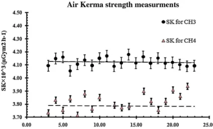
4.3. GZP6HDRbrachytherapy
Air kerma strength (SK) for GZP6 HDR brachytherapy sys-tem from Monte Carlo code was obtained 4414×10−6 and
4077×10−6Gym2h−1forsourceofCH
3andCH4,respectively.
The value of measured air kerma strength was
obtained by takinginto account the date ofmeasurement 4207×10−6±1%Gym2h−1 for CH
3 and 3864.5×10−6±
1%Gym2h−1forCH
4.Fig.4showsresultsofSKmeasurement byamultipledistancemethod.Thereisabout5.4%difference betweenthemeasurementandsimulation.
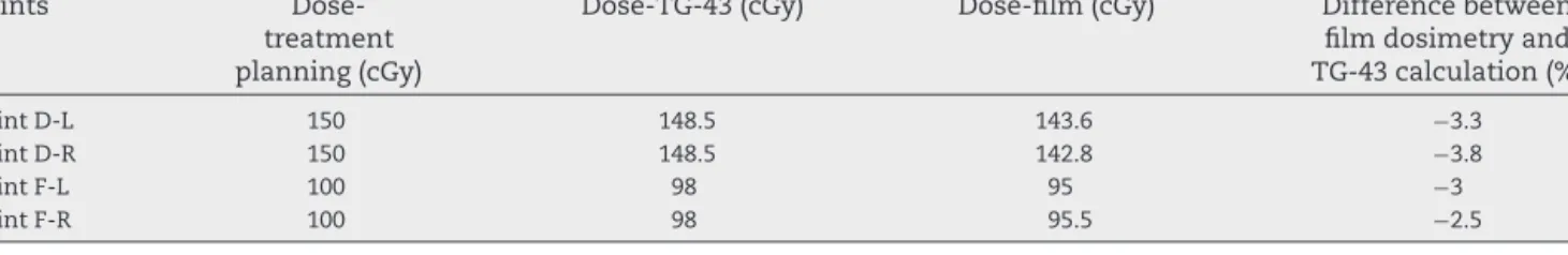
Table2indicatestheresultsofTG-43dosecalculationsand filmdosimetry.Maximumdifferencebetweenfilmdosimetry andTG-43wasto3.8%.
Fig.4–Plotoftheairkermarate×d2vs.distanced(cm)fromthesourcecenter.Theerrorbarsonthisdatapointsrepresent 5%statisticalfluctuationoftheFarmerdosimetry.
Table2–Comparisonbetweenthemeasuredandcalculateddosevaluesatdifferentpointsaroundatypicalapplicatorin GYNimplantofGZP660CoHDRsource.
Points
Dose-treatment planning(cGy)
Dose-TG-43(cGy) Dose-film(cGy) Differencebetween filmdosimetryand TG-43calculation(%) PointD-L 150 148.5 143.6 −3.3 PointD-R 150 148.5 142.8 −3.8 PointF-L 100 98 95 −3 PointF-R 100 98 95.5 −2.5
5.
Conclusions
AlthoughtheAmericanAssociationofPhysicistsinMedicine TaskGroupreports56and59providereasonableguidanceon specificprocessofQAinbrachytherapy,26,27 improved
guid-anceisneededforanytreatmentdeliverysystemtominimize each individual treatment failure. In this project, the dose distributions around the GYNapplicators for twodifferent machines,SelectronLDRsourceandGZP6HDRsystemwere measuredusingEBTGafchoromicfilmforasampleGYNsetup. Theresultshowsgoodagreementbetweenourfilmdosimetry, TG-43calculationandtreatmentplanning(totaldifferenceis upto6%).Inthis study,ithasbeendemonstratedthat Per-spexcanbeusedasphantommaterialinbrachytherapy.The Perspexmaterialisdenserthan water,soitproducesmore attenuationofprimaryradiationbutontheotherhand,this iscompensatedbyanincreaseinscatterradiationunderfull scatteringconditions.28
These investigations have verified that using the
Gafchromic film dosimetry technique together with the
new phantom; one could examine the accuracy of dose
calculationatdifferentdistancestotheGYNapplicators.This systemhasshownthatinahighdosegradientregionaround compoundapplicators,userscouldverifytheaccuracyofdose distributionwithatechniquethat isclinicallyrelevantand practical.Recently,therearesomestudiesrelatedtoproper phantomsforQAofbrachytherapysystems.29,30 Comparing
them with this phantom, different models of Gafchromic filmscanbeusedwhichareveryconvenientandreliablefor routinequalityassurance.Thisassemblycanbeusedathigh gradientregionsforperiodicQAprocedures.
Conflict
of
interest
None.
Financial
disclosure
None.
Acknowledgements
I hereby acknowledge Clinical Research Development Unit (CRDU)ShohadaTajrish hospitalwhich wassupportedthis research.
The Radiotherapy Department of Shahid Beheshti
Uni-versityatShohada hospitalsponsoredthe purchaseofthe phantommaterialsandfilmsusedintheinvestigations.The
authorswouldliketopresenttheirappreciationforDrMehdi Ghorbaniforhisinvaluablecontributionsatdifferentphases ofthisproject.Inaddition,theeditorialcommentsand sug-gestionsbyDrCourtneyKnaupforenhancingthequalityof themanuscriptaregreatlyappreciated.
r
e
f
e
r
e
n
c
e
s
1. AsnaashariK,GholamiS,KhosraviH.Lessonslearntfrom errorsinradiotherapycenters.IntJRadiatRes
2014;12(4):361–7.
2. GerbauletA.TheGECESTROhandbookofbrachytherapy;2002.
3. Niroomand-RadA,BlackwellCR,CourseyBM,etal. Radiochromicfilmdosimetry:recommendationsofAAPM radiationtherapycommitteetaskgroup55.MedPhys
1998;25(11):2093–115.
4. Chiu-TsaoS-T,MedichD,MunroIIIJ.Theuseofnew GAFCHROMIC®EBTfilmforI125seeddosimetryinSolid
Water®phantom.MedPhys2008;35(8):3787–99.
5. PapagiannisP,AngelopoulosA,PantelisE,SakelliouL, KaraiskosP,ShimizuY.MonteCarlodosimetryof60CoHDR
brachytherapysources.MedPhys2003;30(4):712–21.
6. SinaS,FaghihiR,MeigooniAS,MehdizadehS,ShiraziMAM, ZehtabianM.Impactofthevaginalapplicatoranddummy pelletsonthedosimetryparametersofCs-137brachytherapy source.JACMP2011;12(3).
7. ToossiMTB,GhorbaniM,MowlaviAA,etal.Airkerma strengthcharacterizationofaGZP6Cobalt-60brachytherapy source.RPOR2010;15(6):190–4.
8. NaseriA,MesbahiA.ApplicationofMonteCarlocalculations forvalidationofatreatmentplanningsysteminhighdose ratebrachytherapy.RPOR2009;14(6):200–4.
9. MesbahiA.RadialdosefunctionsofGZP6intracavitary brachytherapy60Cosources:treatmentplanningsystem
versusMonteCarlocalculations.IJRR2008;5(4):181–6.
10. FragosoM,LoveP,VerhaegenF,etal.Thedosedistributionof lowdoserateCs-137inintracavitarybrachytherapy:
comparisonofMonteCarlosimulation,treatmentplanning calculationandpolymergelmeasurement.PMB
2004;49(24):5459.
11. GrigsbyPW,WilliamsonJF,PerezCA.Sourceconfiguration anddoseratesfortheSelectronafterloadingequipmentfor gynecologicapplicators.IntJRadiatOncol1992;24(2): 321–7.
12. LiuL,PrasadSC,BassanoDA.Determinationof137Cs
dosimetryparametersaccordingtotheAAPMTG-43 formalism.MedPhys2004;31(3):477–83.
13. SaidiP,SadeghiM,HosseiniH,TenreiroC. ThermoluminescentandMonteCarlodosimetryof IR06-103Pdbrachytherapysource.JACMP2011;12(4).
14. SarfehniaA,KawrakowI,SeuntjensJ.Directmeasurementof absorbeddosetowaterinHDRI192rbrachytherapy:water calorimetry,ionizationchamber,GafchromicFilm,and TG-43.MedPhys2010;37(4):1924–32.
15. MuenchPJ,MeigooniAS,NathR,McLaughlinW.Photon energydependenceofthesensitivityofradiochromicfilm andcomparisonwithsilverhalidefilmandLiFTLDsusedfor brachytherapydosimetry.MedPhys1991;18(4):769–75.
16. Chiu-TsaoS-T,HoY,ShankarR,WangL,HarrisonLB.Energy dependenceofresponseofnewhighsensitivityradiochromic filmsformegavoltageandkilovoltageradiationenergies.Med Phys2005;32(11):3350–4.
17. ButsonMJ,PeterK,MetcalfePE.Effectsofread-outlight sourcesandambientlightonradiochromicfilm.PMB
1998;43(8):2407.
18. RivardMJ,CourseyBM,DeWerdLA,etal.UpdateofAAPM TaskGroupNo.43Report:arevisedAAPMprotocolfor brachytherapydosecalculations.MedPhys2004;31(3):633–74.
19. MeigooniA.Recentdevelopmentsinbrachytherapysource dosimetry.IJRR2004;2(3):97–105.
20. Perez-CalatayudJ,BallesterF,DasRK,etal.Dosecalculation forphoton-emittingbrachytherapysourceswithaverage energyhigherthan50keV:reportoftheAAPMandESTRO.
MedPhys2012;39(5):2904–29.
21. MelhusCS,RivardMJ.ApproachestocalculatingAAPMTG-43 brachytherapydosimetryparametersfor137Cs,125I,192Ir, 103Pd,and169Ybsources.MedPhys2006;33(6):
1729–37.
22. ToossiM,GhorbaniM,MowlaviA,MeigooniA.Dosimetric characterizationsofGZP660Cohighdoseratebrachytherapy
sources:applicationofsuperimpositionmethod.RadiolOncol
2012;46(2):170–8.
23. SinaS,FaghihiR,MeigooniA,MehdizadehS,ZehtabianM, Mosleh-ShiraziM.Simulationoftheshieldingeffectsofan applicatorontheAAPMTG-43parametersofCS-137 SelectronLDRbrachytherapysources.IJRR2009;7(3):135–40.
24. TECDOCI.1274.Calibrationofphotonandbetaraysourcesused inbrachytherapy.Guidelinesonstandardizedproceduresat SecondaryStandardsDosimetryLaboratories(SSDLs)and Hospitals.IAEA;2002.
25. DevicS,SeuntjensJ,HegyiG,etal.Dosimetricpropertiesof improvedGafChromicfilmsforsevendifferentdigitizers.Med Phys2004;31(9):2392–401.
26. NathR,AndersonLL,MeliJA,OlchAJ,StittJA,WilliamsonJF. Codeofpracticeforbrachytherapyphysics:reportofthe AAPMRadiationTherapyCommitteeTaskGroupNo.56.Med Phys1997;24(10):1557–98.
27. KuboHD,GlasgowGP,PethelTD,ThomadsenBR,Williamson JF.Highdose-ratebrachytherapytreatmentdelivery:reportof theAAPMRadiationTherapyCommitteeTaskGroupNo.59.
MedPhys1998;25(4):375–403.
28. MeliJA,MeigooniAS,NathR.Onthechoiceofphantom materialforthedosimetryof192Irsources.IntJRadiatOncol
1988;14(3):587–94.
29. Lipi ´nskaJ,ZwierzchowskiG.Dosimetricverificationofthe dosedistributioninpulseddoseratebrachytherapy.RPOR
2006;11(5):223–8.
30. KohrP,SiebertF-A.Qualityassuranceofbrachytherapy afterloadersusingamulti-slitphantom.PMB