INTRODUCTION
The vertebrate forebrain is subdivided into the telencephalon, which gives rise to the cerebral cortex, and the diencephalon, which comprises the future thalamus, prethalamus (PT) and hypothalamus. The blueprint for these subdivisions is established early in development as patterned gene expression (Wilson and Houart, 2004; Rhinn et al., 2006). Although many of the genes play key roles in forebrain development, regulatory relationships between these genes are poorly understood. Hedgehog (Hh) proteins secreted from the ventral diencephalic midline play a central role in patterning the early forebrain primordium (Bertrand and Dahmane, 2006; Ingham and Placzek, 2006). Hh function results in transcriptional activation of several targets, many of which encode transcription factors (TFs). The homeobox TF gene six3is transcriptionally activated by Hh signaling and directly activates shhtranscription (Geng et al., 2008; Jeong et al., 2008). six3function is essential for correct development of all forebrain derivatives, as mice and human with compromised six3or shhfunction develop with a profound forebrain defect termed holoprosencephaly (HPE) (Krauss, 2007).
In addition to regulating genes important for development of the forebrain proper, Hh signaling regulates the transcription of genes involved in retinal development. The neural retina is initially part of the forebrain primordium, but separates from it early in development through evagination to form bilateral optic vesicles. A transient optic stalk (OS) connects each optic vesicle to the forebrain (Schmitt and Dowling, 1994). Owing to their common origin, the early OS and retinal domains are contiguous. Hh proteins secreted from the ventral-diencephalic-midline pattern the initially uniform OS/retinal
field (Ekker et al., 1995; Varga et al., 2001; Schimmenti et al., 2003; Lee, J. et al., 2008), partitioning it into three domains: the OS, the ventral retina and the dorsal retina (Lupo et al., 2005). The future OS lies closest to the Hh source and expresses several Hh-dependent homeobox TF genes, including pax2a(Macdonald et al., 1995; Take-uchi et al., 2003; Mui et al., 2005). The optic vesicles, distal to the Hh source, express pax6, a gene with key regulatory roles during retinal development (Gehring, 1996).
The zinc-finger TF Zic2 plays a key role during early forebrain development. Mutations in mammalian ZIC2can cause HPE (Brown et al., 1998), and the zebrafish ortholog zic2ais required for correct formation of the ventral diencephalon (Sanek and Grinblat, 2008). In the developing retina, Zic2 and other Zic family members are required for retinal axon guidance in mouse and chick (Herrera et al., 2003; Zhang et al., 2004; Lee, R. et al., 2008). Although essential functions for Zic genes are well documented, the molecular basis for these functions during forebrain and eye development is unclear.
In this study, we further examine the function of zebrafish zic2a during forebrain development, in particular its regulatory relationship with Hh signaling and six3b. We show that zebrafish Zic2a negatively modulates Hh-dependent transcription of six3bin the forebrain. Zic2a repression of six3bis essential for the correct formation of the PT, a diencephalic derivative. Zic2a is also required in the OS and retinal primordium, where it inhibits Hh-dependent expression of pax2a. These data identify a novel role for Zic2a in the regulatory network, coordinated by Hh signaling, that patterns the vertebrate forebrain.
MATERIALS AND METHODS Zebrafish strains and embryo culture
Adult zebrafish were maintained according to established methods (Westerfield, 1995). Embryos were obtained from natural matings and staged according to Kimmel (Kimmel et al., 1995). The following zebrafish strains were used: wild-type AB, smob641 (Varga et al., 2001), syut4 (Odenthal et al., 2000), pax2ab593(Erickson et al., 2007), Tg(pou4f3:gap43-GFP)s356t(Xiao et al., 2005), and Tg(–8.0cldnb:lynGFP)zf106 (Lecaudey et al., 2008).
Zebrafish
zic2a
patterns the forebrain through modulation
of Hedgehog-activated gene expression
Nicholas A. Sanek1,2,*, Aaron A. Taylor1,†, Molly K. Nyholm1,†and Yevgenya Grinblat2,‡
Holoprosencephaly (HPE) is the most common congenital malformation of the forebrain in human. Several genes with essential roles during forebrain development have been identified because they cause HPE when mutated. Among these are genes that encode the secreted growth factor Sonic hedgehog (Shh) and the transcription factors Six3 and Zic2. In the mouse, Six3 and Shh activate each other’s transcription, but a role for Zic2 in this interaction has not been tested. We demonstrate that in zebrafish, as in mouse, Hh signaling activates transcription of six3bin the developing forebrain. zic2ais also activated by Hh signaling, and
represses six3bnon-cell-autonomously, i.e. outside of its own expression domain, probably through limiting Hh signaling. Zic2a repression of six3bis essential for the correct formation of the prethalamus. The diencephalon-derived optic stalk (OS) and neural retina are also patterned in response to Hh signaling. We show that zebrafish Zic2a limits transcription of the Hh targets pax2aand
fgf8ain the OS and retina. The effects of Zic2a depletion in the forebrain and in the OS and retina are rescued by blocking Hh
signaling or by increasing levels of the Hh antagonist Hhip, suggesting that in both tissues Zic2a acts to attenuate the effects of Hh signaling. These data uncover a novel, essential role for Zic2a as a modulator of Hh-activated gene expression in the developing forebrain and advance our understanding of a key gene regulatory network that, when disrupted, causes HPE.
KEY WORDS: Zic2, Six3, Hedgehog signaling, Pax2, Prethalamus, Optic stalk, Retina, Zebrafish
Development 136, 3791-3800 (2009) doi:10.1242/dev.037820
1Departments of Zoology and Anatomy and 2The Genetics Ph.D. Training Program,
University of Wisconsin, 1117 West Johnson Street, Madison, WI 53706, USA. *Present address: National Institutes of Mental Health, Bethesda, MD 20892-4483, USA
†These authors contributed equally to this work ‡Author for correspondence ([email protected])
Accepted 1 September 2009
D
E
V
E
LO
P
M
E
N
3792
Mutant genotyping
smob641 homozygous mutant embryos were positively identified by characteristic morphological defects and a lack of ptc1expression. pax2ab593 embryos were identified by a lack of fgf8aexpression in the mid-hindbrain boundary (MHB), and by morphological defects.
In situ hybridization (ISH)
Antisense RNA probes were transcribed using the MAXIscript Kit (Ambion, Foster City, CA, USA) from the following plasmid templates: arx(Miura et al., 1997), atoh7(Masai et al., 2000), efna5a(Picker and Brand, 2005), fgf8a (Furthauer et al., 1997), foxg1(Rohr et al., 2001), pax2a(Hoyle et al., 2004), pax2b(Pfeffer et al., 1998), pax6a(Krauss et al., 1991), rx3(Jeong et al., 2007), six3b(Seo et al., 1998), il17rd(Tsang et al., 2002), spry4(Furthauer et al., 2001), vax1(Take-uchi et al., 2003), vax2(Gross and Dowling, 2005) and zic2a(Grinblat and Sive, 2001). Single-color ISH was carried out as previously described (Gillhouse et al., 2004). Double fluorescent analysis was carried out using digoxigenin (DIG)-labeled probes (Roche, Basel, Switzerland) detected with anti-DIG Fab fragments (Roche) and Fast Red substrate (Sigma, St Louis, MO, USA), combined with fluorisceinated-dextran (FLU-DEX) (Invitrogen, Carlsbad, CA, USA) detected with anti-FLU Fab fragments (Roche) and enzyme-labeled fluorescence (ELF)-97 substrate (Invitrogen). Differential interference contrast (DIC) and epifluorescent imaging was carried out on a Zeiss Axioskop2 Plus microscope with AxioVision 3.0 software (Zeiss, Oberkochen, Germany). Confocal imaging was carried out on an Olympus FV1000 microscope with FV10-ASW software (Olympus, Tokyo, Japan).
Quantitative PCR (qPCR) analysis Preparation of cDNA
Total RNA was extracted from embryos using Trizol (Invitrogen). The RNA was then cleared of genomic DNA with a Turbo DNA-free Kit (Ambion), and 250 ng was reverse-transcribed in duplicate using an iScript Select cDNA Synthesis Kit (BioRad) with oligo(dT) primers. Duplicate reactions were pooled prior to qPCR.
Real-time PCR methods
Primers were designed against sequences that spanned an intron near the 3⬘
end of each gene and produced an amplicon of ~200 bp. Primer specificity was checked by agarose gel electrophoresis and melting curve analysis.
The 20 µl reactions comprised: 1⫻Power SYBR Green Master Mix (Applied Biosystems), 200 nM of each primer and 2 µl of a 1:10 dilution of cDNA template. Components shared across reactions were mixed first and then aliquoted to ensure equivalent reaction conditions. Each template type was analyzed in triplicate during each ‘technical replicate’. No RT (reverse transcriptase) and no template-control reactions were performed for each template type. Reactions were run on an Applied Biosystems ABI 7500 as follows: denaturation at 94°C for 4 minutes; amplification at 94°C for 30 seconds, 58°C for 1 minute and 72°C for 1 minute (40⫻). Two technical replicates were performed for each template type.
Real-time PCR data analysis
Cycle threshold (Ct) values were filtered such that values greater than two standard deviations from the average Ct of each technical replicate were excluded from analysis. The remaining Ct values were analyzed using the ‘relative standard curve’ method as outlined in the ABI Prism 7700 User Bulletin #2. Relative standard curves were produced using 1:4 serial dilutions of cDNA as a template and the amplification for each primer set was linear in log space (R2>0.997 in each case; primer efficiency was nearly 2; data not shown). six3b primer sequences were 5⬘ -ATCGAAGACA-GAGGGACAGG-3⬘ and 5⬘-ATACTGGAGACGCTGGTCGT-3⬘;gapdh primer sequences were 5⬘-ATCAAGAAAGTCGTCAAGGCTG-3⬘and 5⬘ -ATCTACTCCTTGGAGGCCATGT-3⬘.
Knockdown assays
Three antisense morpholino oligomers (MOs) were used to knock down expression of zic2ain this study: two translation-blocking MOs (zic2a AUG, CGATGAAGTTCAATCCCCGCTCACA; zic2a PROX, CTCTTTCAAGCAGTCTATTCACGGC) and a splice-blocking MO (zic2aMO, CTCACCTGAGAAGGAAAACATCATA) (Nyholm et al.,
2007). Six3bMO (CTCTAAAGGAGACCTGAAAACCATG) was purchased from Open Biosystems (Ando et al., 2005; McCollum et al., 2007). Control morphants were generated using the standard control MO (conMO; Genetools, Philomath, OR, USA). MOs were diluted in 1⫻Danieau buffer (Nasevicius and Ekker, 2000) to 1-2 ng/nl (zic2aMO), 8 ng/nl (six3MO), 4-6 ng/nl (zic2aAUG and PROX), or 3-4 ng/nl (conMO), and 1 nl was injected per embryo at the 1- to 2-cell stage. Cyclopamine (Toronto Research Chemicals, North York, Ontario, Canada) was used at 100 M as previously described (Tyurina et al., 2005). SU5402 (Calbiochem, Darmstadt, Germany) treatments were carried out on embryos with chorions at embryonic day 3 (E3). SU5402 was dissolved in DMSO and used at 10 M (Yamauchi et al., 2006).
Overexpression assays
Sense RNA transcripts were made with the mMessage mMachine Kit (Ambion) from the following templates: shha(Ungar and Moon, 1996) and hhip(Ochi et al., 2006). mRNA was purified using spin columns (Bio-Rad, Hercules, CA, USA) and diluted in RNAse-free water. Injections of 12.5 or 25 pg of shhamRNA and 3.8 pg of hhipmRNA were administered per embryo.
RESULTS
six3btranscription is activated by Hh signaling in
the zebrafish forebrain
An important regulatory relationship between Hh signaling and six3 has been demonstrated in the mouse forebrain (Geng et al., 2008; Jeong et al., 2008). To test whether this relationship exists in zebrafish, we asked whether Hh signaling is required to activate six3b transcription in the zebrafish forebrain and retina. Embryos treated with cyclopamine to block Hh signaling showed reduced six3b expression in the forebrain (Fig. 1A,B), but not in the retina. Similarly, smob641mutants, deficient in Hh signaling (Varga et al., 2001), showed strongly reduced six3bexpression in the forebrain (Fig. 1C,D), but continued to express six3bin the retina (Fig. 1C⬘,D⬘). These results demonstrate that Hh signaling is required for proper levels of six3b expression in the zebrafish telencephalon and diencephalon.
We next asked whether Hh signaling is sufficient to activate six3b transcription in the zebrafish forebrain. When six3bexpression was assayed by ISH in embryos microinjected with low amounts of shha RNA, mildly expandedsix3bexpression was consistently observed in the telencephalon and diencephalon (Fig. 1G-H). Notably, this low level of shhaRNA did not trigger ectopic expression of ptc1, a direct target of Hh signaling (Fig. 1E-F), suggesting that six3bis a more sensitive indicator of Hh signaling levels than ptc1. When shh -overexpressing embryos were assayed quantitatively by real-time PCR, six3btranscript levels were not found to increase significantly in proportion to increasing shha levels (see Fig. S1 in the supplementary material), suggesting that Hh signaling is not sufficient to activate six3b transcription. Together, these results indicate that, in zebrafish, Hh signaling is required for six3b transcription in the telencephalon and diencephalon and does not control six3btranscription in the retina.
Zic2a represses six3btranscription in the forebrain in an Hh-dependent manner
six3and zic2play crucial roles during forebrain development in mammals, yet a regulatory relationship between them has not been tested in this tissue. To determine whether six3btranscription is regulated by Zic2a in zebrafish, we employed antisense MO knockdown assays as previously described (Nyholm et al., 2007; Sanek and Grinblat, 2008). Zebrafish six3bis expressed in the anterior forebrain primordium from early somitogenesis (Seo et al., 1998). In Zic2a-depleted morphants, six3bwas expressed normally
RESEARCH ARTICLE Development 136 (22)
D
E
V
E
LO
P
M
E
N
at the 10-somite (10s) stage (Fig. 2A-B). However, at the 15s and prim-5 stages, zic2amorphants upregulated expression of six3b throughout the forebrain (Fig. 2C-F), suggesting that Zic2a functions to repress six3b starting at mid-somitogenesis. This upregulation was confirmed by qPCR analysis (Fig. 2G).
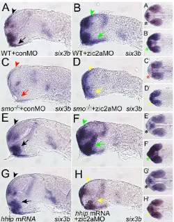
zic2ais expressed in the telencephalon and dorsal diencephalon (Sanek and Grinblat, 2008) (see Fig. S2 in the supplementary material), consistent with the possibility that Zic2a could act cell-autonomously, and perhaps directly, to repress six3bin these cells. However, in the diencephalon, zic2ais restricted to the distal OS and is not expressed in cells that ectopically activate six3bin morphants (see Fig. S2 in the supplementary material), suggesting that Zic2a functions indirectly in this area. As shhaexpression appears to be unaffected in Zic2a-depleted embryos (Sanek and Grinblat, 2008), we hypothesized that Zic2a might control transcription of a secreted factor that acts as an Hh agonist or antagonist to modulate the transcriptional activation of six3b. This hypothesis predicts that expression of six3bshould be enhanced in zic2amorphants in an Hh signaling-dependent manner. six3bexpression is greatly decreased, but not eliminated, in Hh signaling-defective smob641embryos (Fig.
3A,C); the low level of six3bexpression remaining in these mutants is likely to be due to residual Hh signaling through the maternally provided smo (Varga et al., 2001). Zic2a depletion in smob641 embryos did not result in increased six3bthroughout the forebrain as is typical of zic2amorphants (Fig. 3A-D). An increase in six3b expression was observed relative to smob641 embryos, but this expression was weak and restricted anteriorly, consistent with an enhanced transcriptional response to residual Hh signaling in smob641embryos. Similar results were obtained in zic2amorphants treated with cyclopamine to block Hh signaling (data not shown). These findings support the hypothesis that Zic2a functions to regulate six3btranscription in an Hh signaling-dependent manner.
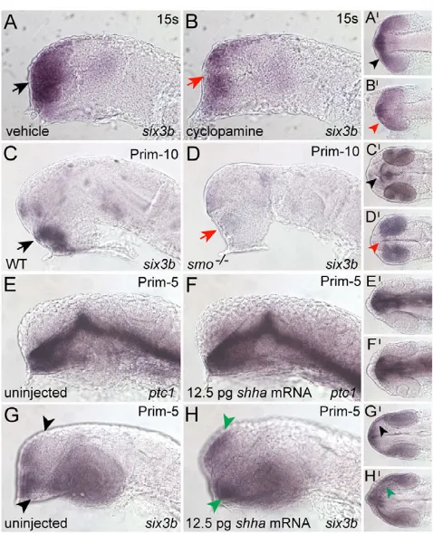
[image:3.612.55.294.55.350.2]If Zic2a functions to control the level or extent of Hh signaling, the addition of an exogenous Hh antagonist should also rescue the zic2a morphant defect. To test this prediction, we used RNA overexpression of Hhip, a membrane-bound and secreted Hh-binding protein that antagonizes Hh signaling in mice and zebrafish (Chuang and McMahon, 1999; Coulombe et al., 2004; Ochi et al., 2006). Although Zic2a depletion resulted in six3b expansion throughout the forebrain (Fig. 3E-F), and hhipmRNA injection did Fig. 1. Hh signaling activates six3bexpression in the developing
zebrafish forebrain. (A,B)At 15s, six3bis expressed normally in the anterior forebrain of vehicle-treated embryos (black arrowheads in A, 15/15 embryos) and dramatically reduced in cyclopamine-treated embryos (red arrowheads in B, 14/14 embryos treated from the 2- to 4-cell stage onwards). (C,D)At prim-10, ventral forebrain six3b expression is strong in wild-type (WT) embryos (black arrowheads in C, 67/92 embryos) and absent or greatly reduced in smo–/–mutants (red
arrowheads in D, 25/92 embryos). (E-H)Embryos injected with a low amount of shhamRNA (12.5 pg) show expanded six3bexpression [green arrowheads in H, 10/23 embryos, 2 experiments (n2)], but no change in ptc1(F, 31/31 embryos, n2). Embryos shown in panels A-H are lateral views. Panels A⬘-H⬘are ventral views of the same embryos, anterior to the left.
Fig. 2. Zic2a restricts six3bexpression throughout the forebrain beginning at mid-somitogenesis.(A,B)six3bis similarly expressed in conMO- and zic2aMO-injected embryos at 10s-12s (A, 41/41 embryos, n2; B, 57/57 embryos, n2). (C,D)At 12s-15s, six3bexpression is normal in control morphants (black arrowheads in C, 38/38 embryos, n3), and expanded throughout the forebrain in zic2amorphants (green arrowheads in D, 58/58 embryos, n3). (E,F)six3bexpansion in zic2aMO-injected embryos persists at prim-5 (black arrowheads in E, 42/42 control morphants, n3; green arrowheads in F, 40/41 embryos, n3). (G)Real-time PCR showed that six3blevels, normalized to gapdh, were increased (P0.0001) in ZicMO-injected embryos relative to control morphants (based on three biological replicates, with two technical replicates per biological replicate). A-F are lateral views; eyes have been removed from embryos in C-F. A⬘-B⬘are ventral views of the same embryos. C⬘-F⬘are dorsal views of the same embryos, anterior to the left.
D
E
V
E
LO
P
M
E
N
[image:3.612.311.564.56.332.2]3794
not affect six3bexpression (Fig. 3G), embryos co-injected with hhip mRNA and zic2aMO showed significant rescue of six3b expression in the telencephalon and diencephalon (Fig. 3H). Together, these data suggest that Zic2a functions as a negative modulator of Hh-induced gene expression in the zebrafish forebrain.
Zic2a function in the prethalamus requires six3b repression
We have previously shown that Zic2a depletion causes defects in the ventral diencephalon-derived PT (Sanek and Grinblat, 2008). To test whether ectopic expression of six3b in zic2a morphants was responsible for this defect, we knocked down Zic2a and Six3b simultaneously using a six3-specific MO (Ando et al., 2005;
McCollum et al., 2007), and assayed the resulting embryos for expression of dlx2aand arx, markers of the PT (arrowheads in Fig. 4). As previously described, dlx2awas reduced in the zic2amorphant PT (Fig. 4A,B). dlx2awas unaffected in six3bmorphants (Fig. 4C). In zic2a; six3bdouble morphants, the dlx2aPT domain was similar to that in the controls in the amount of staining, although it was somewhat mispatterned, indicating that Six3b depletion partially rescues the PT defect in zic2amorphants (Fig. 4D,E). arx expression, reduced in zic2a morphants, was similarly rescued by Six3b knockdown (Fig. 4F-J). These data suggest that Zic2a promotes PT development through repression of six3b, and suggest that Six3 might play a role in the development of the diencephalon.
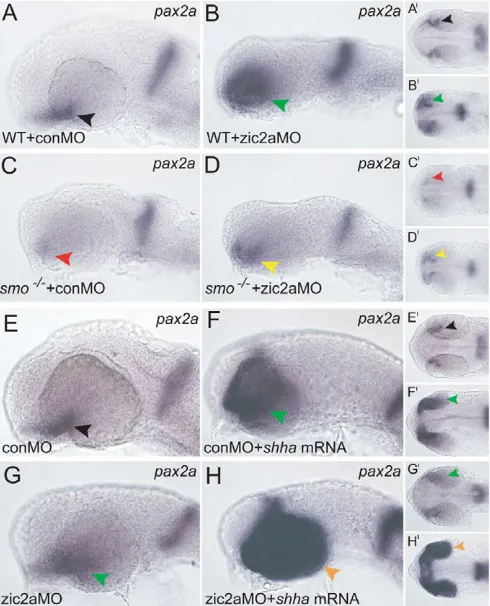
Zic2a restricts the expression of optic stalk markers
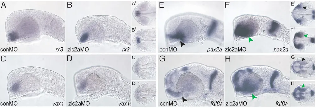
Because Zic2a modulates the transcriptional readout of Hh signaling in the developing forebrain, and another major role for Hh signaling is to pattern the adjacent OS and retinal precursors, we next asked whether Zic2a is involved in the development of the OS and retina. Zic2a-depleted embryos were examined at the prim-5 stage for expression of several regional markers. rx3, which marks the pre-optic area of the anterior diencephalon adjacent to the OS, was expressed normally (Fig. 5A,B). The expression of the proximal OS-restricted vax1was also unaffected (Fig. 5C,D). By contrast, pax2a (Fig. 5E,F) and fgf8a(Fig. 5G,H), distal OS markers, were strongly expanded into the ventral retina, as were OS markers pax2band il17rd(data not shown). The specificity of the pax2aexpansion phenotype was confirmed in two ways: by using non-overlapping translation-blocking MOs against zic2ato produce the same defect
[image:4.612.316.564.56.220.2]RESEARCH ARTICLE Development 136 (22)
Fig. 3. Reduced Hh signaling in zic2amorphants prevents six3b expansion.(A-D)Zic2a depletion in smo–/–mutant embryos.
Zic2a-depleted WT siblings show expanded six3bin the telencephalon and diencephalon (green arrowheads in B, 120/151 embryos, n2), whereas smo–/–mutants injected with conMO lose six3bexpression (C, 25/25
smo–/–embryos, n2). zic2aMO-injected smo–/–mutants show
upregulated, but anteriorly restricted, six3bexpression (yellow arrowheads in D, 14/17 smo–/–embryos, n2). (E-H)Zic2a depletion in
[image:4.612.50.300.57.376.2]the presence of exogenous Hhip. Embryos injected with zic2aMO alone show expanded six3b(green arrowheads in F, 45/45 embryos, n2), whereas embryos injected with hhipmRNA express six3bnormally (black arrowheads in G, 43/43 embryos, n2). Embryos co-injected with zic2aMO and hhipmRNA show rescued six3bexpression (yellow arrowheads in H, 17/40 embryos, n2). A-H are lateral views, anterior to the left. A⬘-H⬘are ventral views of the same embryos, anterior to the left. Arrowheads point to the telencephalon and arrows to the diencephalon. Asterisks in A⬘-H⬘illustrate six3bexpression in the optic stalks. All embryos are shown at prim-5.
Fig. 4. Six3b depletion rescues prethalamic patterning defects in Zic2a-depleted embryos. (A)Uninjected embryos show normal dlx2a expression in the diencephalon (29/29 embryos, n2). (B)zic2a morphants exhibit a strong reduction of diencephlic dlx2a(18/19 embryos, n2). (C)six3bmorphants show normal dlx2apatterning (23/23 embryos, n2). (D)Embryos co-injected with six3bMO and zic2aMO exhibit partially rescued prethalamic dlx2aexpression (30/52 embryos, n2). (E)A graphic summary of the dlx2arescue experiments. (F)Normal arxexpression is observed in the PT of uninjected control embryos. (G)arxexpression is strongly reduced in zic2amorphants (13/15 embryos, n2). (H)six3bmorphants show normal arx expression. (I)Embryos co-injected with six3bMO and zic2aMO show partial rescue of arx(10/23 embryos, n2). (J)A graphic summary of the arxdomain rescue experiments. Red, strongly reduced dlx2a orarxin PT; blue, normal or nearly normal expression of dlx2aor arx in PT. Arrowheads point to the diencephalic domains of dlx2a orarxexpression. Lateral views with anterior to the left shown at prim-5 (A-D) or 20s (F-I).
D
E
V
E
LO
P
M
E
N
(data not shown), and by knocking down Zic2a together with p53, to show that the pax2adefect in zic2amorphants was not due to increased cell death (see Fig. S3 in the supplementary material). The pax2apatterning defect first manifested in Zic2a-depleted embryos by 19s (see Fig. S4 in the supplementary material). By contrast, fgf8a was correctly patterned at 19s (see Fig. S4 in the supplementary material), and did not become expanded until 21s (data not shown).
The regulatory relationships between zic2a, fgf8aand pax2awere further analyzed using Zic2a MO assays in combination with pharmacological inhibition of fibroblast growth factor (FGF) signaling and pax2a mutants. This analysis demonstrated that zic2a acts upstream of pax2aand fgf8a(see Fig. S5 in the supplementary material), and that fgf8apromotes pax2aexpression downstream of Zic2a function (see Fig. S6 in the supplementary material), which is consistent with previous findings (Nakayama et al., 2008).
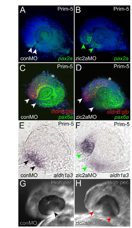
We next asked whether early retinal development was disrupted in Zic2a-depleted embryos concomitant with expansion of OS markers into the retina. During normal development, the OS-retinal border is marked by expression of pax2a. In zic2amorphants, pax2a was expanded into the ventral retina and the OS-retinal border did not form properly (Fig. 6A,B), indicative of the incomplete retinal closure defect coloboma. pax6a expression in the morphant retina, assayed relative to nasally localized green fluorescent protein (GFP) in the Tg(–8.0cldnb:lynGFP)zf106 transgenic line (Lecaudey et al., 2008), was largely unaffected (Fig. 6C,D). The naso-temporal markersfoxg1and efna5a were also patterned correctly (see Fig. S7 in the supplementary material); however, the ventral retinal marker aldh1a3was mildly expanded in zic2amorphants (Fig. 6E,F). Analysis of these retinal markers showed that retinal patterning is largely unaffected by Zic2a depletion, except in the ventral region. However, the morphological defect (coloboma) that was evident by prim-5 (Fig. 6A-F) persisted until later stages (Fig. 6G,H). Expression of atoh7 in the first differentiating neurons of the ventral retina was absent in zic2a morphants, although neurogenesis at later stages was not grossly affected (see Fig. S7 in the supplementary material). Together, these
data show that Zic2a is dispensable for overall retinal patterning and neurogenesis, but functions to promote normal development of the ventral retina and to repress OS marker transcription in the ventral retina at stages prior to 19s.
To better understand the regulatory relationships between zic2a and the other OS markers, we mapped their expression relative to each other at two stages: at 16s, soon after these genes are first expressed in the presumptive OS-retina, and at 23s, near the end of somitogenesis. At 16s, pax2awas expressed in a broad domain that included the OS, the proximal optic vesicle and part of the distal optic vesicle (Fig. 7A). pax6aexpression was limited to the distal optic vesicle (Fig. 7B), where it overlapped extensively with pax2a. fgf8awas expressed in the anterior forebrain but, at this stage, did not extend into the OS (Fig. 7C). zic2awas expressed strongly in the OS and weakly in the distal retina (Fig. 7D). vax1was expressed broadly in the OS and in the presumptive retina at this stage (Fig. 7E). These expression patterns, summarized in Fig. 7F, suggest that the OS-retinal border is already established by 16s, with the expression of zic2arestricted on the OS side and pax6aon the retinal side; however, expression of pax2aand vax1 is not yet restricted at this border. By 23s, zic2a,pax2aand vax1 expression domains were largely restricted to the OS, where they overlapped almost completely (Fig. 7G,H; data not shown). Together, these data are consistent with a role for Zic2a in OS-retinal border formation during somitogenesis.
Hh activates zic2atranscription and Zic2a
modulates Hh-activated gene transcription in the optic stalk and retinal primordium
[image:5.612.53.561.59.233.2]Because Hh signaling is an essential activator of OS gene expression (Ekker et al., 1995; Macdonald et al., 1995; Varga et al., 2001; Stenkamp and Frey, 2003), we asked whether it was also required to activate zic2aexpression in the OS. Hh signaling-deficient smob641mutants showed a marked reduction in OS zic2a expression (Fig. 8A,B,D,E). To determine whether Hh signaling was sufficient to activate zic2aexpression, wild-type embryos were injected with shha mRNA. zic2a expression was Fig. 5. Zic2a limits distal OS marker expression in the ventral retina.The effect of Zic2a depletion was examined using ISH. (A-D)The following markers were expressed normally in control and zic2amorphants: rx3in the pre-optic area of the hypothalamus (A, 36/36 control morphants, n3; B, 42/44 zic2amorphants, n3); vax1in the proximal OS (C, 50/50 control morphants, n4; D, 58/68 zic2amorphants, n4). (E,F)pax2ais expressed normally in control morphants (E, 51/52 embryos, n4), and is expanded into the retina in zic2amorphants (F, 123/131 embryos, n6). (G,H)fgf8ais restricted to the OS in control morphants (G, 20/20 embryos, n2), and expanded into the retina when Zic2a is depleted (H, 44/53 embryos, n3). Green arrowheads in F,H point to abnormal expression in the ventral retina. A-H are lateral views, anterior to the left. A⬘-H⬘are ventral views of the same embryos, anterior to the left. All embryos are at prim-5.
D
E
V
E
LO
P
M
E
N
3796
dramatically expanded in the retina of embryos overexpressing shha, whereas zic2a expression outside of the OS and retina appeared reduced (Fig. 8C,F). These results show that Hh signaling is both necessary and sufficient to activate zic2a expression in the OS and retinal precursors.
Because Hh signaling activates OS gene expression and Zic2a represses it, we hypothesized that zic2a might negatively modulate the transcriptional outcome of Hh signaling in the OS. If this hypothesis were correct, OS marker expansion in response to Zic2a depletion should require intact Hh signaling. Zic2a-depleted embryos with functional Hh signaling (i.e. wild-type embryos and smob641heterozygotes) showed expanded pax2a
relative to control morphants (Fig. 9A-B), and homozygous smob641 mutants had little or no pax2a expression (Fig. 9C). Although smob641homozygotes depleted of Zic2a expressed some pax2ain the ventral retina, this expression was weaker, and the domain less expanded, than in zic2amorphants (Fig. 9D). Similar results were obtained for fgf8a expression in Zic2a-depleted smob641 mutants and for pax2a expression in Zic2a-depleted shhat4mutants (data not shown). Together, these results show that zic2afunction during OS and retinal patterning is dependent on the presence of an intact Hh signaling pathway.
If Zic2a functions as a negative modulator of transcription downstream of Hh signaling, Zic2a depletion should sensitize the embryo to increased Hh signaling, i.e. exogenous Hh should activate pax2aexpression more strongly in zic2amorphants than in controls. As shown in Fig. 9E-H, simultaneous introduction of zic2aMO and shhamRNA resulted in a more robust ectopic expansion of pax2a in the OS and retina than did shhamRNA injection alone. Similar results were obtained for OS markers fgf8aand spry4(see Fig. S8 in the supplementary material). These results further support a role for Zic2a as a negative modulator of Hh-induced gene expression during OS and retinal patterning.
Zic2a function is required in the ventral retina before 19s, when pax2aexpansion is first observed in zic2amorphants. However, zic2ais not transcribed in the ventral retina until late somitogenesis, after 23s (Fig. 7; see Fig. S2 in the supplementary material). These observations argue that in the ventral retina, as in the diencephalon, Zic2a acts to restrict expression of Hh target genes outside of its expression domain. Furthermore, this model predicts that an Hh antagonist should rescue patterning defects in Zic2a-depleted
[image:6.612.51.269.51.427.2]RESEARCH ARTICLE Development 136 (22)
Fig. 6. Ventral retinal defects in zic2amorphants.(A,B)pax2ais normally expressed at the OS-retinal border of control morphants (A) and is expanded into the ventral retina of zic2amorphants (B). (C,D)pax6aexpression is seen throughout the retina of Tg(–8.0cldnb:lynGFP)zf106embryos injected with conMO (C) or zic2aMO (D). Arrowheads in C,D point to the anterior limit of pax6a expression, and asterisks mark the posterior limit of cldnb:gfp expression in the nasal retina. (E,F)aldh1a3expression is normal in conMO-injected embryos (arrowheads in E, 44/44 embryos, n2). The zic2amorphant retina fails to close and aldh1a3expression is expanded (arrowheads in F, 35/38 embryos, n2). The choroid fissure is closed in uninjected embryos (arrowhead in G), but open in zic2a morphants (red arrowheads in H, 21/28 embryos, n2). A-F show dissected retinae at prim-5, anterior to the left. A-D are confocal z-stacks. Embryos in G,H are at the high-pec stage.
Fig. 7. Patterned gene expression in OS and retinal precursors. (A-F)Wild-type embryos at 16s stained using wholemount in situ hybridization (WISH, green) for expression of pax2a(A), pax6a(B), fgf8a(C), zic2a(D) and vax1(E). Nuclei were counterstained using DAPI (red). The schematic (F) illustrates the overlap between zic2aand pax2a expression domains in the presumptive OS, and the overlap between pax2aand pax6adomains in the presumptive retina. (G-I)Expression of zic2a(G), pax2a(H) and pax6a(I) detected at 23s. Expression patterns, imaged using confocal microscopy, are shown as z-stacks in ventral view, anterior up.
D
E
V
E
LO
P
M
E
N
[image:6.612.310.553.55.293.2]embryos. We have shown that Hhip can rescue six3bexpansion in the forebrain (Fig. 3), and have similarly tested the ability of Hhip to rescue pax2aexpansion in the ventral retina. Although zic2a morphants showed the typical pax2aexpansion (Fig. 10A-B) and embryos injected with zebrafish hhipmRNA developed normally (Fig. 10C), embryos co-injected with zic2aMO and hhipmRNA showed a nearly normal expression of pax2a(Fig. 10D), indicating a strong rescue of the OS-retinal defect in zic2amorphants by Hh antagonism. Taken together, these results argue that Zic2a controls OS and retinal patterning non-cell-autonomously and in an Hh-dependent manner.
DISCUSSION
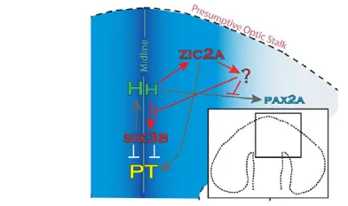
Hh signaling, an essential regulator of vertebrate forebrain development (Bertrand and Dahmane, 2006; Fuccillo et al., 2006; Ingham and Placzek, 2006), functions, in part, through coordinating expression patterns of several target genes, among them six3in the forebrain and pax2 in the OS and retina. Data presented here identify zebrafish zic2aas an Hh-regulated OS marker, and as an essential novel modulator of Hh-activated gene expression in the forebrain, OS and retina. We show that Zic2a restricts transcription of known Hh target genes: six3b in the telencephalon and diencephalon, and pax2ain the ventral retina. We propose that zic2afunctions in the OS to regulate transcription of an Hh agonist or antagonist (Fig. 11). This novel regulatory mechanism is important for the formation of the PT and correct patterning of the ventral retina in zebrafish, and could possibly be conserved in mammals.
Zic2a: a step towards understanding the molecular basis of HPE
The majority of mutations identified in HPE patients are found in three genetic loci – SHH,ZIC2and SIX3 (Cohen, 2006; Dubourg et al., 2007; Krauss, 2007) – suggesting that these genes may be part of a common regulatory network. In support of this idea, transcriptional crossregulation between Six3and Shhwas recently documented in the mouse forebrain and shown to be essential for correct formation of the ventral diencephalic midline (Geng et al., 2008; Jeong et al., 2008), but a role for Zic2in this network has not been tested in the mouse model.
The central role of Hh signaling during forebrain development is well documented in non-mammalian vertebrates, particularly in the zebrafish (Karlstrom et al., 1999; Tyurina et al., 2005). Similarly, mammalian Six3 and its two zebrafish orthologs, six3aand six3b, play essential roles in the developing forebrain and retina (Kobayashi et al., 1998; Lagutin et al., 2003; Inbal et al., 2007; Lavado et al., 2008). Although Six3 has been shown to act indirectly upstream of Zic2 transcription during early Xenopusdevelopment (Gestri et al., 2005), the regulatory relationship between Hh signaling, Six3 and Zic2 has not been extensively explored. In this study, we show that the relationship between Hh and six3is at least partly conserved in zebrafish, as Hh is required for six3b expression. We further show that zic2ais an essential modulator of Hh-induced expression of six3bin the zebrafish forebrain. Although we have not analyzed six3a expression in zic2a morphants, the six3MO used here has been reported to inhibit both six3aand six3b(Ando et al., 2005), raising the possibility that Zic2a regulates both six3orthologs.
[image:7.612.51.302.58.168.2]The forebrain of Zic2a-depleted embryos develops surprisingly normally given the extent of six3b expansion throughout the telencephalon and diencephalon, perhaps because this derepression Fig. 8. Hh is necessary and sufficient for zic2aexpression in the
OS and ventral retina.(A-F)zic2ais expressed in the OS at 16s (A). In smo–/–mutants, zic2aexpression is reduced in the OS at this stage (B,
14/14 embryos, n2), but not elsewhere. shhamRNA-injected WT embryos express zic2aectopically in the OS and retina at 16s (C, 13/13 embryos, n1). zic2aexpression is still reduced in smo–/–mutants at 19s
(D,E, 30/30 embryos, n2), and expanded in shha-injected retina (F, 19/19 embryos, n1). The same result was observed at prim-5 (39/42 embryos, n2, data not shown). Arrowheads point to the OS. Embryos are in lateral view, anterior to the left.
Fig. 9. Zic2a patterns the OS and retina in an Hh-dependent manner.(A-D)Zic2a-depleted WT siblings show expanded pax2a(B, 99/99 embryos, n4), whereas smo–/–mutants injected with conMO
lose pax2aexpression (C, 12/12 embryos, n2). zic2aMO-injected smo–/–mutants show a rescued pax2aphenotype (D, 30/30 embryos,
n4). (E-H)Co-injection of conMO and shhamRNA, or injection of zic2aMO alone, causes moderately expanded pax2aexpression (F, 20/34 embryos, n2; G, 30/30 embryos, n2, respectively). Embryos injected with zic2aMO and shhamRNA show strong pax2a
expansion (H, 23/33 embryos, n2). Arrowheads mark the OS. Panels A-H are lateral view, anterior to the left. Panels A⬘-H⬘are ventral views of the same embryos, anterior to the left. All embryos are at prim-5.
D
E
V
E
LO
P
M
E
N
[image:7.612.316.561.60.363.2]3798
takes place relatively late in development, at mid-somitogenesis stages. Our data show that the forming PT is particularly sensitive to Six3b levels at mid-somitogenesis, consistent with the late role of mouse Six3 in the diencephalon (Lavado et al., 2008). These data suggest that Zic2 similarly modulates the regulatory relationship between Hh signaling and Six3 during mammalian embryogenesis, and offer a novel explanation of how ZIC2mutations cause HPE.
Another Zic family member, zic1, was recently shown to play an essential, yet distinct, role during zebrafish forebrain development (Maurus and Harris, 2009). By contrast to Zic2a, which limits Hh signaling in the ventral forebrain, Zic1 promotes Hh expression and Zic1 depletion leads to loss of ventral forebrain tissue (cyclopia). This functional difference can be explained either by distinct DNA binding specificities of Zic1 and
Zic2a or by differences in where and when they function during development. In support of the latter, the OS is likely to be the major site of Zic2a function in the forebrain, as zic2ais strongly expressed there beginning at mid-somitogenesis. In contrast, zic1 is not found in the OS until late-somitogenesis, when it is only weakly expressed there (Grinblat and Sive, 2001). The more severe phenotype caused by Zic1 depletion in zebrafish suggests that ZIC1 is more broadly required during mammalian development than ZIC2, and is consistent with the fact that ZIC1 mutations have not been identified in HPE patients.
Is the retinal function of Zic2a conserved?
Zebrafish embryos depleted of Zic2a develop a characteristic retinal defect. A similar defect, non-syndromic ocular colobomas (NSOC), is frequently associated with HPE; moreover, mutations in SHH have been correlated with this defect (Schimmenti et al., 2003). Human ZIC2mutations have not been associated with NSOC, and the underlying genetic causes of most ocular colobomas are not well understood (Schimmenti et al., 2003). Ocular defects, that may include coloboma, have been reported in homozygous Zic2a–/–mice, but not characterized (Elms et al., 2003; Herrera et al., 2003). The data presented here offer a potential explanation for these ocular defects and suggest ZIC2as a candidate gene in human colobomas. Coloboma in zic2amorphants may result from misregulation of either pax2aor six3b, as both genes have important functions in the developing OS and retina. Several lines of evidence argue that Zic2a function in the retina is not mediated through Six3b. First, OS and retinal mispatterning in zic2amorphants is not rescued by Six3 depletion (see Fig. S9 in the supplementary material). Second, retinal expression of six3bis not dependent on Hh signaling in mice (Geng et al., 2008) or zebrafish (Fig. 1). Third, six3boverexpression leads to a larger retina, and we do not observe an increase in retinal size in zic2amorphants. Although these data show that Zic2a is not a major regulator of retinal six3b, further analysis is required to determine whether Zic2a contributes to this regulation. Conversely, misregulation of PAX2 has been causally linked to coloboma in human (Gregory-Evans et al., 2004) and chick (Sehgal et al., 2008), suggesting pax2aexpansion in zic2amorphants as the likely cause of ventral retinal defects.
zic2ais expressed in the zebrafish retina, as is Zic2in higher vertebrates (Nagai et al., 1997; Brown et al., 2003; Toyama et al., 2004). The relatively late onset of zic2aexpression in the ventral retina allows us to distinguish its function in the OS precursors, as described in this manuscript, from its later functions. Retinal Zic2 controls guidance of ipsilaterally projecting retinal axons in mouse (Herrera et al., 2003; Garcia-Frigola et al., 2008; Lee, R. et al., 2008). This function of zic2is not conserved in zebrafish, where all retinal ganglion cell axons project contralaterally (Sakai and Halloran, 2006). Further studies to address retinal-specific functions of zebrafish zic2awill probably require conditional overexpression and genetic mutant analyses.
How does zic2a function during forebrain patterning?
Hh growth factors act as morphogens in the OS and retinal primordium. They are secreted from the diencephalic midline and diffuse laterally, instructing cells in proximal positions to develop as OS and cells located more distally to develop as ventral retina (Ekker et al., 1995; Macdonald et al., 1995; Take-uchi et al., 2003; Lupo et al., 2005). Hh-induced pax2a, vax1and vax2promote OS development, at least in part through repression of the retinal marker pax6(Macdonald et al., 1995; Schwarz et al., 2000; Take-uchi et al.,
[image:8.612.49.300.58.215.2]RESEARCH ARTICLE Development 136 (22)
Fig. 10. Exogenous Hh antagonist rescues OS and retinal patterning in Zic2a-depleted embryos.(A)Wild-type pax2a expression. (B-D)Embryos injected with zic2aMO show expanded pax2a(B, 43/43 embryos, n3), whereas embryos injected with hhip mRNA show normal pax2aexpression (C, 8/8 embryos, n1). Embryos co-injected with zic2aMO and hhipmRNA show normal pax2a expression (D, 30/41 embryos, n3). A-D are lateral views, anterior to the left. A⬘-D⬘are ventral views of the same embryos, anterior to the left. Arrowheads mark the posterior limit of pax2aexpression in the retina. All embryos are at prim-5.
Fig. 11. A model of Zic2a function in the zebrafish forebrain primoridum.zic2a is required to restrict expression of six3bin the forebrain and pax2ain the ventral retina. We propose that, in both tissues, zic2aacts non-cell-autonomously through negative modulation of Hh signaling. Hh ligands are produced at the diencephalic midline and trigger a signaling cascade in neighboring cells that activates transcription of several targets, including zic2ain the OS. Zic2a, in turn, may control transcription of a secreted Hh agonist or antagonist in the OS. These data suggest that Zic2a and Hh signaling are engaged in a
novel negative-feedback loop in the developing forebrain.
D
E
V
E
LO
P
M
E
N
[image:8.612.53.249.516.632.2]2003; Mui et al., 2005). We have now shown that zic2a is also activated by Hh signaling in the OS, but unlike the other targets, does not promote OS or inhibit retinal fates. Instead, Zic2a acts to modulate the Hh-regulated pattern of gene expression. Zic2a function provides the means for Hh signaling to negatively modulate, and thus refine, its downstream signaling activity. Although negative-feedback regulation such as this has been described for several signaling pathways, including Hh signaling (Chuang and McMahon, 1999; Ochi et al., 2006; Lee, J. et al., 2008) and Fgf signaling (Furthauer et al., 2001), this negative regulation is not typically mediated via induction of a transcription factor.
As a transcription factor, Zic2a probably acts cell-autonomously to directly activate or repress transcription of its target genes. However, our data clearly indicate that Zic2a downregulates the transcription of several genes outside of its own expression domain. A plausible explanation is that Zic2a directly regulates transcription of a secreted factor, which then acts on neighboring cells to modulate Hh signaling. Although zic2ais expressed in several domains in the forebrain, the OS is the most likely site for this function because of its proximity to the tissues affected in morphants. Likely candidate targets of Zic2a include genes that facilitate or hinder the transport of Hh ligands, e.g. the Hh antagonist hhip(Chuang and McMahon, 1999; Coulombe et al., 2004; Ochi et al., 2006), and extracellular proteins such as Glypican or Megalin (Low-density lipoprotein-related protein 2 – Zebrafish Information Network), which can aide or hinder transcytosis and affect long-range signaling (Willnow et al., 1996; McCarthy et al., 2002; Beckett et al., 2008). The zebrafish megalingene is not expressed in the OS (McCarthy et al., 2002) and therefore may be repressed by Zic2a. The known zebrafish hhipgene is not expressed in the OS and is not a candidate target for Zic2a (data not shown), but at least two other hhipgenes are likely to exist (Katoh and Katoh, 2006; Ochi et al., 2006). A concerted effort to identify direct transcriptional targets of Zic2a is in progress and is likely to uncover novel Hh modulators that act downstream of zic2ain OS and retinal patterning. This novel Hh negative-feedback control might also be used in other developmental contexts where Hh signaling is involved, such as the limb bud.
Acknowledgements
We thank Steve Wilson, Jeff Gross, Mary Halloran, Mary Ellen Lane, Ann Ungar, Monte Westerfield and the Zebrafish International Resource Center (ZIRC) for providing plasmids and zebrafish lines; and Jessica Pierson and other members of the Grinblat laboratory for insightful discussions throughout the course of this work and for constructive comments during preparation of this manuscript. This work was funded by an NIH RO1 grant to Y.G. Deposited in PMC for release after 12 months.
Author contributions
N.A.S. carried out the bulk of data collection and analysis; A.B.T. performed real-time PCR and analyzed the data; M.K.N. assisted with morphant analysis and real-time PCR experiments; and Y.G. participated in data analysis. All four authors contributed to manuscript preparation.
Supplementary material
Supplementary material for this article is available at http://dev.biologists.org/cgi/content/full/136/22/3791/DC1
References
Ando, H., Kobayashi, M., Tsubokawa, T., Uyemura, K., Furuta, T. and Okamoto, H.(2005). Lhx2 mediates the activity of Six3 in zebrafish forebrain growth. Dev. Biol.287, 456-468.
Beckett, K., Franch-Marro, X. and Vincent, J. P.(2008). Glypican-mediated endocytosis of Hedgehog has opposite effects in flies and mice. Trends Cell Biol. 18, 360-363.
Bertrand, N. and Dahmane, N.(2006). Sonic hedgehog signaling in forebrain development and its interactions with pathways that modify its effects. Trends Cell Biol.16, 597-605.
Brown, L. Y., Kottmann, A. H. and Brown, S.(2003). Immunolocalization of Zic2 expression in the developing mouse forebrain. Gene Expr. Patterns3, 361-367.
Brown, S. A., Warburton, D., Brown, L. Y., Yu, C. Y., Roeder, E. R., Stengel-Rutkowski, S., Hennekam, R. C. and Muenke, M.(1998). Holoprosencephaly due to mutations in ZIC2, a homologue of Drosophila odd-paired. Nat. Genet. 20, 180-183.
Chuang, P. T. and McMahon, A. P.(1999). Vertebrate Hedgehog signaling modulated by induction of a Hedgehog-binding protein. Nature397, 617-621.
Cohen, M. M., Jr (2006). Holoprosencephaly: clinical, anatomic, and molecular dimensions. Birth Defects Res. A Clin. Mol. Teratol.76, 658-673.
Coulombe, J., Traiffort, E., Loulier, K., Faure, H. and Ruat, M.(2004). Hedgehog interacting protein in the mature brain: membrane-associated and soluble forms. Mol. Cell. Neurosci.25, 323-333.
Dubourg, C., Bendavid, C., Pasquier, L., Henry, C., Odent, S. and David, V.
(2007). Holoprosencephaly. Orphanet. J. Rar. Dis.2, 8.
Ekker, S. C., Ungar, A. R., Greenstein, P., von Kessler, D. P., Porter, J. A., Moon, R. T. and Beachy, P. A.(1995). Patterning activities of vertebrate hedgehog proteins in the developing eye and brain. Curr. Biol.5, 944-955.
Elms, P., Siggers, P., Napper, D., Greenfield, A. and Arkell, R.(2003). Zic2 is required for neural crest formation and hindbrain patterning during mouse development. Dev. Biol.264, 391-406.
Erickson, T., Scholpp, S., Brand, M., Moens, C. B. and Waskiewicz, A. J.
(2007). Pbx proteins cooperate with Engrailed to pattern the midbrain-hindbrain and diencephalic-mesencephalic boundaries. Dev. Biol.301, 504-517.
Fuccillo, M., Joyner, A. L. and Fishell, G.(2006). Morphogen to mitogen: the multiple roles of hedgehog signalling in vertebrate neural development. Nat. Rev. Neurosci.7, 772-783.
Furthauer, M., Thisse, C. and Thisse, B.(1997). A role for FGF-8 in the dorsoventral patterning of the zebrafish gastrula. Development124, 4253-4264.
Furthauer, M., Reifers, F., Brand, M., Thisse, B. and Thisse, C.(2001). sprouty4 acts in vivo as a feedback-induced antagonist of FGF signaling in zebrafish.
Development128, 2175-2186.
Garcia-Frigola, C., Carreres, M. I., Vegar, C., Mason, C. and Herrera, E.(2008). Zic2 promotes axonal divergence at the optic chiasm midline by EphB1-dependent and -inEphB1-dependent mechanisms. Development135, 1833-1841.
Gehring, W. J.(1996). The master control gene for morphogenesis and evolution of the eye. Genes Cells1, 11-15.
Geng, X., Speirs, C., Lagutin, O., Inbal, A., Liu, W., Solnica-Krezel, L., Jeong, Y., Epstein, D. J. and Oliver, G.(2008). Haploinsufficiency of Six3 fails to activate Sonic hedgehog expression in the ventral forebrain and causes holoprosencephaly. Dev. Cell15, 236-247.
Gestri, G., Carl, M., Appolloni, I., Wilson, S. W., Barsacchi, G. and
Andreazzoli, M.(2005). Six3 functions in anterior neural plate specification by promoting cell proliferation and inhibiting Bmp4 expression. Development132, 2401-2413.
Gillhouse, M., Wagner Nyholm, M., Hikasa, H., Sokol, S. Y. and Grinblat, Y.
(2004). Two Frodo/Dapper homologs are expressed in the developing brain and mesoderm of zebrafish. Dev. Dyn.230, 403-409.
Gregory-Evans, C. Y., Williams, M. J., Halford, S. and Gregory-Evans, K.
(2004). Ocular coloboma: a reassessment in the age of molecular neuroscience.
J. Med. Genet.41, 881-891.
Grinblat, Y. and Sive, H.(2001). zic Gene expression marks anteroposterior pattern in the presumptive neurectoderm of the zebrafish gastrula. Dev. Dyn. 222, 688-693.
Gross, J. M. and Dowling, J. E.(2005). Tbx2b is essential for neuronal differentiation along the dorsal/ventral axis of the zebrafish retina. Proc. Natl. Acad. Sci. USA102, 4371-4376.
Herrera, E., Brown, L., Aruga, J., Rachel, R. A., Dolen, G., Mikoshiba, K., Brown, S. and Mason, C. A.(2003). Zic2 patterns binocular vision by specifying the uncrossed retinal projection. Cell114, 545-557.
Hoyle, J., Tang, Y. P., Wiellette, E. L., Wardle, F. C. and Sive, H.(2004). nlz gene family is required for hindbrain patterning in the zebrafish. Dev. Dyn.229, 835-846.
Inbal, A., Kim, S. H., Shin, J. and Solnica-Krezel, L.(2007). Six3 represses nodal activity to establish early brain asymmetry in zebrafish. Neuron55, 407-415.
Ingham, P. W. and Placzek, M.(2006). Orchestrating ontogenesis: variations on a theme by sonic hedgehog. Nat. Rev. Genet.7, 841-850.
Jeong, J. Y., Einhorn, Z., Mathur, P., Chen, L., Lee, S., Kawakami, K. and Guo, S.(2007). Patterning the zebrafish diencephalon by the conserved zinc-finger protein Fezl. Development134, 127-136.
Jeong, Y., Leskow, F. C., El-Jaick, K., Roessler, E., Muenke, M., Yocum, A., Dubourg, C., Li, X., Geng, X., Oliver, G. et al.(2008). Regulation of a remote Shh forebrain enhancer by the Six3 homeoprotein. Nat. Genet.40, 1348-1353.
Karlstrom, R. O., Talbot, W. S. and Schier, A. F.(1999). Comparative synteny cloning of zebrafish you-too: mutations in the Hedgehog target gli2 affect ventral forebrain patterning. Genes Dev.13, 388-393.
Katoh, Y. and Katoh, M.(2006). Comparative genomics on HHIP family
orthologs. Int. J. Mol. Med.17, 391-395.
D
E
V
E
LO
P
M
E
N
3800
Kimmel, C. B., Ballard, W. W., Kimmel, S. R., Ullmann, B. and Schilling, T. F.
(1995). Stages of embryonic development of the zebrafish. Dev. Dyn.203, 253-310.
Kobayashi, M., Toyama, R., Takeda, H., Dawid, I. B. and Kawakami, K.
(1998). Overexpression of the forebrain-specific homeobox gene six3 induces rostral forebrain enlargement in zebrafish. Development125, 2973-2982.
Krauss, R. S.(2007). Holoprosencephaly: new models, new insights. Expert Rev. Mol. Med.9, 1-17.
Krauss, S., Johansen, T., Korzh, V. and Fjose, A.(1991). Expression pattern of zebrafish pax genes suggests a role in early brain regionalization. Nature353, 267-270.
Lagutin, O. V., Zhu, C. C., Kobayashi, D., Topczewski, J., Shimamura, K., Puelles, L., Russell, H. R., McKinnon, P. J., Solnica-Krezel, L. and Oliver, G.
(2003). Six3 repression of Wnt signaling in the anterior neuroectoderm is essential for vertebrate forebrain development. Genes Dev.17, 368-379.
Lavado, A., Lagutin, O. V. and Oliver, G.(2008). Six3 inactivation causes progressive caudalization and aberrant patterning of the mammalian diencephalon. Development135, 441-450.
Lecaudey, V., Cakan-Akdogan, G., Norton, W. H. and Gilmour, D.(2008). Dynamic Fgf signaling couples morphogenesis and migration in the zebrafish lateral line primordium. Development135, 2695-2705.
Lee, J., Willer, J. R., Willer, G. B., Smith, K., Gregg, R. G. and Gross, J. M.
(2008). Zebrafish blowout provides genetic evidence for Patched1-mediated negative regulation of Hedgehog signaling within the proximal optic vesicle of the vertebrate eye. Dev. Biol.319, 10-22.
Lee, R., Petros, T. J. and Mason, C. A.(2008). Zic2 regulates retinal ganglion cell axon avoidance of ephrinB2 through inducing expression of the guidance receptor EphB1. J. Neurosci.28, 5910-5919.
Lupo, G., Liu, Y., Qiu, R., Chandraratna, R. A., Barsacchi, G., He, R. Q. and Harris, W. A.(2005). Dorsoventral patterning of the Xenopus eye: a collaboration of Retinoid, Hedgehog and FGF receptor signaling. Development 132, 1737-1748.
Macdonald, R., Barth, K. A., Xu, Q., Holder, N., Mikkola, I. and Wilson, S. W.
(1995). Midline signalling is required for Pax gene regulation and patterning of the eyes. Development121, 3267-3278.
Masai, I., Stemple, D. L., Okamoto, H. and Wilson, S. W.(2000). Midline signals regulate retinal neurogenesis in zebrafish. Neuron27, 251-263.
Maurus, D. and Harris, W. A.(2009). Zic-associated holoprosencephaly: zebrafish Zic1 controls midline formation and forebrain patterning by regulating Nodal, Hedgehog, and retinoic acid signaling. Genes Dev.23, 1461-1473.
McCarthy, R. A., Barth, J. L., Chintalapudi, M. R., Knaak, C. and Argraves, W. S.(2002). Megalin functions as an endocytic sonic hedgehog receptor. J. Biol. Chem.277, 25660-25667.
McCollum, C. W., Amin, S. R., Pauerstein, P. and Lane, M. E.(2007). A zebrafish LMO4 ortholog limits the size of the forebrain and eyes through negative regulation of six3b and rx3. Dev. Biol.309, 373-385.
Miura, H., Yanazawa, M., Kato, K. and Kitamura, K.(1997). Expression of a novel aristaless related homeobox gene ‘Arx’ in the vertebrate telencephalon, diencephalon and floor plate. Mech. Dev.65, 99-109.
Mui, S. H., Kim, J. W., Lemke, G. and Bertuzzi, S.(2005). Vax genes ventralize the embryonic eye. Genes Dev.19, 1249-1259.
Nagai, T., Aruga, J., Takada, S., Gunther, T., Sporle, R., Schughart, K. and Mikoshiba, K.(1997). The expression of the mouse Zic1, Zic2, and Zic3 gene suggests an essential role for Zic genes in body pattern formation. Dev. Biol. 182, 299-313.
Nakayama, K., Satoh, T., Igari, A., Kageyama, R. and Nishida, E.(2008). FGF induces oscillations of Hes1 expression and Ras/ERK activation. Curr. Biol.18, R332-R334.
Nasevicius, A. and Ekker, S. C.(2000). Effective targeted gene ‘knockdown’ in zebrafish. Nat. Genet.26, 216-220.
Nyholm, M. K., Wu, S. F., Dorsky, R. I. and Grinblat, Y.(2007). The zebrafish zic2a-zic5 gene pair acts downstream of canonical Wnt signaling to control cell proliferation in the developing tectum. Development134, 735-746.
Ochi, H., Pearson, B. J., Chuang, P. T., Hammerschmidt, M. and Westerfield, M.(2006). Hhip regulates zebrafish muscle development by both sequestering Hedgehog and modulating localization of Smoothened. Dev. Biol.297, 127-140.
Odenthal, J., van Eeden, F. J., Haffter, P., Ingham, P. W. and Nusslein-Volhard, C.(2000). Two distinct cell populations in the floor plate of the zebrafish are induced by different pathways. Dev. Biol.219, 350-363.
Pfeffer, P. L., Gerster, T., Lun, K., Brand, M. and Busslinger, M.(1998). Characterization of three novel members of the zebrafish Pax2/5/8 family: dependency of Pax5 and Pax8 expression on the Pax2.1 (noi) function.
Development125, 3063-3074.
Picker, A. and Brand, M.(2005). Fgf signals from a novel signaling center determine axial patterning of the prospective neural retina. Development132, 4951-4962.
Rhinn, M., Picker, A. and Brand, M.(2006). Global and local mechanisms of forebrain and midbrain patterning. Curr. Opin. Neurobiol.16, 5-12.
Rohr, K. B., Barth, K. A., Varga, Z. M. and Wilson, S. W.(2001). The nodal pathway acts upstream of hedgehog signaling to specify ventral telencephalic identity. Neuron.29, 341-351.
Sakai, J. A. and Halloran, M. C.(2006). Semaphorin 3d guides laterality of retinal ganglion cell projections in zebrafish. Development133, 1035-1044.
Sanek, N. A. and Grinblat, Y.(2008). A novel role for zebrafish zic2a during forebrain development. Dev. Biol.317, 325-335.
Schimmenti, L. A., de la Cruz, J., Lewis, R. A., Karkera, J. D., Manligas, G. S., Roessler, E. and Muenke, M.(2003). Novel mutation in sonic hedgehog in non-syndromic colobomatous microphthalmia. Am. J. Med. Genet. A116A, 215-221.
Schmitt, E. A. and Dowling, J. E.(1994). Early eye morphogenesis in the zebrafish, Brachydanio rerio. J. Comp. Neurol.344, 532-542.
Schwarz, M., Cecconi, F., Bernier, G., Andrejewski, N., Kammandel, B., Wagner, M. and Gruss, P.(2000). Spatial specification of mammalian eye territories by reciprocal transcriptional repression of Pax2 and Pax6.
Development127, 4325-4334.
Sehgal, R., Karcavich, R., Carlson, S. and Belecky-Adams, T. L.(2008). Ectopic Pax2 expression in chick ventral optic cup phenocopies loss of Pax2 expression.
Dev. Biol.319, 23-33.
Seo, H. C., Drivenes, Ellingsen, S. and Fjose, A.(1998). Expression of two zebrafish homologues of the murine Six3 gene demarcates the initial eye primordia. Mech. Dev.73, 45-57.
Stenkamp, D. L. and Frey, R. A.(2003). Extraretinal and retinal hedgehog signaling sequentially regulate retinal differentiation in zebrafish. Dev. Biol.258, 349-363.
Take-uchi, M., Clarke, J. D. and Wilson, S. W.(2003). Hedgehog signalling maintains the optic stalk-retinal interface through the regulation of Vax gene activity. Development130, 955-968.
Toyama, R., Gomez, D. M., Mana, M. D. and Dawid, I. B.(2004). Sequence relationships and expression patterns of zebrafish zic2 and zic5 genes. Gene. Expr. Patterns4, 345-350.
Tsang, M., Friesel, R., Kudoh, T. and Dawid, I. B.(2002). Identification of Sef, a novel modulator of FGF signalling. Nat. Cell Biol.4, 165-169.
Tyurina, O. V., Guner, B., Popova, E., Feng, J., Schier, A. F., Kohtz, J. D. and Karlstrom, R. O.(2005). Zebrafish Gli3 functions as both an activator and a repressor in Hedgehog signaling. Dev. Biol.277, 537-556.
Ungar, A. R. and Moon, R. T.(1996). Inhibition of protein kinase A phenocopies ectopic expression of hedgehog in the CNS of wild-type and cyclops mutant embryos. Dev. Biol.178, 186-191.
Varga, Z. M., Amores, A., Lewis, K. E., Yan, Y. L., Postlethwait, J. H., Eisen, J. S. and Westerfield, M.(2001). Zebrafish smoothened functions in ventral neural tube specification and axon tract formation. Development128, 3497-3509.
Westerfield, M.(1995). The Zebrafish Book. Eugene, OR. University of Oregon Press.
Willnow, T. E., Hilpert, J., Armstrong, S. A., Rohlmann, A., Hammer, R. E., Burns, D. K. and Herz, J.(1996). Defective forebrain development in mice lacking gp330/megalin. Proc. Natl. Acad. Sci. USA93, 8460-8464.
Wilson, S. W. and Houart, C.(2004). Early steps in the development of the forebrain. Dev. Cell6, 167-181.
Xiao, T., Roeser, T., Staub, W. and Baier, H.(2005). A GFP-based genetic screen reveals mutations that disrupt the architecture of the zebrafish retinotectal projection. Development132, 2955-2967.
Yamauchi, H., Hotta, Y., Konishi, M., Miyake, A., Kawahara, A. and Itoh, N.
(2006). Fgf21 is essential for haematopoiesis in zebrafish. EMBO Rep.7, 649-654.
Zhang, J., Jin, Z. and Bao, Z. Z.(2004). Disruption of gradient expression of Zic3 resulted in abnormal intra-retinal axon projection. Development131, 1553-1562.
RESEARCH ARTICLE Development 136 (22)