Menny, A. and Serna, M. and Boyd, C.M. and Gardner, S. and Joseph, A.P.
and Morgan, B.P. and Topf, Maya and Brooks, N.J. and Bubeck, D. (2018)
CryoEM reveals how the complement membrane attack complex ruptures
lipid bilayers. Nature Communications 9 (1), ISSN 2041-1723.
Downloaded from:
Usage Guidelines:
Please refer to usage guidelines at
or alternatively
CryoEM reveals how the complement membrane
attack complex ruptures lipid bilayers
Anaïs Menny
1
, Marina Serna
1,2
, Courtney M. Boyd
1
, Scott Gardner
1
, Agnel Praveen Joseph
3,6
, B. Paul Morgan
4
,
Maya Topf
3
, Nicholas J. Brooks
5
& Doryen Bubeck
1
The membrane attack complex (MAC) is one of the immune system
’
s
fi
rst responders.
Complement proteins assemble on target membranes to form pores that lyse pathogens and
impact tissue homeostasis of self-cells. How MAC disrupts the membrane barrier remains
unclear. Here we use electron cryo-microscopy and
fl
icker spectroscopy to show that MAC
interacts with lipid bilayers in two distinct ways. Whereas C6 and C7 associate with the outer
lea
fl
et and reduce the energy for membrane bending, C8 and C9 traverse the bilayer
increasing membrane rigidity. CryoEM reconstructions reveal plasticity of the MAC pore and
demonstrate how C5b6 acts as a platform, directing assembly of a giant
β
-barrel whose
structure is supported by a glycan scaffold. Our work provides a structural basis for
under-standing how
β
-pore forming proteins breach the membrane and reveals a mechanism for
how MAC kills pathogens and regulates cell functions.
https://doi.org/10.1038/s41467-018-07653-5
OPEN
1Department of Life Sciences, Sir Ernst Chain Building, Imperial College London, London SW7 2AZ, UK.2Spanish National Cancer Research Centre, CNIO,
Melchor Fernández Almagro, 3.28029 Madrid, Spain.3Institute of Structural and Molecular Biology, Department of Biological Sciences, Birkbeck, University of London, Malet Street, London WC1E 7HX, UK.4Division of Infection and Immunity, School of Medicine, Cardiff University, Heath Park, Cardiff CF14 4XN,
UK.5Department of Chemistry, Imperial College London, London SW7 2AZ, UK.6Present address: Scienti
fic Computing Department, Science and Technology Facilities Council, Research Complex at Harwell, Didcot OX11 0FA, UK. These authors contributed equally: Marina Serna, Courtney M. Boyd. Correspondence and requests for materials should be addressed to D.B. (email:[email protected])
123456789
T
he multiprotein membrane attack complex is a bactericidal
weapon of the innate immune system that also modulates
inflammation and proliferation when formed on self-cells
1.
The MAC pore targets a wide range of pathogens, forming on and
killing Gram-negative bacteria
2, enveloped viruses
3, and
para-sites
4. This innate immune effector is essential for
fighting
bac-teria of the genus
Neisseria
;
5with genetic deficiencies in
component proteins leading to recurrent infections
6. Host cells
are protected from bystander damage by the GPI-anchored
receptor CD59, the only membrane-bound inhibitor of MAC
7.
Deficiency of CD59 causes the lethal blood disorder Paroxysmal
Nocturnal Hemoglobinuria, a disease characterized by
thrombo-sis and chronic hemolythrombo-sis
8. Complement activation and MAC
formation also contributes to killing of cancer cells during
immunotherapy treatments
9. Therefore, developing a molecular
model of how MAC forms on target cells is essential for
under-standing the immune response to microbes and for the
devel-opment of therapeutics that regulate complement activity.
MAC assembles from soluble complement proteins in an
obligate sequential pathway
10. In the presence of membranes,
C5b6 (a complex comprised of C5b and C6
11) binds C7 to form
the lipophilic MAC precursor C5b7
12. C8 irreversibly binds the
nascent complex, resulting in the membrane-inserted C5b8
13. C9
molecules associate with C5b8 in the membrane to form C5b9
and polymerize to complete the MAC pore
14. Previous electron
cryo-microscopy (cryoEM) reconstructions of the complex
revealed that the
final MAC is comprised of 6 polypeptide chains
(C5b, C6, C7, C8
α
, C8
β
, and C8
γ
) together with 18 C9 monomers
that are arranged in a split-washer configuration
15,16. Based on
structural similarity with bacterial homologs, the giant (110 Å
diameter)
β
-barrel pore is formed when helical bundles in the
Membrane Attack Complex-Perforin (MACPF) domains
trans-form into transmembrane
β
-hairpins (TMH)
17–19, although a
molecular mechanism for how this occurs is not currently
understood.
In this study, we used cryoEM to determine the structures of
two MAC conformations at near atomic resolution and derive a
nearly complete atomic model for the pore. In combination with
flicker spectroscopy, we show how MAC assembly impacts
bio-physical properties of the bilayer and resolve the mechanisms of
membrane interaction and MAC activity.
Results
CryoEM structure of the MAC. The human MAC pore was
formed on liposomes from individual complement proteins. The
lipid composition of vesicles was selected based on the
stoichio-metric homogeneity of deposited pores. MAC was solubilized with
detergent and purified for structural studies, as described
pre-viously
15. By integrating newly collected data across multiple
electron microscopes (Supplementary Table 1), we were able to
improve the overall resolution of the MAC from 8.5 Å
15to 4.9 Å;
however, density corresponding to the interface between C6 and
the terminal C9 was still poorly resolved. We used 3D
classifica-tion procedures to computaclassifica-tionally isolate two stoichiometrically
identical conformations, open and closed, which varied in the
extent of
β
-barrel closure (Fig.
1
and Supplementary Fig. 1). 2D
classification of negatively stained complexes inserted into a lipid
monolayer confirmed the presence of these states in a membrane
environment (Supplementary Fig. 2a), in agreement with
cryo-tomography structures of MACs in liposomes
16. The maps were
further subdivided into three components: an asymmetric region
(C5b, C6, C7, and C8), a hinge region (C7, C8, and two C9
molecules), and a C9 oligomer. Using a masked refinement
strategy coupled with signal subtraction
20, we improved
the resolution of the asymmetric regions for each conformation to
4.7 Å and 5.9 Å (Fig.
1
, Supplementary Figs. 1, and 2b). The hinge
region of the open conformation was resolved to 4.9 Å
(Supple-mentary Figs. 1 and 2b). Masked refinement from
signal-subtracted images followed by sub-volume averaging was used
to resolve the averaged C9 monomer from the open conformation
at 4.4 Å (Fig.
1
a, and Supplementary Figs. 1, 2b, 3c). A similar
analysis of the C9 oligomer from the closed conformation resulted
in a lower resolution map. Therefore, we focused our
interpreta-tion of C9 on density derived from the open conformainterpreta-tion
oli-gomer. The new maps enabled us to build an atomic model that
includes the irregular and asymmetric
β
-barrel pore
(Supple-mentary Table 1 and Supple(Supple-mentary Fig. 3). Although density is
lacking for many side-chains within C5b and the lower half of the
central
β
-barrel, we have imposed experimental restraints that
justify their register in the atomic model. Crystal structures for
soluble components (C5b6
11and C8
21, a heterotrimeric complex
consisting of
α
,
β
, and
γ
polypeptide chains) together with
homology models for C7 and C9 were
fitted into the density.
Domains of these structures were
first refined as rigid bodies, with
disulfide bond restraints. Models were further refined restraining
secondary structure and side-chain geometry to higher resolution
crystallographic structures.
β
-strands that comprise the central
barrel were initiated where side-chain density was visible and
extended imposing idealized backbone geometry constraints. The
trajectory for each strand is linear and the register was confirmed
by correlating glycan density with the position of the modified
residue in the sequence (Supplementary Fig. 4).
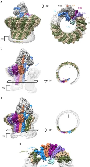
MAC is a
fl
exible immune pore. We used 3D classification to
resolve MAC conformational
flexibility (Fig.
1
and
Supplemen-tary Fig. 1). The open conformation is characterized by a 30 Å
wide chasm that runs the length of the complex (Fig.
1
b). Lipid
molecules likely
fill the opening on the wall of the pore,
remi-niscent of arc pores observed for both mammalian and bacterial
β
-pore forming proteins
22,23. The asymmetric region juts into the
lumen of the barrel like a
“
paddle
”
, exaggerating the MAC
’
s split
washer shape (Fig.
1
b and Supplementary Movie 1). C8
γ
is
wedged in the crease between the rotated asymmetric component
and adjacent C9 oligomer, and may limit the rotation of the
paddle. While curvature of C9 arcs vary at either end, the central
section is near-circular with monomers equally spaced ~16
˚
apart
(Fig.
1
b, c), reminiscent of the arrangement observed for a C9
homo-oligomer
24. Although the chasm is sealed in the closed
conformation (Fig.
1
c), interfaces mediating the MACPF-rim and
transmembrane regions are not
flush. The asymmetric region
swings back and meets C9 in a noncanonical
MAPCF-thrombospondin (C6-TSP3) interaction with limited buried
sur-face area (Fig.
1
d). Despite a contiguous extracellular
β
-barrel,
there remains a gap within the transmembrane pore where the
shorter hairpins of C6, C7, and C8 abut those of C9 (Fig.
1
c). In
the open conformation, the
first and terminal C9 are latterly
shifted by ~20 Å, while the ring of the closed conformation
remains planar (Fig.
1
). Therefore, conformational
flexibility of
the assembly may impact local curvature of the membrane.
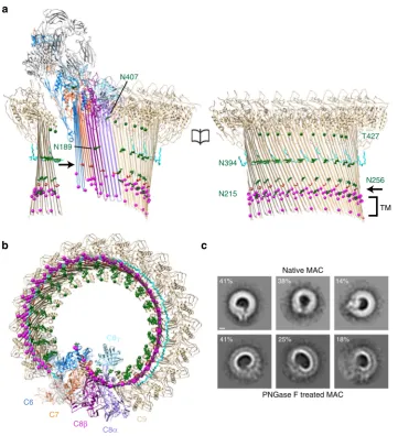
The MAC is a highly-glycosylated assembly with all
comple-ment components post-translationally modified
11,25–27. We
observe density for many of the reported glycans on C7
25,
C8
27, and C9
26, which line the
β
-barrel
’
s concave face (Figs.
1
b, c,
C5b
C6
C7
C8β
C8α
C8γ
C9
C9 90°
90°
90°
C6TSP3
C9MACPF
C6MACPF
TM TM
a
b
c
d
TM
[image:4.595.138.454.49.631.2]C5b6 is a spatial platform for MAC assembly. Limited
resolu-tion of previous MAC reconstrucresolu-tions
15,16prevented any
struc-tural analysis of rearrangements within C5b6 during pore
formation. We therefore investigated how C7-binding to C5b6
triggers the lipophilic transition using our high-resolution maps
(Fig.
3
). Our data reveal that the C5b6 complex serves as a spatial
platform directing MAC assembly. C5b MG domains 1, 4, and 5,
together with the
“
link
”
domain, bridge a cluster of arches
com-prised of the lipoprotein receptor class A (LDL) domains of C6, C7,
and C8
β
(Fig.
3
a and Supplementary Fig. 5). While the core of C5b
remains largely unchanged during the transition, C6 undergoes
marked domain rearrangements upon integration into the MAC.
C6 is comprised of 10 individual domains that can be classified
into three functional parts: (1) those that mediate the interaction
with C5b, (2) regulatory auxiliary modules, and (3) the
pore-forming MACPF domain. Large structural re-arrangements of C6
auxiliary domains accompany conformational changes within the
pore-forming MACPF (Fig.
3
d and Supplementary Fig. 5a).
Superposition of the soluble and MAC-incorporated forms of
C5b6 show that although the relative orientation of the C5b
thioester-like domain (TED) and C6 C-terminal complement
control protein (CCP) domains remains unchanged, the C6 LDL
is displaced by C7-binding. The two N-terminal TSP domains
(TSP1 and TSP2) undergo a concerted rotation with respect to
the core MACPF, resulting in a
final position near-perpendicular
to the plane of the membrane, stabilizing the newly formed
β
-sheet. Rotations of regulatory auxiliary modules coincide with an
unbending and untwisting of the C6 MACPF
β
-sheet. Movement
of the C6 epidermal growth factor (EGF) domain and MACPF
helix-turn-helix (CH3) motif release the pore-forming TMH
regions, in agreement with lower resolution structures of
pore-forming toxin homologs
28.
Auxiliary domains mediate the lipophilic transition. We next
explored whether conformational changes incurred by C6 were
C9
a
c
b
N394
C6
C7
C8β C8α
C8γ
N256
N215
TM
T427
41% 38% 14%
41% 25% 18%
Native MAC
PNGase F treated MAC
N189
N407
[image:5.595.119.481.48.445.2]conserved across complement proteins. N- and C-terminal TSP
domains of C7, C8 (TSP1 and TSP2), and C9 (TSP1), overlay
with those of C6 (TSP2 and TSP3) in its membrane-inserted
“
active
”
conformation (Supplementary Fig. 6a). Furthermore, the
position of CH3 relative to the MACPF
β
-sheet is the same for all
MAC components (Supplementary Fig. 6b). Although the core
MACPF domains of all MAC components overlay well, the
glycine-rich hinge enables a wide range of angles to accommodate
the varied curvature of the barrel. Similar to the pore transition of
C6, C8 regulatory auxiliary modules (EGF and TSP2) release
TMH regions of both
α
and
β
chains (Supplementary Fig. 6c).
However, unlike the dramatic C6 MACPF unbending, the angle
of the C8
β
MACPF sheet remains constant (Supplementary
Fig. 6d). Component strands untwist to align with the C7
β
-sheet
and pack against the C7 CH3 latch. Surprisingly, C8
β
CH3 does
not undergo a lateral shift during pore formation, suggesting that
the C8
β
MACPF is already primed for membrane insertion.
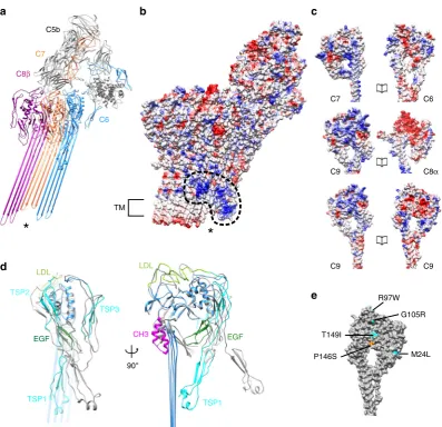
To investigate the molecular basis underpinning the
direction-ality of MAC
’
s sequential assembly, we compared electrostatic
surface potentials of complement proteins (Fig.
3
c). The interface
between C6-C7, C8
α
-C9, and C9-C9 is formed mainly by polar
and charge interactions (Supplementary Table 2). Incorporation
of C8
α
exposes a negatively charged patch that may influence
recruitment of the positively charged face of C9. C9
oligomeriza-tion could be propagated by electrostatic complementarity
between leading and lagging faces, consistent with CDC pore
formation
29. Mutations that alter polymerization of C9 are
implicated in some cases of age-related macular degeneration
(AMD)
30. We are now able to assign disease-related variants on
the human C9 structure, and
find that three of these
AMD-associated mutations (P146S, G105R, and T149I) would likely
impact the negatively charged patch that drives oligomerization.
G105R and T149I variants decrease polymerization of C9
30and
would reduce the footprint of the negatively charged interface. By
TM*
a
b
C5b
C8β
C7
C6
90°
d
LDLCH3
TSP1 EGF
TSP1
TSP2
TSP3
EGF LDL
C7 C6
C9 C8α
C9 C9
c
e
R97WG105R
M24L T149I
P146S
*
[image:6.595.99.497.46.430.2]contrast, variant P146S increases self-polymerization of C9
30and
may act by increasing polarity of the surface.
Interactions with the lipid bilayer. Complement proteins within
the MAC interact with the lipid bilayer in two distinct ways.
Transmembrane hairpins of MAC components vary in length
and amino acid composition. C6 and C7 hairpins contain a single
band of aromatic residues (Fig.
2
a). A patch of positively charged
residues resides proximal to the tips of the short C6 and C7
β
-hairpins (Fig.
3
a, b), suggesting interaction with phospholipid
headgroups of the outer leaflet. Similar to other
β
-barrel pore
assemblies
31, two rings of aromatic residues separated by the
width of the bilayer anchor C8 and C9 within the membrane
(Fig.
2
a). The longer hairpins of C8 and C9 expose hydrophobic
residues on the
β
-barrel
’
s outer surface (Fig.
3
b), consistent with
an interface with lipid tails.
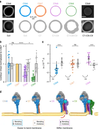
To investigate how these two types of interactions impact
mechanical properties of the target cell membrane, we performed
fluctuation analysis (flicker spectroscopy) of giant unilamellar
vesicles (GUVs). In
flicker spectroscopy, the magnitude of
thermally induced
fluctuations in a GUV membrane are
quantified by tracking the membrane contour in a series of
video-microscopy images. This data is
fitted to a two-parameter
model to extract the membrane bending rigidity and tension
32.
We used phase contrast light microscopy to track individual
GUVs at each step of MAC assembly and confirmed the presence
of functional pores by diffusion of sucrose across the bilayer
(Fig.
4
a). We recorded membrane
fluctuations after the addition
of C5b6, C7, C8, and C9, or when buffer replaced C5b6 in the
sequence. When C5b6 was absent, the amplitude of the GUV
membrane
fluctuations remained constant throughout the
experiment (Fig.
4
a, b and Supplementary Movie 2). The small
increase in
fluctuations observed at later time points is likely to be
due to slight increases in temperature caused by lamp heating. By
contrast, membrane
fluctuations of GUVs that supported MAC
assembly were reduced upon C8-binding and were diminished to
negligible levels with the addition of C9 (Fig.
4
a, b and
Supplementary Movie 2). These data suggest that large MAC
lesions in cells may lead to rigidification of the membrane by the
insertion of
β
-hairpins across the bilayer and by equilibration of
the extracellular milieu across the pore.
Where
fluctuation amplitudes could be parameterized, we
analyzed changes in bending modulus and tension during MAC
assembly (Fig.
4
c and Supplementary Fig. 7). Bending modulus is
an intrinsic property of the membrane and describes the amount
of energy required to change the mean curvature of a lipid bilayer.
It can be affected by thickness of the bilayer
33, membrane
asymmetry
34, and lipid composition
35. C5b6 is a soluble complex
that ionically associates with membranes
36. Upon C7-binding, the
complex becomes lipophilic and is irreversibly tethered
12.
Although this study and previous work are consistent with an
interaction of C7 with lipids
12,37,38, the ability of C5b6 to alter the
biophysical properties of the membrane was unanticipated. C5b6
reduced the bending modulus of GUVs without affecting tension
(Fig.
4
c and Supplementary Fig. 7). The reduction in bending
modulus was maintained upon C7 binding and anchoring of the
C5b7 complex to the GUV. As C8 incorporates into the MAC to
form C5b8, bending modulus increases (Fig.
4
c). By comparison,
tension remains largely unchanged (Supplementary Fig. 7a).
Taken together, MAC specifically impacts the energy required to
bend the membrane in a way that changes during the sequential
assembly pathway. While our experimental system assumes a
uniform distribution of pores on GUVs, MAC formation on
target cells is heavily influenced by activation of upstream
complement pathways and opsonization of bacterial cells. These
deposition hotspots may therefore influence changes in bending
rigidity coefficients in a concentration-dependent manner.
Discussion
We have presented here cryoEM structures of two MAC
con-formations and derived an atomic model for the transmembrane
complex. Furthermore, we have shown corroborating
flicker
spectroscopy data that explain how MAC assembly precursors
alter physical properties of the membrane to prime the bilayer for
attack. Our results provide a structural framework on which to
interpret earlier biological data and inform future mechanistic
models. Our structure has revealed a number of features that
increase our understanding of the complement system, and
informs a general mechanism for how
β
-barrel pore-forming
proteins rupture lipid bilayers.
Previous high resolution structures of
β
-pore forming proteins
were based on the assumption that the oligomeric assemblies are
both rigid and symmetric
29,39, however we discovered that these
proteins can be
flexible within the membrane. Here we
demon-strate that complement proteins within the C5b8 paddle rotate
with respect to the nascent C9 oligomer, which in itself varies in
curvature between the two observed MAC conformations
(Sup-plementary Movie 1). A glycan scaffold that lines the inner wall of
the giant
β
-barrel constrains the range of movement (Fig.
2
).
Together with C8
γ
, wedged between C5b8 and C9, this scaffold
provides a structural support that maintains the curvature of an
otherwise unsupported tall and
flexible giant
β
-barrel pore. We
propose that rotation of pore
β
-hairpins within the bilayer could
provide an additional level of membrane destabilization that
contributes to lytic activity of
β
-pore forming proteins. Bacterial
membranes are complex targets, densely packed with both
polysaccharides and porins
40. Flexibility of a growing MAC pore
would allow short stretches of
β
-hairpins to move within the
plane of the bilayer to accommodate the compositional
com-plexity of its membrane environment.
complement-mediated lysis. It will be interesting to understand the interplay
between MAC components and its inhibitor within the context of
the membrane. In addition to complement proteins, CD59
also acts as a co-receptor for the cholesterol-dependent
β
-pore
forming toxin Intermedilysin
44. CD59 binds a
β
-hairpin of
the toxin
’
s auxiliary domain that contains the partially inserted
cholesterol recognition loops
45. Intriguingly, we have found that
residues of MAC components that interact with CD59
46,47lie within
β
-hairpins of C8
α
and C9. As changes in membrane
fluctuations impact diffusion of GPI-anchored proteins
48,
we speculate that CD59 may respond to changes in physical
properties of the membrane and capture the newly formed
β
-hairpins of complement proteins during their helix-to-hairpin
transition.
0 1 2
C5b6 C5b7 C5b8 C5b9
C7+C8+C9
C7 C7+C8
a
Contour rmsd normalized to C5b7
b
–4 –2 0 2 4
C5b6 C5b7 C5b8
Δκ
(10
–19
J)
Δκ Δκ Δκ
c
C5b6 C5b9
C7+C8+C9
Salt
+ C7
Bending modulus
Bending modulus
Bending modulus
Easier to bend membrane Stiffer membrane
=
d
0.5 μm
+ C9
***
ns***
C5b6 + C8
C5b6 C5b7 C5b8 C5b9
*
ns
****
[image:8.595.133.466.51.481.2]Our structural and biophysical data inform a general
mechanism for
β
-pore forming proteins (Fig.
4
d). Large-scale
rotations of regulatory domains accompany the
aqueous-to-transmembrane transition of soluble monomers. We propose that
partially inserted proteins initiate interaction with the outer leaflet
and prime the membrane for attack by lowering the energy for
bending the lipid bilayer, a mechanism exploited by malarial
parasites during cell invasion
49and by the insertion and folding
of
β
-barrel proteins
50. Subsequent protein conformational
re-arrangements distort the lamellar structure of the bilayer, causing
it to stiffen and ultimately rupture. Changes in mechanical
properties of the lipid environment could explain the nuanced
cellular response to membrane damage by both MAC and
bac-terial pore-forming toxins.
In conclusion, we show that the MAC is a
flexible immune
pore that interacts with the lipid bilayer in two distinct ways.
These interactions govern how MAC initiates membrane-binding
and ultimate rupture. Our observations provide a general
mechanism for pore formation and explain how MAC function
can be tuned to different targets and cellular contexts.
Methods
Purification of MAC pores. A lipid mix consisting of 1,2-dioleoyl-sn-glycero-3-phosphocholine (DOPC) and 1,2-dioleoyl-sn-glycero-3-phosphoethanolamine (DOPE) (6:4 w/w, Anatrace) in chloroform was dried under nitrogen gas and rehydrated in buffer containing 20 mM HEPES-NaOH, 150 mM NaCl at pH 7.4. Rehydrated liposomes were extruded through a 100 nm polycarbonate membrane (Whatman) to produce a monodisperse solution of unilamellar liposomes, whose local membrane curvature is negligible on the length scale of the complete pore. Complement proteins C5b6, C7, C8, and C9 (Complement Technology) were sequentially incubated with liposomes at a molar ratio of 1:1:1:18. A 5 min incu-bation step was allowed between each component addition followed by incuincu-bation for 1 h at 37 °C to optimize for assembly completion before transferring to 4 °C overnight. Assembled MAC complexes were solubilized in 1.5 % Cymal-5 (Ana-trace) in the presence of DOPC (1 mg/ml) and glycerol (10 %) for 1 hour at room temperature. Solubilized complexes were purified using density centrifugation in a sucrose solution (5–20%) containing 0.004 % Cymal-7 NG (Anatrace). Samples were spun for 4 hours at 45,000 rpm using an SW60Ti rotor. Fractions were screened using negative stain EM and those containing complete MAC pores were pooled, concentrated and sucrose removed using a ZebaSpin desalting column (Thermofisher Scientific).
De-glycosylation of C8 and C9. C8 and C9 were incubated with PNGase F (N-Glycosidase F, Roche) at the ratio 1 µg:0.67 U and diluted in protein buffer (120 mM NaCl, 20 mM Hepes pH 7.4) to afinal concentration of 0.3 mg/ml. Under control conditions C8 and C9 were mixed in identical ratios with buffer (50 mM sodium phosphate pH 7.3, 12.5 mM EDTA, 50% glycerol) and diluted to 0.3 mg/ml. Reaction mixes were incubated for 48 h at 37 °C.
Liposome lysis assay. A DOPC:DOPE lipid mix (6:4 mol:mol) was resuspended in calcein solution (50 mM calcein, 150 mM NaCl, 20 mM Hepes pH 7.4), freeze-thawed 6 times (−196 °C to 65 °C) and extruded through a 100 nm polycarbonate membrane to form calcein encapsulated liposomes. Liposomes were purified on a gravityflow Sephadex-G50 (GE Healthcare) column to remove non-encapsulated calcein. Calcein is self-quenched at 50 mM and is un-quenched through its dilution in the outer solution following MAC lysis of liposomes. Liposomes lysis was per-formed by sequential addition of C5b6 (5 min, 37 °C), C7 (5 min, 37 °C), C8 and C9 at a mass ratio of 1:1:1:1. Fluorescence was recorded immediately following C9 addition and every minute for 60 min on a SpectraMax M2fluorometer (Molecular Devices) with monochromator excitation at 490 nm and emission recorded at 520 nm. For each independent recording, backgroundfluorescence (calcein encapsu-lated liposomes alone) was measured and subtracted from the data before nor-malizing to the maximalfluorescence value. Maximal lysisfluorescence was recorded after bursting the liposomes through a freeze-thaw cycle. MAC lysis of liposomes was measured in three independent replicates.
Negative stain EM of MAC pores on lipid monolayers. To form lipid mono-layers, 4 mm diameter wells in a Teflon plate werefilled with 9 µl of buffer (120 mM NaCl, 20 mM Hepes pH 7.4) and overlaid with 2 µl of DOPC:DOPE (60:40 mol:mol) at 1 mg/ml in chloroform. Chloroform was allowed to evaporate for 1 min and a CF400-CU grid (Agar Scientific) was deposited at the surface of the well with the carbon side facing the solution. To form MAC pores, all steps were performed at 37 °C and the Teflon plate was kept on buffer soaked tissue in a closed container to maintain constant humidity. With minimal perturbation to the
monolayer C5b6, C7, C8 (60 nMfinal concentration), and C9 (1.2 µM) were sequentially added to the solution at molar ratios of 1:1:1:20. A 5 min incubation step was allowed between each component addition followed by 15 min incubation after C9 addition. Grids with adherent monolayers were then gently peeled off the solution and directly stained in uranyl acetate 2% (w:v). Images were acquired on a Tecnai F20 electron microscope (Thermo Fisher Scientific) with a Falcon II camera at ×50,000 magnification (2.05 Å/pixel), 0.75–1.5 µm underfocus. Grids were imaged across 4 quadrants of the grid to control for local variations in monolayer composition. For PNGase F digested samples, images were acquired on a Tecnai T12 electron microscope (Thermo Fisher Scientific) with a F216 camera (TVIPS) at ×42,000 magnification (3.71 Å/pixel).
CryoEM. In order to obtain a sufficient proportion of intact pores on cryoEM grids, freshly purified solublized MAC (2.5 µl) was applied to glow-discharged holey carbon grids (Quantifoil R 1.2/1.3). Samples wereflash frozen in liquid ethane cooled in liquid nitrogen using a Vitrobot Mark III (Thermo Fisher Scientific) and stored under liquid nitrogen until use. Screening of cryoEM conditions was per-formed on a 120 kV Tecnai T12 (Thermo Fisher Scientific). Eight datasets were collected on 300 kV Titan Krios microscopes (Thermo Fisher Scientific) equipped with Falcon II or Falcon III direct electron detectors (Thermo Fisher Scientific), at a defocus range of 1.75 to 4 µm underfocus. Exposures were recorded as movies comprising of 32–39 frames. Due to theflexibility and low concentration of detergent solubilized pores, large datasets were collected to obtain sufficient populations of homogenous particles. Particle distribution was highly dependent on ice-thickness, therefore only holes that allowed us to obtain both sufficient contrast and monodisperse particles were selected. Some datasets included the use of carbon-coated holey carbon grids to improve the distribution of particles. A summary of imaging conditions is provided in Supplementary Table 1.
Image processing. Electron micrograph movie frames were aligned by Motion-Cor251, discarding thefirst and last frames. CTF parameters were estimated using CTFFIND452. Any movies containing lowfigure of merit scores, substantial drift, low contrast, thick ice, or crystalline ice were discarded from further analysis. Particles were manually selected and extracted from high-quality aligned movies using RELION53. Particles were subjected to iterative rounds of 2D classification to improve the homogeneity of the dataset. The published MAC reconstruction was strongly low-passfiltered (60 Å) to prevent model bias and used as a starting model for a gold-standard 3D autorefinement of images. 231,767 selected particles con-tributed to a consensus MAC structure whose resolution was determined at 4.9 Å. These orientations served as the starting point for tracking beam-induced move-ment of individual particles, which was corrected using particle polishing within RELION.
3D classification of images revealed conformational heterogeneity of the MAC (Supplementary Fig. 1). Particles containing an intact closedβ-barrel (23%) were grouped and subjected to an additional round of 3D autorefinement (5.6 Å resolution). Those that were stoichiometrically identical to the closed conformation but had an openβ-barrel (35%) were grouped separately and independently refined (5.6 Å resolution). It was not possible to improve the maps as a whole for the two conformations because of a continuous relative rotation between the asymmetric region (C5b6, C7, C8, and neighboring C9) and the C9 oligomer. We therefore solved the structures of the asymmetric region (4.7 Å and 5.9 Å, open and closed respectively), hinge of the open conformation (4.9 Å), and C9 oligomer of the open conformation (4.4 Å) separately. The global open and closed maps were used as a reference to assemble the parts together. To improve the alignment of the asymmetric and hinge regions, density corresponding to the C9 oligomer was subtracted from the original images. Orientation parameters for the asymmetric and hinge regions were refined from density-subtracted images by applying a mask based on its position in maps generated from the original images. Masks were optimized to include regions of the structure that moved together and were included in subsequent rounds of 3D autorefinement. A similar procedure was adopted to focus the refinement on the C9 oligomer. Afinal resolution of 4.4 Å was achieved for the C9 oligomer within the open conformation by averaging density for 8 neighboring copies in Chimera54. Resolution of all maps was determined using the masking-effect corrected Fourier Shell Correlation (FSC) as implemented in RELION post-processing. Local resolution estimates were calculated within a soft spherical mask that is translated across the map, using phase-randomization to assess the convolution effects of the mask and locally low-passfiltered, as implemented within RELION.
density were also removed from the model. Truncated crystal structures for C5b6 and C8αβγwere initially placed manually in to the map using Chimera followed by rigid bodyfitting using the Fit in Map tool. These atomic models were split into domains, which were real-space refined as rigid bodies in Coot55. Domains that comprised the primary interaction interface between C5b and C6 were grouped as a single body (C6: CCP1, CCP2, TSP3, and C5b: TED) in thefirst instance. C5b MG domains were also grouped in the early stages of refinement. As there are no crystal structures available for C7 or C9, homology models were generated using MODELLER56. C8βwas chosen as template for C9 (26.76% sequence identity), while the C7 model was based on the crystal structure of soluble C6 (31.87% sequence identity) together with coordinates for the C7 Factor I-like domains (PDB:2WCY)57. CH3 helices of MAC proteins were remodeled with MODELLER and the 15 C-terminal residues of C9 were manually built in Coot. For C9, the LDL, EGF, and TSP domains wereflexiblyfitted in a stepwise, iterative process whereby large-scale movements were refinedfirst using iMODFIT58followed by further local real-space refinement using Flex-EM59. Here, segments of the structure were restrained based either on user-defined rigid bodies or those defined by RIB-FIND60. Thefitting progress was analyzed by local scoring using the Segment Based Manders’Overlap Coefficient (SMOC)61, as implemented in the TEMPy software62. Refinement was carried out iteratively until the CCC between the map and model stabilized. Once each component wasfitted, interfaces were assessed for clashes with Chimera followed by further refinement of the sub-complexes, as necessary. Loops connecting domains were refined in Coot or removed if density was not apparent. Models were further optimized using PHENIX real-space refinement63with secondary structure element and disulfide bond restraints. Where crystal structures were available, reference-based restraints were also imposed. The MAC poreβ-hairpins were extended with idealizedβ-strands, in which main-chain geometry was generated using an in-house program to set amino acid (phi, psi) angles to (−140, 135). A full atomic model was then gen-erated by adding side-chains to backbone atoms using the SCWRL4 program64. Modeled strands were least squares-fitted onto existingβ-strands of core MACPF domains and manually adjusted to minimize clashes using Coot. Overlapping residues were removed. Models were merged and linking residues were real-space refined in Coot. Known glycosylation sites were added to the extended model in Coot, with placement guided both by residue position and visible density. Thefinal models were subjected to afinal refinement using global minimization in PHENIX with secondary structure and di-sulfide bond restraints. The quality of thefinalfits between maps and models per residue was assessed using TEMPy SMOC score (Supplementary Fig. 3). Statistics of overall model quality and geometry outliers for
final models were reported using MolProbity65(Supplementary Table 1).
Map visualization and analysis. Density maps and models were visualized in Chimera. Local resolution of the maps and angular distribution of the particles were assessed in RELION and visualized in Chimera. Coulombic potentials of interaction interfaces were calculated and visualized in Chimera. Maps sharpened with a global B-factor and low passfiltered according to local resolution estimates were used forfitting and refinement. Interaction interfaces and structural re-arrangements of complement proteins were analyzed in Coot. Structural movie and
figures were generated in Chimera.
Flicker spectroscopy. DOPC:DOPE (60:40 mol:mol) was dissolved at 1 mg/ml in chloroform and coated onto the conductive side of an indium tin oxide coated glass slide (Sigma-Aldrich). Following chloroform evaporation for 30 min, two glass slides (one coated with lipid, one without) were placed either side of a custom-made Polydimethylsiloxane (PDMS) o-ring (Sigma-Aldrich) with the conductive sides of the slides facing inwards to form a chamber. The chamber wasfilled with a solution of 290 mM sucrose, 1 mM Hepes pH 7.4 and the conductive slides were connected to a TG315 signal generator (Aim-TTi Instruments). To electro-form GUVs, an alternating potential of 1 V at 10 Hz was applied through the slides for 2 h, followed by 1 V at 2 Hz for 1 h to detach the GUVs from the glass. The sucrose containing GUVs were then diluted in a hyperosmotic solution of 1% bovine serum albumin (BSA), 360 mM glucose, 50 mM NaCl, and 10 mM Hepes pH 7.4 and imaged on the day they were produced.
GUVs were imaged in CoverWell perfusion chambers (Grace Bio-Labs) attached to BSA-coated glass slides, allowing the sequential injection of MAC components while continuously tracking individual GUVs. Fluctuation videos were recorded on an Eclipse TE2000-E microscope (Nikon Instruments) at ×30 or ×60 magnification using a Zyla sCMOS camera (Andor) at a frame rate of ~30 frames per second and an exposure of 0.5 ms. MAC formation was achieved through sequential addition of C5b6, C7, C8, and C9 in 1:1:1:21 molar ratios allowing for a 5 min incubation between each addition. Thirtysecondfluctuation videos were recorded prior to C5b6 addition and following the addition and incubation of each component. Loss of GUV contrast was used as an indicator of full MAC deposition and lysis of the GUV, and was observed 8–12 min post C9 addition. GUVs were chosen at random from those that were visiblyfluctuating and all GUVs that lysed post-C9 addition were used for analysis (>95% of GUVs). In control experiments, C5b6 was replaced by protein buffer (120 mM NaCl, 10 mM Hepes pH 7.4); the rest of the protocol was identical.
Analysis of bending modulus and tension variations was performed using a custom-built LabView program (National Instruments)66. Briefly, GUV contour
coordinates were extracted with subpixel resolution from each video frame and Fourier transformed to extractfluctuation modes. Thefluctuation mode amplitudes were averaged across all frames in a particular video to give mean square amplitudes at the GUV equator (h2(q
x,y=0)). These were plotted as a function of the mode
wavenumber (qx) and the following model wasfitted to the data to extract bending
modulus (κ) and tension (σ) values as described in equation (1):
hðqx;y¼0Þ2¼1
L kBT
2σ ð 1 qx
1 ffiffiffiffiffiffiffiffiffiffiffiffiffi
σ kcþqx
q Þ ð1Þ
wherekBis the Boltzmann constant,Ttemperature, andLthe mean GUV contour
circumference. Fluctuation data werefitted from mode 4 to mode 20
(Supplementary Fig. 7b). Failure offits were mostly a result of contrast loss due to GUV lysis after C9 addition, bending modulus variations were hence never extracted for this step. In some cases,fitting was not possible at earlier steps of MAC formation as a result of poor contour extraction, due to image interference caused by neighboring GUVs or significant deformation of the GUV leading to unreliable contour tracking. For these GUVs, bending and tension were not extracted at the step offit failure but were analyzed at previous steps and integrated into the data set. In addition, changes in the extent offluctuation in each GUV were quantified following the addition of each MAC component by calculating the contour RMSD (averaged over all frames for each video).
For allflicker spectroscopy experiments, 18 individual GUVs were tracked through MAC formation over three independent experiments. By measuring changes in bending modulus across the same vesicle throughout the assembly process, these measurements are independent of intrinsic small variations in GUV mechanical properties within a population of vesicles. Control experiments were performed on the same day and same batch of GUVs. As some data sets did not follow a normal distribution, as defined by the D’Agostino & Pearson and Shapiro–Wilk tests, all variation significances were assessed with double-sided non-parametric tests (Mann–Whitney and Wilcoxon match-pair tests). All statistics were computed using Prism (GraphPad Software),figure plots were generated using DataGraph (Visual Data Tools).
Code availability. Custom software for membranefluctuation analysis will be provided upon request.
Data availability
CryoEM data and corresponding atomic models have been deposited in public repositories. Seven maps have been deposited in the Electron Microscopy Data Bank with accession codes:EMD-0106,EMD-0107,EMD-0109,EMD-0110, EMD-0111,EMD-0112,EMD-0113. Atomic coordinates have been deposited in the Protein Data Bank with accession codes PDB:6H03,6H04. Other data are available from the corresponding author upon reasonable request. A reporting summary for this Article is available as a Supplementary Informationfile.
Received: 17 August 2018 Accepted: 12 November 2018
References
1. Morgan, B. P., Boyd, C. & Bubeck, D. Molecular cell biology of complement membrane attack.Semin. Cell Dev. Biol.72, 124–132 (2017).
2. Tomlinson, S., Taylor, P. W., Morgan, B. P. & Luzio, J. P. Killing of gram-negative bacteria by complement. Fractionation of cell membranes after complement C5b-9 deposition on to the surface of Salmonella minnesota Re595.Biochem. J.263, 505–511 (1989).
3. Nakamura, M. et al. Quantification of the CD55 and CD59, membrane inhibitors of complement on HIV-1 particles as a function of complement-mediated virolysis.Microbiol. Immunol.40, 561–567 (1996).
4. Hoover, D. L., Berger, M., Nacy, C. A., Hockmeyer, W. T. & Meltzer, M. S. Killing of Leishmania tropica amastigotes by factors in normal human serum. J. Immunol.132, 893–897 (1984).
5. Harriman, G. R. et al. The role of C9 in complement-mediated killing of Neisseria.J. Immunol.127, 2386–2390 (1981).
6. Nagata, M. et al. Inherited deficiency of ninth component of complement: an increased risk of meningococcal meningitis.J. Pediatr.114, 260–264 (1989). 7. Meri, S. et al. Human protectin (CD59), an 18,000-20,000 MW complement
lysis restricting factor, inhibits C5b-8 catalysed insertion of C9 into lipid bilayers.Immunology71, 1–9 (1990).
8. Takeda, J. et al. Deficiency of the GPI anchor caused by a somatic mutation of the PIG-A gene in paroxysmal nocturnal hemoglobinuria.Cell73, 703–711 (1993).
10. Podack, E. R. Molecular composition of the tubular structure of the membrane attack complex of complement.J. Biol. Chem.259, 8641–8647 (1984). 11. Hadders, M. A. et al. Assembly and regulation of the membrane attack
complex based on structures of C5b6 and sC5b9.Cell Rep.1, 200–207 (2012). 12. DiScipio, R. G., Chakravarti, D. N., Muller-Eberhard, H. J. & Fey, G. H. The structure of human complement component C7 and the C5b-7 complex.J. Biol. Chem.263, 549–560 (1988).
13. Steckel, E. W., Welbaum, B. E. & Sodetz, J. M. Evidence of direct insertion of terminal complement proteins into cell membrane bilayers during cytolysis. Labeling by a photosensitive membrane probe reveals a major role for the eighth and ninth components.J. Biol. Chem.258, 4318–4324 (1983). 14. Podack, E. R., Tschoop, J. & Müller-Eberhard, H. J. Molecular organization of
C9 within the membrane attack complex of complement. Induction of circular C9 polymerization by the C5b-8 assembly.J. Exp. Med.156, 268–282 (1982). 15. Serna, M., Giles, J. L., Morgan, B. P. & Bubeck, D. Structural basis of
complement membrane attack complex formation.Nat. Commun.7, 10587 (2016).
16. Sharp, T. H., Koster, A. J. & Gros, P. Heterogeneous MAC initiator and pore structures in a lipid bilayer by phase-plate cryo-electron tomography.Cell Rep. 15, 1–8 (2016).
17. Shatursky, O. et al. The mechanism of membrane insertion for a cholesterol-dependent cytolysin: a novel paradigm for pore-forming toxins.Cell99, 293–299 (1999).
18. Shepard, L. A. et al. Identification of a membrane-spanning domain of the thiol-activated pore-forming toxin Clostridium perfringens perfringolysin O: an alpha-helical to beta-sheet transition identified byfluorescence spectroscopy.Biochemistry37, 14563–14574 (1998).
19. Rosado, C. J. et al. A common fold mediates vertebrate defense and bacterial attack.Science317, 1548–1551 (2007).
20. Nguyen, T. H. D. et al. Cryo-EM structure of the yeast U4/U6.U5 tri-snRNP at 3.7 Å resolution.Nature530, 298–302 (2016).
21. Lovelace, L. L., Cooper, C. L., Sodetz, J. M. & Lebioda, L. Structure of human C8 protein provides mechanistic insight into membrane pore formation by complement.J. Biol. Chem.286, 17585–17592 (2011).
22. Leung, C. et al. Real-time visualization of perforin nanopore assembly.Nat. Nanotechnol.12, 467–473 (2017).
23. Sonnen, A. F., Plitzko, J. M. & Gilbert, R. J. Incomplete pneumolysin oligomers form membrane pores.Open Biol.4, 140044 (2014).
24. Dudkina, N. V. et al. Structure of the poly-C9 component of the complement membrane attack complex.Nat. Commun.7, 10588 (2016).
25. Liu, T. et al. Human plasma N-glycoproteome analysis by immunoaffinity subtraction, hydrazide chemistry, and mass spectrometry.J. Proteome Res.4, 2070–2080 (2005).
26. Franc, V., Yang, Y. & Heck, A. J. Proteoform profile mapping of the human serum complement component C9 revealing unexpected new features of N-, O-, and C-glycosylation.Anal. Chem.89, 3483–3491 (2017).
27. Franc, V., Zhu, J. & Heck, A. J. R. Comprehensive proteoform characterization of plasma complement component C8αβγby hybrid mass spectrometry approaches.J. Am. Soc. Mass Spectrom.29, 1099–1110 (2018).
28. Lukoyanova, N. et al. Conformational changes during pore formation by the perforin-related protein pleurotolysin.PLoS Biol.13, e1002049 (2015). 29. van Pee, K. et al. CryoEM structures of membrane pore and prepore complex
reveal cytolytic mechanism of Pneumolysin.eLife6, e23644 (2017). 30. Kremlitzka, M. et al. Functional analyses of rare genetic variants in
complement component C9 identified in patients with age-related macular degeneration.Hum Mol Genet.27, 2678–2688 (2018).
31. Bokori-Brown, M. et al. Cryo-EM structure of lysenin pore elucidates membrane insertion by an aerolysin family protein.Nat. Commun.7, 11293 (2016). 32. Yoon, Y. Z., Hale, J. P., Petrov, P. G. & Cicuta, P. Mechanical properties of
ternary lipid membranes near a liquid-liquid phase separation boundary.J. Phys. Condens. Matter22, 062101 (2010).
33. Bermúdez, H., Hammer, D. A. & Discher, D. E. Effect of bilayer thickness on membrane bending rigidity.Langmuir20, 540–543 (2004).
34. Elani, Y. et al. Measurements of the effect of membrane asymmetry on the mechanical properties of lipid bilayers.Chem. Commun. (Camb.)51, 6976–6979 (2015).
35. de Meyer, F. J., Benjamini, A., Rodgers, J. M., Misteli, Y. & Smit, B. Molecular simulation of the DMPC-cholesterol phase diagram.J. Phys. Chem. B114, 10451–10461 (2010).
36. Silversmith, R. E. & Nelsestuen, G. L. Interaction of complement proteins C5b-6 and C5b-7 with phospholipid vesicles: effects of phospholipid structural features.Biochemistry25, 7717–7725 (1986).
37. Hu, V. W., Esser, A. F., Podack, E. R. & Wisnieski, B. J. The membrane attack mechanism of complement: photolabeling reveals insertion of terminal proteins into target membrane.J. Immunol.127, 380–386 (1981). 38. Esser, A. F., Kolb, W. P., Podack, E. R. & Müller-Eberhard, H. J. Molecular
reorganization of lipid bilayers by complement: a possible mechanism for membranolysis.Proc. Natl Acad. Sci. USA76, 1410–1414 (1979).
39. Ruan, J., Xia, S., Liu, X., Lieberman, J. & Wu, H. Cryo-EM structure of the gasdermin A3 membrane pore.Nature557, 62–67 (2018).
40. Konovalova, A., Kahne, D. E. & Silhavy, T. J. Outer membrane biogenesis. Annu. Rev. Microbiol.71, 539–556 (2017).
41. Moskovich, O., Herzog, L. O., Ehrlich, M. & Fishelson, Z. Caveolin-1 and dynamin-2 are essential for removal of the complement C5b-9 complex via endocytosis.J. Biol. Chem.287, 19904–19915 (2012).
42. Sinha, B. et al. Cells respond to mechanical stress by rapid disassembly of caveolae.Cell144, 402–413 (2011).
43. Romero, M. et al. Intrinsic repair protects cells from pore-forming toxins by microvesicle shedding.Cell Death Differ.24, 798–808 (2017).
44. Giddings, K. S., Zhao, J., Sims, P. J. & Tweten, R. K. Human CD59 is a receptor for the cholesterol-dependent cytolysin intermedilysin.Nat. Struct. Mol. Biol.11, 1173–1178 (2004).
45. Johnson, S., Brooks, N. J., Smith, R. A., Lea, S. M. & Bubeck, D. Structural basis for recognition of the pore-forming toxin intermedilysin by human complement receptor CD59.Cell Rep.3, 1369–1377 (2013).
46. Husler, T., Lockert, D. H. & Sims, P. J. Role of a disulfide-bonded peptide loop within human complement C9 in the species-selectivity of complement inhibitor CD59.Biochemistry35, 3263–3269 (1996).
47. Lockert, D. H. et al. Identity of the segment of human complement C8 recognized by complement regulatory protein CD59.J. Biol. Chem.270, 19723–19728 (1995).
48. Reister-Gottfried, E., Leitenberger, S. M. & Seifert, U. Diffusing proteins on a
fluctuating membrane: analytical theory and simulations.Phys. Rev. E81, 031903 (2010).
49. Koch, M. et al. erythrocyte-binding antigen 175 triggers a biophysical change in the red blood cell that facilitates invasion.Proc. Natl Acad. Sci. USA114, 4225–4230 (2017).
50. Marsh, D., Shanmugavadivu, B. & Kleinschmidt, J. H. Membrane elastic
fluctuations and the insertion and tilt of beta-barrel proteins.Biophys. J.91, 227–232 (2006).
51. Zheng, S. Q. et al. MotionCor2: anisotropic correction of beam-induced motion for improved cryo-electron microscopy.Nat. Methods14, 331–332 (2017). 52. Rohou, A. & Grigorieff, N. CTFFIND4: fast and accurate defocus estimation
from electron micrographs.J. Struct. Biol.192, 216–221 (2015).
53. Fernandez-Leiro, R. & Scheres, S. H. W. A pipeline approach to single-particle processing in RELION.Acta Crystallogr. D. Struct. Biol.73, 496–502 (2017). 54. Pettersen, E. F. et al. UCSF Chimera--a visualization system for exploratory
research and analysis.J. Comput. Chem.25, 1605–1612 (2004).
55. Emsley, P., Lohkamp, B., Scott, W. G. & Cowtan, K. Features and development of Coot.Acta Crystallogr. D. Biol. Crystallogr.66, 486–501 (2010). 56. Webb, B. & Sali, A. Protein structure modeling with MODELLER.Methods
Mol. Biol.1654, 39–54 (2017).
57. Phelan, M. M. et al. Solution structure of factor I-like modules from complement C7 reveals a pair of follistatin domains in compact pseudosymmetric arrangement.J. Biol. Chem.284, 19637–19649 (2009). 58. Lopéz-Blanco, J. R. & Chacón, P. iMODFIT: efficient and robustflexiblefitting
based on vibrational analysis in internal coordinates.J. Struct. Biol.184, 261–270 (2013).
59. Topf, M. et al. Protein structurefitting and refinement guided by cryo-EM density.Structure16, 295–307 (2008).
60. Pandurangan, A. P. & Topf, M. RIBFIND: a web server for identifying rigid bodies in protein structures and to aidflexiblefitting into cryo EM maps. Bioinformatics28, 2391–2393 (2012).
61. Joseph, A. P. et al. Refinement of atomic models in high resolution EM reconstructions using Flex-EM and local assessment.Methods100, 42–49 (2016). 62. Farabella, I. et al. a Python library for assessment of three-dimensional
electron microscopy densityfits.J. Appl. Crystallogr.48, 1314–1323 (2015). 63. Afonine, P. V. et al. Real-space refinement in PHENIX for cryo-EM and
crystallography.Acta Crystallogr. D. Struct. Biol.74, 531–544 (2018). 64. Krivov, G. G., Shapovalov, M. V. & Dunbrack, R. L. Improved prediction of
protein side-chain conformations with SCWRL4.Proteins77, 778–795 (2009). 65. Williams, C. J. et al. MolProbity: more and better reference data for improved
all-atom structure validation.Protein Sci.27, 293–315 (2018). 66. Purushothaman, S., Cicuta, P., Ces, O. & Brooks, N. J. Influence of high
pressure on the bending rigidity of model membranes.J. Phys. Chem. B119, 9805–9810 (2015).
Acknowledgements
N.J.B. is supported by a EPSRC Programme Grant (EP/ J017566/1); A.P.J. and M.T. are supported by MRC (MR/M019292/1).
Author contributions
M.S. and A.M. prepared samples. M.S., A.M., C.M.B., and D.B. performed the electron microscopy. M.S., A.M., D.B., C.M.B., and S.G. contributed to the image processing. M.S., C.M.B., A.P.J., D.B., and M.T. performed molecular modeling. A.M. and N.J.B. per-formedflicker spectroscopy. B.P.M. contributed to discussions and analysis of MAC structure. All authors contributed to the experimental design, data analysis, and pre-paration of the manuscript. M.S. and C.M.B. contributed equally.
Additional information
Supplementary Informationaccompanies this paper at https://doi.org/10.1038/s41467-018-07653-5.
Competing interests: The authors declare no competing interests.
Reprints and permissioninformation is available online athttp://npg.nature.com/ reprintsandpermissions/
Publisher’s note:Springer Nature remains neutral with regard to jurisdictional claims in published maps and institutional affiliations.
Open Access This article is licensed under a Creative Commons Attribution 4.0 International License, which permits use, sharing, adaptation, distribution and reproduction in any medium or format, as long as you give appropriate credit to the original author(s) and the source, provide a link to the Creative Commons license, and indicate if changes were made. The images or other third party material in this article are included in the article’s Creative Commons license, unless indicated otherwise in a credit line to the material. If material is not included in the article’s Creative Commons license and your intended use is not permitted by statutory regulation or exceeds the permitted use, you will need to obtain permission directly from the copyright holder. To view a copy of this license, visithttp://creativecommons.org/ licenses/by/4.0/.