Journal of Chemical and Pharmaceutical Research, 2015, 7(5):109-116
Research Article
CODEN(USA) : JCPRC5
ISSN : 0975-7384
Phytochemical studies and biological activity of Dodonaea Viscosa
flowers extract
Reham Ezzat Shafek
†, Nabila Helmy Shafik
†, Helana Naguib Michael
†, Ali Mohamed
El-Hagrassi* and Abeer Fouad Osman**
†Chemistry of Tanning Materials and Leather Technology Department, National Research Centre, Dokki, Cairo,
Egypt
* Phytochemistry and Plant Systematic Department, National Research Centre, Dokki, Cairo, Egypt ** Chemistry of Natural Compounds Department, National Research Centre, Dokki, Cairo, Egypt
_____________________________________________________________________________________________
ABSTRACT
The main objective of this present work is to find out good pharmacological activities in herbal source with their phytochemical study, which is aimed to investigate of 80% ethanolic extract of Dodonaea Viscosa flowers for their antioxidant, analgesic and cytotoxic activities. Fractionation of this extract led to the isolation of fourteen natural
bioactive compounds, among them a novel one was isolated and identified as;
6-OH-kaempferol-3,6-dimethylether-4`-glucopyranoside besides 13 known compounds including one kaempferol glycoside, three methylated kaempferol,onequercetin glycoside, five methylated quercetin and three aglycones. All the pure compounds were identified by chromatographic methods, chemical degradation and spectral data (1H NMR, 13C NMR). The 300 µL
ethanolic extract concentration showed the most potent antioxidant activity with inhibition % 82.09 ± 0.15 using the
DPPH radical scavenging method which is higher than the standard BHA (68.2) and lower than the standard TBHQ (98.2). Its cytotoxic activity against breast carcinoma cell line (MCF7) showed IC50 19.4 µg/ml compared with the
standard used (Cisplatin), which its IC50 is 5.48µg/ml. Its anti-nociceptive properties showed that the 200 µL
ethanolic extract concentration significantly (p<0.05) increased the response time in hot plate method and
decreased the number of writhes in acetic acid writhing test when comparing with the control group.
Keywords: Dodonaea Viscosa, Phytochemicals, pharmacological activities, antioxidant, analgesic, cytotoxic activity.
_____________________________________________________________________________________________
INTRODUCTION
______________________________________________________________________________
[12]. The flowers are used as a “home-brew “substitute to bestow a bitter flavor, and also as a tonic [13]. The knowledge of individual chemical constituents of a medical plant is essential for optimizing extraction procedures, understanding pharmacological activity as well as potential toxicity. In general the species contains di- and triterpenes, saponins, flavonoids and a complex mixture of other phenolic compounds. It is that any therapeutic activity in the herb is associated with polyvalent pharmacological effects brought on by synergistic combination of several constituents rather than any single isolated one [14]. Ghilbert [15] identified 23 flavones from seeds, bark, flowers and leaves of D.viscosa, characterized by oxygenation at C-3 and in almost 50% of cases, methoxylation at C-6. Siddiqui’s review [16] makes reference to eighteen flavonoids including glycosides of quercetin (e.g. rutin) and isorhamnetin these were isolated previously by Nair and Subramanian in 1975. More recently Getie et al [17] isolated relatively large concentrations of quercetin, kaempferol and isorhamnetin in D.viscosa crude leaf extract. Previous chemical studies on this species resulted in the isolation and characterization of several flavonoids [18], diterpenoid acids [19,20], some biologically active saponins [21,22] and plant acids [23] a novel p-coumaric acid ester [24], essential oils [25], sterols [26,19] and tannins [23] from the aerial parts of D.viscosa and saponin esters from the seeds of D.viscosa [22]. The present study was carried out to investigate the phytoconstituents and the evaluation of the antioxidant, analgesic and cytotoxic activities of Dodonaea Viscosa flowers powder extract where one new compound was isolated and identified as 6-OH kaempferol-3,6-di methylether-4`-O-glucopyranoside besides 13 known compounds. The structures of the isolated pure compounds have been established using conventional methods of chemical and physical analysis and confirmed by 1H and 13C-NMR.
EXPERIMENTAL SECTION
General methods: 1
H (500 MHz) and 13C (125 MHz) NMR: Jeol spectrometer (Kyoto, Japan) in DMSO-d6; UV: Shimadzu spectrophotometer model UV-240 (Kyoto, Japan); Polyamide 6S (Riedel, De Häen), Cellulose (Merck) and Sephadex LH-20 (Pharmacia); paper chromatography (PC): Whatman No. 1 and preparative (PPC) on 3 MM paper using the following solvent systems: (1) BAW (n-BuOH/AcOH/H2O, 6:1:2); (2) H2O; (3) AcOH/H2O (15:85), (4) 6% AcOH (AcOH/H2O, 06:94) and (5) Forestal (AcOH/ Conc. HCl/ H2O, 30:3:10).
Drugs and chemicals for antioxidant test
DPPH (2,2-diphenyl-2-picrylhydrazyl hydrate), BHA (Butylated Hydroxyl Anisol) and TBHQ (Tert-Butylated Hydroxyl Qunione); from Sigma-Aldrich Chemie, Steinheim, Germany; as standard antioxidants.
Potential cytotoxicity measurements by SRB assay.
One tumor cell line - MCF7 (breast carcinoma), was obtained from the National Cancer Institute, Cairo, Egypt, by serial sub-culturing. Potential cytotoxicity of the 80% EtOH extract of Dodonaea Viscosa flowers was tested using (SRB) assay of cytotoxic activity according to the method of Skehan et al. [27].
Drugs and chemicals for analgesic test
Acetic acid was purchased from Sigma-Aldrich® Co. and diluted in 0.9 % sodium chloride (Sigma-Aldrich®, MO, USA) to prepare 0.6 % acetic acid. Diclofenac sodium was purchased from Novartis Co. (Novartis Co., Cairo, Egypt).
Animals for analgesic test:-
Male Swiss albino mice weighing 20-25 g were purchased from Modern Veterinary Office for Laboratory Animals (Cairo, Egypt). Mice were housed in polyethylene cages under controlled laboratory conditioning (25 ± 1°C temperature, 55 % relative humidity and normal dark/light cycles). Food and water were provided ad libitum. All the experimental protocols were approved by The Animal Care and Use Committee at the Faculty of Pharmacy, Suez Canal University.
Plant material:
The cultivated Dodonaea Viscosa was collected from Agriculture museum, Egypt. A voucher specimen is deposited in the National Research Centre Herbarium.
Extraction and fractionation:
flavonoid compounds namely : 6-OH- kaempferol-4`- glucopyranoside (1), quercetin-3-O-α-L-rhamnopyranoside (2), quercetin-3`-methyl ether (4), quercetin-4`-methyl ether (5), kaempferol-3-methyl ether (6), 6-OH kaempferol- 3,6-dimethyl ether (7), kaempferol-3,6-dimethyl ether (8), quercetin-3,6-dimethyl ether (9), quercetin-6,4`- dimethyl ether (10), quercetin-3,6,4`-trimethyl ether (11), 6-OH kaempferol (12), kaempferol (13), quercetin (14) and the new natural flavanoid compound; 6-OH kaempferol-3,6-dimethylether-4`-glucopyranoside (3). Their chemical structure have been established by conventional methods of chemical and physical analysis and confirmed by 1H and 13C NMR spectroscopy.
Evaluation of antioxidant activity DPPH radical scavenging assay:
Radical scavenging activity of plant extract against stable DPPH was determined spectrophotometrically in comparison with BHA and TBHQ as standard antioxidants. When DPPH reacts with an antioxidant compound, which can donate hydrogen, it is reduced. The changes in color (from deep violet to light yellow) were measured at 517nm on a Shimadzu Spectrophotometer (UV-1601 PC). Radical scavenging activity of plant extract was measured by method of Brand-Williams et al.[28], as described below.
- Plant solutions were prepared by dissolving a known weight of plant in 10 ml of methanol (HPLC grade). - The solution of DPPH in methanol (6 x 10⁵ M) was prepared freshly, before UV measurements.
- 2.9 ml of this solution were mixed with 100, 200 and 300 µL of plant solution in 1 cm path length cuvette.
- The samples were kept in the dark for 15 min. at room temperature and then the decrease in adsorption was measured.
- The reference cuvette contained DPPH blank.
- The radical scavenging activity of the samples (antioxidant activity) was expressed as percent inhibition of DPPH radical as the following equation:
Inhibition %= [(A control –A treatment) / A control)] x 100
Where:
A control: is the absorbance of the control. A treatment: is the absorbance of the treatment.
Evaluation of potential cytotoxicity:
The main objective of cancer therapy is to achieve maximum therapeutic destroy of tumor cells using the minimal concentration of the drug. This can be achieved, in principle, via selective antitumor preparations. While 100% selectivity may be impractical, achievement of reasonably high selectivity seems to be aim [29].
Potential cytotoxicity of the ethanolic extract of Dodonaea viscosawas tested using the method of Skehan et al. [27].
- Cells were plated in 96-multiwell plate (104cells/well) for 24 hrs before treatment with the extract to allow attachment of cell to the wall of the plate
- Different concentrations of the extract under test (0, 1, 2.5, 5 and 10 µg/ml) were added to the cell monolayer, triplicate wells were prepared for each individual dose.
- Monolayer cells were incubated with the extracts for 48 hrs at 37ºC and in atmosphere of 5% CO2. - After 48 hrs, cells were fixed, washed and stained with Sulpho-Rhodamine- B stain.
- Excess stain was washed with acetic acid and attached stain was recovered with Tris EDTA buffer. - Color intensity was measured in an ELISA reader.
- The relation between surviving fraction and extract concentration is plotted to get the survival curve of each tumor cell line after the specified extract.
Principle:
The ethanolic extract (1-10 µg/ml) of Dodonaea viscose was tested for any cytotoxic activity against breast carcinoma cell line (MCF7), using the SRB assay. SRB is a bright pink aminoxanthrene dye with two sulphonic groups. It is a protein stains that binds to the amino groups of intracellular proteins under mildly acidic conditions to provide a sensitive index of cellular protein content.
Evaluation of Analgesic activity Experimental design
The animals were divided into 4 groups of 6 animals each and dose given as follows:
______________________________________________________________________________
Group 2: served as standard & received Diclofenac sodium in Tween 80 suspension with water at a dose of 10 mg/kg body weight orally.
Group 3: served as test & received Dodonaea viscosa extract in Tween 80 suspension with water at a dose of 100 mg/kg body weight orally.
Group 4: served as test & received Dodonaea viscosa extract in Tween 80 suspension with water at a dose of 200 mg/kg body weight orally.
Hot plate test
The animals were placed individual in Hot plate regulated at temperature (55±0.5ºC) before the treatment & its reaction time was determined. After noting the initial reaction time, the treatment should be given to each mouse. Then each animal was placed in the Eddy’s hot plate under regulated temp. The response time was noted at the time at which animals reacted to the pain stimulus either by paw licking or jump response, whichever appeared first [30-33]. Mice with baseline latencies of <5s or >30s were eliminated from the study. The reaction time is noted by stop-watch and then the reaction time was determined after 0, 15, 30, 45 & 60 min. after oral administration of standard and test drug [34].
Acetic acid writhing test
Koster method [35] was used. The mice were injected intraperitoneally with 0.1 ml of 0.6% acetic acid solution 30 min. after treatment with the extract, which induced the characteristic writhing. The number of writhing was observed between (5-15min). The data were collected and computed according to the following formula:
Inhibition % = [Mean of writhing test (control) – Mean writhing test (test) / Mean number of writhing test (control)] X 100.
Statistical Analysis for analgesic
Results are expressed as mean ± S.E.M. Data were analyzed using one-way analysis of variance (ANOVA) followed by Bonferroni’s post-hoc test. All statistical tests were done employing the Statistical Package for Social Sciences, version 19 (SPSS Software, SPSS Inc., Chicago, USA) and the differences were considered significant at P< 0.05.
New natural 6-OH kaempferol-3,6-di mehylether-4`-glucopyranoside:-
Rf-values x100: (1) 64, (2) 19, (3) 34 ; UV λmax nm (MeOH): 242sh, 250, 268, 290, 325sh, 345; +NaOMe: 272, 302sh, 385; + NaOAc: 270sh, 267, 328, 397; +NaOAc/H3BO3: 252sh, 270, 350; + AlCl3: 266sh, 272, 295, 360, 392; +AlCl3 / HCl: 262sh, 275, 295, 350, 382; 1H-NMR: δ(ppm) 7.88 (d, J= 8.5 Hz, H-2',6`), 6.91 (d, J= 8.5 Hz, H-3',5`), 6.47 (s, H-6), 5.04 (d, J= 7.5 Hz H-1 of the glucose), 3.1-3.6 (m, sugar proton), 3.8, 3.7 (s, 2-OCH3 ). 13C-NMR spectral data: Aglycone moiety: δ(ppm) 151.4 (C-2), 137.5 (C-3), 178.2 (C-4), 151.9 (C-5), 131.2 (C-6), 155.1 (C-7), 94.1 (C-8), 157.5 (C-9), 104.5 (C-10), 124.3 (C-1`), 129.2 (C-2`), 116.6 (C-3`), 157.9 (C-4`), 116.6 (C-5`), 129.3 (C-6`), 59.6, 56.0 (2OCH3), 4`-O-glucoside; 99.8 1``), 73.3 2``), 76.7 3``), 69.8 4``), 77.3 (C-5``), 60.7 (C-6``).
6-OH kaempferol 1H-NMR: δ (ppm) 8.01 (d, J= 8.5 Hz, H-2', 6`), 6.91 (d, J= 8.5 Hz, H-3`, 5'), 6.50 (s, H-8). 6-OH kaempferol-3,6-di mehylether1H-NMR: δ (ppm) 7.98 (d, J= 8.5 Hz, H-2',6`), 7.10 (d, J= 8.5 Hz, H-3`, 5'), 6.89 (s, H-8).
RESULTS AND DISCUSSION
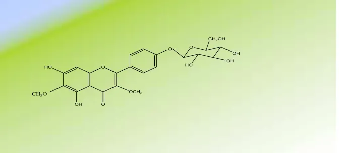
the aglycone 6-OH kaempferol (identified through Rf-values and 1H–NMR spectroscopy). The structure of the new compound (3) was confirmed through 1H–NMR spectral data which gave rise to the signals of kaempferol whereby two doublet signals appeared at δ 7.88 and 6.91ppm assigned to H-2`, 6` and H-3`, 5` with absence of H-3 and H-6 signals(which appeared at 6.7, 6.2) indicating their substitution at these positions and a doublet signals at 5.04 with J = 7.5Hz were assignable to an α-anomeric proton of a glucose moiety, beside the presence of two singlet signals at δ
3.7 and 3.8 ppm were assignable to the presence of 2 methoxy groups and the remaining sugar protons resonance in the spectrum appeared in the region from δ ppm 3.1-3.6. The chemical shift value of the anomeric glucose proton ensure that this moiety is not sited at the C-3 position of the aglycone moiety (it must be more downfield at ≈ 5.5 ppm or more)[38].The site of attachment of the two methoxy groupsto the aglycone moiety and for final confirmation of the chemical structure for this compound; 13C NMR analysis was therefore performed. The recorded spectrum revealed the presence of the two signals at 59.6 and 56.0 ppm proved the presence of two methoxy groups, in addition to signal of the anomeric glucose carbon at δ ppm 99.8 (glucose at 4`-position) and a downfield shift for C-6 at 131.2 sure that the methylation at that position and not glycosylation that appeared at 129.2 ppm. Also the upfield shift of C-4` at 157.9 ppm than the unsubstituted one (159.9) ppm , with the downfield shift of both 3` and 5`at 116.6 ppm than the unsubstituted one (115.4 ppm) confirmed the substitution of position-4`and the other carbon resonances in this spectrum possessed chemical shifts (see exp.) which also fitted well with the structure of (3) as 6-OH kaempferol-3,6-dimehylether-4`-glucopyranoside which is the first reported natural occurrence of this new compound.
Structure of 6-OH kaempferol-3,6-dimehylether-4`-glucopyranoside (3)
Results and discussion of the antioxidant activity:-
The results of DPPH inhibition by plant extract in comparison with BHA and TBHQ as standard antioxidants are represented in Table 1 and Fig. 1. Among the three doses used for Dodonaea viscosa extract (100, 200 and 300 µL), the results showed that a maximum scavenging activity was offered by 300 µL of Dodonaea viscosa extract with inhibition % of 82.09 ± 0.15, followed by 200 µL with inhibition % of 81.02 ± 0.11, and finally 100 µL with inhibition % of 79.91 ± 0.16.
Antioxidant activity of the extract with the three concentrations (100, 200 and 300 µL) was compared with the two standards (BHA and TBHQ), and from these results, we can conclude that the three concentrations of Dodonaea
viscosa extract showed strong antioxidant activity, higher than the standard BHA (inhibition % of 68.2), and lower
than the standard TBHQ (inhibition % of 98.2).
[image:5.595.162.498.290.442.2]Polyphenolic compounds are a class of antioxidant agents which act as free radical terminators [39,40]. The highest concentration (300 ml) of 80% ethanolic extract of Dodonaea viscosa had the highest antioxidant activity ensure that its phenolic compounds are responsible for its antioxidant activity.
Table 1: DPPH inhibition (%) of different concentrations of Dodonaea viscosa extract in comparison with BHA and TBHQ
Drug DPPH inhibition (%)
Standard antioxidant (BHA) 68.2
Standard antioxidant (TBHQ) 98.2
Dodonaea viscoseextract, 100 µL 79.91 ± 0.16 Dodonaea viscosa extract, 200 µL 81.02 ± 0.11 Dodonaea viscosa extract, 300 µL 82.09 ± 0.15
O
O
O
OH HO
OCH3
CH3O
O
OH
HO CH2OH
______________________________________________________________________________
0 10 20 30 40 50 60 70 80 90 100
Dodonaea viscosa
extract
BHA TBHQ
[image:6.595.138.489.92.254.2]100 200 300
Fig. 1: DPPH inhibition (%) of different concentrations of Dodonaea viscosa extract in comparison with BHA and TBHQ
Results And Discussion of the cytotoxic activity:-
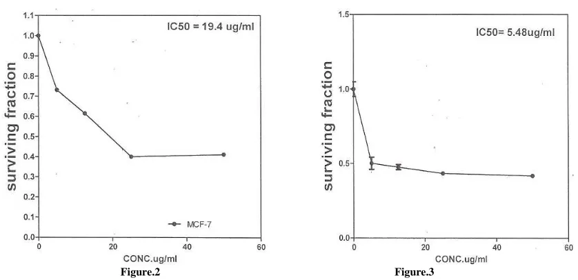
[image:6.595.216.396.381.490.2]The results of the cytotoxic activity on human tumor cell lines was determined according to the dose values of drug exposure required to reduce survival in the cell lines to 50% (IC50). Cytotoxic activities were examined on breast carcinoma cell line (MCF7). The results for ethanolic extract of Dodonaea viscosa compared with that of the standard used (Cisplatin) were recorded in Table 2. Plots of surviving fraction vs concentration in micrograms of the ethanolic extract of Dodonaea viscosa are shown in Fig.2, and that of the standard (Cisplatin) in Fig. 3. The results showed that the 80% ethanolic extract of Dodonaea viscosa has strong cytotoxic activity, as its IC50 is 19.4 µg/ml, compared with the standard used (Cisplatin), which its IC50 is 5.48 µg/ml.
Table 2: Cytotoxic activity of the ethanolic extract of Dodonaea viscose and the standard drug (Cisplatin)
MCF-7 Conc.µ/ml
Drugs
1.000 0.000
Standard drug (Cisplatin)
0.501 5.000
0.476 12.500
0.434 25.000
0.419 50.000
1.000 0.000
0.733 5.000
0.616 12.500
0.401 25.000
0.413 50.000
[image:6.595.105.524.518.721.2]Figure.2: Cytotoxic activity of the ethanolic extract of Dodonaea viscose Figure 3: Cytotoxic activity of the standard drug (Cisplatin)
Figure.2 Figure.3
Results and discussion for analgesic:-
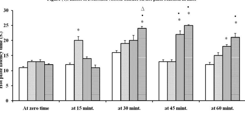
organized response and is useful in elucidating centrally mediated anti-nociceptive responses, which focuses mainly on changes above the spinal cord level. The anti-nociceptive properties of Dodonaea viscosa extract was investigated on mice using the hot plate test as the highest dose (200 mg/kg) used from the extract significantly (P< 0.05) increased hot plate time at 30, 45 and 60 min., when compared to control group as shown in Table 3 and Figure 4 which this indicated the analgesic activity of the extract.
The abdominal constrict ion response induced by acetic acid is a sensitive procedure to evaluate peripherally acting analgesics[41].In general, acetic acid causes pain by liberating endogenous substances such as serotonin, histamine, prostaglandins (PGs), bradykinins and substance P, endings[42].Results were obtained using this method as shown in Table 4 that the high dose 200 mg/kg of the extract exhibited analgesic as it significantly ( P<0.05) decreased the number of writhing response when compared with the control and the other groups.Phytochemical screening of the ethanolic extract of Dodonaea viscosa gave rise to the isolation of 14 flavonoids compounds as described before, which might be responsible for the analgesic activity Yadav et al., 2011[43] reported in the current investigation. Therefore, the overall results obtained suggested that the ethanolic extract of Dodonaea viscosemight relieve pain.
Table- 3: Effect of Dodonaea viscosa extract on hot plate reaction in mice
Group Dose (mg/kg)
P.O.
0 min
15 min
30 min
45
min 60 min
Group I Vehicle 11 ± 0.37 12 ± 0.56 16 ± 0.63 13 ± 0.63 12 ± 0.76
Group II (Diclofenac sodium) 10 13 ± 0.42 20 ± 1.28* 19 ± 0.97 13 ± 0.63 15 ± 0.97
Group III (Dodonaea viscosa extract) 100 13 ± 0.63 14 ± 0.56 20 ± 1.69 22 ± 1.17*● 18 ± 0.63*
Group IV (Dodonaea viscosa extract) 200 12 ± 0.37 11 ± 0.84 24 ± 0.65*●∆ 25 ± 0.21*● 21 ± 1.38*●
* Control ● Diclofenac ∆ Dose 100
[image:7.595.99.516.346.542.2]Figure (4): Effect of Dodonaea viscosa extract on hot plate reaction in mice
Table- 4: Effect of Dodonaea viscosa extract on acetic acid-induced writhings in mice
Treatment Dose (mg/kg, p.o.)a Writhing (n)b Inhibition (%)
Control (Vehicle, 10 ml/kg, p.o.) ….. 27 ± 0.42 ……
Diclofenac 10 19 ± 0.45* 29.63
Dodonaea viscosa extract 100 22 ± 1.48* 18.51 Dodonaea viscosa extract 200 14 ± 0.5*●∆ 48.15
a Administered 30 min before 0.6% acetic acid (60 mgykg, i.p.). b
Counted for 20 min after acetic acid injection; values are mean"S.E.M.; n=6. * Control ● Diclofenac ∆ Dose 100
REFERENCES
[1]AG Fakim. Mol. Aspect Med., 2006, 27(1), 1-93. [2]A Pasquale. J Ethnopharmacol,1984; 11(1):1-16.
[3]AS Rojas, S Cruz, H Ponce-Monter, R Mata. Planta Med,1996; 62:154-159.
[image:7.595.157.458.597.659.2]______________________________________________________________________________
[5]B Anilreddy.J Pharmaceut Sci Tech, 2009; 1:1-9.
[6]MS Rani, SP Rao , K Mohan. J Pharmaceut Res Health Care, 2009; 1:97-112. [7]A Shanmugavasana, T Ramachandran. J. Ethnopharmacol, 2011; 137:1172-1176.
[8]A Pengelly. Medicinal activity of Dodonaea viscosa—A preliminary study. Regional Industries Research and Development Corporation, RIRDC Publication No 08/172, RIRDC Project No PRJ-000703Australia, 2008.
[9]MN Khalil, SJ Sperotto, PM Manfron.Fitoterapia,2006; 77: 478 – 480. [10]J Meenu, S Sunil, K Manoj. Int J Pharm Pharmaceut Sci, 2011; 3: 69-74.
[11]VPVeerapur, KR Prabhakar, VK Parihar, PBansal, KK Srinivasan, KI Priyadarsini, MK Unnikrishnan. Int J
Phytomed, 2010; 2: 59–70.
[12]AB Cribb, JW Cribb. Wild medicine in Australia,collins, Sydney, 1981, pp. 228-237. [13]H Wagner, C Ludwig, L Grotjahn, MSY Khan. Phytochemistry, 1987; 26: 697-702.
[14]H Wagner. In Handbook of Medicinal plants(Ed, Yaniv, Z. B., U.) Haworth press, New York, 2005. [15]E L Ghisalberti. Fitotherapia, 1998; LXIX: 99-113.
[16]A SiddiquiA. Asian J. of Chemistry, 1998; 10: 14-16.
[17]M G Getie, R Rietz, RH Neubert.Ethiopian Pharmacy .J, 2000; 18: 25-34. [18]HY Hsu, Y P Chen, H Kakisawa.Phytochemistry, 1971; 10: 2813. [19]K Sachdev, DK Kulshreshtha.Planta Medica, 1984; 50: 448-449. [20]M Dimbi, C Delaude,M Kapundo, Bull.Soc.Chem. Belg,1985; 44: 141. [21]K N S Sastry, Y Nayudamma, Leather Sci, 1996; 13:174.
[22]H Wagner, C Ludwig, L Grotjahn, MSY Khan.Phytochemistry, 1987; 26: 697-702. [23]M Rachel, LC Jose. J. of Natural Products, 1991; 54: 913-917.
[24]A G Mekkawi, J S Mossa.Pharmazie, 1981; 36: 517. [25]R Hegnauer. Chemotaxonomie der Pflanzen, 1964, Vol. VI.
[26]K Sachdev, DK Kulshreshtha. Phytochemistry, 1983; 22: 1253-1256. [27]P Skehan, R Storeng.J Natl Cancer Inst, 1990; 82: 1107-1112.
[28]W Brand-Williams, ME Cuvelier, C Berset. Lebens. Wisse Techn, 1995; 28: 25–30.
[29]AM Badawi, MA Mohamed, MZ Mohamed, MM Khowdairy.J. Cancer Res. Ther, 2007; 3(4): 198-206. [30]GM Behera, M Baidya, BN Satish, S Bilal, S Panda. Res J Pharm Biol ChemSci,2011; 2(1):283-296. [31]RD Yadav, SK Jain, S Alok, S Sharma. Der Pharmacia Lettre,2011; 3(5):179-182.
[32]LT Nikajoo. Int J Pharm Pharm Sci,2009; 1(1):33-37.
[33]M Mohan, VS Gulecha, VM Aurangabadkar, R Balaraman, AAustin, SThirugnanasampathan. Oriental Pharm
Exp Med,2009; 9(3):232-237.
[34]HXiaojia, JHuizi, XWenzheng, ZZhang Wei, LXiaohua, YShikai, CMing, LJianqiang, ZWei-dong.Journal of
Ethanopharmacology, 2008; 120: 118-122.
[35]Koster AM,De Beer EJ: Acetic acid analgesic screening. Federation Proceedings, 1959; 18, 412. [36]CW Glennie, JB Harborne. Phytochemistry, 1971; 10:1325.
[37]El Sayed IE, Abd El Rahman AH, Richard R. Ann. Chem., 1990: 877.
[38]JB Harborne. The Flavonoids Advances in Research Since 1986. In: Markham KR, Geiger H (eds.). 1H nuclear magnetic resonance spectroscopy of flavonoids and their glycosides in hexadeuterodimethylsulfoxide, Chapman and Hall, London: 1993,pp. 441- 497.
[39]F Shahidi, PK wanasundara .J. Food sci. Nutr., 1992; 32:67-103. [40]D Huang, RL prior. J. Agric. Foodchem, 2005; 53:1841-1856. [41]RM Gené, L Segura, T Adzet. J. Ethnopharmacol,1989; 60: 157. [42]GA Bentley, SH Newton, J Starr. J. Pharmacol,1983; 79: 125.