The Helicobacter pylori VacA toxin is a urea
permease that promotes urea diffusion across
epithelia
Francesco Tombola, … , Mario Zoratti, Emanuele Papini
J Clin Invest.
2001;
108(6)
:929-937.
https://doi.org/10.1172/JCI13045
.
Urease and the cytotoxin VacA are two major virulence factors of the human pathogen
Helicobacter pylori
, which is responsible for severe gastroduodenal diseases. Diffusion of
urea, the substrate of urease, into the stomach is critically required for the survival of
infecting
H. pylori
. We now show that VacA increases the transepithelial flux of urea across
model epithelia by inducing an unsaturable permeation pathway. This transcellular pathway
is selective, as it conducts thiourea, but not glycerol and mannitol, demonstrating that it is
not due to a loosening of intercellular junctions. Experiments performed with different cell
lines, grown in a nonpolarized state, confirm that VacA permeabilizes the cell plasma
membrane to urea. Inhibition studies indicate that transmembrane pores formed by VacA act
as passive urea transporters. Thus, their inhibition by the anion channel blocker
5-nitro-2-(3-phenylpropylamino) benzoic acid significantly decreases toxin-induced urea fluxes in both
polarized and nonpolarized cells. Moreover, phloretin, a well-known inhibitor of eukaryotic
urea transporters, blocks VacA-mediated urea and ion transport and the toxin’s main
biologic effects. These data show that VacA behaves as a low-pH activated, passive urea
transporter potentially capable of permeabilizing the gastric epithelium to urea. This opens
the novel possibility that in vivo VacA may favor
H. pylori
infectivity by optimizing urease
activity.
Article
Find the latest version:
http://jci.me/13045/pdf
Introduction
Infection by Helicobacter pylori (1, 2) determines an increased incidence of gastritis, gastroduodenal ulcer, and gastric adenocarcinoma and lymphomas in humans. The colonization of the stomach by this microorganism requires complex adaptation process-es. Among them, a key feature is acid tolerance allow-ing the bacterium to cross the stomach lumen (pH 1–2) and to survive within the mucous layer covering the gastric epithelium (pH range, 1–2 to 6). Cell death by acidification is prevented by a cytosolic urease, which converts urea into ammonia and bicarbonate (1–6). These products of urea hydrolysis are also metabolic substrates for H. pylori(7, 8). The importance of urea availability for H. pyloriis confirmed by the presence of an acid-activated passive urea transporter (UreI) in the bacterial inner membrane (9, 10) and by positive chemotaxis of the parasite toward urea (11, 12).
Besides urease, efficient gastric colonization by H. pylorialso requires the cytotoxin VacA (13). This approx-imately 88-kDa secreted protein is a major virulence and pathogenic factor, linked to severe gastric tissue damage
(14). In vitro, VacA needs activation by acidic pH pre-treatment (15) and induces cell vacuolation and various cytotoxic effects (2, 14, 16). VacA forms anion-selective channels in model and cell membranes (17–20). Howev-er, the in vivo relevance of these phenomena for H. pylori
gastric infection and alteration is still undefined. We investigated whether VacA cooperates with urease by increasing the diffusion of urea from the mucosal tissue to the stomach lumen. This hypothesis is partly based on previous studies suggesting that VacA’s func-tion in vivo is the selective permeabilizafunc-tion of the gas-tric epithelium to nutrients and other useful mole-cules, by altering cell-cell junctions (21, 22) and/or cell membranes (17, 20). By using polarized and nonpolar-ized cells and model membranes, and by combining functional, pharmacological, and electrophysiological approaches, it is shown that VacA determines a rapid and selective increase of epithelial permeability to urea, through the generation of a passive and unsaturable transcellular pathway. Evidence is provided that for-mation of VacA pores in the cell plasma membrane is responsible for this increased urea permeability.
The Helicobacter pylori VacA toxin is a
urea permease that promotes urea
diffusion across epithelia
Francesco Tombola,
1Laura Morbiato,
1Giuseppe Del Giudice,
2Rino Rappuoli,
2Mario Zoratti,
1and Emanuele Papini
31Centro Consiglio Nazionale delle Ricerche Biomembrane, Department of Biomedical Sciences,
University of Padova, Padova, Italy
2Immunobiology Research Institute Siena, Siena, Italy
3Department of Biomedical Sciences and Human Oncology, Section of General Pathology,
University of Bari, Bari, Italy
Address correspondence to: Emanuele Papini, Department of Biomedical Sciences and Human Oncology, Section of General Pathology, University of Bari, Piazza Giulio Cesare 11, 70124 Bari, Italy.
Phone: 39-080-5478465; Fax: 39-080-5478471; E-mail: [email protected].
Received for publication April 16, 2001, and accepted in revised form July 24, 2001.
Urease and the cytotoxin VacA are two major virulence factors of the human pathogen Helicobacter pylori, which is responsible for severe gastroduodenal diseases. Diffusion of urea, the substrate of ure-ase, into the stomach is critically required for the survival of infecting H. pylori. We now show that VacA increases the transepithelial flux of urea across model epithelia by inducing an unsaturable per-meation pathway. This transcellular pathway is selective, as it conducts thiourea, but not glycerol and mannitol, demonstrating that it is not due to a loosening of intercellular junctions. Experiments per-formed with different cell lines, grown in a nonpolarized state, confirm that VacA permeabilizes the cell plasma membrane to urea. Inhibition studies indicate that transmembrane pores formed by VacA act as passive urea transporters. Thus, their inhibition by the anion channel blocker 5-nitro-2-(3-phenylpropylamino) benzoic acid significantly decreases toxin-induced urea fluxes in both polarized and nonpolarized cells. Moreover, phloretin, a well-known inhibitor of eukaryotic urea transporters, blocks VacA-mediated urea and ion transport and the toxin’s main biologic effects. These data show that VacA behaves as a low-pH activated, passive urea transporter potentially capable of permeabi-lizing the gastric epithelium to urea. This opens the novel possibility that in vivo VacA may favor H. pyloriinfectivity by optimizing urease activity.
J. Clin. Invest. 108:929–937 (2001). DOI: 10.1172/JCI200113045.
Methods
Reagents. VacA from strain CCUG 17874 was purified as described previously (23) and was stored at 4°C in PBS (pH 7.4). VacA was activated by treatment at pH 2.0, 37°C for 3 minutes. AGS and MKN28 cells were a kind gift from V. Ricci (Institute of Human Physiolo-gy, University of Pavia, Pavia, Italy). Plastic-ware, cul-ture media, and FCS were from Life Technologies (Milan, Italy). Transwell polycarbonate filters for in vitro epithelial differentiation (pore diameter, 0.4 µm) were from Corning-Costar Corp. (Cambridge, Massa-chusetts, USA). [14C]Urea (57 mCi/mmol), D-[14C]mannitol (56 mCi/mmol), and [1(3)-3H]glycerol (3 Ci/mmol) were from Amersham Pharmacia Biotech (Milan, Italy). [14C]Thiourea (53 mCi/mmol) was from NEN Life Science Products (Boston, Massachusetts, USA). The 5-nitro-2-(3-phenylpropylamino) benzoic acid (NPPB), phloretin, parachloromercury-benzen-sulfonate (pCMBS), ouabain, 8Br-cAMP, 8Br-cGMP, collagenase A1, thiourea, and urea were from Sigma-Aldrich srl (Milan, Italy). Synthetic diphytanoyl-phos-phatidylcholine (purity >99%) was from Avanti Polar Lipids (Alabaster, Alabama, USA). Urea solutions were made freshly before experiments.
Cell culture. Cells were grown on plastic at 37°C in a humidified 5% (vol/vol) CO2atmosphere in DMEM containing 10% (vol/vol) FCS and gentamycin. Trypsin-EDTA detached cells were seeded on polycarbonate
fil-ters or plastic wells at the density of 0.5 ×10⁶/cm2and further kept in culture medium as indicated above. Epithelial differentiation was obtained after culturing on filters for 14 days, whereas nondifferentiated cells were cultured on plastic for 2 days before experiments.
Measurement of transepithelial fluxes. Monolayers on fil-ters were washed with fresh culture medium and treat-ed with acid-activattreat-ed or nonactivattreat-ed VacA (15–125 nM) diluted in the same medium. Controls were treat-ed in the same way with equivalent aliquots of PBS. After 0.25–27.0 hours of incubation, unbound toxin was washed out by rinsing with DMEM without phe-nol red, 0.023% BSA (wt/vol), 10 mM Na-HEPES (pH 7.4), and incubation was continued in the same medi-um at 37°C, in normal atmosphere, after placing radi-olabeled tracers (45 µM solutes or 2 µCi/ml in the case of [3H]water) in the basolateral chamber. After the desired time intervals, the apical medium was retrieved and replaced by prewarmed medium. Radioactivity was determined by liquid scintillation and flows expressed as pmol/hcm2of cell monolayer. Ion con-ductivity was measured at 37°C with a Millipore Corp. apparatus (Bedford, Massachusetts, USA). In some experiments, unlabeled urea and/or thiourea (100–400 mM), NPPB, pCMBS, or phloretin (50–500 µM) was included in both apical and basolateral chambers dur-ing the transepithelial diffusion assays, or durdur-ing VacA intoxication, as specified.
Measurement of transmembrane fluxes. Cells in plastic wells were intoxicated, or not, with VacA as described above. In some cases, cells were washed twice with ice-cold PBS (pH 7.4), containing 0.023% (wt/vol) BSA (PBS-BSA) and further incubated with radiolabeled solutes (45 µM) in the same medium at 4°C for differ-ent time intervals (2.5–15 minutes). Radioactivity asso-ciated to cells was determined after three washings (washing time, ∼20 seconds) with PBS-BSA (4°C) and cell lysis in PBS-BSA plus 0.5% (wt/vol) SDS. Alterna-tively, after intoxication, cells were washed with cul-ture medium and further incubated at 37°C in the same conditions for 1 hour in the presence of labeled tracers (45 µM). Cells were then rapidly washed and further incubated in PBS-BSA at 4°C. The radioactiv-ity present in the extracellular medium was deter-mined at various time points (2.5–15 minutes). In some experiments, AGS and HeLa cells were depleted of ATP (24): cells were pretreated at 37°C with 1 mg/ml NaN3for 30 minutes and for an additional 30 minutes with 1 mg/ml NaN3and 13 mM 2-deoxy-D-glucose in 2-deoxy-D-glucose- and pyruvate-free DMEM plus 10% (vol/vol) dialyzed FCS, and further incubated with VacA in the same conditions, before determining transmembrane urea fluxes.
[image:3.576.60.289.54.242.2]In other experiments, control or VacA-intoxicated HeLa cells were pretreated with 1 mM 8Br-cAMP or 1 mM 8Br-cGMP or 0.250 mM ouabain for 30 minutes, and urea fluxes were then measured in the presence of the same agonists. Alternatively, during urea flux measurement, NaCl in the extracellular medium was
Figure 1
replaced by isoosmotic mannitol (25). Oocytes isolat-ed from Xenopus laevis ovary were treated with 2 mg/ml collagenase A1 in OR-2 buffer for 2 hours at 25°C and then defolliculated manually. After an overnight recovery at 20°C, oocytes were treated or not with acid activated VacA at 24°C for 4 hours, and the uptake and release of 14C-urea were determined as described (25) in Barth’s buffer (88 mM NaCl, 1 mM KCl, 0.82 mM MgSO4, 0.33 mM Ca(NO3)2, 0.41 mM CaCl2, 2.4 mM NaHCO3, 10 mM HEPES/Na [pH 7.4], 10 U/ml penicillin, and 10 µg/ml streptomycin) at 24°C. Phloretin (200 µM) was included in the urea flux experiments in some cases.
Cell vacuolation. Cells were seeded in 24-wells trays at the density of 103/cm22 days before experiments and then treated with 50 nM VacA preactivated at pH 2.0 in DMEM containing 5 mM NH4Cl and various concen-trations of phloretin (0–480 µM) at 37°C. After 6 hours, cell vacuolation was quantified using the neu-tral red uptake assay as described previously (15).
Measurement of VacA-induced ion current in planar lipid bilayers. Experiments were performed as described pre-viously (17), with 0.5 M KCl, 0.5 mM CaCl2, 0.5 mM MgCl2, and 10 mM HEPES/K (pH 7.2) in both cham-bers. VacA (2–4 nM) activated at a low pH (2.0) was added to the ciscompartment, and current was
deter-mined with or without addition to either the cis or the
transside of inhibitors (0–200 µM phloretin; 0–500 µM pCMBS; 0–400 mM thio-urea and urea). The current was carried mainly by Cl–, as VacA is anion selective (17).
Results
VacA increases the transepithelial diffusion of urea. Polar-ized epithelia in vitro mimic the watertight epithelia present in the kidney, the bladder and, most relevant for the present study, the gastric mucosa (26). Tight-ness is associated with the development of a
differen-Table 1
Effect of VacA on the diffusion rate (pmol/hcm2) of various mole-cules across MDCK II monolayers
CTRL VacA VacA-CTRL
Urea 597 ± 45 1681 ± 98 1084 ± 143
Thiourea 841 ± 89 2848 ± 103 2007 ± 192
Glycerol 601 ± 67 701 ± 18 100 ± 85
Mannitol 840 ± 61 862 ± 72 22 ± 133
[image:4.576.132.461.49.314.2]The medium bathing the basolateral side of MDCK II monolayers was sup-plemented with the indicated radioactive tracers (45 µM), and radioactivity in the apical chamber was measured as a function of time. Transepithelial dif-fusion of control- and 125 nM of VacA–treated monolayers and their differ-ence are reported, expressed as pmol/hcm2± SE (n= 3 or 4).
Figure 2
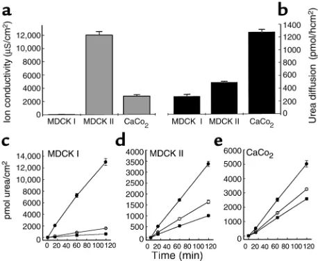
[image:4.576.304.536.621.686.2]tiated apical membrane, enriched in cholesterol and sphingolipids (27, 28). Polarized MDCK I, MDCK II, and CaCo2monolayers were used to test the effect of VacA on the transepithelial diffusion of urea and other small neutral molecules. Their basal resistance to urea diffusion was compared with transepithelial electrical resistance, a parameter largely dependent on the tightness of cell-cell junctions (the paracellular pathway). Data show that the epithelial permeabilities to urea and to ions do not correlate (Figure 1, a and b), strongly suggesting that the epithelial barrier to
urea in our model systems is mostly represented by cell membranes, i.e., the transcellular pathway, in agreement with other studies (26, 28).
After intoxication for 3 hours with low pH–activated VacA (125 nM), added to the apical compartment to mimic the in vivo conditions, urea diffusion increased in all tested epithelia (Figure 1, c, d, and e). The strongest effect (∼20-fold increment with respect to control epithe-lia) was recorded with MDCK I monolayers, whereas with MDCK II and CaCo2 cells, the effect was less pro-nounced (approximately two and three times the control value, respectively) but still significant. In absolute terms, the VacA effect with these two latter cell lines was 20–24% of that observed with MDCK I cells. Nonacti-vated VacA was much less effective in all cases.
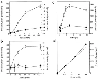
Dose-response analysis of the effect of VacA on urea transepithelial flux and on the epithelial ion conductiv-ity was performed with MDCK I and MDCK II epithelia after a 3-hour intoxication (Figure 2, a and b). In the for-mer case, the VacA effect reached a maximal value at approximately 130 nM, and the half-maximal increase was obtained at approximately 40 nM. In the latter, a plateau was reached at approximately 60 nM, and the half-maximal concentration was approximately 20 nM. The epithelial ion conductivity of MDCK I monolayers increased by a factor of four with 125 nM VacA, in agree-ment with previous observations (21, 22). However, at lower VacA doses (<16 nM), the increase of ion conduc-tivity was negligible, whereas urea flux was already four times higher than in control. In agreement with our pre-vious data (21), VacA did not alter the TER of CaCo2 (data not shown) and MDCK II monolayers. These data suggest that VacA increases the transepithelial diffusion of urea by activating a transcellular pathway, although some contribution of the paracellular pathway probably occurs with MDCK I cells at high VacA doses (see below). Urea diffusion through MDCK II epithelia increased immediately after VacA addition and reached a maxi-mum (threefold increase) after approximately 7–8 hours. Urea flow remained high for the following approximately 15 hours (Figure 2c). Nonactivated VacA had a lower effect even after a long incubation time.
To test whether the urea transport system induced by VacA was saturable, the flow of [14C]urea was measured with MDCK II monolayers in the presence of increas-ing concentrations of unlabeled urea (Figure 2d). Urea diffusion increased almost linearly up to 400 mM, con-sistent with a passive unsaturable urea transport.
Table 1 compares the effect of VacA on the transep-ithelial flow of urea and other small neutral molecules across MDCK II monolayers. The diffusion of thiourea was efficiently increased by VacA, whereas that of glyc-erol and mannitol was scarcely affected. As expected (21) mannitol diffusion was increased in MDCK I monolayers, but with a much lower efficacy (∼6% of urea flow) (data not shown). The transepithelial flow of 3H-H
2O was not modified (data not shown).
[image:5.576.63.287.51.403.2]VacA increases the plasma membrane permeability to urea. The data described above suggest a VacA-elicited
Figure 3
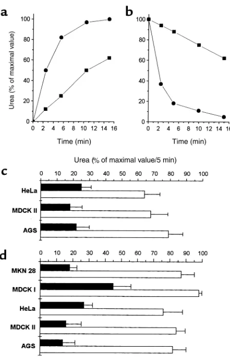
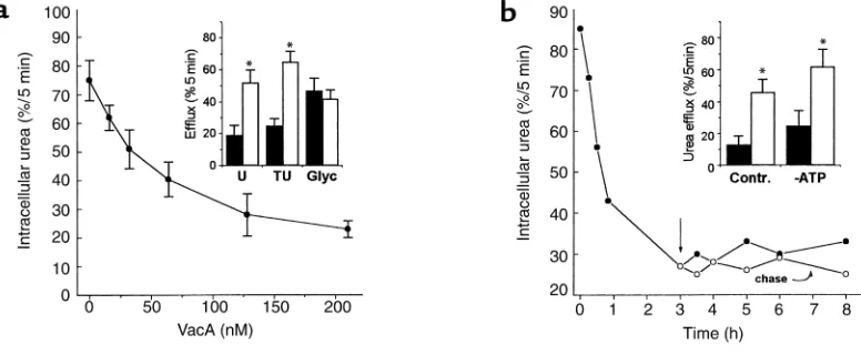
increase of the plasma membrane permeability to urea. Indeed, VacA increased the rate of urea penetration into nonpolarized human gastric cells (AGS), as well as its efflux after previous equilibration (Figure 3, a and b). The permeability to urea of the plasma membrane of nonpolarized MDCK I, MDCK II, HeLa, and MKN28 cells was also increased by VacA treatment (Figure 3, c and d). A dose response performed with AGS cells, after 3 hours of intoxication (Figure 4a), showed that VacA-induced permeabilization of the plasma membrane reached a plateau at approximately 125 nM, with a half maximal effect at approximately 30 nM. Similar data were obtained with nonpolarized MDCK II (data not shown). As in polarized epithelia, VacA also increased the transmembrane diffusion of thiourea, but not of glycerol (Figure 4a, inset). The effect of VacA on nonpo-larized AGS cells (Figure 4b) was already strong after 15 minutes and almost maximal after 1 hour. In the fol-lowing 7 hours, urea transmembrane flux remained constant, and it was not reversed by toxin washout (chase). A similar time course of permeabilization was found when urea uptake by cells was monitored (data not shown). The effect of VacA on AGS (Figure 4b, inset) and HeLa cells (data not shown) was not inhibited by the block of its endocytosis, after ATP depletion (24). Neither the basal nor the VacA-induced urea fluxes could be modified by the presence of ouabain (0.25 mM) or of 8Br-cAMP (1 mM) and 8Br-cGMP (1 mM) (data not shown). Replacement of NaCl (150 mM) in the urea assay medium with 300 mM mannitol did not affect VacA-induced urea fluxes (data not shown).
VacA-induced increase of transepithelial and transmembrane urea diffusion is due to the VacA pore. Anion-channel and urea transporter blockers were used to investigate the
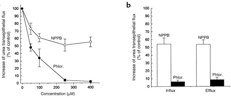
[image:6.576.104.492.466.626.2]mechanism of VacA-induced urea permeation. NPPB, a blocker of the VacA pore (20, 29), inhibited toxin-induced transepithelial flux of urea in MDCK II cell monolayers to a maximal extent of about 55% of con-trols, with a half-maximal concentration of about 50 µM (Figure 5a). Similarly, NPPB partially inhibited the plasma membrane permeabilization to urea induced by VacA in nonpolarized AGS cells (Figure 5b). Phloretin, a blocker of eukaryotic urea transporters (30), effi-ciently inhibited the increase of both transepithelial and transmembrane urea diffusion induced by VacA (Figure 5, a and b). The transmembrane urea flux of isolated oocytes from Xenopus laevis, intrinsically devoid of specific urea transport (31), was also increased (35 ± 4%; n = 3) by VacA (700 nM for 4 hours at 24°C), and this effect was phloretin sensitive (data not shown). Phloretin inhibited cell vacuolation of nonpolarized cells treated with VacA in the presence of ammonium ions (Figure 6a). The pH of acidic compartments in untreated cells was not altered by phloretin, as judged from the basal uptake of neutral red (data not shown). This compound also prevented the VacA-induced increase of ion conductivity of MDCK I cell monolay-ers (Figure 6b). In this epithelial system, urea flux was also inhibited by phloretin, with characteristics depending on the intoxication protocol (Figure 6c). If phloretin was coincubated with VacA and then washed out, or if it was added after intoxication, during the assay for urea transepithelial flux, a partial inhibition was observed. On the other hand, if it was maintained during both the intoxication and assay steps, a strong inhibition was observed, roughly corresponding to the sum of the effects elicited by the other protocols. This is consistent with inhibition by phloretin of both the
Figure 4
VacA-induced transcellular urea diffusion pathway and alteration of the paracellular pathway.
These data strongly suggest that phloretin directly interacts with the toxin. To test this possibility, its effect on the VacA anion channel was assessed in planar lipid bilayer experiments. Figure 7a shows that phloretin inhibited the VacA-induced ion current, with an IC50of approximately 15 µM. Thiourea (Figure 7b) and pCMBS (data not shown), two other molecules known to affect passive urea transporters (32–34), were also tested. Thiourea (400 mM) determined a 30% (trans) or
a 45% (cis) inhibition of ion conduction by VacA chan-nels in planar lipid bilayers, depending on the side of addition. Urea (400 mM) produced a weaker 5% inhibi-tion. Consistently, 400 mM thiourea inhibited VacA-induced [14C]urea diffusion across MDCK II monolay-ers by 70%, whereas 400 mM urea was ineffective. pCMBS (up to 250 µM) had no effect in either assay.
Discussion
Although recent data indicate that H. pylori relays in part upon the cytotoxin VacA for a successful
coloniza-Figure 5
[image:7.576.107.482.50.209.2]Effect of NPPB and phloretin (Phlor.) on the increase of transepithelial and transmembrane urea flux due to VacA. (a) Polarized MDCK II monolayers were treated with 125 nM activated VacA for 3 hours and then washed and further assayed for [14C]urea transepithelial flow in the presence of the indicated concentrations of NPPB or phloretin (added to both apical and basolateral compartments). (b) Nonpolarized AGS cells were intoxicated by 125 nM of VacA at 37°C for 3 hours, and assayed for initial rate of [14C]urea cell influx and efflux, in the pres-ence of 250 µM phloretin or NPPB. Data are expressed as percent of the increase observed in controls (+VacA, no inhibitor). Data are the mean of three experiments run in duplicate ± SE.
Figure 6
[image:7.576.73.508.458.635.2]tion of the stomach (17), a clear link between the toxin biologic effects in vitro and its role in vivo is missing. The data presented here suggest that, in vivo, VacA action consists of enhancing the diffusion of urea from the under-mucosal tissue toward the stomach lumen. In fact, the epithelial barrier to urea diffu-sion is selectively decreased by VacA through the generation of a passive, unsaturable transport sys-tem. This action is not related to a decrease of the resistance of the paracellular pathway and is observed not only in high-resistance epithelia, such as MDCK I monolayers, but also in low-resistance ones, such as CaCo2 and MDCK II monolayers. These data indirectly suggest that, upon VacA intox-ication, transcellular urea flow is increased. Experi-ments conducted with nonpolarized cells indeed indicate that VacA permeabilizes the cell plasma membrane to urea with negligible nonspecific mem-brane lesion, as the diffusion of other neutral mole-cules, such as is much less affected.
The rapid onset of the effect, not compatible with the endocytic rate of the toxin (24), and the lack of inhibi-tion by block of endocytosis, indicate that VacA acts directly on the plasma membrane.
Although we cannot exclude that in polarized epithelial cells the basolateral membrane is also per-meabilized by VacA, any such effect is likely to be irrel-evant. This is because it is the apical membrane domain that poses the highest resistance to the flow of small nonelectrolytes (26, 35), owing to its external emi-leaflet particularly enriched in cholesterol, sphin-gomyelin, and glycosphingolipids (26–28, 35, 36). In
MDCK cells, it has been estimated that the intrinsic resistance to urea of the apical emi-leaflet is about 50 times higher than that of the cytosolic emi-leaflet and much higher than that of the basolateral membrane (28). This implies that permeabilization of this lipid layer by VacA is necessary and sufficient to enhance urea flow through the entire epithelium.
[image:8.576.100.508.442.615.2]In theory, VacA might permeabilize the plasma mem-brane to urea by activating an endogenous, urea trans-porter. Alternatively, given that low-pH activated VacA inserts into the lipid bilayer to form a transmembrane pore (17, 18, 20), VacA itself may act as a passive urea transporter. In fact, the pharmacology of the VacA-induced effect, that is, the sensitivity to phloretin and the insensitivity to PCMBS, is compatible with both possibilities (30). The data presented in this work sup-port this second view. In fact, inhibition of the VacA channel by NPPB (20, 29) substantially reduces also the urea transepithelial and transmembrane permeability increases determined by the toxin. Phloretin, a blocker of urea transporters, strongly reduces both urea diffu-sion and ion conduction due to the toxin, with compa-rable dose responses. As indicated by the lack of inhibitory effect of ouabain and of the absence of extra-cellular ions on VacA-dependent urea fluxes, there is no coupling between ion and urea transport, thus ruling out the indirect activation of an ion-coupled urea transporter by VacA. A further indication that VacA-induced urea flow is not coupled to Na+comes from the similarity of influx and efflux kinetics in cellular systems (Figure 3), despite the presence of a Na+ elec-trochemical gradient.
Figure 7
As in the case of Xenopusoocytes, in which urea trans-porters are often expressed and characterized, no evi-dence of cAMP-activatable urea transporters was found in HeLa (see Results) or in MDCK cells (28). The VacA-induced urea flux was not modulated by cAMP either. Experiments on oocytes, believed to lack specific urea transporters, show that VacA induces a phloretin-sen-sitive increase of urea transmembrane flux also in this system, strengthening the notion that it acts as an autonomous transporter. In addition, the indirect acti-vation of an aquaporin able to conduct also urea seems unlikely. The only mammalian aquaporin known not to be inhibited by thiol reagents is AQP-4, which does not conduct urea (37). In our model epithelial system, the transepithelial flow of water was not detectably increased by VacA intoxication.
In agreement with current ideas on VacA action (1), phloretin also interferes with VacA-induced cell vac-uolation and TER decrease in MDCK I monolayers. Thiourea is conducted by the VacA-induced trans-port system and, at high concentrations, interferes with urea flow, suggesting that the two molecules enter the same pathway. Similarly, thiourea and, to minor extent, urea reduce ion conduction by VacA, indicating that these molecules engage the lumen of the VacA anion channel, interfering with ion flow.
A question that needs to be addressed is whether urea is limiting for H. pyloriin the gastric environ-ment. Although the stomach contains 1- to 3-mM urea, time- and space-dependent fluctuations of urea concentration may occur, as suggested by positive chemotaxis toward urea by H. pylori(11, 12). More-over, urease activity itself is expected to determine a local urea shortage, especially in case of acid stress (pH < 6.5) (9). Although an accurate analysis of the system is impossible at present, the surface density of
H. pyloricells in mouse models (105–106 CFU/stom-ach; ref. 13) and the measured rate of urea consump-tion by maximally activated urease (0.38–0.6 µmol/min 108 CFU; ref. 6) are compatible with a decrease of urea concentration within the thin mucous layer (< 50 µm) where bacterial cells reside. Under conditions of local urea depletion, the diffu-sion rate through the gastric barrier would be expect-ed to be limiting, as our data show that basal urea equilibration through polarized cells takes hours. Hence, it is possible that the establishment of a high density of infecting cells is favored by an increased urea diffusion through the gastric mucosa.
These observations and considerations support the novel hypothesis that VacA, by increasing urea diffu-sion into the stomach, is part of an adaptation system, also comprising the urea transporter UreI and urease, aimed at enabling H. pylorito survive in the stomach. The evolution of pore-forming toxins as permeases selective for crucial substrates, capable of inserting in the membrane of host cells with no acute cytotoxicity, may be a relevant strategy also of other human pathogens associated to mucosal epithelia.
Gastric mucosa permeabilization may have other implications. Besides urea, the VacA transport system also conducts electrolytes and possibly other neutral solutes. As suggested by the loss of gastric integrity induced by barrier-breaking agents (ethanol, aspirin, detergents, phospholipases), this might predispose to tissue damage, given that the gastric epithelial barrier preserves the entire mucosa from the potentially dele-terious effects of ingested food of extremely variable chemical composition and osmolarity (35). In conclu-sion, apical membrane permeabilization by VacA may favor H. pylori-associated diseases both by promotion of bacterial infection and by impairment of the capa-bility of gastric cells to regulate their cytosolic milieu upon environmental variations.
Acknowledgments
The authors are grateful to Stefano Piccolo and his lab-oratory (University of Padua) for help with performing experiments with Xenopusoocytes. This research was supported by Cofin MURST 2000, by the University of Bari, and by CNR target project Biotechnology (97.01168.PF 49).
1. Montecucco, C., and Rappuoli, R. 2001. Living dangerously: how Heli-cobacter pylorisurvives in the human stomach. Nat. Rev. Mol. Cell Biol. 2:457–466.
2. Achtman, M., and Suerbaum, S. 2001. Helicobacter pylori: molecular and cel-lular biology. Horizon Scientific Press. Wymondham, United Kingdom. 3. Marshall, B.J., Barrett, L.J., Prakash, C., McCallum, R.W., and Guerrant, R.L. 1990. Urea protects Helicobacter(Campylobacter) pylorifrom the bac-tericidal effect of acid. Gastroenterology.99:697–702.
4. Bauerfeind, P., Garner, R., Dunn, B.E., and Mobley, L.T. 1997. Synthesis and activity of Helicobacter pyloriurease and catalase at low pH. Gut.
40:25–30.
5. Scott, D.R., et al. 1998. The role of internal urease in acid resistance of
Helicobacter pylori. Gastroenterology. 114:58–67.
6. Stingl, K., et al. 2000. Prolonged survival and cytoplasmic pH home-ostasis of Helicobacter pyloriat pH 1. Infect. Immun.69:1178–1181. 7. Garner, R.M., Fulkerson, J., Jr, and Mobley, H.L. 1998. Helicobacter pylori
glutamine synthetase lacks features associated with transcriptional and posttranslational regulation. Infect. Immun.66:1839–1847.
8. Burns, B.P., Hazell, S.L., and Mendz, G.L. 1995. Acetyl-CoA carboxylase activity in Helicobacter pyloriand the requirement of increased CO2for
growth. Microbiology.141:3113–3118.
9. Gordon, D. 2000. A pH-sensitive channel regulates urea access to Heli-cobacter pyloriurease. Gastroenterology. 118:249–250.
10. Weeks, D.L., Eskandari, S., Scott, R.D., and Sachs, G. 2000. A H+-gated
urea channel: the link between Helicobacter pyloriurease and gastric col-onization. Science. 287:482–485.
11. Nakamura, H., et al. 1998. Urease plays an important role in the chemo-tactic motility of Helicobacter pyloriin a viscous environment. Infect. Immun.66:4832–4837.
12. Mizote, T., Yoshiyama, H., and Nakazawa, T. 1997. Urease-independent chemotactic responses of Helicobacter pylorito urea, urease inhibitors, and sodium bicarbonate. Infect. Immun. 65:1519–1521.
13. Salama, N.R., Otto, G., Tompkins, L., and Falkow, S. 2001. Vacuolating cytotoxin of Helicobacter pyloriplays a role during colonization in a mouse model of infection. Infect. Immun. 69:730–736.
14. Montecucco, C., Papini, E., de Bernard, M., Telford, J.L., and Rappuoli, R. 1999. Helicobacter pylorivacuolating cytotoxin and associated patho-genic factors. In The comprehensive sourcebook of bacterial protein toxins. J.E. Alouf and J.H. Freer, editors. Academic Press Inc. San Diego, California, USA. 264–283.
15. de Bernard, M., et al. 1995. Low pH activates the vacuolating toxin of
Helicobacter pylori, which becomes acid and pepsin resistant. J. Biol. Chem.
270:23937–23940.
16. Reyrat, J.-M., et al. 1999. Towards deciphering the Helicobacter pylori cyto-toxin. Mol. Microbiol.34:197–204.
1999. VacA from Helicobacter pylori: a hexameric chloride channel. FEBS Lett. 450:101–104.
19. Czajkowsky, D.M., Iwamoto, H., Cover, T.L., and Shao, Z. 1999. The vac-uolating toxin from Helicobacter pyloriforms hexameric pores in lipid bilayers at low pH. Proc. Natl. Acad. Sci. USA. 96:2001–2006.
20. Szabò, I., et al. 1999. Formation of anion-selective channels in the cell plasma membrane by the toxin VacA of Helicobacter pyloriis required for its biological activity. EMBO J.18:5517–5527.
21. Papini, E., et al. 1998. Selective increase of the permeability of polarized epithelial cell monolayers by Helicobacter pylorivacuolating toxin. J. Clin. Invest. 102:813–820.
22. Pelicic, V., et al. 1999. Helicobacter pyloriVacA cytotoxin associated with the bacteria increases epithelial permeability independently of its vac-uolating activity. Microbiology.145:2043–2050.
23. Manetti, R., et al. 1995. Helicobacter pyloricytotoxin: importance of native conformation for induction of neutralizing antibodies. Infect. Immun. 63:4476–4480.
24. McClain, M.S., Schraw, W., Ricci, V., Boquet, P., and Cover, T.L. 2000. Acid activation of Helicobacter pylorivacuolating cytotoxin (VacA) results in toxin internalization by eukaryotic cells. Mol. Microbiol.37:433–442. 25. You, G., et al. 1993. Cloning and characterisation of the
vasopressin-reg-ulated urea transporter. Nature. 365:844–847.
26. Zeidel, M.L. 1996. Low permeabilities of apical membranes of barrier epithelia: what makes watertight membranes watertight? Am. J. Physiol.
271:F243–F245.
27. Lavelle, J.P., et al. 1997. Low permeabilities of MDCK cell monolayers: a model barrier epithelium. Am. J. Physiol. 273:F67–F75.
28. Hill, W.G., and Zeidel, M.L. 2000. Reconstituting the barrier properties
of water-tight epithelial membrane by design of leaflet-specific lipo-somes. J. Biol. Chem.275:30176–30185.
29. Tombola, F., et al. 1999. Inhibition of the vacuolating and anion chan-nel activities of the VacA toxin of Helicobacter pylori. FEBS Lett.
460:221–225.
30. Sands, J.M., Timmer, R.T., and Gunn, R.B. 1997. Urea transporters in kidney and erythrocytes. Am. J. Physiol.273:F321–F339.
31. Zhang, R., and Verkman, A.S. 1991. Water and urea permeability prop-erties of Xenopusoocytes: expression of mRNA from toad urinary blad-der. Am. J. Physiol. 260:C26–C34.
32. Couriaud, C., Ripoche, P., and Rousselet, G. 1996. Cloning and func-tional characterisation of a rat urea transporter: expression in the brain.
Biochim. Biophys. Acta.1309:197–199.
33. Martial, S., et al. 1996. Functional differentiation of human red blood cell and kidney urea transporters. Am J. Physiol.271:F1264–F1268. 34. Couriaud, C., et al. 1999. Molecular and functional characterization of
an amphibian urea transporter. Biochim. Biophys. Acta.1421:347–352. 35. Priver, N.A., Rabon, E., and Zeidel, M.L. 1993. Apical membrane of the gastric parietal cell: water, proton, and nonelectrolyte permeabilities. Bio-chemistry. 32:2459–2468.
36. Van Meer, G., and Simons, K. 1986. The function of tight junctions in maintaining differences in lipid composition between the apical and the basolateral cell surface domains of MDCK cells. EMBO J.5:1455–1464. 37. Borgnia, M., Nielsen, S., Engel, A., and Agre, P. 1999. Cellular and molec-ular biology of the aquaporin water channels. Annu. Rev. Biochem.
68:425–458.