0095-1137/97/$04.00
1
0
Copyright © 1997, American Society for Microbiology
Evaluation of a Fluorescence-Labelled Oligonucleotide Probe
Targeting 23S rRNA for In Situ Detection of
Salmonella
Serovars in Paraffin-Embedded Tissue Sections and
Their Rapid Identification in Bacterial Smears
STEEN NORDENTOFT,
1* HENRIK CHRISTENSEN,
2AND
HENRIK CASPAR WEGENER
1Danish Veterinary Laboratory, DK-1790 Copenhagen V,
1and Department of Veterinary Microbiology,
Royal Veterinary and Agricultural University, DK-1870 Frederiksberg C,
2Denmark
Received 24 January 1997/Returned for modification 3 March 1997/Accepted 7 July 1997
A method for the detection of
Salmonella
based on fluorescence in situ hybridization (FISH) has been
developed and applied for the direct detection of
Salmonella
in pure cultures and in formalin-fixed,
paraffin-embedded tissue sections. On the basis of the 23S rRNA gene sequences representing all of the
S. enterica
subspecies and
S. bongori
, an 18-mer oligonucleotide probe was selected. The specificity of the probe was tested
by in situ hybridization to bacterial cell smears of pure cultures. Forty-nine of 55 tested
Salmonella
serovars
belonging to subspecies I, II, IIIb, IV, and VI hybridized with the probe. The probe did not hybridize to serovars
from subspecies IIIa (
S. arizonae
) or to
S. bongori
. No cross-reaction to 64 other strains of the family
Enterobacteriaceae
or 18 other bacterial strains outside this family was observed. The probe was tested with
sections of formalin-fixed, paraffin-embedded tissue from experimentally infected mice or from animals with
a history of clinical salmonellosis. In these tissue sections the probe hybridized specifically to
Salmonella
serovars, allowing for the detection of single bacterial cells. The development of a fluorescence-labelled specific
oligonucleotide probe makes the FISH technique a promising tool for the rapid identification of
S. enterica
in
bacterial smears, as well as for the detection of
S. enterica
in histological tissue sections.
Different serovars of
Salmonella
have long been recognized
as major causes of infections in humans and animals, causing a
variety of clinical manifestations ranging from mild
gastroen-teritis to severe sepsis (13). Clinical infection is often followed
by a subclinical carrier state, in which the convalescing
indi-vidual may continue to shed salmonellae in feces for a longer
period (11).
The genus
Salmonella
can be divided into seven homology
groups by DNA-DNA hybridization studies (12, 19); however,
the genus is a large group with respect to antigenicity,
consist-ing of more than 2,300 described serovars (25). As DNA
ho-mology group V diverges from the others, it has been proposed
as a new species,
Salmonella bongori
(28). The remaining
sub-species, subspecies I, II, IIIa, IIIb, IV, and VI, belong to the
species
Salmonella enterica
. Subspecies I is the largest group,
with about 59% of the described
Salmonella
serovars (25),
including all the clinically most relevant serovars (18).
The interactions of single bacterial strains in complex
eco-logical systems have been studied by fluorescence in situ
hy-bridization (FISH) techniques. By hybridizing with
fluores-cence-labelled oligonucleotide probes specifically targeting
ribosomes in
Escherichia coli
or
Salmonella typhimurium
, the
spatial distribution of bacteria in the intestinal flora of
strep-tomycin-treated mice has been established (20, 27). However,
to be able to detect a broad range of clinically relevant
Salmo-nella
serovars by the FISH technique under natural conditions,
a genus-specific RNA-targeted probe is necessary. Such a
probe has so far not been available.
The ribosomal genes are universally distributed and contain
highly conserved regions as well as variable regions, toward
which oligonucleotide probes with various specificities can be
developed (26, 32). In growing bacteria the gene is transcribed
into a high number of ribosomes, and following fixation these
can be used as targets for hybridization with short
oligonucle-otide probes (16). By end labelling such probes with
fluores-cent molecules, single bacterial cells can be identified by
epi-fluorescence microscopy (14).
The purpose of this study was to generate a specific
oligo-nucleotide probe targeting the different serovars of
S. enterica
by sequencing and aligning 23S rRNA gene sequences and to
evaluate the applicability of the probe for FISH identification of
Salmonella
in bacterial smears and detection in formalin-fixed,
paraffin-embedded specimens from animals with clinical
Salmo-nella
infections.
MATERIALS AND METHODS
Bacterial strains and culture methods.The strains used to test the specificity
of the oligonucleotide probe are listed in Tables 2 and 3.Salmonellastrains were
either type strains or clinical isolates identified by serotyping at the Danish
Veterinary Laboratory (DVL). Other representatives of the family
Enterobacte-riaceaewere type strains or clinical isolates identified by using the API 20E
system (bioMe´rieux, Marcy, France). Gram-positive test strains were all type
strains. The bacteria used to test probe specificity and for sequencing were all cultured overnight at 37°C on blood agar (BA; CM331 [Oxoid] supplemented with 5% bovine blood). The strains used to inoculate the experimentally infected mice were grown in nutrient broth (CM1 [Difco]) at 37°C overnight. Samples from lung and liver were plated on BA and Drigalski agar (17), and the plates were incubated overnight at 37°C.
Fixation of bacteria for smear hybridization.A single colony from an
over-night culture was picked and resuspended in 800ml of 10% buffered formalin,
and the mixture was incubated for 1 h at room temperature. The bacteria were
pelleted by centrifugation at 5,0003gfor 5 min, washed in 500ml of 0.1%
Nonidet P-40 (Sigma Chemical, St. Louis, Mo.), and resuspended in a 1:1 mix-ture of a storage buffer (40 mM Tris-HCl [pH 7.5], 0.2% Nonidet P-40) and 96%
ethanol. The fixed bacteria were stored at220°C until use.
Sequencing of rRNA genes of the bacteria.S. typhimuriumDVL 3389-1,
Proteus mirabilisSN157, andCitrobacter amalonaticusUX31 were used for par-tial sequencing of the 23S rRNA gene. For each isolate a single colony was picked and the bacteria were washed in 1 ml of phosphate-buffered saline (140
* Corresponding author. Mailing address: Danish Veterinary
Labo-ratory, Hangøvej 2, DK-8200 Aarhus N, Denmark. Phone: 4535300100.
Fax: 4535300448. E-mail: [email protected].
2642
on May 15, 2020 by guest
http://jcm.asm.org/
mM NaCl, 22.5 mM phosphate buffer [pH 7.2]) and pelleted at 5,0003gfor 5
min. The bacteria were resuspended in 100ml of TE (10 mM Tris-HCl [pH 8.0],
1 mM EDTA), lysed by boiling for 10 min, and immediately cooled on ice. The
lysates were diluted 1:100 in double-distilled H2O (ddH2O), and 2ml of this
solution was added to 50ml of a PCR mixture consisting of 50 mM KCl, 2.5 mM
MgCl2, 10 pmol of each primer, 100mM (each) dATP, dCTP, dGTP, and dTTP,
and 0.5 U of polymerase (Amplitaq; Perkin-Elmer) in 10 mM Tris-HCl (pH 8.3).
Primers with homology toE. coliwere used for PCR amplification of a segment
in the 23S rRNA gene; the forward primer, 59-TCAGAAGTGCGAATGC-39,
was located at position 1243 (E. coli numbering), and the reverse primer,
59-AACTTACCCGACAAGG-39, was located at position 1941 (9).
Amplifica-tion was carried out on a thermocycler, with initial denaturaAmplifica-tion at 94°C for 2 min and subsequent amplification for 34 cycles, with each cycle consisting of anneal-ing at 45°C for 1 min, extension at 72°C for 2 min, and denaturation at 94°C for 2 min. The 698-base PCR product was purified by using QIAquick spin columns (QIAGEN, Hilden, Germany), according to the manufacturer’s instructions, and was verified by gel electrophoresis and ethidium bromide staining.
The nucleotide sequence of the PCR product was determined by cycle se-quencing (31) with an Amplitaq FS dye terminator kit and a 373A automatic DNA sequencer (Applied Biosystems/Perkin-Elmer, Foster City, Calif.) by using the same primers used for the PCR amplification. The segments were sequenced twice in both directions.
Selection of probe sequence.The sequences obtained were aligned with the
publishedE. coli23SrrnBsequence (7), together with sequences from
Salmo-nellaserovars representing all subspecies (8, 10, 37),Yersinia enterocolitica(10), andCitrobacter freundii(10), by using the Sequence Navigator software (Applied Biosystems). An 18-mer oligonucleotide probe sequence complementary to the
region from positions 1713 to 1730 (E. colinumbering) was selected as being
specific forSalmonellasubspecies I, IIIb, and VI. The theoretical specificity was
checked by using the CHECK_PROBE program in the RDP Database (24) and in the EMBL database by using the Fasta search tool.
Oligonucleotide probes.ASalmonellaoligonucleotide probe (Sal3; 59-AATC
ACTTCACCTACGTG-39) the universal bacterial probe (Eub338; 59-GCTGCC
TCCCGTAGGAGT-39) (3), and a nonsense probe with a sequence
complemen-tary to Eub338 (Non338; 59-CGACGGAGGGCATCCTCA-39) (35) were used.
The probes were all synthesized and labelled with fluorescein (Sal3 and Non338)
or the red fluorescent dye Cy3 (Eub338) at the 59end (Hobolth DNA syntese,
Hillerød, Denmark). The systematic name of the salmonella probe was L-S-Sal-1713-a-A-18 (2); however, for reasons of simplicity it is designated Sal3 in this report.
Whole-cell hybridization.The specificity of Sal3 was tested by hybridization with whole bacterial cells. The universal bacterial probe Eub338 served as a positive control. Six-well Teflon-coated slides (NovaKemi, Enskede, Sweden)
were coated with poly-L-lysine (Sigma Chemical) according to the
manufactur-er’s instructions. A 1-ml solution of fixed bacteria was spotted in the wells and air
dried. Bacteria were dehydrated in 70 and 96% ethanol for 2 min at each
concentration. After drying, 8ml of hybridization solution (0.7 M NaCl, 0.1 M
Tris-HCl [pH 8.0], 0.1% sodium dodecyl sulfate, 10 mM EDTA) containing 5 ng
of probe perml was added, and the slide was placed in a moisture chamber and
incubated for 3 h at 45°C. Washing was performed by immersing the slide gently
in ddH2O, and this was followed by incubation in preheated hybridization buffer
at 45°C for 20 min. Finally, the slide was rinsed in ddH2O and air dried. The
slides were mounted in a phosphate-buffered (pH 8.5) medium; the medium
contained 20 mg ofn-propyl gallate (Sigma Chemical) per ml to prevent fading
of the fluorescence. The hybridized bacteria were visualized by epifluorescence
microscopy at3400 or31,000 magnification on a Leica DMRB microscope
equipped with a 100-W mercury lamp and an I3 filter (Leica, Wetzlar, Germany) for excitation at 450 to 490 nm. Nonhybridized bacteria were hardly detectable and were identified by phase-contrast microscopy. Images were captured with a Sony (Tokyo, Japan) DXC-930P 3CCD video camera and a Oculus TCX frame grabber (Coreco Inc.). Pictures were processed as tagged-image file format (TIFF) files on a personal computer running Image-Pro software, version 1.3 (Media Cybernetics, Silver Springs, Md.).
Murine experimental infections and clinical material.Monoinfected murine lung and liver tissue samples were prepared by inoculation of bacteria in the
lateral tail vein as described by Tegtmeier et al. (33).S. typhimuriumDVL 3389-1
andE. coliATCC 25299 were used for the inoculations. Bacteria from 10 ml of nutrient broth were harvested and washed in normal saline (0.9% NaCl). The
suspensions were adjusted to 109CFU/ml by resuspending them in normal saline,
and the mice received a dose of 0.5 ml. Six-week-old female Balb/c mice were
inoculated with eitherE. coliorS. typhimurium; controls received only normal
saline. After 10 min, the mice were sacrificed by cervical dislocation and their lungs and livers were aseptically removed and placed in a sterile petri dish. To verify the monoinfection with the inoculated strain, the organism was reisolated from each organ by plating on BA and Drigalski agar. A sample from each organ
was fixed in 10% buffered formalin for 3 days, embedded in paraffin, cut in 3-mm
slides, and mounted on coated microscope slides (Superfrost1; Menzel-Gla¨ser,
Braunschweig, Germany).
Samples from pigs and calves with pneumonia or enterocolitis were received at the pathological laboratory at DVL for diagnostic purposes. The samples were delivered unfixed by ordinary mail and were fixed in buffered formalin upon receipt.
In situ hybridization of tissue specimens.Tissue sections from the experimen-tally infected mice or histological sections of clinical material were tested for
Salmonellaby hybridization with Sal3 and Eub338. To test for nonspecific bind-ing the nonsense probe Non338 was used. The slides were dewaxed twice in xylol for 5 min each time and in 99% ethanol for 5 min. With a DAKO-pen (DAKO,
Glostrup, Denmark) a circle was drawn around the tissue specimen, and 20ml of
hybridization buffer (0.7 M NaCl, 0.1 M Tris-HCl [pH 8.0], 0.1% sodium dodecyl
sulfate, 10 mM EDTA) containing 5 ng of oligonucleotide probe perml was
added. For labelling of all bacteria in the clinical specimen a mixture of fluores-cein-labelled Sal3 and Cy3-labelled Eub338 was added to the same hybridization buffer. The slides were placed in a humid chamber and incubated for 16 h at 45°C. Washing, mounting, and detection were performed as described above for whole-cell hybridization. Fluorescent bacteria were detected in the tissue by epifluorescense microscopy with the G/R filter combination (Leica) for the simultaneous excitation of fluorescein and Cy3 at 490 and 575 nm.
Nucleotide sequence accession numbers.The sequence data have been
depos-ited in GenBank under accession nos. U88706 (S. typhimuriumNVL3389-1),
U88707 (C. amalonaticusUX31), and U88708 (Proteus mirabilisSN157).
RESULTS
rRNA gene sequencing.
The middle parts of the 23S rRNA
genes (
E. coli
positions 1243 to 1841) of
S. typhimurium
,
C.
amalonaticus
, and
P. mirabilis
were sequenced and aligned with
homologous sequences from other serotypes and species of the
family
Enterobacteriaceae
. Two regions with high sequence
variability were identified at positions 1470 to 1590 and
posi-tions 1710 to 1750, respectively. The homologous sequence
from each strain showed a high degree of similarity to the
sequence of the
E. coli
23S
rrnB
gene (7), confirming the close
relationship among members of the family
Enterobacteriaceae
.
When calculating absolute similarity, including the similarities
of hypervariable regions, the
S. typhimurium
sequence was the
most closely related to the
C. amalonaticus
sequence, with a
similarity of 98%, and to the
E. coli
sequence, with a similarity
of 97%, while it was more divergent from the
P. mirabilis
sequence, with a similarity of 90%. When the
S. typhimurium
sequence was aligned with homologous sequences from the
other
Salmonella
subspecies, the absolute similarity, including
the similarities of hypervariable regions, were 97 to 99.5%,
with the highest similarity being to the sequence of subspecies
I.
It was not possible to generate a single probe with a
se-quence that matched those of serovars of
S. enterica
for this
part of the 23S gene. Instead, a probe matching the sequences
of
Salmonella
subspecies I, IIIb, and VI but with a single
mismatch for
Salmonella
subspecies II and IV and two
mis-matches for
Salmonella
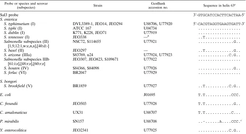
subspecies IIIa was selected. A list of
the base compositions within the target area is presented in
Table 1. The target sequences were similar within each
sub-species with the exception of the sequences of
S. tennessee
and
Salmonella
subspecies II (
S. basel
), which each diverged by one
base. The sequences of nonsalmonellae all diverged by at least
two mismatches.
Whole-cell hybridization.
The sensitivity and specificity of
the probe were tested by hybridization with
fluorescence-la-belled probes on smears of formalin-fixed whole cells (Fig.
1A). All strains except
Staphylococcus aureus
were able to
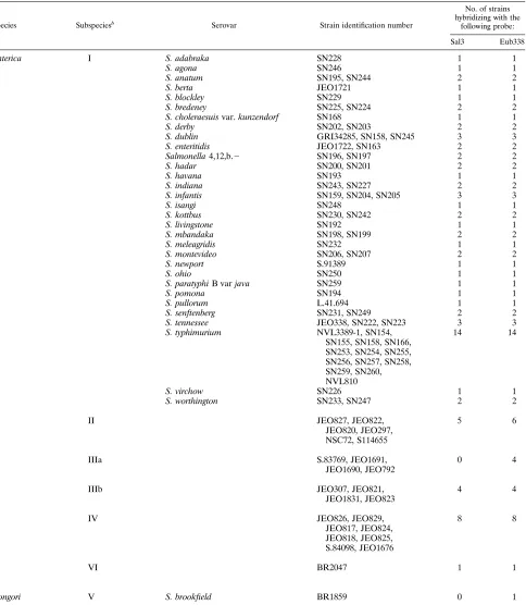
hybridize with Eub338. Sal3 was tested against 86
Salmonella
isolates covering 55 serovars from subspecies I to VI. By using
low-stringency hybridization and washing conditions, the probe
hybridized and yielded a strong signal for 80 of 86
Salmonella
isolates tested (Table 2). As predicted from the alignment of
the sequence in the target area (Table 1), the probe hybridized
to the sequences of the tested serovars of subspecies I, II, IIIb,
IV, and VI, although the sequences of serovars of subspecies II
and IV had one mismatch. No hybridization was obtained with
the four strains of subspecies IIIa, one strain of subspecies V,
and one strain of subspecies II (
S. basel
), which all had two to
on May 15, 2020 by guest
http://jcm.asm.org/
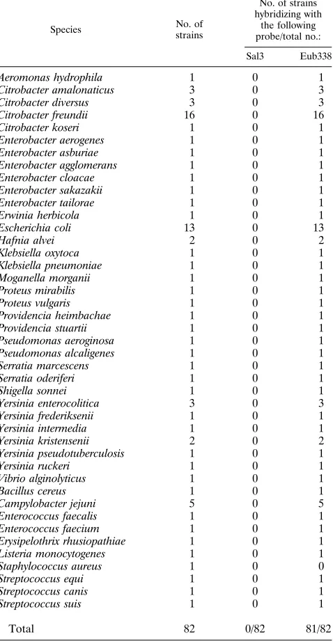
three mismatches (Table 1). No cross hybridization was
ob-served to any of 64 other representatives of the family
Entero-bacteriaceae
tested or to 18 bacterial strains outside of the
family
Enterobacteriaceae
which might occur in the intestinal
tract of pigs and humans (Table 3).
In situ hybridization of tissue specimens.
To test the
abili-ties of the probes to hybridize in situ in paraffin-embedded
material, murine tissue experimentally infected with either
S.
typhimurium
or
E. coli
was prepared. Only tissue from
monoin-fected organs verified by bacterial culturing was used in the
test. In tissue sections from
S. typhimurium
-infected mice, a
distinct fluorescent signal from rod-shaped bacteria could be
detected when hybridization with Sal3 was performed. Bacteria
could be identified as single cells in the liver sinusoids or in the
vessels of the lung, as expected from the hematogenous spread
of the bacteria (Fig. 1B). The same result was achieved when
hybridization with Eub338 was performed. In the tissue from
E. coli
-infected mice, only Eub338 hybridized to bacteria in the
tissue, and no signal was obtained when hybridization with Sal3
was performed. To test for nonspecific binding of the probe,
tissue sections were incubated with a nonsense probe, probe
Non338, which contains the a base sequence complementary to
that of Eub338 and which is unable to hybridize to ribosomes.
When using this probe no signal was detected at all, confirming
the specificities of the probes.
The probes were also tested with formalin-fixed histological
sections from 15 pigs and calves with histories of pneumonia or
enterocolitis in which
S. typhimurium
or
S. dublin
was found by
traditional culture methods (data not shown). For histological
sections from all animals the salmonella probe hybridized
spe-cifically, and
Salmonella
bacteria could be seen either as
clus-ters of cells in the necrotic debris or as single cells scattered
throughout the inflamed tissue. In a few instances single
bac-teria were detected intracellularly as well. In order to show the
spatial localization of salmonella in relation to the indigenous
microflora, the fluorescein-labelled Sal3 probe was used
to-gether with a Cy3-labelled universal probe in the same slide
(Fig. 1C).
Salmonella
bacteria were detected deep in the
lam-ina propria and in the superficial debris, while the indigenous
microflora were shown to be localized only superficially in the
debris. The number of
Salmonella
bacteria detected in each
section varied. In some sections only a few microcolonies were
found, while in others numerous bacterial clusters were
de-tected.
For five samples in which
Salmonella
had not been detected
by culturing, only Eub338 hybridized to the bacteria. No
un-specific binding of the probe was observed when hybridization
with the nonsense probe in the clinical material was
per-formed.
DISCUSSION
[image:3.612.58.557.82.354.2]In the present study a specific oligonucleotide probe was
developed for the in situ detection of
S. enterica
serovars in
bacterial smears and in tissue sections by the FISH technique.
Selection of the probe sequence was based upon alignment of
a sequence segment in the 23S rRNA gene from several
sal-monella serotypes and other enterobacterial strains. This
seg-ment was selected because it contains two regions which have
previously been shown to exhibit high degrees of sequence
variability in bacteria (9, 34). The stringency of the
hybridiza-tion condihybridiza-tions was chosen so that it would not discriminate
between strains with one mismatch, because this allows for the
specific detection of the largest group of
S. enterica
but does
TABLE 1. Sequence variation in the 23S rRNA genes of different
Enterobacteriaceae
species at
E. coli
positions 1713 to 1730
Probe or species and serovar
(subspecies) Strain
GenBank
accession no. Sequence in helix 63
a
Sal3 probe
3
9
-
GTGCATCCACTTCACTAA
-5
9
S. enterica
S. typhimurium
(I)
DVL3389-1, JEO14, JEO294
U88706, U77920
5
9
-
CACGTAGGTGAAGTGATT
-3
9
S. typhi
(I)
ATCC 167
U04734
...
S. dublin
(I)
K771, K228, JEO71
U77919
...
S. tennessee
(I)
JEO338
—
b..T...
Salmonella
subspecies (II)
[1,9,12:1,w:e,n,x],[40:d:-]
NSC72, S114655
U77921
...G..
S. basel
(II)
JEO297
—
..T...G..
S. arizona
(IIIa)
S83769, u24
U77924, U77923
...C.G..
Salmonella
subspecies IIIb
[61:i:z],[48:r:z],[60:r:z]
JEO307, JEO823, S109671
U77922
...
S. houten
(IV)
S84366, S84098
U77926
...G..
S. ferlac
(VI)
BR2047
U77929
...
S. bongori
S. brookfield
(V)
BR1859
U77927
..T...C.G..
E. coli
J01695
T.T...CCC.
C. freundii
JEO503
U77928
T.T...G..
C. amalonaticus
UX31
U88707
T.T...C...
P. mirabilis
SN157
U88708
..-...A....CCC.
cY. enterocolitica
JEO2341
U77925
...C.G..
aE. colinumbering (positions 1713 to 1730) was used.
b—, strains were from reference 8.
c—, gap in the sequence made by a deletion in the gene.
on May 15, 2020 by guest
http://jcm.asm.org/
not allow for cross hybridization to any of the closely related
Citrobacter
species or
E. coli
. Most important, all tested
sero-vars from subspecies I hybridized with Sal3, because members
of this subspecies constitute the most commonly encountered
and the most pathogenic serovars (5, 18).
The separation of serovars into seven homology groups or
subspecies is based on DNA-DNA hybridization studies (12,
19). The same separation of serovars has been found when
comparing 23S rRNA gene sequences (10); the variations were
low within genes from serovars of the same subspecies, but
they were three times higher when serovars from different
subspecies were compared. On the basis of the assumption that
the genetic sequence is highly conserved within each
subspe-cies, the specificity of the probe was tested against 55 serovars
representing all seven subspecies. This test showed a high
degree of conservation in the probe target area, because the
probe hybridized only to the subspecies expected from the
alignment shown in Table 1. However, one strain from
sub-species II failed to hybridize because this strain contained two
mismatches instead of one, as for the other subspecies II
strains.
Several DNA probes used for the detection of
Salmonella
in
food or feces have been published (1, 15, 23). Most probes
target chromosomal or plasmid genes, but oligonucleotide
probes targeting the 16S and 23S rRNA have also been
pub-lished (21, 30, 36). Those probes have been applied in
detec-tion assays based on denaturated DNA or RNA bound to
membranes, eliminating the possibility of the detection and
spatial localization of single bacterial cells in tissue samples.
Hybridization to ribosomes in situ is highly dependent on
target accessibility (4). Secondary and tertiary structures in the
cells, as well as protein binding, might prevent hybridization to
the ribosome (4), thus making specific probes meant for other
applications useless for the detection of bacteria by the FISH
technique. The target sequence of Sal3 is located at helix 63,
and in this study we have shown it to be accessible for in situ
hybridization of
Salmonella.
Fixation is another crucial step for in situ hybridization (4,
22). The fixative serves the dual purposes of opening up the
bacterial wall for probe penetration and protecting the
ribo-somes from degradation by endogenous RNase activity.
Alde-hyde fixatives are most frequently used with gram-negative
bacteria (14), whereas alcohol fixation is recommended for
gram-positive bacteria (6, 29). In our hands, formalin fixation
enabled hybridization of both gram-negative and gram-positive
bacteria. The exception was
S. aureus
, for which hybridization
with Eub338 was not achieved, probably due to the low level of
permeability of the cell wall.
The use of short oligonucleotide probes reduces the time
and temperature required for hybridization. By increasing the
probe concentration the velocity of the hybridization is further
increased; however, this also increases nonspecific staining
(35). When using an intermediate probe concentration of 1 to
5 ng/
m
l, the ribosomes in
E. coli
were saturated after 2 h of
incubation with Eub338 (35). When hybridizing to pure
cul-tures, 3 h of incubation was used, with excellent results.
How-ever, for reasons of convenience, the tissue samples were
hy-bridized overnight.
To enhance the level of discrimination of the signal from
single bacteria in tissue sections, we applied a
narrow-band-FIG. 1. (A) A smear of S. typhimuriumhybridized with the
fluorescein-labelled salmonella probe (Sal3). (B) Formalin-fixed tissue section of murine
lung tissue experimentally infected withS. typhimuriumand hybridized with Sal3,
showing rod-shaped green fluorescent bacteria in the small capillaries of the lung tissue and red autofluorescing erythrocytes. (C) Tissue section from colon of a pig with salmonellosis. By hybridizing with a mixture of Sal3 labelled with fluo-rescein and the universal bacterial probe (Eub338) labelled with Cy3, the spatial
localization of salmonella (yellow) can be determined in proportion to the indigenous microflora (red). The color of salmonella is changed as it hybridizes
with both probes. Magnifications,3450.
on May 15, 2020 by guest
http://jcm.asm.org/
[image:4.612.61.296.72.673.2]pass filter combination which allows for simultaneous
excita-tion in two separate bands. This filter allowed for the
simulta-neous spatial visualization of both salmonella bacteria and the
indigenous microflora when hybridization was done with two
probes. It also reduced autofluorescence from tissue
[image:5.612.73.557.81.637.2]consider-ably by turning the color of the tissue red and thereby allowing
for the detection of single bacteria (Fig. 1B). When a standard
fluorescein isothiocyanate filter was used, only clusters of
bac-teria could be detected against a background of green
autofluorescence.
TABLE 2. Strains of
Salmonella
tested by in situ hybridization with fluorescence-labelled probes
aSpecies Subspeciesb Serovar Strain identification number
No. of strains hybridizing with the
following probe:
Sal3 Eub338
S. enterica
I
S. adabraka
SN228
1
1
S. agona
SN246
1
1
S. anatum
SN195, SN244
2
2
S. berta
JEO1721
1
1
S. blockley
SN229
1
1
S. bredeney
SN225, SN224
2
2
S. choleraesuis
var.
kunzendorf
SN168
1
1
S. derby
SN202, SN203
2
2
S. dublin
GRI34285, SN158, SN245
3
3
S. enteritidis
JEO1722, SN163
2
2
Salmonella
4,12,b.
2
SN196, SN197
2
2
S. hadar
SN200, SN201
2
2
S. havana
SN193
1
1
S. indiana
SN243, SN227
2
2
S. infantis
SN159, SN204, SN205
3
3
S. isangi
SN248
1
1
S. kottbus
SN230, SN242
2
2
S. livingstone
SN192
1
1
S. mbandaka
SN198, SN199
2
2
S. meleagridis
SN232
1
1
S. montevideo
SN206, SN207
2
2
S. newport
S.91389
1
1
S. ohio
SN250
1
1
S. paratyphi
B var
java
SN259
1
1
S. pomona
SN194
1
1
S. pullorum
L.41.694
1
1
S. senftenberg
SN231, SN249
2
2
S. tennessee
JEO338, SN222, SN223
3
3
S. typhimurium
NVL3389-1, SN154,
SN155, SN158, SN166,
SN253, SN254, SN255,
SN256, SN257, SN258,
SN259, SN260,
NVL810
14
14
S. virchow
SN226
1
1
S. worthington
SN233, SN247
2
2
II
JEO827, JEO822,
JEO820, JEO297,
NSC72, S114655
5
6
IIIa
S.83769, JEO1691,
JEO1690, JEO792
0
4
IIIb
JEO307, JEO821,
JEO1831, JEO823
4
4
IV
JEO826, JEO829,
JEO817, JEO824,
JEO818, JEO825,
S.84098, JEO1676
8
8
VI
BR2047
1
1
S. bongori
V
S. brookfield
BR1859
0
1
aA total of 55 serovars were tested.; 80 of 86 and 86 of 86 strains hybridized with probes Sal3 and Eub338, respectively.
bAs proposed by Reeves et al. (28).
on May 15, 2020 by guest
http://jcm.asm.org/
The theoretical sensitivity of detection by the FISH
tech-nique is one bacterium in a 3-
m
m-thick tissue section; however,
the number of bacteria in each tissue section from the clinical
material varied significantly. By performing hybridization with
tissue sections, we were able to detect salmonella bacteria in all
the samples which were found to be positive by traditional
culture methods. However, when dealing with very low
num-bers of bacteria, the culture method might still be the most
sensitive method because it contains a preenrichment step.
Direct detection of bacteria is highly dependent on the
amount and stability of the target, because the intensity of the
signal is proportional to the rRNA content (14). Used as a
research tool, the FISH technique has previously been applied
for studying the spatial distribution of
E. coli
or
S. typhimurium
in the intestines of monoinfected mice (20, 27). This was done
under optimal research conditions. We found that this
tech-nique is also applicable for the detection of
Salmonella
in
clinical samples. The detection of
Salmonella
bacteria in tissue
was possible even after the paraffin-embedding process and in
the presence of indigenous microflora. Autolysis of the tissue,
as a result of the material being shipped unfixed to the
labo-ratory, did not hinder the detection of the bacteria in our study.
In this study we have developed and tested a
fluorescence-labelled oligonucleotide probe for the specific detection of
S.
enterica
in clinical samples by in situ hybridization. The stability
of the ribosome target allowed for the detection of single cells
even in clinical material, as well as identification in smears of
pure cultures. The results obtained by use of a readily
synthe-sized fluorescence-labelled oligonucleotide probe, combined
with a simple hybridization protocol, therefore suggest that the
FISH technique can be used for the detection of
S. enterica
serovars.
ACKNOWLEDGMENTS
We thank Peter Ahrens for technical support with the sequencing
and Henning Rasmussen for serotyping, while we thank Anni Ravn
and Ulla Andreasen for technical assistance in preparing the tissue
sections. We thank John E. Olsen, Department of Veterinary,
Micro-biology at the Royal Veterinary and Agricultural University,
Copen-hagen, Denmark, for donating bacterial strains.
The work was supported by grant 9307926 from the Danish
Agri-cultural and Veterinary Research Council.
REFERENCES
1.Aabo, S., A. Thomas, M. L. M. Hall, H. R. Smith, and J. E. Olsen.1992. Evaluation of a Salmonella-specific DNA probe by colony hybridization
using non-isotopic and isotopic labeling. APMIS100:623–628.
2.Alm, E. W., D. B. Oerther, N. Larsen, D. A. Stahl, and L. Raskin.1996. The
oligonucleotide probe database. Appl. Environ. Microbiol.62:3557–3559.
3.Amann, R. I., B. J. Binder, R. J. Olson, S. W. Chisholm, R. Devereux, and D. A. Stahl.1990. Combination of 16S rRNA-targeted oligonucleotide probes with flow cytometry for analyzing mixed microbial populations. Appl.
Environ. Microbiol.56:1919–1925.
4.Amann, R. I., W. Ludwig, and K. H. Schleifer.1995. Phylogenetic identifi-cation and in situ detection of individual microbial cells without cultivation.
Microbiol. Rev.59:143–169.
5.Anonymous.1996. Survey of serovars from human cases of salmonellosis. State Serum Institute, Copenhagen, Denmark.
6.Beimfohr, C., A. Krause, R. Amann, W. Ludwig, and K. H. Schleifer.1993.
In situidentification of lactococci, enterococci and streptococci. Syst. Appl.
Microbiol.16:450–456.
7.Brosius, J., T. J. Dull, D. D. Sleeter, and H. F. Noller.1981. Gene
organi-zation and primary structure of a ribosomal RNA operon fromEscherichia
coli. J. Mol. Biol.148:107–127.
8.Christensen, H.Unpublished data.
9.Christensen, H., M. Boye, L. K. Poulsen, and O. F. Rasmussen.1994. Analysis of fluorescent pseudomonads based on 23S ribosomal DNA
se-quences. Appl. Environ. Microbiol.60:2196–2199.
10. Christensen, H., S. Nordentoft and J. E. Olsen.23S ribosomal DNA
nucle-otide sequences ofSalmonella entericaandS. bongoriin order to establish
phylogenetic relations. Submitted for publication.
11. Clarke, R. C., and C. L. Gyles.1993.Salmonella, p. 133–153.InC. L. Gyles and C. O. Thoen (ed.), Pathogenesis of bacterial infections in animals, 2nd. ed. Iowa State University Press, Ames.
12. Crosa, J. H., D. J. Brenner, W. H. Ewing, and S. Falkow.1973. Molecular
relationships among the salmonellae. J. Bacteriol.115:307–315.
13. D’Aoust, J. Y.1991. Pathogenicity of foodborneSalmonella. Int. J. Food
Microbiol.12:17–40.
14. DeLong, E. F., G. S. Wickham, and N. R. Pace.1989. Phylogenetic strains: ribosomal RNA-based probes for the identification of single cells. Science
243:1360–1363.
15. Fitts, R., M. Diamond, C. Hamilton, and M. Neri.1983. DNA-DNA
hybrid-ization assay for detection ofSalmonellaspp. in foods. Appl. Environ.
Mi-crobiol.46:1146–1151.
16. Giovannoni, S. J., E. F. DeLong, G. J. Olsen, and N. R. Pace.1988. Phylo-genetic group-specific oligonucleotide probes for identification of single
mi-crobial cells. J. Bacteriol.170:720–726.
[image:6.612.60.299.105.566.2]17. Kaufmann, F.1961. Die bakteriologie der Salmonella-species. Munksgaard, Copenhagen, Denmark.
TABLE 3. Members of the family
Enterobacteriaceae
and selected
gram-positive strains tested by whole-cell hybridization with
fluorescence-labelled oligonucleotide
Species No. of
strains
No. of strains hybridizing with
the following probe/total no.:
Sal3 Eub338
Aeromonas hydrophila
1
0
1
Citrobacter amalonaticus
3
0
3
Citrobacter diversus
3
0
3
Citrobacter freundii
16
0
16
Citrobacter koseri
1
0
1
Enterobacter aerogenes
1
0
1
Enterobacter asburiae
1
0
1
Enterobacter agglomerans
1
0
1
Enterobacter cloacae
1
0
1
Enterobacter sakazakii
1
0
1
Enterobacter tailorae
1
0
1
Erwinia herbicola
1
0
1
Escherichia coli
13
0
13
Hafnia alvei
2
0
2
Klebsiella oxytoca
1
0
1
Klebsiella pneumoniae
1
0
1
Moganella morganii
1
0
1
Proteus mirabilis
1
0
1
Proteus vulgaris
1
0
1
Providencia heimbachae
1
0
1
Providencia stuartii
1
0
1
Pseudomonas aeroginosa
1
0
1
Pseudomonas alcaligenes
1
0
1
Serratia marcescens
1
0
1
Serratia oderiferi
1
0
1
Shigella sonnei
1
0
1
Yersinia enterocolitica
3
0
3
Yersinia frederiksenii
1
0
1
Yersinia intermedia
1
0
1
Yersinia kristensenii
2
0
2
Yersinia pseudotuberculosis
1
0
1
Yersinia ruckeri
1
0
1
Vibrio alginolyticus
1
0
1
Bacillus cereus
1
0
1
Campylobacter jejuni
5
0
5
Enterococcus faecalis
1
0
1
Enterococcus faecium
1
0
1
Erysipelothrix rhusiopathiae
1
0
1
Listeria monocytogenes
1
0
1
Staphylococcus aureus
1
0
0
Streptococcus equi
1
0
1
Streptococcus canis
1
0
1
Streptococcus suis
1
0
1
Total
82
0/82
81/82
on May 15, 2020 by guest
http://jcm.asm.org/
18. Le Minor, L.1992. The genusSalmonella, p. 2761–2774.InA. Balows, H. G.
Tru¨per, M. Dworkin, W. Harder and K.-H. Schleifer (ed.), The prokaryotes,
2nd ed. Springer Verlag, New York, N.Y.
19. Le Minor, L., M. Ve´ron, and M. Y. Popoff.1982. Proposition pour une
nomenclature desSalmonella. Ann. Microbiol. Inst. Pasteur133B:245–254.
20. Licht, T. R., K. A. Krogfelt, P. S. Cohen, L. K. Poulsen, J. Urbance, and S. Molin.1996. Role of lipopolysaccharide in colonization of the mouse
intes-tine bySalmonella typhimuriumstudied by in situ hybridization. Infect.
Im-mun.64:3811–3817.
21. Lin, C. K., and H. Y. Tsen.1995. Development and evaluation of two novel oligonucleotide probes based on 16S rRNA sequence for the identification ofSalmonellain foods. J. Appl. Bacteriol.78:507–520.
22. Macnaughton, S., A. G. O’Donnell, and T. M. Embley.1994.
Permeabiliza-tion of mycolic-acid-containing actinomycetes forin situhybridization with
fluorescently labelled oligonucleotide probes. Microbiology140:2859–2865.
23. Maddox, C. W., and W. H. Fales.1991. Use of aSalmonella typhimurium
-derived virulence probe in the detection ofSalmonellasp. and in the
char-acterization ofS. cholerae-suisvirulence plasmids. J. Vet. Diagn. Invest.
3:218–222.
24. Maidak, B. L., N. Larsen, M. J. McCaughey, R. Overbeek, G. J. Olsen, K. Fogel, J. Blandy, and C. R. Woese.1994. The ribosomal database project.
Nucleic Acids Res.22:3485–3487.
25. Popoff, M. Y., and L. Le Minor.1992. Antigenic formulas of theSalmonella
serovars. WHO Collaborating Center for Reference and Research on Sal-monella, Institut Pasteur, Paris, France.
26. Poulsen, L. K., G. Ballard, and D. A. Stahl.1993. Use of rRNA fluorescence in situ hybridization for measuring the activity of single cells in young and
established biofilms. Appl. Environ. Microbiol.59:1354–1360.
27. Poulsen, L. K., F. Lan, C. S. Kristensen, P. Hobolth, S. Molin, and K. A. Krogfelt.1994. Spatial distribution ofEscherichia coliin the mouse large
intestine inferred from rRNA in situ hybridization. Infect. Immun.62:5191–
5194.
28. Reeves, M. W., G. M. Evins, A. A. Heiba, B. D. Pilkaytis, and J. J. Farmer III.
1989. Clonal structure ofSalmonella typhiand its genetic relatedness to other
salmonellae as shown by multilocus enzyme electrophoresis, and proposal of
Salmonella bongoricomb. nov. J. Clin. Microbiol.27:313–320.
29. Roller, C., M. Wagner, R. Amann, W. Ludwig, and K. H. Schleifer.1994.In
situprobing of gram-positive bacteria with high DNA G1C content using
23S rRNA-targeted oligonucleotides. Microbiology140:2849–2858.
30. Ro¨nner, S. G. E., and E. Stackebrandt.1994. Development of 23S
rDNA-oligonucleotide probes for the identification ofSalmonellaspecies. Syst.
Appl. Microbiol.17:257–264.
31. Sears, L. E., L. S. Moran, C. Kissinger, T. Creasey, O. Perry, M. Roskey, E. Sutherland, and B. E. Slatko.1992. CircumVent thermal cycle sequencing and alternative manual and automated DNA sequencing protocols using the
highly thermostable VentR (exo-) DNA polymerase. BioTechniques13:626–
633.
32. Stahl, D. A., B. Flesher, H. R. Mansfield, and L. Montgomery.1988. Use of phylogenetically based hybridization probes for studies of ruminal microbial
ecology. Appl. Environ. Microbiol.54:1079–1084.
33. Tegtmeier, C., N. E. Jensen, and H. E. Jensen.1995. Development of a
peroxidase-antiperoxidase (PAP) technique for the identification of
Hae-mophilus somnusin pneumonic calf lungs in Denmark. APMIS103:540–547. 34. Van Camp, G., S. Chapelle, and R. De Wachter.1993. Amplification and sequencing of variable regions in bacterial 23S ribosomal RNA genes with
conserved primer sequences. Curr. Microbiol.27:147–151.
35. Wallner, G., R. Amann, and W. Beisker.1993. Optimizing fluorescentin situ
hybridization with rRNA-targeted oligonucleotide probes for flow
cytomet-ric identification of microorganisms. Cytometry14:136–143.
36. Wilson, S. G., S. Chan, M. Deroo, M. Vera-Garcia, A. Johnson, D. J. Lane, and D. Halbert.1990. Development of a colorimetric, second generation
nucleic acids hybridization method for detection ofSalmonellain foods and
a comparison with conventional culture procedure. J. Food Sci.55:1394–
1398.
37. Zhu, Q., C. K. Lim, and Y. N. Chan.1996. Detection ofSalmonella typhiby
polymerase chain reaction. J. Appl. Bacteriol.80:244–251.