Open Access
Research article
Knee loading stimulates cortical bone formation in murine femurs
Ping Zhang
1,2, Min Su
1, Shigeo M Tanaka
3and Hiroki Yokota*
1,2Address: 1Departments of Biomedical Engineering, Indiana University – Purdue University Indianapolis, Fesler Hall 115, 1120 South Drive,
Indianapolis IN 46202, USA, 2Department of Anatomy and Cell Biology, Indiana University School of Medicine, Fesler Hall 115, 1120 South
Drive, Indianapolis IN 46202, USA and 3Graduate School of Natural Science and Technology, Kanazawa University, Ishikawa, Japan
Email: Ping Zhang - [email protected]; Min Su - [email protected]; Shigeo M Tanaka - [email protected]; Hiroki Yokota* - [email protected]
* Corresponding author
Abstract
Background: Bone alters its architecture and mass in response to the mechanical environment, and thus varying loading modalities have been examined for studying load-driven bone formation. The current study aimed to evaluate the anabolic effects of knee loading on diaphyseal cortical bone in the femur.
Methods: Using a custom-made piezoelectric loader, 0.5-N loads were laterally applied to the left knee of C57/BL/6 mice at 5, 10, 15, and 20 Hz for 3 minutes per day for 3 consecutive days. Animals were sacrificed for examination 13 days after the last loading. The contralateral femur was used as a non-loading control, and the statistical significance of loading effects was evaluated with p < 0.05.
Results: Although diaphyseal strains were measured as small as 12 μstrains, bone histomorphometry clearly demonstrated frequency-dependent enhancement of bone formation. Compared to a non-loading control, bone formation on the periosteal surface was significantly enhanced. The loading at 15 Hz was most effective in elevating the mineralizing surface (1.7 x; p < 0.05), mineral apposition rate (1.4 x; p < 0.001), and bone formation rate (2.4 x; p < 0.01). The loading at 10 Hz elevated the mineralizing surface (1.4 x; p < 0.05), mineral apposition rate (1.3 x; p < 0.01), and bone formation rate (1.8 x; p < 0.05). The cross-sectional cortical area and the cortical thickness in the femoral diaphysis were significantly increased by loading at 10 Hz (both 9%) and 15 Hz (12% and 13%, respectively).
Conclusion: The results support the anabolic effects of knee loading on diaphyseal cortical bone in the femur with small in situ strain, and they extend our knowledge on the interplay between bone and joints. Strengthening the femur contributes to preventing femoral fractures, and the discovery about the described knee loading might provide a novel strategy to strengthen osteoporotic bones. Further analyses are required to understand the biophysical and molecular mechanism behind knee loading.
Background
Osteoporosis as a skeletal disorder is characterized by compromised bone strength, which leads to an increased
risk of fracture. Patients with femoral fracture account for a substantial volume of orthopaedic hospitalizations. Thus, strengthening femurs is a keen healthcare issue
par-Published: 19 September 2006
BMC Musculoskeletal Disorders 2006, 7:73 doi:10.1186/1471-2474-7-73
Received: 22 May 2006 Accepted: 19 September 2006
This article is available from: http://www.biomedcentral.com/1471-2474/7/73 © 2006 Zhang et al; licensee BioMed Central Ltd.
ticularly among the aging population [1]. Load-imposing exercises are recommended as a protective measure [2], but they are often limited to healthy individuals who can afford physical activities [3]. Varying loading modalities have therefore been developed in an attempt to enhance femoral bone formation, and biomechanical factors such as loading frequencies, strain and strain-induced fluid flow, and alteration in intramedullary pressure are inves-tigated to understand load-driven anabolic responses [4-7]. Whole body vibrations, for instance, are effective in elevating bone mass in femoral trabecular bone [8]. Few mild, non-invasive loading modalities, however, have been shown to stimulate formation of cortical bone in the femur. In this report, one form of joint loading – knee loading – is described as a potential means to provoke anabolic responses in femoral cortical bone.
Joint loading is a relatively new form of mechanical stim-ulus in which loads are laterally applied to the epiphyseal bone. Long bones such as an ulna and a femur are appar-ently less stiff in the lateral direction than in the axial direction, and the epiphysis rich in trabecular bone is softer than the diaphysis composed of cortical bone. Therefore, our rationale behind joint loading is that exposing the least rigid part of bone – epiphyseal bone – to lateral loads might effectively trigger load-driven ana-bolic responses in the diaphysis. In fact, elbow loading in our previous mouse study [9,10] revealed enhanced corti-cal bone formation in the ulnar diaphysis. Compared to an axial ulna-bending modality [11], elbow loading required at most one fourth of the loads (0.5-N forces) to induce bone formation with the same loading timetable. The current study extends elbow loading to knee loading and examines the anabolic effects on femoral cortical bone.
We have previously shown that knee loading stimulates bone formation in the tibia [12,13], but no study has been performed to evaluate its effects on the femur. A knee is anatomically a complex structure and application of loads to the knee does not imply that the same effects are expected to the tibia and the femur. With knee loading, loads are applied to the lateral and medial epicondyles of the femur as well as the lateral and medial condyles of the tibia. Since the tibial condyle is positioned closer to the loader than the femoral epocondyle, the femoral epiphy-sis is expected to receive fewer loads than the tibial epiph-ysis. In addition, anabolic responses to knee loading at different frequencies may vary between the tibia and the femur. If knee loading is effective in the femur, it provides a possibility in future to prevent bone loss throughout the femur including the femoral neck. A substantial bone loss or damage to the femoral neck could result in a serious healthcare problem particularly in the aging population.
Two specific questions addressed herein are (1) whether lateral loads applied to the epiphysis enhance cortical bone formation in the femoral diaphysis; and (2) if so, whether bone formation is dependent on loading fre-quencies. Using femurs ex vivo, we previously showed that molecular transport in diaphyseal lacunae was activated with knee loading [14]. However, any in vivo conse-quences or potential mechanisms of enhanced molecular transport were not examined. In this study, we address the question of whether loads applied to the joint site would enhance bone formation in the remote diaphyseal bone.
To answer that question we used a custom-made piezoe-lectric mechanical loader for knee loading. Dependence of loading frequencies on anabolic responses has been pointed out in varying loading studies [11,15], and we employed loading frequencies at 5, 10, 15 and 20 Hz. The results in this study clearly support that knee loading can effectively enhance bone formation in a loading fre-quency-dependent manner.
Methods
Experimental animals
Sixty-one female C57/BL/6 mice ~14 weeks of age (body weight of ~20 g) were used (Harlan Sprague-Dawley, Inc., Indianapolis, IN). Five animals were housed per cage at the Laboratory Animal Resource Center of Indiana Uni-versity School of Medicine, and they were fed with stand-ard laboratory chow and water ad libitum. The animals were allowed to acclimate for 2 weeks before the experi-ment. All procedures, performed in this study, were in accordance with the Institutional Animal Care and Use Committee guidelines at Indiana University School of Medicine and approved.
Mechanical loading
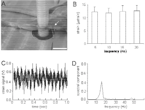
The mouse was placed in an anesthetic induction-cham-ber to induce sedation and mask-anesthetized using 2% isoflurane. Using the custom-made piezoelectric mechan-ical loader, mechanmechan-ical loads were applied for 3 minutes per day for 3 consecutive days to the left knee through the lateral-medial direction (Fig. 1). The mice were randomly divided into four groups for four loading frequencies (5, 10, 15 or 20 Hz, N = 8), and the loads with a peak-to-peak force of 0.5 N were applied. Using the same procedure, the right knee was placed under the loader without oscillatory loading and used as the non-loading control. After load-ing, the mouse was allowed normal cage activity and any abnormal behavior, weight loss or a diminished food intake was monitored.
Piezoelectric joint loader
board (PCI-6052E; National Instruments, Austin, TX) and a piezo-driver (model PZD 700-L; Trek, New York, NY) (Fig. 1). Before the loading experiment, loads applied to the knee were calibrated using an anesthetized mouse. A strain gauge (CEA-06-062UW; Vishay Measurements Group, Raleigh, NC) was attached on the aluminum can-tilever connected to the stator and known forces from 0 to 10 N were given. To position the knee properly, the lower end of the loading rod and the upper end of the supporter were designed to form a pair of semispherical cups. The lateral and medial epicondyles of the femur together with the lateral and medial condyles of the tibia were confined in the cups. Because of the complex anatomy of the knee, the lateral side of the femur is less projected outwards. Therefore, the femoral epicondyle apparently receives fewer loads than the tibial condyle. The tip of the loader
had a contact area of 4 mm diameter. To avoid stress con-centration between the knee and the loader, the loading surface and the supporter were covered with silicon rub-ber.
Measurement of strain
Five animals were used for strain measurements (Fig. 2). After euthanasia, the periosteal surface of a left femur was exposed. A strain gauge (Model EA-06-015DJ-120, Meas-urements Group Inc., NC) was trimmed into 0.5 mm in width and 2.2 mm in length, and attached with glue to the mid-diaphysis (50% along the length of the femur). Note that it was unavoidable to remove a part of surrounding tissue for attachment of the strain gauge but the loading site in the knee was kept intact. Bone histomorphometry was conducted using 0.5-N loads, and therefore the strain
Setup of a mechanical loader for knee loading used in this study
Figure 1
was also measured with the same loading conditions. The distal epiphysis was loaded at 5, 10, 15 and 20 Hz with 0.5 N. Voltage signals from the strain gauge were processed with a signal conditioning amplifier (2210, Measurement Group Inc., NC), and Fourier transform was conducted. The measurement was repeated 5 times, and the peak-to-peak voltage was converted to the actual strain value using the standard calibration line [15,16]. Loads were confined with the loading rod, and no bending moment was pre-sumably generated to the femur or the tibia.
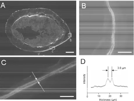
Fluorescence labeling and sample preparation
Mice were given an intraperitoneal injection of calcein (Sigma, St. Louis, MO), a fluorochrome dye, at 30 μg/g
body mass on days 2 and 6 after the last loading (Fig. 3). Animals were sacrificed 13 days after the last loading, and femurs were harvested for histomorphometric analyses. The isolated bones were cleaned of soft tissues, and the distal and proximal ends were cleaved to allow infiltration of the fixatives with 10% neutral buffered formalin. After 48 h in the fixatives, bones were transferred to 70% alco-hol for storage. Specimens were dehydrated in a series of graded alcohols and embedded in methyl methacrylate (Aldrich Chemical Co., Milwaukee, WI). The transverse sections (~80 μm in thickness) were removed from the mid-diaphysis, ~8 mm distant from the distal end using a diamond-embedded wire saw (Delaware Diamond
[image:4.612.63.540.90.450.2]Strain measurement with knee loading
Figure 2
Knives, Wilmington, DE), and they were mounted on standard microscope slides.
Bone histomorphometry
Using a Nikon Optiphot microscope (Nikon, Inc., Garden City, NY) and a Bioquant digitizing system (R & M Bio-metrics, Nashville, TN), we measured total perimeter (B.Pm), endocortical perimeter, single-labeled perimeter (sL.Pm), labeled perimeter (dL.Pm), and double-labeled area (dL.Ar). From these measurements we derived mineralizing surface (MS/BS = [1/2 sL.Pm + dL.Pm]/B.Pm in %), mineral apposition rate (MAR =
dL.Ar/dL.Pm/4 in μm/day), and bone formation rate
(BFR/BS = MAR × MS/BS × 365 in μm3/μm2 per year). To
evaluate the effects of the loading frequencies the relative
parameters such as rMS/BS, rMAR, and rBFR/BS were derived as differences between the loaded and the control femurs. In drawing a fluorescent intensity curve, a Meta-Morph Imaging System (version 3.6, Universal Imaging Co.) was used. Note that the fluorescent labeling data rep-resent bone formation during the days 2 and 6 after the last loading.
The total bone cross-sectional area (mm2), bone
medul-lary area (mm2), and cortical thickness (mm) were also
measured. The cross-sectional cortical area was deter-mined by subtracting the bone medullary area from the total bone cross-sectional area. The cortical thickness was defined as the mean distance between the endosteal and the periosteal surfaces on both anterior and posterior
[image:5.612.59.518.88.433.2]Femoral cross-sections with knee loading
Figure 3
sides. The measurements were taken at the middle of each side, and the mean value was determined from two inde-pendent measurements. The relative alteration was calcu-lated as differences between loaded (L) and control (C) femurs such as ([L - C]/C × 100 in %).
Bone porosity
Intracortical porosity was determined from the tibial and femoral transverse sections of the non-loaded limbs in the mid-diaphysis (~50 μm thickness; N = 24). Using a Nikon Optiphot microscope and a Bioquant digitizing system, we measured cross-sectional cortical area (mm2), total
porous area (mm2), and the number of pores whose area
was larger than an identifiable threshold of 11 μm2 with
the optical system. From these measurements, we derived intracortical porosity (ratio of the porous area to the total bone area in %) and pore density (number/mm2). The
mean value was calculated from 2 independent measure-ments.
Microcomputed tomography (μCT)
Micro-CT was performed using a desktop μCT-20 (Scanco Medical AG, Auenring, Switzerland). The hindlimb was harvested keeping the intact knee. The sample was placed in a plastic tube filled with 70% ethanol and centered in the gantry of the machine. A series of cross-sectional images were captured in an 8-mm segment including the knee at 30-μm resolution. Images were imported into Scion Image software (Scion Corp., Fredrick, MD, USA), and three-dimensional reconstruction was conducted.
Statistical analysis
The data were expressed as mean ± SEM. Statistical signif-icance among groups was examined using ANOVA, and a post-hoc test was conducted using a Fisher's protected least significant difference (PLSD) for all the pairwise comparisons. A paired t-test was employed to evaluate sta-tistical significance between the loaded and control femurs. All comparisons were two-tailed and statistical significance was evaluated with p < 0.05.
Results
The animals used for bone histomorphometry tolerated the procedures, and any abnormal behavior including weight loss or diminished food intake was not observed. No bruising or other damages were detected at the loading site.
In situ strain in the femoral mid-diaphysis with knee loading
In response to knee loading with 0.5 N forces, the strains at the mid-diaphysis of the femur were measured at 5, 10, 15 and 20 Hz loading frequencies (Fig. 2). The measured strains were in the range from 8 to 19 μstrain, and the mean value was ~12 μstrain regardless of the loading fre-quencies. No statistically significance was observed among the four loading frequencies (ANOVA, p = 0.99).
Enhancement of cortical bone formation on periosteal surface
[image:6.612.56.561.473.719.2]Bone formation on the periosteal surface was significantly enhanced (Table 1, Fig. 3), especially with knee loading at
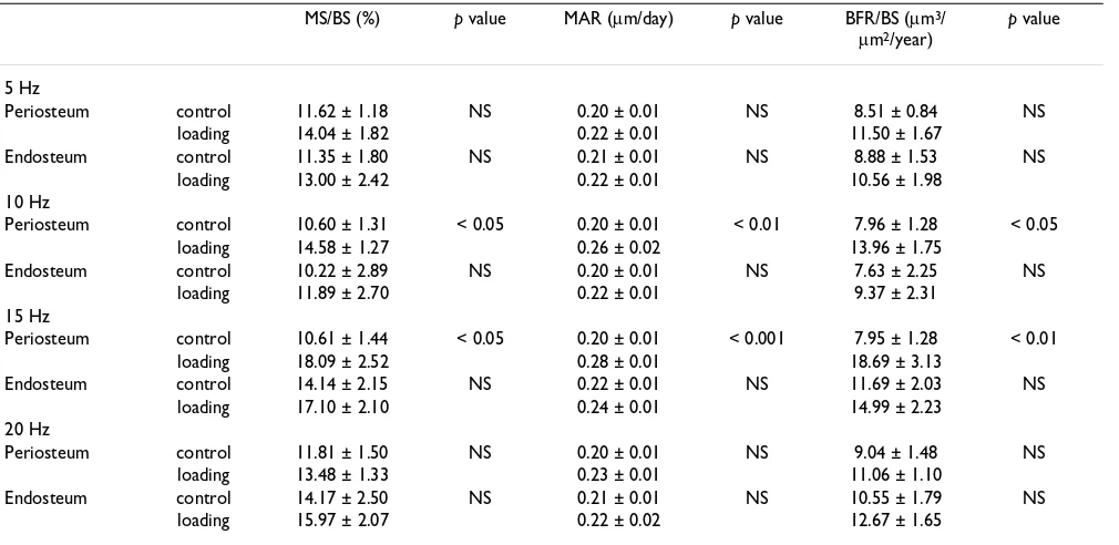
Table 1: Bone formation in periosteum and endosteum with knee loading
MS/BS (%) p value MAR (μm/day) p value BFR/BS (μm3/
μm2/year)
p value
5 Hz
Periosteum control 11.62 ± 1.18 NS 0.20 ± 0.01 NS 8.51 ± 0.84 NS
loading 14.04 ± 1.82 0.22 ± 0.01 11.50 ± 1.67
Endosteum control 11.35 ± 1.80 NS 0.21 ± 0.01 NS 8.88 ± 1.53 NS
loading 13.00 ± 2.42 0.22 ± 0.01 10.56 ± 1.98
10 Hz
Periosteum control 10.60 ± 1.31 < 0.05 0.20 ± 0.01 < 0.01 7.96 ± 1.28 < 0.05
loading 14.58 ± 1.27 0.26 ± 0.02 13.96 ± 1.75
Endosteum control 10.22 ± 2.89 NS 0.20 ± 0.01 NS 7.63 ± 2.25 NS
loading 11.89 ± 2.70 0.22 ± 0.01 9.37 ± 2.31
15 Hz
Periosteum control 10.61 ± 1.44 < 0.05 0.20 ± 0.01 < 0.001 7.95 ± 1.28 < 0.01
loading 18.09 ± 2.52 0.28 ± 0.01 18.69 ± 3.13
Endosteum control 14.14 ± 2.15 NS 0.22 ± 0.01 NS 11.69 ± 2.03 NS
loading 17.10 ± 2.10 0.24 ± 0.01 14.99 ± 2.23
20 Hz
Periosteum control 11.81 ± 1.50 NS 0.20 ± 0.01 NS 9.04 ± 1.48 NS
loading 13.48 ± 1.33 0.23 ± 0.01 11.06 ± 1.10
Endosteum control 14.17 ± 2.50 NS 0.21 ± 0.01 NS 10.55 ± 1.79 NS
loading 15.97 ± 2.07 0.22 ± 0.02 12.67 ± 1.65
10 and 15 Hz. Compared to the non-loading control, bone formation on the periosteal surface using the com-bined data for 4 loading groups was significantly enhanced in terms of MS/BS, MAR, and BFR/BS (1.4 x, 1.3 x, and 1.7 x, all p < 0.001). The loading at 10 Hz elevated the three morphometric parameters (MS/BS, MAR, and BFR/BS) by 1.4-fold (p < 0.05), 1.3-fold (p < 0.01), and 1.8-fold (p < 0.05) on the periosteal bone, respectively. Similarly, the loading at 15 Hz resulted in an increase in these parameters by 1.7-fold (MS/BS; p < 0.05), 1.4-fold (MAR; p < 0.001) and 2.4-fold (BFR/BS; p < 0.01). Unlike the periosteal surface, although bone formation on the endosteal surface was increased, no statistically significant effects were observed on the endosteal surface.
Dependence of anabolic responses on loading frequencies Dependence on the loading frequencies was also promi-nent in the relative parameters defined on the periosteal surface (Fig. 4). In order to further evaluate the effects of knee loading on the periosteal and endosteal surfaces, rMS/BS, rMAR, and rBFR/BS were determined. Compared to the loading at 5 Hz, the loading at 15 Hz resulted in a significant increase in three morphometric parameters (p
< 0.01 for rMS/BS and rBFR/BS; and p < 0.05 for rMAR). Furthermore, compared to the loading at 20 Hz, the load-ing at 15 Hz resulted in an increase in these parameters (p
< 0.01 for rMS/BS and rBFR/BS; p < 0.05 for rMAR). In contrast, no significant alterations were observed on the endosteal surface at any loading frequency (p = 0.75 – 0.99; data not shown).
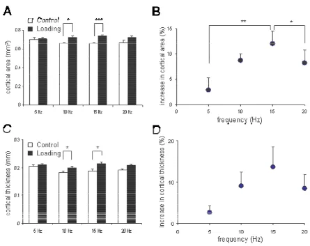
Elevation in cortical bone area and thickness
A frequency-dependent enhancement of the cortical area and the cortical thickness was observed (Fig. 5). First, the cross-sectional cortical area was increased from 0.66 ± 0.01 mm2 (control) to 0.74 ± 0.01 mm2 (loading) at 15
Hz (p < 0.001), and from 0.66 ± 0.01 mm2 (control) to
0.72 ± 0.02 mm2 (loading) at 10 Hz (p < 0.05). No
signif-icant alteration was observed at 5 Hz (p = 0.43) or 20 Hz (p = 0.56). Second, the cortical thickness was also elevated from 0.188 ± 0.007 mm (control) to 0.213 ± 0.008 mm (knee loading) at 15 Hz (p < 0.05), and from 0.183 ± 0.005 mm (control) to 0.199 ± 0.005 mm (knee loading) at 10 Hz (p < 0.05). No significant changes were observed at 5 Hz (p = 0.45) or 20 Hz (p = 0.06). Among the four loading frequencies, the loading at 15 Hz generated a cross-sectional cortical area significantly greater than that at 5 Hz (p < 0.05). Regarding the cortical thickness, there was no statistical difference among the four loading fre-quencies (p = 0.19).
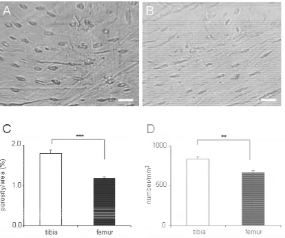
Bone porosity in the tibia and the femur
Bone porosity in the cortical bone was significantly differ-ent between the tibia and the femur (Fig. 6). Regarding intracortical porosity, the tibia (1.79 ± 0.10 in %) was
more porous than the femur (1.18 ± 0.04) (p < 0.001). Similarly, the porosity density (number/mm2) was higher
in the tibia (831 ± 42) than the femur (666 ± 20) (p < 0.01).
Discussion
The histomorphometric results reveal that knee loading – lateral loads applied to the femoral distal epiphysis – increase formation of diaphyseal cortical bone in the femur. The enhancement was most significant with a 15 Hz loading frequency. Unlike a spring loading device [17], hip loading with insertion of a pair of wires [18], or femoral vein ligation [19], the described knee loading modality is non-invasive. Strain has been considered a potential determinant for load-driven bone formation [20,21], and a strain threshold required to induce ana-bolic responses was modeled as a function of the number
Alteration in the histomorphometric parameters on the peri-osteal and endperi-osteal surfaces with knee loading at 5, 10, 15, and 20 Hz
Figure 4
of daily loading cycles [4]. With 0.5-N loads in the current study, an in situ strain was measured as small as 12 μstrain at the site of bone formation in the femoral mid-diaphy-sis. Since loads were applied 3 min per day, the number of daily loading cycles was 2,700 with the most effective loading frequency at 15 Hz. These results indicate that cor-tical bone formation can be enhanced with an in situ
strain of ~10 μstrain at 1,000 – 10,000 loading cycles. Given the same loading cycles, the estimated in situ strain was approximately 1/10 of the predicted threshold value of 1000 μstrain [4].
Knee loading was reported as an effective means to enhance bone formation in the tibia without inducing sig-nificant in situ strain at the site of bone formation
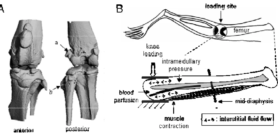
[12,13,22]. In the present study we demonstrated that it is effective to stimulate bone formation in the femoral dia-physis, although the most effective loading frequency dif-fers. Many factors can contribute to the observed bone formation including load-driven blood perfusion, muscle contraction, and strain induced fluid flow (Fig. 7). One of the hypotheses is that the applied loads generate an oscil-latory alteration in intramedullary pressure and the induced pressure gradient stimulates fluid flow and ana-bolic responses. Trabecular bone in the epiphysis is less stiff than cortical bone in the diaphysis [23,24], and Young's modulus in the lateral direction is smaller than that in the axial direction [25]. Therefore, lateral loads with knee loading to the femoral epiphysis may effectively modulate intramedullary pressure in the femoral bone
[image:8.612.75.520.88.441.2]Alteration in the cross-sectional cortical area and the cortical thickness with and without knee loading at 5, 10, 15 and 20 Hz
Figure 5
cavity. Furthermore, modulation of intramedullary pres-sure may produce a local prespres-sure gradient in the diaphy-sis and induce interstitial fluid flow on the cortical bone surface.
The results of bone formation in the current study are con-sistent with the reported enhancement of molecular
trans-port ex vivo with knee loading [14]. Load-driven
interstitial fluid flow was monitored using dye migration in histological bone sections [5] and visualized through fluid displacements in cortical and trabecular bones [26]. Recently a fluorescence-based technique, "fluorescence after photo-bleaching," was employed to evaluate molec-ular transport in a lacuna [14,27,28]. In order to examine the possibility of induction of molecular transport in the diaphysis with knee loading, the photo-bleaching experi-ments were conducted using a mouse femur ex vivo [14].
A fluorescently labeled lacuna in cortical bone was photo-bleached and a rate of fluorescence recovery was meas-ured with and without loads on the epiphysis. It was reported that knee loading shortened the fluorescent recovery approximately by one quarter [14]. A further linkage analysis between alteration in intramedullary pressure and shortening of the recovery time would con-tribute to our understanding of the mechanism behind the anabolic responses with knee loading.
Differential sensitivity of the periosteal and endosteal sur-faces was observed in response to knee loading. Although bone formation increased on both the periosteal and the endosteal surfaces, in contrast to the marked enhance-ment of bone formation on the periosteal surface, the endosteal surface exhibited no significant increase in any of the morphometric parameters. Our observations are
[image:9.612.89.503.88.433.2]Bone porosity in the tibia and the femur
Figure 6
consistent with some of the previous studies using other loading modalities such as tibia bending [29-31], ulna axial loading [32], and loading with a spring device [17], although strain distributions are different among loading modalities. The results may suggest a differential role of alterations in intramedullary pressure on the periosteal and endosteal surfaces, and differences in anatomical and physiological microenvironments. For instance, the non-uniform osteogenic potentials could also result from local variations in cellular and molecular compositions, and/or heterogeneity in load-driven pressure gradient and inter-stitial fluid flow on these two surfaces [5-7].
We examined porosity of cortical bone in the tibia and the femur as a potential cause of differential frequency dependence in response to knee loading. As reported by Qin et al. with their pressure experiments, we postulated that the observed frequency dependence with knee load-ing could be caused by interactions of microstructures in the lacunocanalicular network and intramedullary pres-sure [6,7]. The histological analysis revealed that cortical bone in the tibia is more porous than that in the femur. Our porosity observation indicates that femoral micro-structure might be sensitive to higher frequencies that tib-ial microstructure. A further bone microstructures analysis
would help to elucidate the role of load frequency in the mechanism of joint loading.
Limitations inherent in the present study include the restricted loading frequencies and varying numbers of daily loading cycles among the loading groups. In whole-body vibrations, it has been suggested that higher fre-quencies (> 30 Hz) can stimulate bone formation with a lower level of strain [33,34]. Although the anabolic responses at 20 Hz were insignificant, we do not have any information on the effects of loading frequencies above 20 Hz. Dependence of the observed anabolic responses on loading frequencies requires further investigations since a daily loading cycle and a strain rate are considered important factors, and mechanical testing should be con-ducted to examine strengthening of bones with knee load-ing. Note that although loading at 20 Hz has the highest cycle number as well as the strain rate, it did not exhibit superior capability of bone formation. We did not detect any morphological damage on loaded knees, but more careful inspection using molecular markers such as expression and activities of matrix metalloproteinases will be useful for detecting the maximum loads that do not cause any inflammation or evaluating potential regional acceleratory phenomena [35-37]. Our preliminary studies indicate that knee loading is able to enhance bone
[image:10.612.67.543.88.324.2]forma-(A) Micro CT images (anterior and posterior views) of the mouse knee
Figure 7
tion throughout the femur including the femoral neck, and it can stimulate healing of wounded bone. Our future analyses include the role of osteoclastic activities as well as osteoblastic activities with knee loading. A prototype human knee joint loading supporter, designed and fabri-cated for mechanical characterization, could be used in future for a clinical trial [38].
Conclusion
In summary, the current mouse femur study demonstrates that knee loading is an effective means to enhance cortical bone formation on the periosteal surface in the femoral diaphysis with an in situ strain as small as ~10 μstrain at the site of bone formation. Furthermore, the efficacy of the observed anabolic responses was dependent on the loading frequency. The results here extend our knowledge of load-driven bone formation and the interplay between the epiphysis and the diaphysis in the femur. Strengthen-ing femurs contributes to preventStrengthen-ing femoral fractures, and the described knee loading might provide a novel strategy to develop mechanical therapies.
Competing interests
The author(s) declare that they have no competing inter-ests.
Authors' contributions
PZ performed the animal experiments as well as bone his-tomorphometry and drafted the manuscript. MS carried out the strain measurements. ST conducted part of the animal experiments. HY designed the project and edited the manuscript. All authors read and approved the final manuscript.
Acknowledgements
The authors appreciate Dr. G.M. Malacinski for critical reading of the man-uscript. This study was in part supported by NIH R03AG024596 and NIH R01AR52144.
References
1. Iqbal MM, Sobhan T: Osteoporosis: a review. Mo Med 2002,
99:19-24.
2. Turner CH, Robling AG: Mechanisms by which exercise improves bone strength. J Bone Miner Metab 2005, 23:S16-22. 3. Chestnut CH: Bone mass and exercise (review). Amer J of Med
1993, 95:S34-36.
4. Rubin CT, Sommerfeldt DW, Judex S, Qin Y: Inhibition of osteo-penia by low magnitude, high-frequency mechanical stimuli.
Drug Discov Today 2001, 6:848-858.
5. Wang L, Ciani C, Doty SB, Fritton SP: Delineating bone's intersti-tial fluid pathway in vivo. Bone 2004, 34:499-509.
6. Qin YX, Kaplan T, Saldanha A, Rubin CT: Fluid pressure gradients, arising from oscillations in intramedullary pressure, is corre-lated with the formation of bone and inhibition of intracorti-cal porosity. J Biomech 2003, 36:1427-1437.
7. Qin YX, Lin W, Rubin C: The pathway of bone fluid flow as defined by in vivo intramedullary pressure and streaming potential measurements. Ann Biomed Eng 2002, 30:693-702. 8. Flieger J, Karachalios T, Khaldi L, Raptou P, Lyritis G: Mechanical
stimulation in the Form of vibration prevents postmenopau-sal bone loss in ovariectomized rats. Calcif Tissue Int 1998,
63:510-514.
9. Yokota H, Tanaka SM: Osteogenic potential with joint loading modality. J Bone Miner Metab 2005, 23:302-308.
10. Tanaka SM, Sun HB, Yokota H: Bone formation induced by a novel form of mechanical loading on joint tissue. Biol Sci Space
2004, 18:41-44.
11. Warden SJ, Turner CH: Mechanotransduction in cortical bone is most efficient at Loading frequencies of 5–10 Hz. Bone
2004, 34:261-270.
12. Zhang P, Tanaka SM, Jiang H, Su M, Yokota H: Diaphyseal bone for-mation in murine tibiae in response to knee loading. J Appl Physiol 2006, 100:1452-1459.
13. Zhang P, Tanaka SM, Jiang H, Su M, Yokota H: Loading frequency-dependent enhancement of bone formation in mouse tibia with knee-loading modality. J Bone Miner Res 2005,
20:S134-S135.
14. Su M, Zhang P, Cheng T, Yokota H: Load-driven molecular trans-port in response to knee loading. Proc 52nd Annual ORS Meeting
2006:1027.
15. Hsieh YF, Robling AG, Ambrosius WT, Burr DB, Turner CH:
Mechanical loading of diaphyseal bone in vivo: the strain threshold for an osteogenic response varies with location. J Bone Miner Res 2001, 16:2291-2297.
16. Robling AG, Turner CH: Mechanotransduction in bone: genetic effects on mechanosensitivity in mice. Bone 2002, 31:562-569. 17. Meade JB, Cowin SC, Klawitter JJ, Van Buskirk WC, Skinner HB:
Bone remodeling due to continuously applied loads. Calcif Tis-sue Int 1984, 36:S25-30.
18. Yoshida G, Hirana T, Shindo H: Deformation and vascular occlu-sion of the growing rat femoral head induced by mechanical stress. J Orthopaedic Sci 2000, 5:495-502.
19. Bergula AP, Huang W, Frangos JA: Femoral vein ligation increases bone mass in the hindlimb suspended rat. Bone
1999, 24:171-177.
20. Turner CH: Three rules for bone adaptation to mechanical stimuli. Bone 1998, 23:399-407.
21. Judex S, Gross TS, Zernicke RF: Strain gradients correlate with sites of exercise-induced bone-forming surfaces in the adult skeleton. J Bone Miner Res 1997, 12:1737-1745.
22. Warden SJ: Breaking the rules for bone adaptation to mechan-ical loading. J Appl Physiol 2006, 100:1441-1442.
23. Besier TF, Lloyd DG, Ackland TR, Cochrane JL: Anticipatory effects on knee joint loading during running and cutting maneuvers. Med Sci Sports Exercise 2001, 33:1176-1181.
24. Kerin AJ, Wisnom MR, Adams MA: The compressive strength of articular cartilage. Proc Instn Mech Engrs (H) 1998, 212:273-280. 25. Turner CH, Cowin SC, Rho JY, Ashman RB, Rice JC: The fabric
dependence of the orthotropic elastic constants of cancel-lous bone. J Biomech 1990, 23:549-561.
26. Knothe Tate ML, Knothe U: An ex vivo model to study transport processes and fluid flow in loaded bone. J Biomech 2000,
33:247-254.
27. Patel RB, O'Leary JM, Bhatt SJ, Vasnja A, Knothe Tate ML: Determ-ing the permeability of cortical bone at multiple length scales using fluorescence recovery after photobleaching techniques. Proc 51st Annual ORS Meeting 2004:141.
28. Wang L, Wang Y, Han Y, Henderson SC, Majeska RJ, Weinbaum S, Schaffler MB: In situ measurement of solute transport in the bone lacunar-canalicular system. Proc Natl Acad Sci 2005,
102:11911-11916.
29. LaMothe JM, Hamilton NH, Zernicke RF: Strain rate influences periosteal adaptation in mature bone. Med Eng Phys 2005,
27:277-284.
30. Gross TS, Srinivasan S, Liu CC, Clemens TL, Bain SD: Noninvasive loading of the murine tibia: an in vivo model for the study of mechanotransduction. J Bone Miner Res 2002, 17:493-501. 31. Srinivasan S, Weimer DA, Agans SC, Bain SD, Gross TS:
Low-mag-nitude mechanical loading becomes osteogenic when rest is inserted between each load cycle. J Bone Miner Res 2002,
17:1613-1620.
32. Lee KC, Maxwell A, Lanyon LE: Validation of a technique for studying functional adaptation of the mouse ulna in response to mechanical loading. Bone 2002, 31:407-412.
33. Rubin C, Turner AS, Bain S, Mallinckrodt C, McLeod K: Anabolism low mechanical signals strengthen long bones. Nature 2001,
Publish with BioMed Central and every scientist can read your work free of charge "BioMed Central will be the most significant development for disseminating the results of biomedical researc h in our lifetime."
Sir Paul Nurse, Cancer Research UK
Your research papers will be:
available free of charge to the entire biomedical community
peer reviewed and published immediately upon acceptance
cited in PubMed and archived on PubMed Central
yours — you keep the copyright
Submit your manuscript here:
http://www.biomedcentral.com/info/publishing_adv.asp
BioMedcentral
34. Rubin C, Recker R, Cullen D, Ryaby J, McCabe J, McLeod K: Preven-tion of postmenopausal bone loss by a low-magnitude, high-frequency mechanical stimuli: a clinical trial assessing com-pliance, efficacy, and safety. J Bone Miner Res 2004, 19:343-351. 35. Pap G, Eberhardt R, Sturmer I, Machner A, Schwarzberg H, Roessner
A, Neumann W: Development of osteoarthritis in the knee joints of Wistar rats after strenuous running exercise in a running wheel by intracranial self-stimulation. Pathol Res Pract
1998, 194:41-47.
36. Sun HB, Yokota H: Reduction of cytokine-induced expression and activity of MMP-1 and MMP-13 by mechanical strain in MH7A rheumatoid synovial cells. Matrix Biol 2002, 21:263-270. 37. Lee TC, Staines A, Taylor D: Bone adaptation to load: micro-damage as a stimulus for bone remodelling. J Anat 2002,
201:437-446.
38. Su M, Zhang P, Yokota H, Malacinski GM: Development of a knee-loading joint supporter for potential use in preventing bone loss during spaceflight/aging. Biol Sci Space 2005, 19:245-249.
Pre-publication history
The pre-publication history for this paper can be accessed here: