RESEARCH ARTICLE
Neurog1
can partially substitute for
Atoh1
function in hair cell
differentiation and maintenance during organ of Corti development
Israt Jahan*, Ning Pan, Jennifer Kersigo and Bernd Fritzsch*ABSTRACT
Atoh1, a basic helix-loop-helix (bHLH) transcription factor (TF), is essential for the differentiation of hair cells (HCs), mechanotransducers that convert sound into auditory signals in the mammalian organ of Corti (OC). Previous work demonstrated that replacing mouse Atoh1 with the fly ortholog atonal rescues HC differentiation, indicating functional replacement by other bHLH genes. However, replacingAtoh1withNeurog1resulted in reduced HC differentiation compared with transientAtoh1expression in a‘ self-terminating’Atoh1conditional null mouse (Atoh1-Cre; Atoh1f/f). We now show that combining Neurog1 in one allele with removal of floxed Atoh1 in a self-terminating conditional mutant (Atoh1-Cre; Atoh1f/kiNeurog1) mouse results in significantly more differentiated inner HCs and outer HCs that have a prolonged longevity of 9 months compared withAtoh1self-terminating littermates. Stereocilia bundles are partially disorganized, disoriented and not HC type specific. Replacement ofAtoh1withNeurog1maintains limited expression of Pou4f3 and Barhl1 and rescues HCs quantitatively, but not qualitatively. OC patterning and supporting cell differentiation are also partially disrupted. Diffusible factors involved in patterning are reduced (Fgf8) and factors involved in cell-cell interactions are affected (Jag1, Hes5). Despite the presence of many HCs with stereocilia these mice are deaf, possibly owing to HC and OC patterning defects. This study provides a novel approach to disrupt OC development through modulating the HC-specific intracellular TF network. The resulting disorganized OC indicates that normally differentiated HCs act as‘self-organizers’for OC development and thatAtoh1plays a crucial role to initiate HC stereocilia differentiation independently of HC viability.
KEY WORDS: Hair cells, Survival, Basic helix-loop-helix, Transcription factors, Misexpression, Knock-in
INTRODUCTION
Neurosensory development requires the sequential, coordinated activation and cross-regulation of numerous transcription factors (TFs) to define precursors and initiate differentiation of the various cell types of the nervous and sensory system (Fritzsch et al., 2015; Imayoshi and Kageyama, 2014; Reiprich and Wegner, 2015). Molecularly dissecting these interactions requires model systems with limited cellular diversity and stereotyped cellular patterning. The organ of Corti (OC) is such a model system, with hair cells (HCs) and supporting cells (SCs) organized into the most stereotyped cell assembly of vertebrates (Slepecky, 1996), and is
suited to detect minute aberrations (Jahan et al., 2013). The stereotyped cellular pattern may allow the molecular dissection of the intricate interaction of multiple basic helix-loop-helix (bHLH) proteins (Benito-Gonzalez and Doetzlhofer, 2014; Fritzsch et al., 2010b) to define HCs/SCs.
Targeted deletion studies in mice have demonstrated that three bHLH TFs (Neurog1,Neurod1,Atoh1) are essential to differentiate neurons and HCs of the inner ear (Bermingham et al., 1999; Fritzsch et al., 2006, 2010b). These loss-of-function analyses also revealed cross-regulation among these bHLH TFs. For example,Neurog1 null mice showed loss of HCs (Ma et al., 2000; Matei et al., 2005), apparently through alteration ofAtoh1expression (Raft et al., 2007). Absence ofNeurog1 reduced HCs, whereas absence ofNeurod1 resulted in ectopic HCs in ganglia (Jahan et al., 2013). Changes in Atoh1 expression may be mediated through cross-regulation of Neurod1downstream ofNeurog1(Jahan et al., 2010).Neurod1, in turn, may suppressAtoh1in the ear (Jahan et al., 2013), comparable to its role in the cerebellum (Pan et al., 2009).
Other loss-of-function studies showed that Atoh1 drives HC differentiation (Bermingham et al., 1999; Fritzsch et al., 2005; Pan et al., 2011), and overexpression ofAtoh1transformed non-sensory cells into HCs (Kelly et al., 2012; Zheng and Gao, 2000).Atoh1 differentiates HCs in the ear, and the level and duration ofAtoh1 expression regulate different types of HCs and their viability (Jahan et al., 2013; Sheykholeslami et al., 2013). Consistent with these data, self-terminating Atoh1 (Atoh1-Cre; Atoh1f/f) mice initiated near normal HC differentiation but rapidly lost most HCs by 3 weeks (Pan et al., 2012a), a loss also reported in inducible Cre lines (Cai et al., 2013; Chonko et al., 2013). Replacing both alleles ofAtoh1with another bHLH TF,Neurog1(Atoh1kiNeurog1/kiNeurog1), resulted in very few immature HCs bearing microvilli with a central kinocilium (Jahan et al., 2012). This inability of Neurog1 to maintain and differentiate HCs beyond that achieved with even transientAtoh1expression (Pan et al., 2012a) contrasts with work on retinal ganglion cells (RGCs), in which theAtoh1paralog,Atoh7, was replaced byNeurod1(Mao et al., 2008).Neurod1can replace Atoh7, possibly because RGC precursors are pre-programmed to differentiate as RGCs independently of the type of bHLH TF (Mao et al., 2013). In molecular terms, the pairing ofAtoh1/Neurog1is as different as that of Atoh7/Neurod1 (52% versus 60% sequence similarity), suggesting that overall binding differences might not fully explain these very different results in eyes and ears. We reasoned that failure of functional replacement ofAtoh1byNeurog1 in the ear (Jahan et al., 2012), compared withAtoh7byNeurod1in the retina (Mao et al., 2008), orAtoh1by flyatonal(Wang et al., 2002), could relate either to ‘self-regulation’ of Atoh1 via its enhancer (Helms et al., 2000) or to a unique functional requirement of Atoh1/Atonal protein to initiate HC differentiation.
To test these possibilities, we generated a new mouse model (Atoh1-Cre; Atoh1f/kiNeurog1) with insertion ofNeurog1in one allele of Atoh1(Jahan et al., 2012) and removal of the second floxed Received 10 February 2015; Accepted 13 July 2015
Department of Biology, College of Liberal Arts & Sciences, University of Iowa, Iowa City, IA 52242, USA.
*Authors for correspondence ([email protected]; [email protected])
DEVEL
O
Atoh1allele by self-termination withAtoh1-Cre(Pan et al., 2012a). This novel composite mouse mutant can differentiate most HCs and can rescue many HCs for up to 9 months. However, HCs stereocilia are variably disorganized, OC patterning is disrupted, and mice are deaf despite near normal numbers of rather well differentiated HCs.
RESULTS
Neurog1expression in HCs with transient expression of
Atoh1
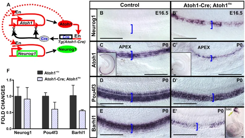
We previously demonstrated that Neurog1 replacement in both alleles ofAtoh1resulted in expression ofNeurog1in HC precursors that differentiate multiple microvilli (Jahan et al., 2012). LikeAtoh1 null mutant mice (Bermingham et al., 1999), homozygousNeurog1 knock-in mice die after birth, precluding postnatal analysis.Atoh1 autoregulates its own expression by activating an Atoh1-specific enhancer (Helms et al., 2000). We speculated that replacement of Atoh1byNeurog1in both alleles might have failed to activate the enhancer for adequateNeurog1expression underAtoh1promoter control (Jahan et al., 2012). To overcome this problem, we generated a novel mouse model that combinedNeurog1knock-in in oneAtoh1 allele with a floxed secondAtoh1allele, the latter to be excised by Atoh1-Cre (Atoh1-Cre; Atoh1f/kiNeurog1; Fig. 1A). This newly developed Atoh1-Cre; Atoh1f/kiNeurog1 model resulted in viable mice, overcoming the neonatal lethality of the homozygous Neurog1 knock-in (Jahan et al., 2012).Atoh1f/+ orAtoh1f/fmice withoutAtoh1-Crewere used as a control in this study except for the RT-qPCR analysis, where Atoh1+/kiNeurog1 mice were used as control for comparison of Neurog1 expression with Atoh1-Cre; Atoh1f/kiNeurog1mice.
In situ hybridization (ISH) showed that Neurog1 knock-in into the Atoh1 locus (Atoh1-Cre; Atoh1f/kiNeurog1) resulted in expression of Neurog1 in OC HCs not detected in control
littermates (Fig. 1B,B′). At P0,Atoh1-Cre; Atoh1f/kiNeurog1mice showed very littleAtoh1expression in the apex (Fig. 1C,C′). By contrast, a downstream target of Atoh1, Pou4f3, was strongly expressed in the HCs of P0 Atoh1-Cre; Atoh1f/kiNeurog1 OC (Fig. 1D,D′), whereas expression of Barhl1 was delayed compared with the vestibular epithelia at P0 (Fig. 1E,E′). At P7, qPCR showed that relative mRNA expression forNeurog1,Pou4f3 and Barhl1 in Atoh1-Cre; Atoh1f/kiNeurog1 cochlea was reduced compared withAtoh1+/kiNeurog1cochlea, but the reduction was not statistically significant (Atoh1+/kiNeurog1 expression was normalized to 1, Fig. 1F). In self-terminatingAtoh1-Cre; Atoh1f/f mice, ISH of bothPou4f3andBarhl1showed patchy loss at P0 (Pan et al., 2012a). By contrast, Neurog1-misexpressing mice revealed expression of bothPou4f3andBarhl1until P7 (Fig. 1F). Partial expression of Pou4f3 and Barhl1 might improve HC viability, as HCs are rapidly lost in eitherPou4f3(Hertzano et al., 2004; Xiang et al., 2003) orBarhl1(Li et al., 2002) null mutants.
Neurog1misexpression with transient expression ofAtoh1
results in HC maintenance
[image:2.612.105.506.428.655.2]Immunohistochemistry of Myo7a (an HC-specific marker) at P7 revealed the formation of an almost continuous row of inner hair cells (IHCs) and two to three rows of outer hair cells (OHCs) in the Neurog1-misexpressing mice (Fig. 2). There were occasionally two rows of IHCs. Extra rows of IHCs coincide with absence of inner pillar (IP) cells, as shown by immunohistochemistry of acetylated tubulin (which is abundant in SCs), next to the extra row of IHCs (Fig. 2B-C‴). There was no indication that the length of the cochlea was changed (supplementary material Table S1). The third row of OHCs disappeared toward the base of the cochlea, including the formation of some gaps in OHCs (Fig. 2C-D‴). Tubulin immunohistochemistry labeled differentiated IP cells, outer pillar
Fig. 1. Our mouse model combinesAtoh1-Cre, a floxedAtoh1allele and anAtoh1allele replaced byNeurog1.(A)Atoh1-Creuses anAtoh1enhancer (En) activated by Atoh1 protein to generate Cre recombinase to excise the floxedAtoh1allele. (B-E′) ISH shows misexpression ofNeurog1in mutant HCs (B′) and its absence in control littermate (B). By contrast,Atoh1shows weak expression in one row of cells in the apical tip of the cochlea inAtoh1-Cre; Atoh1f/kiNeurog1mice (C′and inset) compared with broad expression in control littermate (C and inset).Pou4f3shows near identical expression in mutant and control littermates (D,D′). By contrast,Barhl1is reduced in the cochlea compared with vestibular epithelia inAtoh1-Cre; Atoh1f/kiNeurog1mice (E′, yellow arrow in inset) compared with the control
littermate (E). Brackets mark the OC. (F) qPCR analysis in P7 cochlea indicates that relative expression ofNeurog1,Pou4f3andBarhl1are reduced inAtoh1-Cre; Atoh1f/kiNeurog1cochlea compared withAtoh1+/kiNeurog1cochlea, but are not statistically significant (P=0.8, 0.07 and 0.2, respectively). (Atoh1+/kiNeurog1expression was normalized to 1). Each qPCR data represents the mean of two to three biological and three technical replicates. Error bars represent s.d. Scale bars: 100 µm.
DEVEL
O
(OP) cells and many Deiters’cells at P7 (Fig. 2). Overall,Neurog1 misexpression resulted in a considerable increase in HC and SC formation as compared with Atoh1-Cre; Atoh1f/f mice, which mostly had one row of OHCs and only a few IHCs at P7 (Fig. 3).
We conclude thatNeurog1misexpression combined with transient Atoh1expression can partially substitute forAtoh1to differentiate and maintain HCs, as compared with the massive loss of nearly all HCs in self-terminating mice (Pan et al., 2012a). Diminished long-term viability of HCs correlates with reduced expression of TFs known to be essential for HC viability (Pou4f3,Barhl1).
More HCs form inAtoh1-Cre; Atoh1f/kiNeurog1than in
Atoh1-Cre; Atoh1f/fmice
We next quantified the Myo7a-positive HCs, identifying IHCs as being medial and OHCs lateral to the tubulin-immunopositive IP/OP cells. We counted all cells in a 300 µm stretch near the apex (10%), near the middle (50%) or near the base of the cochlea (90%) in comparable segments from control, Atoh1-Cre; Atoh1f/kiNeurog1 and Atoh1-Cre; Atoh1f/f littermates (Fig. 3D,E; supplementary
material Table S2;n=6). The numbers of IHCs were significantly higher inAtoh1-Cre; Atoh1f/kiNeurog1than inAtoh1-Cre; Atoh1f/fmice (Fig. 3D;P<0.01). IHC counts betweenAtoh1-Cre; Atoh1f/kiNeurog1 mice and control littermates showed no significant difference (Fig. 3D). In summary, replacement of one allele of Atoh1 by Neurog1 combined with a self-terminating second Atoh1 allele rescued most IHCs as compared with the massive loss of IHCs in the Atoh1-Cre; Atoh1f/fmouse.
The quantification of OHCs demonstrated that significantly more OHCs form in Atoh1-Cre; Atoh1f/kiNeurog1 compared with
[image:3.612.219.556.55.474.2]Atoh1-Cre; Atoh1f/f mice (Fig. 3E; in the apex, P<0.05; in the
Fig. 2. Misexpression ofNeurog1results in differentiation of HCs and SCs.Myo7a (HC) and tubulin (SC) immunohistochemistry in P7Atoh1-Cre; Atoh1f/kiNeurog1mice shows one row of IHCs and two to three rows of OHCs
with near normal SCs (A-D‴). There is loss of the third row of OHCs (D-D‴) and gaps in OHCs (brackets in C-D‴) towards the base of the cochlea. Some additional IHCs are associated with a loss of IP cells (arrows in B-C‴). Scale bars: 10 µm.
Fig. 3. Misexpression ofNeurog1rescues IHC formation.(A-C) Myo7a-positive HCs inAtoh1-Cre; Atoh1f/kiNeurog1cochlea are compared with those in equivalent segments ofAtoh1-Cre; Atoh1f/fand control littermates at P7. The
Atoh1-Cre; Atoh1f/kiNeurog1cochlea has more HCs (B), in contrast to a few patchy IHCs and one row of OHCs in theAtoh1-Cre; Atoh1f/fcochlea (C). Brackets highlight gaps in HCs. (D) Quantification of HCs reveals that the numbers of IHCs inAtoh1-Cre; Atoh1f/kiNeurog1mice (red bar) are significantly
higher than those inAtoh1-Cre; Atoh1f/fmice (green bar) but are comparable to those of control littermates (blue bar) in all three areas. (E) However, OHC numbers are reduced inAtoh1-Cre; Atoh1f/kiNeurog1mice compared with the control (blue bar), but are significantly above those inAtoh1-Cre; Atoh1f/fmice
(green bar). Misexpression ofNeurog1preferentially rescues IHC formation. Each bar represents the mean of six cochleae. Error bars represent s.d. *P<0.05, **P<0.01. Scale bars: 10 µm.
DEVEL
O
[image:3.612.65.378.58.446.2]middle and base of the cochlea,P<0.01). In contrast to IHCs, OHCs in Atoh1-Cre; Atoh1f/kiNeurog1 mice were significantly reduced compared with control littermates (Fig. 3E; in the apex,P<0.05; in the middle and base of the cochlea, P<0.01). Misexpression of Neurog1increased survival preferentially of IHCs, with the greatest reduction in the third row of OHCs (Fig. 2C-D‴, Fig. 3B,E). Previous work on self-terminatingAtoh1conditional null mice (Pan et al., 2012a) andAtoh1hypomorphs (Sheykholeslami et al., 2013) showed a differential loss of OHCs, with the third row being most susceptible, whereas tamoxifen-inducedAtoh1-Cre wiped out all HCs rapidly (Cai et al., 2013; Chonko et al., 2013).
HCs survive longer inAtoh1-Cre; Atoh1f/kiNeurog1than in
Atoh1-Cre; Atoh1f/fmice
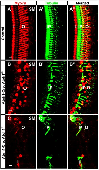
We next investigated the longevity of these HCs using Myo7a and tubulin immunohistochemistry (Fig. 4). At 9 months, many Myo7a-positive HCs remained in theAtoh1-Cre; Atoh1f/kiNeurog1mice. In particular, many IHCs (medial to the tubulin-positive IP cells) survived, but OHCs showed patchy loss (Fig. 4B-B″). This contrasted with severe HC loss inAtoh1-Cre; Atoh1f/fmice, where only a few HCs survived up to 9 months (Fig. 4C-C″), essentially being largely lost by 3 weeks (Pan et al., 2012a). In addition to HCs, SCs also persisted longer inAtoh1-Cre; Atoh1f/kiNeurog1mice and many more tubulin-positive pillar cells and Deiters’ cells were observed compared withAtoh1-Cre; Atoh1f/fmice (Fig. 4B′,B″,C′,C″).
Atoh1-Cre; Atoh1f/kiNeurog1
mice are deaf, despite the presence of most HCs
The proper arrangement of cells is essential for the function of the OC to enable sound perception (Cai et al., 2003; Jacobo and Hudspeth, 2014). To assess hearing function in Neurog1 -misexpressing mice, we measured the auditory brainstem response (ABR) to click stimuli at P30 (Fig. 5).Atoh1-Cre; Atoh1f/kiNeurog1 mice showed no ABR, in contrast to control littermates (Fig. 5). We concluded that the expression of Neurog1 facilitates the development and maintenance of many HCs in a non-functional OC. We next investigated possible reasons for this functional defect, focusing first on the stereocilia of HCs.
ReplacingAtoh1byNeurog1results in aberrant stereocilia bundle formation
HC function depends on the normal development of the stereocilia bundles that mediate mechanotransduction (Hudspeth, 2014; Jacobo and Hudspeth, 2014; Sienknecht et al., 2014). Loss, alteration or reduction of Atoh1 results in abnormal stereocilia bundle formation (Chonko et al., 2013; Pan et al., 2012a), if any bundle forms at all (Pan et al., 2011).Atoh1dosage and timing of expression are important for proper stereocilia development and maturation (Jahan et al., 2013). Several mutations that interfere with stereocilia bundle organization and homeostasis are known (Hertzano et al., 2008; Kitajiri et al., 2010; Mogensen et al., 2007; Sekerková et al., 2011) and many of these result not only in aberrant bundle morphology but also in the death of HCs (Kersigo and Fritzsch, 2015; Self et al., 1998; Ueyama et al., 2014). Consistent with a role ofAtoh1in stereocilia differentiation, forced expression ofAtoh1can restore hair bundles (Yang et al., 2012).
[image:4.612.77.273.341.676.2]To extend our findings beyond the obvious increase in HC survival (Figs 2–4) and to better understand the apparent deafness (Fig. 5), we next investigated how replacement of Atoh1 with Neurog1 influences stereocilia formation and thus the function of HCs. We examined at least three cochlea of the Atoh1-Cre; Atoh1f/kiNeurog1mutant and littermate controls, each at three different stages (P1, P7 and P22), by scanning electron microscopy (SEM) (Figs 6 and 7). We found that the onset of stereocilia bundle differentiation in Neurog1-misexpressing mice was delayed (Fig. 6A-B″). The bundles were immature, with the formation of
Fig. 4.Neurog1misexpression improves longevity of IHCs.
Immunohistochemistry of Myo7a and tubulin shows that, compared with the control (A-A″), both IHCs and OHCs and many SCs survive until 9 months in
[image:4.612.349.524.513.704.2]Atoh1-Cre; Atoh1f/kiNeurog1mice (B-B″). By contrast, only some rare OHCs and SCs survive in theAtoh1-Cre; Atoh1f/flittermate (C-C″). IP, inner pillar cell; I, IHC; O, OHC; P, pillar cell (inner/outer). Scale bars: 10μm.
Fig. 5.Atoh1-Cre; Atoh1f/kiNeurog1mice show no ABR response.P30
Atoh1-Cre; Atoh1f/kiNeurog1mice show no click response in ABR (B), in contrast to the control (A), indicating severe deafness over the intensity range tested.
DEVEL
O
microvilli that were all similar in length surrounding a central kinocilium at P1 (Fig. 6B-B″). Control littermates already displayed the staircase pattern of progressively increasing length of stereocilia modiolar to an acentric kinocilium (Fig. 6A). This stunted stereocilia growth was more obvious in OHCs than IHCs, especially in the second and third rows of OHCs ofAtoh1-Cre; Atoh1f/kiNeurog1mice. In Atoh1-Cre; Atoh1f/kiNeurog1 mice at P7, the bundles formed a nearly normal staircase pattern in the apex of the cochlea, whereas the basal HCs displayed aberrations such as fusion or loss of stereocilia, predominantly in the third row of OHCs (Fig. 6C-D″).
SEM revealed progressive deformations of the stereocilia bundles in Atoh1-Cre; Atoh1f/kiNeurog1 mice (Fig. 7). Some HCs in the
Neurog1-misexpressing mice had stereocilia bundles that lacked the staircase organization, and some stereocilia were fused with each other or showed stunted growth (cyan arrows in Fig. 7). SEM also revealed that the thickness of the stereocilia of some IHCs in Atoh1-Cre; Atoh1f/kiNeurog1mice was altered to resemble OHCs, indicating partial cell fate switching of some IHCs to OHCs (red arrows in Fig. 7). Beyond the fusion and ectopic stereocilia formation, SEM in the Neurog1-misexpressing mice also revealed disruption of the boundaries between adjacent HCs (Fig. 7). Some HCs touched each other without intervening SCs inAtoh1-Cre; Atoh1f/kiNeurog1mice (green arrows in Fig. 7). At P7 and P22, we found ectopic stereocilia bundles protruding from the apical surfaces of the IP cells (yellow arrows, Fig. 7). This altered pattern of HCs and SCs indicated disruptions of cell-cell interactions. We therefore further analyzed the molecular basis of OC pattern formation (Fritzsch et al., 2014b; Groves and Fekete, 2012), focusing on genes known to affect OC development.
We previously demonstrated conversion of OHCs into IHCs in Neurod1conditional deletion mice (Jahan et al., 2010). Absence of Neurod1 resulted in premature and altered Atoh1 and Fgf8 expression and transformation of thin OHC stereocilia into thick IHC stereocilia (Jahan et al., 2010, 2013). Essentially,Atoh1and Fgf8expression failed to be suppressed byNeurod1inNeurod1 conditional deletion mice (Jahan et al., 2010). We investigated the expression ofNeurod1andFgf8in theAtoh1-Cre; Atoh1f/kiNeurog1 mice, as they showed the opposite effect, i.e. conversion of stereocilia of IHCs to OHCs. Misexpression ofNeurog1resulted in stronger and earlier expression of Neurod1 in E16.5 Atoh1-Cre; Atoh1f/kiNeurog1 cochlea as compared with control littermates
(Fig. 8A-B″). Fgf8 expression was patchy and much reduced in some IHCs of the E16.5Atoh1-Cre; Atoh1f/kiNeurog1mice (Fig. 8D′; supplementary material Fig. S1B′). The alteration of HC stereocilia diameter in Atoh1-Cre; Atoh1f/kiNeurog1 mice might relate to the locally altered level ofAtoh1/Neurog1signaling that distorted the specificity of HC types. How alteration of Atoh1 signal level mechanistically regulates the thickness of the stereocilia bundles, possibly through modulation of whirlin (Mogensen et al., 2007), requires further work.
Among microRNAs (miRNAs), miR-96 was specifically reported as being required for the proper maturation and organization of the stereocilia bundle (Kuhn et al., 2011; Lewis et al., 2009). Single base mutation in theMIR96gene results in non-syndromic, progressive hearing loss in humans (Kuhn et al., 2011; Lewis et al., 2009). miR-96 is expressed in the sensory epithelia of the mouse cochlea (Weston et al., 2011). We previously reported very limited miR-96 expression only in the apex of Atoh1kiNeurog1/kiNeurog1 cochlea (Jahan et al., 2012) or its near absence in conditional Atoh1 null mice (Pan et al., 2011). Both mutants showed no HC differentiation beyond precursors cells. In theAtoh1-Cre; Atoh1f/kiNeurog1mice, miR-96 expression was almost normal, except for the basal hook region where it showed some gaps (Fig. 8E-F′). How possible alterations in some miRNAs tie into stereocilia development through the repression of relevant genes remains speculative.
Neurog1misexpression alters OC patterning and SC
differentiation
[image:5.612.49.357.57.275.2]Given the formation of stereocilia bundles on IP cells (Fig. 7), we next investigated the distribution of an IP cell-specific marker,p75 (Ngfr) (von Bartheld et al., 1991). ISH ofp75confirmed its selective expression in IP cells in P0 control mice (Fig. 9A). In P0Atoh1-Cre; Atoh1f/kiNeurog1 mice, p75 expression was patchy near the base (Fig. 9B′,B″) but continuous in the apical half (Fig. 9B) of the cochlea. Consistent with stereocilia bundles on IP cells (Fig. 7A-C, E), immunohistochemistry for the HC-specific Myo7a subsequent top75ISH showed that the gaps in thep75-positive IP cells were filled with Myo7a-positive HCs (Fig. 9B″). We also performed dual immunohistochemistry using p75 and Myo7a antibodies with different fluorophore labeling of the secondary antibodies. This also showed that the gaps in p75-immunopositive IP cells were filled
Fig. 6. Delayed onset of maturation of stereocilia bundles inAtoh1-Cre; Atoh1f/kiNeurog1mice.SEM shows that stereocilia bundles remain immature with central kinocilia at P1 inAtoh1-Cre; Atoh1f/kiNeurog1mice (B-B″) and there is no staircase pattern, in contrast to control littermates (A). This delay is most evident in OHCs (B-B″). In P7Atoh1-Cre; Atoh1f/kiNeurog1cochlea, the stereocilia bundles in the apical HCs form a near normal staircase pattern (D,D′), as compared with the control (C), whereas the third row of basal OHCs shows aberration (D″). A portion of HCs are color coded (A-B″) for better visibility: IHCs in cyan and OHCs in yellow. I, IHC; O1-O3, 1-3 rows of OHCs. Scale bars: 5 µm in A-B″,D-D″; 10 µm in C.
DEVEL
O
with Myo7a-positive putative HCs (Fig. 9C-D″). Combined with stereocilia formation in the apical surfaces of the IP cells of Atoh1-Cre; Atoh1f/kiNeurog1 mice, our data suggested that expression of
Neurog1transformed some IP cells into a hybrid of IP and HC. To further assess this possibility, we quantified the tubulin-immunopositive IP cells in P7 Atoh1-Cre; Atoh1f/kiNeurog1 mice (Fig. 9E). There was a reduction of tubulin-positive IP cells in the Neurog1-misexpressing mice relative to control littermates in the apex (P<0.05) and middle (P<0.01) turn of the cochlea. Despite this presumed transformation, overall IP cell formation was increased in Atoh1-Cre; Atoh1f/kiNeurog1 relative to Atoh1-Cre; Atoh1f/f mice (P<0.01). Although p75 is an excellent marker for IP cells, differentiation of IP cells remains normal inp75null mice and its function in IP cells remains obscure (Tan et al., 2010).
Pillar cell formation is abnormal inFgf8null mice (Jacques et al., 2007; Mueller et al., 2002). As previously reported (Jacques et al., 2007; Pirvola et al., 2000), Fgf8 is expressed in all IHCs. In the Atoh1-Cre; Atoh1f/kiNeurog1 mice, downregulation of Fgf8
expression might affect the strength of diffusible signal from IHCs to nearby SCs (Groves and Fekete, 2012). Given the defects in IP cells, we next performed double ISH for Fgf8 and p75 and revealed that the reduction ofFgf8expression correlated with that of p75 in Atoh1-Cre; Atoh1f/kiNeurog1 mice (supplementary material Fig. S1A-C′). Previous work also demonstrated that Atoh1 is transiently expressed in IP cells (Driver et al., 2013; Matei et al., 2005). Signals that prevent IP cells from differentiating as HCs might be disrupted in theAtoh1-Cre; Atoh1f/kiNeurog1mice, resulting in partial transdifferentiation of some IP cells into HCs (Fig. 7).
We also investigated theFgf10andBmp4expression that flanks the medial and lateral borders of the OC, respectively (supplementary material Fig. S1D,D′). In P0Atoh1-Cre; Atoh1f/kiNeurog1mice,Fgf10 was drastically reduced and Bmp4 showed expanded expression toward OC, with gaps where OC cells were replaced by simple epithelium (supplementary material Fig. S1D,D′).
To obtain further molecular insight into SC differentiation, we studied Prox1 expression by ISH in the Neurog1-misexpressing mice (Fritzsch et al., 2010a). Prox1 was expressed in almost all SCs, except for some reduction in the IP cells in P0Atoh1-Cre; Atoh1f/kiNeurog1 mice (Fig. 10A-B″). These data suggested that several reliable markers of SC differentiation were altered, lost or even replaced by HC markers, indicating that expression ofNeurog1 in HCs affected other cells of the OC.
Neurog1expression alters Notch ligand and Notch effector
gene expression
Atoh1expression in HCs regulates the expression of genes in the Delta/Notch signaling pathway that are necessary for the development of the surrounding SCs and maintains the proper patterning of the OC (Kobayashi and Kageyama, 2014; Sprinzak et al., 2011; Yamamoto et al., 2014). Lack of the Notch ligandJag1 results in extra rows of IHCs and the loss of OHCs (Brooker et al., 2006; Kiernan et al., 2006). The Hes/Hey factors, which are downstream target genes of Notch, play important roles in the proper specification of SCs by negatively regulating pro-neuronal bHLH TFs (Doetzlhofer et al., 2009; Zine and de Ribaupierre, 2002). Given the lack of proper patterning of HCs/SCs in the OC, we investigated the expression ofJag1andHes5by ISH in Atoh1-Cre; Atoh1f/kiNeurog1 mice (Fig. 10). In the P0 control mice,Jag1 was widely expressed in the greater epithelial ridge (GER) and in SCs, particularly in the IP cells (Fig. 10C,C′). InAtoh1-Cre; Atoh1f/kiNeurog1mice, the expression ofJag1was slightly reduced in both the GER and SCs, with some patchy loss toward the cochlear base (Fig. 10D,D′).
At E14.5, Hes5 was found in the mid-base of control
cochlea (supplementary material Fig. S2A). E14.5 Atoh1-Cre; Atoh1f/kiNeurog1mice had delayedHes5expression (supplementary material Fig. S2D); Hes5 was upregulated at later stages
(supplementary material Fig. S2E,F) and showed patchy
expression in the GER and in SCs (Fig. 10E-F″; supplementary
material Fig. S2E,F). We combined Hes5 ISH with Myo7a
[image:6.612.53.298.58.381.2]immunohistochemistry to detect whether the downregulation of Hes5 was associated with HC differentiation in the Neurog1 -misexpressing mice (supplementary material Fig. S2G-H″), as previously reported afterHes5loss (Zine and de Ribaupierre, 2002). Myo7a-positive HCs showed no correlation with the patchy loss of Hes5expression in this mutant (supplementary material Fig. S2G-H″). Alteration of Jag1 and Hes5 expression in Atoh1-Cre; Atoh1f/kiNeurog1mice indicated the potential effects ofNeurog1on the expression of these genes, and thus on Delta/Notch signaling. This altered HC-SC communication might disrupt OC patterning
Fig. 7.Neurog1misexpression results in alteration in stereocilia bundle thickness, cell fate changes and disruption of cell-cell interactions.
AlthoughNeurog1misexpression shows near normal HC formation (Figs 2-4), HC type and topology-specific stereocilia bundle formation at P7 (A,D,G) and P22 (B,C,E,F,H,I) are disorganized. Ectopic HCs form in the position of IP cells, as revealed by formation of stereocilia bundles from their apical surfaces (yellow arrows in A-C,E). In addition,Neurog1misexpression results in transdifferentiation of IHCs into OHCs, as demonstrated by the appearance of thin stereocilia of OHCs in the position of IHCs (red arrows in B,D-F). Some OHCs are in broad continuity with each other (green arrows in A,G,I) and some stereocilia bundles are fused or stunted in growth (cyan arrows in C,F,H).
Neurog1misexpression results in irregular stereocilia formation in most HCs, that gradually deteriorate with age. I, IHC; O, OHC; P, pillar cell. Scale bars: 5 µm in A,C-E,G,H; 10 µm in B,F; 3 µm in I.
DEVEL
O
(Fig. 7), but did not result in an overproduction of HCs (supplementary material Fig. S2H-H″), unlike in Hes/Hey mutants (Benito-Gonzalez and Doetzlhofer, 2014; Zine et al., 2001). How changes in intracellular HC gene expression through replacement ofAtoh1byNeurog1alter intercellular signaling and disrupt the mosaic of the OC remains unclear.
In conclusion, near normal numbers of HCs can be generated by combining transientAtoh1expression withNeurog1misexpression, but these HCs have variably defective stereocilia bundles and the OC is disorganized. Despite long-term viable HCs, the abnormal OC organization causes deafness. OC disorganization may be a consequence of changes inFgf8expression, which might lead to the formation of secondary signaling centers comparable to the midbrain hindbrain boundary (Fritzsch et al., 2014a; Lee et al., 1997), or may also be due to altered cellular interactions (Groves and Fekete, 2012).
DISCUSSION
A fundamental aim in the study of neurosensory development, and in the context of regeneration in particular, is understanding the molecular basis for the generation of topologically distinct cell fates from uniform progenitor populations (Fritzsch et al., 2014a; Imayoshi and Kageyama, 2014; Reiprich and Wegner, 2015). Precise spatial and temporal control of gene expression by different combinations of TFs establish the molecular code that determines cell fate (Guillemot, 2007). Molecular dissection of this complexity requires models of limited cellular diversity in a stereotyped arrangement to evaluate minute deviations from normal; for example, in the ommatidia of flies (Johnston and Desplan, 2014). The OC of the mammalian inner ear is another excellent model organ with stereotyped cellular patterning that allows the exploration of molecularly induced deviations of developmental processes mediated
by intracellular and extracellular patterning processes (Fritzsch et al., 2014b; Groves and Fekete, 2012). Our data suggest a profound effect of intracellular signals via cell-cell interactions and alterations in diffusible factors on the cellular and organ patterning process.
Neurog1cooperates with transientAtoh1expression to
develop and maintain HCs
One approach to probing the signal specificity of a given TF in developing gene regulation networks is to replace them by closely related TFs (Guillemot, 2007). ReplacingAtoh7withNeurod1leads to normal differentiation of RGCs, whereas replacingNeurod1byAtoh7 alters the cell fate of amacrine and photoreceptor cells into RGCs (Mao et al., 2013, 2008), indicating context dependency of gene actions. We knockedNeurog1into theAtoh1locus to test whether a bHLH TF that is exclusively associated with proliferative precursors in the ear and brain (Imayoshi and Kageyama, 2014; Ma et al., 2000) can function in differentiation to maintain or alter HC precursor differentiation. We previously showed that Atoh1kiNeurog1/kiNeurog1 can effectively driveNeurod1in HC precursors (Jahan et al., 2012) but can neither initiate normal HC development nor maintain the viability of HC precursors. We also showed that self-terminating Atoh1 results in very limited viability of HCs, with incomplete stereocilia differentiation (Pan et al., 2012a). Our quantitative assessment of HC formation inAtoh1-Cre; Atoh1f/kiNeurog1mice in this study indicates thatNeurog1misexpression partially rescues HCs and maintains HCs for a longer period, as compared with self-terminatingAtoh1conditional null mice (Pan et al., 2012a).Neurog1 cannot maintain the basal third row of OHCs that depends on Fgf20 released from SCs (Huh et al., 2012), a factor that might be affected in our mice due to alteration in SC development.
[image:7.612.50.357.56.370.2]Our data suggest that HC precursors behave like RGCs (Mao et al., 2008) and show limited flexibility to respond to the distantly
Fig. 8.Neurog1misexpression induces premature
Neurod1upregulation and suppressesFgf8but not miR-96 expression.ISH shows that expression ofNeurod1
expands both longitudinally and radially in the HCs of
Atoh1-Cre; Atoh1f/kiNeurog1cochlea as compared with the control littermate, in which expression is limited to IHCs (A-B″).
Fgf8expression displays patchy downregulation (arrows) in the E16.5Atoh1-Cre; Atoh1f/kiNeurog1cochlea (C-D′). This suppression ofFgf8might in part be regulated byNeurod1
overexpression inAtoh1-Cre; Atoh1f/kiNeurog1mice (B,B′,D,D′),
as previously suggested (Jahan et al., 2010). miR-96, an essential miRNA for stereocilia differentiation, shows no expression changes in HCs (E-F′). Scale bars: 100 µm.
DEVEL
O
related bHLH TF Neurog1 beyond enhanced HC differentiation relative to that of transientAtoh1expression. This differs from spiral ganglion neurons, which differentiate readily as HCs if suppression ofAtoh1is removed by eliminatingNeurod1(Jahan et al., 2010). Atoh1 is required for an uncharacterized initial step of HC differentiation that neitherNeurog1norNeurod1can replace, and thus no HCs differentiate in homozygotic Atoh1kiNeurog1/kiNeurog1 mice (Jahan et al., 2012). The flyatonalgene can replaceAtoh1 (Wang et al., 2002) and HCs can partially differentiate with the reduced (Sheykholeslami et al., 2013) or transient (Pan et al., 2012a) expression of Atoh1, or even in the absence ofAtoh1 in chimeric mice (Du et al., 2007). Elucidating the molecular basis of this critical step of downstream gene activation (Cai et al., 2015; Pan et al., 2012b), in whichAtoh1expression cannot be substituted by
Neurog1, could help to transform stem cells more effectively into HCs (Ronaghi et al., 2014; Zine et al., 2014).
Atoh1 cooperates with unknown factors to fully expressPou4f3 (Ahmed et al., 2012), which, in turn, cooperates with Atoh1 to maintain HCs (Chen et al., 2015; Hertzano et al., 2004; Masuda et al., 2012; Xiang et al., 2003). In contrast toPou4f3,Barhl1expression depends exclusively onAtoh1(Chellappa et al., 2008).Barhl1 is required for HC survival even in the presence ofAtoh1andPou4f3 (Li et al., 2002).Atoh1-Cre; Atoh1f/kiNeurog1mice have near normal expression ofPou4f3, but show a delay and progressive reduction of Barhl1(Fig. 1). We suggest thatNeurog1might activate a second pathway for near normalPou4f3expression (Ahmed et al., 2012; Masuda et al., 2012), but fails to fully activateBarhl1needed for HC maintenance (Fig. 11). Our mouse model will prove useful in testing the ability of putative regulators of downstream genes to enhance HC viability through the regulation ofPou4f3andBarhl1expression.
Replacement ofAtoh1byNeurog1alters stereocilia differentiation
Closer investigation of the cochlea ofNeurog1-misexpressing mice revealed various irregularities in stereocilia bundles and their distribution in the OC (Figs 6 and 7): the irregular length of individual stereocilia within the bundles; the uncoupling of stereocilia diameter from HC type; the appearance of ectopic stereocilia bundles on IP cells; and stereocilia bundles being abnormally fused with each other.
Stereocilia are necessary for hearing (Müller and Barr-Gillespie, 2015). Despite the rescue of overall HC formation, Atoh1-Cre; Atoh1f/kiNeurog1mice show no ABR and are deaf (Fig. 5). Whether this is due to some defect in the HCs or to the disorganization of the OC requires future single-cell recordings on isolated HCs. We presume that the irregularities in stereocilia bundles, which are essential for mechanoelectric transduction (Hudspeth, 2014), preclude the normal function of many HCs. Loss ofPou4f3, but not ofBarhl1, causes stereocilia bundle aberrations (Chellappa et al., 2008; Hertzano et al., 2004). A reduced level ofPou4f3 transcripts at or after P7 might contribute to the bundle aberration that develops mostly in late postnatal stages inNeurog1-misexpressing mice (Fig. 7).
Actin is a major protein component of stereocilia (Müller and Barr-Gillespie, 2015) and stereocilia homeostasis is essential for HC function and viability (Kersigo and Fritzsch, 2015; Self et al., 1999; Ueyama et al., 2014). Dysregulation of actin bundling has been associated with HC dysfunction and loss (Mogensen et al., 2007; Perrin et al., 2010; Rzadzinska et al., 2009; Taylor et al., 2015). Various myosins, actins and a rich variety of actin-bundling proteins are regulated downstream of Atoh1 to transform microvilli into stereocilia during development (Kitajiri et al., 2010; Schwander et al., 2010). Loss of miRNAs is also associated with derailed stereocilia development (Weston et al., 2011). Neurog1 misexpression results in near normal miR-96 expression, but other miRNAs could be altered that also affect stereocilia formation. Once causality between Atoh1 and downstream signals associated with various bundle aberrations is clearer, our mouse model could help to eliminate spurious relationships as it has partially uncoupled stereocilia differentiation from HC topology.
Replacement ofAtoh1byNeurog1alters the patterning of the OC
[image:8.612.61.289.54.451.2]The OC is highly stereotyped in organization, with two distinctly patterned compartments separated by a single row of adjacent IP cells, which express Atoh1 (Driver et al., 2013; Fritzsch et al., 2014b; Matei et al., 2005) without differentiating into HCs.
Fig. 9. Myo7a-positive‘HCs’fill gaps inp75-negative IP cells in
Atoh1-Cre; Atoh1f/kiNeurog1mice.ISH ofp75shows continuous expression in IP cells in the apical half (mid), comparable to the control littermates, but the presence of gaps in the base (A-B″). Bracket specifies the OC. Combining Myo7a immunohistochemistry with thep75ISH-positive cochlea shows that some Myo7a-positive‘HCs’form in the gaps betweenp75-positive IP cells (arrows in B″), as further confirmed (arrows in D,D′) by double
immunohistochemistry of p75 and Myo7a (C-D′). Quantification of IP cells (E) demonstrates decreased numbers inAtoh1-Cre; Atoh1f/kiNeurog1mice compared with control littermates, but significantly increased numbers compared with those inAtoh1-Cre; Atoh1f/flittermates. *P<0.05, **P<0.01. Each bar represents the mean of six cochleae. Error bars represent s.d. Scale bars: 10μm.
DEVEL
O
Expression of multiple Hes/Hey factors (Benito-Gonzalez and Doetzlhofer, 2014; Doetzlhofer et al., 2009; Petrovic et al., 2015) may block these cells from HC development. Notch inhibition (and thus reduced expression of Hes/Hey) may lead to HC differentiation of IP cells (Mizutari et al., 2013).Neurog1misexpression affects IP cells in multiple ways, including the formation of HC-like stereocilia bundles and replacement by the HC marker Myo7a in p75-positive IP cells. These instabilities in IP cells might relate to the alteration in Notch signaling, as observed (Figs 9 and 10).
In addition, the disruption of OC patterning is in part due to alterations in the Delta/Notch signaling pathway. For example, expression of Jag1and of the downstream target geneHes5 are altered. Previous work onJag1loss showed a reduction in the total number of HCs (Kiernan et al., 2006). Neurog1 misexpression results in discontinuity of Jag1 and delayed upregulation and differential downregulation of Hes5 in the cochlea. These
expression changes vary radially and longitudinally, providing additional modulations of the variable signals of diffusible factors such as Fgf8, Fgf10 and Bmp4.Combined with altered SC response properties through changes inp75andProx1, these local changes might relate to the random OC patterning defects.
In summary, we demonstrate that misexpression of Neurog1 provides partial functional replacement ofAtoh1in the developing
HC and improves HC maintenance. Neurog1 misexpression
[image:9.612.48.341.57.383.2]changes the signaling pattern of diffusible factors originating from HCs, as well as cell-cell interactions via Delta/Notch; both alterations contribute to the disorganization of the OC. Previous reports on the deletion of different combinations ofHes1,Hes5or Hey2show changes in sensory patterning in support of the lateral inhibition model, but mutants mostly maintain the HC and SC mosaic formation (Doetzlhofer et al., 2009; Zine et al., 2001). Changes of proneural bHLH genes in HCs alters the overall
Fig. 10.Neurog1misexpression in HCs alters SC marker expression pattern.(A-B″) ISH reveals near normal expression ofProx1in SCs, except for some patchy loss in the base of Atoh1-Cre; Atoh1f/kiNeurog1mice. (C-D′)Jag1expression shows patchy loss (arrow in D′) inAtoh1-Cre; Atoh1f/kiNeurog1mice. (E-F″)
Neurog1misexpression results in differential loss ofHes5
expression (arrows in F′,F″). Curved brackets demarcate expression in the GER and square brackets demarcate expression in the OC. Scale bars: 100 µm except 10 µm in C′ and D′.
Fig. 11. Role ofAtoh1andNeurog1in HC fate determination.
Summary flowchart indicating thatAtoh1is essential for HC fate determination, in part mediated by an unknown bHLH gene (Ahmed et al., 2012).Neurog1may in part mimic this unknown bHLH gene to cooperate withAtoh1and maintain the expression of some target genes. However,Neurog1cannot fully regulate downstream HC genes associated with stereocilia maturation and OC patterning, leading to disorganization of the OC in the absence ofAtoh1.
DEVEL
O
[image:9.612.50.352.598.739.2]patterning of the OC, presumably by altering intercellular interactions via diffusible factors (Fgf8) and cell-cell interactions.
Understanding the causalities of these alterations and translating such understanding into regeneration (Zine et al., 2014) could help to restore hearing in deaf patients, with deafness now constituting the fastest growing ailment of the elderly worldwide (Kersigo and Fritzsch, 2015; Müller and Barr-Gillespie, 2015). Beyond the addition or deletion of entire rows of HCs obtained with previous mutations (Doetzlhofer et al., 2009; Zine et al., 2001), we provide here a new model of a dysfunctional OC that can help in OC restoration endeavors. Converting, through additional manipulation, the dysfunctional OC of our new mouse model into a functional OC that is able to restore hearing could provide proof of principle for fledgling attempts that aim to transform a partially defunct elderly OC into a fully functional OC.
Note added in proof
The pattern of the organ of Corti was recently summarized by Jahan et al. (2015).
MATERIALS AND METHODS
Ethics guidelines
All animal procedures were carried out according to the recommendations and guidelines of the University of Iowa Institutional Animal Care and Use Committee (IACUC) under approved protocol ACURF #1309175.
CombiningNeurog1knock-in (Atoh1kiNeurog1) with self-terminatingAtoh1mice (Atoh1-Cre; Atoh1f/kiNeurog1)
Construction of the Neurog1 knock-in plasmid and generation of the
Atoh1kiNeurog1mouse model were described previously (Jahan et al., 2012).
To generate the Atoh1-Cre; Atoh1f/kiNeurog1 line, we bred heterozygous
Neurog1knock-in mice (Atoh1+/kiNeurog1) (Jahan et al., 2012) with mice
carrying theAtoh1-Cretransgene and oneAtoh1floxed allele (Atoh1-Cre;
Atoh1f/+) as described previously (Pan et al., 2012a).
Genotyping
Mice were genotyped using tail DNA for standard PCR amplification as described previously (Jahan et al., 2012; Pan et al., 2012a). For further details, see the supplementary Materials and Methods.
In situhybridization
ISH was performed as previously described (Jahan et al., 2012) using RNA probes labeled with digoxigenin. A detailed description is provided in the supplementary Materials and Methods.
RT-qPCR
For quantitative reverse transcription PCR (RT-qPCR), we used cochlea from P7Atoh1-Cre; Atoh1f/kiNeurog1mice and their heterozygousAtoh1+/kiNeurog1
littermates as control. After sedation with 2,2,2 tribromoethanol, the mice were hemisected and the cochleae were dissected out within 2-3 min in RNase-free conditions and stored in RNAlater (Ambion) at−80°C. Total RNA extraction was performed using the Direct-zol RNA Mini-Prep Kit (Zymo Research) and RNA concentration and 260/280 ratio were obtained with a Nanodrop spectrophotometer. On-column DNase I treatment was performed according to the Direct-zol Kit protocol. 1 µg total RNA was reverse transcribed and cDNA was synthesized with Anchored-oligo (dT)18
primer using the Transcriptor First Strand cDNA Synthesis Kit (Roche). For qPCR, the primers and probes were designed using the Roche Universal ProbeLibrary Assay Design Center and primers were obtained from Integrated DNA Technologies. Primer sequences and priming conditions are listed in supplementary material Table S3. qPCR was performed in a 96-well plate using Roche LightCycler 480 Probes Master Mix and a Roche Light Cycler 480 real-time PCR machine. For all target genes, qPCR was performed for at least three to four biological replicates and three technical replicates including no-template controls for each sample following MIQE
guidelines (Bustin et al., 2009). qPCR data were analyzed in Microsoft Excel andΔΔCTwas calculated to determine the relative expression in
Atoh1-Cre; Atoh1f/kiNeurog1cochlea compared with control normalized to
Actbreference transcript. Statistical analysis was performed with Student’s
t-test in GraphPad Prism 6 software.
Immunohistochemistry and cell counts
Immunohistochemistry was performed as described previously (Jahan et al., 2012). IHCs, OHCs and IP cells were counted in the P7 control,Atoh1-Cre;
Atoh1f/kiNeurog1 and Atoh1-Cre; Atoh1f/f mice in 300 µm stretches in
comparable regions in the apex, middle and base of the cochlea after performing immunohistochemistry for Myo7a and tubulin. Quantification was performed in six independent cochleae from each genotype. For further details, see the supplementary Materials and Methods. The data obtained from the quantification of IHCs, OHCs and IP cells were analyzed using Student’st-test.P<0.05 was considered statistically significant.
Auditory brainstem response (ABR) recording
Following sedation, ABR recording was performed in 1-month-old control
andAtoh1-Cre; Atoh1f/kiNeurog1littermate mice. A loudspeaker was placed
10 cm from the pinna of the test ear and computer-generated clicks were given in an open field environment in a soundproof chamber. Click responses were averaged and recorded signals were bandpass filtered (300 Hz-5 kHz) with a 60 Hz notch filter. The sound level was decreased in 10 dB steps from a 96 dB sound pressure level until there was no noticeable response. For further details, see the supplementary Materials and Methods.
Scanning electron microscopy (SEM)
SEM was performed as previously described (Jahan et al., 2012). A detailed description is provided in the supplementary Materials and Methods.
Acknowledgements
We thank Dr Fernando Giráldez for constructive and valuable comments on the manuscript; Qiufu Ma (Neurog1), Huda Y. Zoghbi (Atoh1), Mengqing Xiang (Barhl1 andPou4f3), Jacqueline E. Lee (Neurod1), Ulla Pirvola (Fgf8), Thomas Gridley (Jag1), Brigid L. M. Hogan (Fgf10), Doris K. Wu (Bmp4), Andy Groves (Hes5) and Guillermo Oliver (Prox1) for providing plasmids forin situhybridization; the Central Microscopy Research Facility for carrying out SEM; the Roy J. Carver Center for Imaging for use of the Leica TCS SP5 confocal microscope; the Roy J. Carver Center for Genomics for use of the Roche 480 LightCycler; and the Iowa Center for Molecular Auditory Neuroscience (P30; ICMAN) for the use of the ABR facility at the University of Iowa.
Competing interests
The authors declare no competing or financial interests.
Author contributions
I.J., N.P. and B.F. conceived the work; N.P. and J.K. performed mouse breeding and genotyping; I.J. collected and analyzed the data; I.J. and B.F. wrote the paper.
Funding
This work was supported by the National Institute on Deafness and Other Communication Disorders (NIDCD) [R03 DC013655 to I.J.]; and the Hearing Health Foundation (Emerging Research Grant to I.J.). We thank the Office of the Vice President for Research (OVPR), University of Iowa College of Liberal Arts and Sciences (CLAS), and the P30 core grant for support [DC 010362]. The funders had no role in study design, data collection and analysis, decision to publish, or preparation of the manuscript. Deposited in PMC for release after 12 months.
Supplementary material
Supplementary material available online at
http://dev.biologists.org/lookup/suppl/doi:10.1242/dev.123091/-/DC1
References
Ahmed, M., Wong, E. Y. M., Sun, J., Xu, J., Wang, F. and Xu, P.-X.(2012). Eya1-Six1 interaction is sufficient to induce hair cell fate in the cochlea by activating Atoh1 expression in cooperation with Sox2.Dev. Cell22, 377-390.
Benito-Gonzalez, A. and Doetzlhofer, A.(2014). Hey1 and hey2 control the spatial and temporal pattern of mammalian auditory hair cell differentiation downstream of hedgehog signaling.J. Neurosci.34, 12865-12876.
DEVEL
O
Bermingham, N. A., Hassan, B. A., Price, S. D., Vollrath, M. A., Ben-Arie, N., Eatock, R. A., Bellen, H. J., Lysakowski, A. and Zoghbi, H. Y.(1999). Math1: an essential gene for the generation of inner ear hair cells. Science 284, 1837-1841.
Brooker, R., Hozumi, K. and Lewis, J.(2006). Notch ligands with contrasting functions: Jagged1 and Delta1 in the mouse inner ear. Development 133, 1277-1286.
Bustin, S. A., Benes, V., Garson, J. A., Hellemans, J., Huggett, J., Kubista, M., Mueller, R., Nolan, T., Pfaffl, M. W., Shipley, G. L. et al.(2009). The MIQE guidelines: minimum information for publication of quantitative real-time PCR experiments.Clin. Chem.55, 611-622.
Cai, H., Richter, C.-P. and Chadwick, R. S. (2003). Motion analysis in the hemicochlea.Biophys. J.85, 1929-1937.
Cai, T., Seymour, M. L., Zhang, H., Pereira, F. A. and Groves, A. K.(2013). Conditional deletion of Atoh1 reveals distinct critical periods for survival and function of hair cells in the organ of Corti.J. Neurosci.33, 10110-10122. Cai, T., Jen, H.-I., Kang, H., Klisch, T. J., Zoghbi, H. Y. and Groves, A. K.(2015).
Characterization of the transcriptome of nascent hair cells and identification of direct targets of the Atoh1 transcription factor.J. Neurosci.35, 5870-5883. Chellappa, R., Li, S., Pauley, S., Jahan, I., Jin, K. and Xiang, M.(2008). Barhl1
regulatory sequences required for cell-specific gene expression and autoregulation in the inner ear and central nervous system.Mol. Cell. Biol.28, 1905-1914.
Chen, K. H., Boettiger, A. N., Moffitt, J. R., Wang, S. and Zhuang, X.(2015). Spatially resolved, highly multiplexed RNA profiling in single cells.Science348, aaa6090.
Chonko, K. T., Jahan, I., Stone, J., Wright, M. C., Fujiyama, T., Hoshino, M., Fritzsch, B. and Maricich, S. M.(2013). Atoh1 directs hair cell differentiation and survival in the late embryonic mouse inner ear.Dev. Biol.381, 401-410. Doetzlhofer, A., Basch, M. L., Ohyama, T., Gessler, M., Groves, A. K. and Segil, N.
(2009). Hey2 regulation by FGF provides a Notch-independent mechanism for maintaining pillar cell fate in the organ of Corti.Dev. Cell16, 58-69.
Driver, E. C., Sillers, L., Coate, T. M., Rose, M. F. and Kelley, M. W.(2013). The Atoh1-lineage gives rise to hair cells and supporting cells within the mammalian cochlea.Dev. Biol.376, 86-98.
Du, X., Jensen, P., Goldowitz, D. and Hamre, K. M.(2007). Wild-type cells rescue genotypicallyMath1-null hair cells in the inner ears of chimeric mice.Dev. Biol. 305, 430-438.
Fritzsch, B., Matei, V. A., Nichols, D. H., Bermingham, N., Jones, K., Beisel, K. W. and Wang, V. Y.(2005). Atoh1 null mice show directed afferent fiber growth to undifferentiated ear sensory epithelia followed by incomplete fiber retention. Dev. Dyn.233, 570-583.
Fritzsch, B., Beisel, K. W. and Hansen, L. A.(2006). The molecular basis of neurosensory cell formation in ear development: a blueprint for hair cell and sensory neuron regeneration?Bioessays28, 1181-1193.
Fritzsch, B., Dillard, M., Lavado, A., Harvey, N. L. and Jahan, I.(2010a). Canal cristae growth and fiber extension to the outer hair cells of the mouse ear require Prox1 activity.PLoS ONE5, e9377.
Fritzsch, B., Eberl, D. F. and Beisel, K. W.(2010b). The role of bHLH genes in ear development and evolution: revisiting a 10-year-old hypothesis.Cell. Mol. Life Sci. 67, 3089-3099.
Fritzsch, B., Jahan, I., Pan, N. and Elliott, K.(2014a). Evolving gene regulation networks into cellular networks guiding adaptive behavior: an outline how single cells could have evolved into a centralized neurosensory system.Cell Tissue Res. 378, 1-19.
Fritzsch, B., Pan, N., Jahan, I. and Elliott, K.(2014b). Inner ear development: building a spiral ganglion and an organ of Corti out of unspecified ectoderm.Cell Tissue Res.361, 7-24.
Fritzsch, B., Jahan, I., Pan, N. and Elliott, K. L.(2015). Evolving gene regulatory networks into cellular networks guiding adaptive behavior: an outline how single cells could have evolved into a centralized neurosensory system.Cell Tissue Res. 359, 295-313.
Groves, A. K. and Fekete, D. M.(2012). Shaping sound in space: the regulation of inner ear patterning.Development139, 245-257.
Guillemot, F. (2007). Spatial and temporal specification of neural fates by transcription factor codes.Development134, 3771-3780.
Helms, A. W., Abney, A. L., Ben-Arie, N., Zoghbi, H. Y. and Johnson, J. E.(2000). Autoregulation and multiple enhancers control Math1 expression in the developing nervous system.Development127, 1185-1196.
Hertzano, R., Montcouquiol, M., Rashi-Elkeles, S., Elkon, R., Yücel, R., Frankel, W. N., Rechavi, G., Möröy, T., Friedman, T. B., Kelley, M. W. et al.(2004). Transcription profiling of inner ears from Pou4f3ddl/ddl identifies Gfi1 as a target of the Pou4f3 deafness gene.Hum. Mol. Genet.13, 2143-2153.
Hertzano, R., Shalit, E., Rzadzinska, A. K., Dror, A. A., Song, L., Ron, U., Tan, J. T., Shitrit, A. S., Fuchs, H., Hasson, T. et al.(2008). A Myo6 mutation destroys coordination between the myosin heads, revealing new functions of myosin VI in the stereocilia of mammalian inner ear hair cells.PLoS Genet.4, e1000207. Hudspeth, A.(2014). Integrating the active process of hair cells with cochlear
function.Nat. Rev. Neurosci.15, 600-614.
Huh, S.-H., Jones, J., Warchol, M. E. and Ornitz, D. M.(2012). Differentiation of the lateral compartment of the cochlea requires a temporally restricted FGF20 signal.PLoS Biol.10, e1001231.
Imayoshi, I. and Kageyama, R.(2014). bHLH factors in self-renewal, multipotency, and fate choice of neural progenitor cells.Neuron82, 9-23.
Jacobo, A. and Hudspeth, A.(2014). Reaction–diffusion model of hair-bundle morphogenesis.Proc. Natl. Acad. Sci. USA111, 15444-15449.
Jacques, B. E., Montcouquiol, M. E., Layman, E. M., Lewandoski, M. and Kelley, M. W.(2007). Fgf8 induces pillar cell fate and regulates cellular patterning in the mammalian cochlea.Development134, 3021-3029.
Jahan, I., Pan, N., Kersigo, J. and Fritzsch, B.(2010). Neurod1 suppresses hair cell differentiation in ear ganglia and regulates hair cell subtype development in the cochlea.PLoS ONE5, e11661.
Jahan, I., Pan, N., Kersigo, J., Calisto, L. E., Morris, K. A., Kopecky, B., Duncan, J. S., Beisel, K. W. and Fritzsch, B.(2012). Expression of Neurog1 instead of Atoh1 can partially rescue organ of Corti cell survival.PLoS ONE7, e30853. Jahan, I., Pan, N., Kersigo, J. and Fritzsch, B.(2013). Beyond generalized hair
cells: molecular cues for hair cell types.Hear. Res.297, 30-41.
Jahan, I., Pan, N., Elliott, K. L. and Fritzsch, B.(2015). The quest for restoring hearing: understanding ear development more completely.Bioessays37, 1-12. Johnston, R. J., Jr and Desplan, C.(2014). Interchromosomal communication
coordinates intrinsically stochastic expression between alleles.Science 343, 661-665.
Kelly, M. C., Chang, Q., Pan, A., Lin, X. and Chen, P.(2012). Atoh1 directs the formation of sensory mosaics and induces cell proliferation in the postnatal mammalian cochlea in vivo.J. Neurosci.32, 6699-6710.
Kersigo, J. and Fritzsch, B. (2015). Inner ear hair cells deteriorate in mice engineered to have no or diminished innervation.Front. Aging Neurosci.7, 33. Kiernan, A. E., Xu, J. and Gridley, T.(2006). The Notch ligand JAG1 is required for
sensory progenitor development in the mammalian inner ear.PLoS Genet.2, e4. Kitajiri, S.-I., Sakamoto, T., Belyantseva, I. A., Goodyear, R. J., Stepanyan, R., Fujiwara, I., Bird, J. E., Riazuddin, S., Riazuddin, S., Ahmed, Z. M. et al. (2010). Actin-bundling protein TRIOBP forms resilient rootlets of hair cell stereocilia essential for hearing.Cell141, 786-798.
Kobayashi, T. and Kageyama, R.(2014). Expression dynamics and functions of Hes factors in development and diseases.Curr. Top. Dev. Biol.110, 263-283. Kuhn, S., Johnson, S. L., Furness, D. N., Chen, J., Ingham, N., Hilton, J. M.,
Steffes, G., Lewis, M. A., Zampini, V., Hackney, C. M. et al.(2011). miR-96 regulates the progression of differentiation in mammalian cochlear inner and outer hair cells.Proc. Natl. Acad. Sci. USA108, 2355-2360.
Lee, S., Danielian, P. S., Fritzsch, B. and McMahon, A. P.(1997). Evidence that FGF8 signalling from the midbrain-hindbrain junction regulates growth and polarity in the developing midbrain.Development124, 959-969.
Lewis, M. A., Quint, E., Glazier, A. M., Fuchs, H., De Angelis, M. H., Langford, C., van Dongen, S., Abreu-Goodger, C., Piipari, M., Redshaw, N. et al.(2009). An ENU-induced mutation of miR-96 associated with progressive hearing loss in mice.Nat. Genet.41, 614-618.
Li, S., Price, S. M., Cahill, H., Ryugo, D. K., Shen, M. M. and Xiang, M.(2002). Hearing loss caused by progressive degeneration of cochlear hair cells in mice deficient for the Barhl1 homeobox gene.Development129, 3523-3532. Ma, Q., Anderson, D. J. and Fritzsch, B.(2000). Neurogenin 1 null mutant ears
develop fewer, morphologically normal hair cells in smaller sensory epithelia devoid of innervation.J. Assoc. Res. Otolaryngol.1, 129-143.
Mao, C.-A., Wang, S. W., Pan, P. and Klein, W. H.(2008). Rewiring the retinal ganglion cell gene regulatory network: Neurod1 promotes retinal ganglion cell fate in the absence of Math5.Development135, 3379-3388.
Mao, C.-A., Cho, J.-H., Wang, J., Gao, Z., Pan, P., Tsai, W.-W., Frishman, L. J. and Klein, W. H. (2013). Reprogramming amacrine and photoreceptor progenitors into retinal ganglion cells by replacing Neurod1 with Atoh7. Development140, 541-551.
Masuda, M., Pak, K., Chavez, E. and Ryan, A. F.(2012). TFE2 and GATA3 enhance induction of POU4F3 and myosin VIIa positive cells in nonsensory cochlear epithelium by ATOH1.Dev. Biol.372, 68-80.
Matei, V., Pauley, S., Kaing, S., Rowitch, D., Beisel, K. W., Morris, K., Feng, F., Jones, K., Lee, J. and Fritzsch, B.(2005). Smaller inner ear sensory epithelia in Neurog 1 null mice are related to earlier hair cell cycle exit.Dev. Dyn.234, 633-650.
Mizutari, K., Fujioka, M., Hosoya, M., Bramhall, N., Okano, H. J., Okano, H. and Edge, A. S. B.(2013). Notch inhibition induces cochlear hair cell regeneration and recovery of hearing after acoustic trauma.Neuron77, 58-69.
Mogensen, M. M., Rzadzinska, A. and Steel, K. P.(2007). The deaf mouse mutant whirler suggests a role for whirlin in actin filament dynamics and stereocilia development.Cell Motil. Cytoskeleton64, 496-508.
Mueller, K. L., Jacques, B. E. and Kelley, M. W.(2002). Fibroblast growth factor signaling regulates pillar cell development in the organ of corti.J. Neurosci.22, 9368-9377.
Müller, U. and Barr-Gillespie, P. G.(2015). New treatment options for hearing loss. Nat. Rev. Drug Discov.14, 346-365.
Pan, N., Jahan, I., Lee, J. E. and Fritzsch, B.(2009). Defects in the cerebella of conditional Neurod1 null mice correlate with effective Tg(Atoh1-cre)
DEVEL
O
recombination and granule cell requirements for Neurod1 for differentiation.Cell Tissue Res.337, 407-428.
Pan, N., Jahan, I., Kersigo, J., Kopecky, B., Santi, P., Johnson, S., Schmitz, H. and Fritzsch, B.(2011). Conditional deletion of Atoh1 using Pax2-Cre results in viable mice without differentiated cochlear hair cells that have lost most of the organ of Corti.Hear. Res.275, 66-80.
Pan, N., Jahan, I., Kersigo, J., Duncan, J. S., Kopecky, B. and Fritzsch, B. (2012a). A novel Atoh1“self-terminating”mouse model reveals the necessity of proper Atoh1 level and duration for hair cell differentiation and viability.PLoS ONE 7, e30358.
Pan, N., Kopecky, B., Jahan, I. and Fritzsch, B.(2012b). Understanding the evolution and development of neurosensory transcription factors of the ear to enhance therapeutic translation.Cell Tissue Res.349, 415-432.
Perrin, B. J., Sonnemann, K. J. and Ervasti, J. M.(2010).β-actin andγ-actin are each dispensable for auditory hair cell development but required for stereocilia maintenance.PLoS Genet.6, e1001158.
Petrovic, J., Galvez, H., Neves, J., Abello, G. and Giraldez, F.(2015). Differential regulation of Hes/Hey genes during inner ear development.Dev. Neurobiol.75, 703-720.
Pirvola, U., Spencer-Dene, B., Xing-Qun, L., Kettunen, P., Thesleff, I., Fritzsch, B., Dickson, C. and Ylikoski, J.(2000). FGF/FGFR-2 (IIIb) signaling is essential for inner ear morphogenesis.J. Neurosci.20, 6125-6134.
Raft, S., Koundakjian, E. J., Quinones, H., Jayasena, C. S., Goodrich, L. V., Johnson, J. E., Segil, N. and Groves, A. K.(2007). Cross-regulation of Ngn1 and Math1 coordinates the production of neurons and sensory hair cells during inner ear development.Development134, 4405-4415.
Reiprich, S. and Wegner, M.(2015). From CNS stem cells to neurons and glia: sox for everyone.Cell Tissue Res.359, 111-124.
Ronaghi, M., Nasr, M., Ealy, M., Durruthy-Durruthy, R., Waldhaus, J., Diaz, G. H., Joubert, L.-M., Oshima, K. and Heller, S.(2014). Inner ear hair cell-like cells from human embryonic stem cells.Stem Cells Dev.23, 1275-1284. Rzadzinska, A. K., Nevalainen, E. M., Prosser, H. M., Lappalainen, P. and Steel,
K. P.(2009). MyosinVIIa interacts with Twinfilin-2 at the tips of mechanosensory stereocilia in the inner ear.PLoS ONE4, e7097.
Schwander, M., Kachar, B. and Müller, U.(2010). Review series: the cell biology of hearing.J. Cell Biol.190, 9-20.
Sekerková, G., Richter, C.-P. and Bartles, J. R.(2011). Roles of the espin actin-bundling proteins in the morphogenesis and stabilization of hair cell stereocilia revealed in CBA/CaJ congenic jerker mice.PLoS Genet.7, e1002032. Self, T., Mahony, M., Fleming, J., Walsh, J., Brown, S. and Steel, K. P.(1998).
Shaker-1 mutations reveal roles for myosin VIIA in both development and function of cochlear hair cells.Development125, 557-566.
Self, T., Sobe, T., Copeland, N. G., Jenkins, N. A., Avraham, K. B. and Steel, K. P. (1999). Role of myosin VI in the differentiation of cochlear hair cells.Dev. Biol.214, 331-341.
Sheykholeslami, K., Thimmappa, V., Nava, C., Bai, X., Yu, H., Zheng, T., Zhang, Z., Li, S. L., Liu, S. and Zheng, Q. Y.(2013). A new mutation of the Atoh1 gene in mice with normal life span allows analysis of inner ear and cerebellar phenotype in aging.PLoS ONE8, e79791.
Sienknecht, U. J., Köppl, C. and Fritzsch, B.(2014). Evolution and development of hair cell polarity and efferent function in the inner ear.Brain Behav. Evol.83, 150-161.
Slepecky, N. B.(1996). Structure of the mammalian cochlea. InThe cochlea, pp. 44-129. New York: Springer.
Sprinzak, D., Lakhanpal, A., LeBon, L., Garcia-Ojalvo, J. and Elowitz, M. B. (2011). Mutual inactivation of Notch receptors and ligands facilitates developmental patterning.PLoS Comput. Biol.7, e1002069.
Tan, J., Clarke, M., Barrett, G. and Millard, R.(2010). The p75 neurotrophin receptor protects primary auditory neurons against acoustic trauma in mice.Hear. Res.268, 46-59.
Taylor, R., Bullen, A., Johnson, S. L., Grimm-Günter, E.-M., Rivero, F., Marcotti, W., Forge, A. and Daudet, N.(2015). Absence of plastin 1 causes abnormal maintenance of hair cell stereocilia and a moderate form of hearing loss in mice. Hum. Mol. Genet.24, 37-49.
Ueyama, T., Sakaguchi, H., Nakamura, T., Goto, A., Morioka, S., Shimizu, A., Nakao, K., Hishikawa, Y., Ninoyu, Y., Kassai, H. et al.(2014). Maintenance of stereocilia and apical junctional complexes by Cdc42 in cochlear hair cells.J. Cell Sci.127, 2040-2052.
von Bartheld, C. S., Patterson, S. L., Heuer, J. G., Wheeler, E. F., Bothwell, M. and Rubel, E. W.(1991). Expression of nerve growth factor (NGF) receptors in the developing inner ear of chick and rat.Development113, 455-470.
Wang, V. Y., Hassan, B. A., Bellen, H. J. and Zoghbi, H. Y.(2002). Drosophila atonal fully rescues the phenotype of Math1 null mice: new functions evolve in new cellular contexts.Curr. Biol.12, 1611-1616.
Weston, M. D., Pierce, M. L., Jensen-Smith, H. C., Fritzsch, B., Rocha-Sanchez, S., Beisel, K. W. and Soukup, G. A.(2011). MicroRNA-183 family expression in hair cell development and requirement of microRNAs for hair cell maintenance and survival.Dev. Dyn.240, 808-819.
Xiang, M., Maklad, A., Pirvola, U. and Fritzsch, B.(2003). Brn3c null mutant mice show long-term, incomplete retention of some afferent inner ear innervation.BMC Neurosci.4, 2.
Yamamoto, S., Schulze, K. L. and Bellen, H. J.(2014). Introduction to Notch signaling. InNotch Signaling, pp. 1-14. New York: Springer.
Yang, S.-M., Chen, W., Guo, W.-W., Jia, S., Sun, J.-H., Liu, H.-Z., Young, W.-Y. and He, D. Z. Z.(2012). Regeneration of stereocilia of hair cells by forced Atoh1 expression in the adult mammalian cochlea.PLoS ONE7, e46355.
Zheng, J. L. and Gao, W.-Q.(2000). Overexpression of Math1 induces robust production of extra hair cells in postnatal rat inner ears.Nat. Neurosci.3, 580-586. Zine, A. and de Ribaupierre, F.(2002). Notch/Notch ligands and Math1 expression patterns in the organ of Corti of wild-type and Hes1 and Hes5 mutant mice.Hear. Res.170, 22-31.
Zine, A., Aubert, A., Qiu, J., Therianos, S., Guillemot, F., Kageyama, R. and de Ribaupierre, F.(2001). Hes1 and Hes5 activities are required for the normal development of the hair cells in the mammalian inner ear.J. Neurosci. 21, 4712-4720.
Zine, A., Löwenheim, H. and Fritzsch, B.(2014). Toward translating molecular ear development to generate hair cells from stem cells. InAdult Stem Cells, pp. 111-161. New York: Springer.