0095-1137/05/$08.00⫹0 doi:10.1128/JCM.43.9.4751–4757.2005
Copyright © 2005, American Society for Microbiology. All Rights Reserved.
Clonal Analysis of
Staphylococcus epidermidis
Isolates
Carrying or Lacking Biofilm-Mediating Genes
by Multilocus Sequence Typing
Svetlana Kozitskaya,
1Michael E. Olson,
3Paul D. Fey,
2,3Wolfgang Witte,
4Knut Ohlsen,
1and Wilma Ziebuhr
1*
Institut fu¨r Molekulare Infektionsbiologie, Universita¨t Wu¨rzburg, Ro¨ntgenring 11, 97070 Wu¨rzburg, Germany1;
Departments of Internal Medicine2and Pathology and Microbiology,3Nebraska Medical Center,
Omaha, Nebraska 68198; and Robert-Koch-Institut Bereich Wernigerode,
Burgstrae 37, 38843 Wernigerode, Germany4
Received 2 February 2005/Returned for modification 9 May 2005/Accepted 21 June 2005
Staphylococcus epidermidisis part of the normal microflora of the human skin but is also a leading cause of device-associated infections in critically ill patients. Commensal and clinicalS. epidermidisisolates differ in their ability to form biofilms on medical devices; the synthesis of biofilms is mediated by theicaADBCoperon. Currently, the epidemiological relatedness betweenica-positive and -negative isolates is not known; neither is it known whether theicagenes can spread to biofilm-negative strains through horizontal gene transfer. In this study, multilocus sequence typing (MLST) was employed for the clonal analysis of 118S. epidermidis ica -positive and -negative strains. MLST revealed that the majority of ica-positive and -negative strains were closely related and formed a single clonal complex. Within this complex one sequence type (ST27) was identified which contained exclusively ica-positive isolates and represented the majority of clinical strains tested. ST27 and relatedica-positive clones carried different SCCmeccassettes (conferring methicillin resis-tance) and the insertion sequence IS256. The findings suggest that theS. epidermidisinfections analyzed in this report are mainly caused by a single clone (ST27) which occurs preferentially in hospitals and differs from clones in the community. It is hypothesized that the successful establishment of ST27 within nosocomial environments has been facilitated by the presence of genes encoding biofilm and resistance traits.
Staphylococcus epidermidisis a bacterium that constitutes a
major component of the normal skin and mucosal microfloras of humans. It is a leading cause of hospital-acquired infections, mostly associated with the use of medical devices in seriously ill or immunocompromised patients. Staphylococcus epidermidis pathogenesis relies on the ability of the bacteria to form thick multilayered biofilms on a wide variety of polymer and metal surfaces (12). In general, biofilms consist of bacteria embedded into a polysaccharide matrix which protects bacteria against hostile environments, including antibiotics and the action of the host immune system, and therefore are an important factor in the development of chronic and recurrent infections (13, 30). Although the production of staphylococcal biofilm is de-pendent upon multiple regulatory proteins, an essential factor is the presence and expression of the four-gene icaADBC operon (15). The operon encodes enzymes necessary for pro-duction of polysaccharide intercellular adhesin (PIA), which is required for biofilm formation and is involved in hemaggluti-nation and bacterial aggregation (7, 19, 20). In animal models, PIA-negative mutants were significantly less likely to cause catheter-associated infections than their PIA-positive isogenic counterparts (27). Epidemiological studies have clearly shown that the majority of commensalStaphylococcus epidermidis iso-lates lack theicaoperon, whereas clinical strains obtained from
device-associated infections harbor the genetic information for biofilm formation in addition to certain insertion sequences (IS256) and genes that mediate methicillin and aminoglycoside resistance (1, 9, 10, 17, 36). Currently, the genetic origin of the icagenes inS. epidermidisis not known, and it is also uncertain how biofilm-forming isolates establish and disseminate within the hospital environment. Occurrence of the ica operon in differentS. epidermidisgenetic backgrounds would suggest mo-bility and horizontal transfer of the biofilm-mediating genes. However, it is also conceivable that theicaoperon imparted to its host a particular evolutionary advantage which allowed a singleS. epidermidisclone to spread within multiple hospital environments. Multilocus sequence typing (MLST) has been used as a tool to study the phylogenetic relationships of a variety of bacterial pathogens, includingS. aureusand S.
epi-dermidis(8, 29, 32). It can be used to elucidate evolutionary
relationships between strains and to identify ancestral geno-types as well as to predict patterns of divergence within groups of related genotypes. In this report, MLST was used to char-acterize a collection of 118 S. epidermidis isolates. BURST analysis suggested that bothica-positive and -negative isolates were derived from a common ancestral clone and that the majority ofica-positive isolates shared one specific sequence type (ST). We conclude from these data that the majority of
theS. epidermidisinfections analyzed in this study are caused
by a single sequence type which is characterized by its biofilm-forming capacity and antibiotic resistance traits. It is tempting to speculate that this sequence type might represent a suitable
* Corresponding author. Mailing address: Institut fu¨r Molekulare Infektionsbiologie, Ro¨ntgenring 11, D-97070 Wu¨rzburg, Germany. Phone: 49 931 31 2154. Fax: 49 931 31 2578. E-mail: w.ziebuhr@mail .uni-wuerzburg.de.
4751
on May 15, 2020 by guest
http://jcm.asm.org/
genetic background for the acquisition of these genes, resulting in a well-adapted clone which spreads in hospital environments.
MATERIALS AND METHODS
Bacterial isolates.A total of 118S. epidermidisstrains, 62ica-positive and 56 ica-negative isolates, were analyzed in this study. The collection consisted of 53 isolates of commensal origin and 65 clinical isolates collected between 1991 and 2004 in six different areas of Germany (89 isolates), one hospital in Ireland (1 isolate), one hospital in Norway (8 isolates), and five different areas in North America (18 isolates) (Table 1).S. epidermidisATCC12228 andS. epidermidis RP62A, two reference strains of which the full-genome nucleotide sequences have recently been published, were also included in the study (11, 34). Clinical strains originated from blood cultures of patients with venous access (n⫽31), prosthetic and native valve endocarditis (n⫽3 andn⫽5, respectively), cere-brospinal fluid (n⫽1), and catheter-related urinary tract infections (n⫽25). Commensal isolates were obtained from nasal swabs of healthy volunteers (n⫽ 41); 11 commensal isolates were taken from nasal swabs of health care workers. Detailed strain information and references are available on the www.mlst.net site in theS. epidermidisdatabase. Isolates were identified to the species level by biochemical characterization using the API-20-Staph system (bioMerieux, Marcy l’Etoile, France). Isolates that exhibited ambiguous results by this method were retested by amplification and direct nucleotide sequencing of PCR fragments coding for 16S rRNA genes using the primers 5⬘-TACGGCTACCTTGTTACG ACTT-3⬘and 5⬘-GAGTTTGATCCTGGCTCA-3⬘(18).
ica-,mecA-, and IS256-specific PCRs and Southern hybridizations.Isolation of chromosomal DNA, restriction enzyme cleavage, and Southern blotting were done as described previously (36). Theicaoperon and IS256were detected by Southern hybridization. For this purposeicaA- and IS256-specific gene probes were amplified using the primers IcaA-forward (5⬘-GAC CTC GAA GTC AAT AGA GGT-3⬘) and IcaA-reverse (5⬘-CCC AGT ATA ACG TTG GAT ACC-3⬘) and the primers IS256-forward (5⬘-TGA AAA GCG AAG AGA TTC AAA
GC-3⬘) and IS256-reverse (5⬘-ATG TAG GTC CAT AAG AAC GGC-3⬘), respectively, withS. epidermidisRP62A chromosomal DNA as the template and conditions as described previously (17, 37). Bacterial strains were tested for the presence of themecAgene by PCR using the primers 5⬘-AAA ATC GAT GGT AAA GGT TGG C-3⬘and 5⬘-AGT TCT GCA GTA CCG GAT TTG C-3⬘ (accession no. X52593) and the following conditions: 1 min at 95°C, 30 s at 52°C, and 45 s at 72°C for 30 cycles. The resulting 532-bp PCR fragment was visualized by agarose gel electrophoresis and ethidium bromide staining.S. epidermidis RP62A andS. carnosusTM300 served as positive and negative controls, respectively.
Determination of SCCmectypes.SCCmectyping was performed by a multiplex PCR approach with subsequent visualization of the amplified DNA fragment patterns by agarose gel electrophoresis and ethidium bromide staining (24). To distinguish between SCCmectypes I to IV, primers and conditions identical to those described by Oliveira et al. (24) were used, with modifications of primers PlsF (5⬘-GGG GTG GTT AAT GGT ATG AAT AAA-3⬘) and PlsR (5⬘-CGG AAT GTT GCT CTT GGT TGT GCG TTT TC-3⬘) (23).
[image:2.585.45.540.90.379.2]MLST.Internal PCR fragments of seven housekeeping genes were amplified using previously described primers (32): for carbamate kinase (arcC), arcC-F (5⬘-TGT GAT GAG CAC GCT ACC GTT AG-3⬘) and arcC-R (5⬘-TCC AAG TAA ACC CAT CGG TC TG-3⬘); for shikimate 5-dehydrogenase (aroE), aroE-F (5⬘-CAT TGG ATT ACC TCT TTG TTC AGC-3⬘) and aroE-R (5⬘-CAA GCG AAA TCT GTT GGG G-3⬘); for glycerol kinase (glpK), glpK-F (5⬘-CATCACCACGGTCAAAACATGC-3⬘) and glpK-R (5⬘-CAG GTC GTC CAA TCT ATC ACG C-3⬘); for guanylate kinase (gmk), gmk-F (5⬘-TCG ATT CTT AGC GAG TTC AAC C-3⬘) and gmk-R (5⬘-CCT TCA GGT GTT GGA AAG GG-3⬘); for phosphate acetyltransferase (pta), pta-F (5⬘-TAC TGC ATC GTA TCC ACC TAA ACG-3⬘) and pta-R (5⬘-TGG TGC TGC ACA TTC TAC TGG AG-3⬘); for triosephosphate isomerase (tpiA), tpiA-F (5⬘-CCA CCA TAT TGA ATA CGT GTA GCG-3⬘) and tpiA-R (5⬘-GCT TAC TTT GAA GAA AGC GGT G-3⬘); and for acetyl coenzyme A acetyltransferase (yqi), yqi-F (5⬘-TGC TGG ACG GAG TTG TGC TAA C-3⬘) and yqi-R (5⬘-ATC CTG CTC GTA TTG CTG CG-3⬘). Nucleotide sequences were determined for both
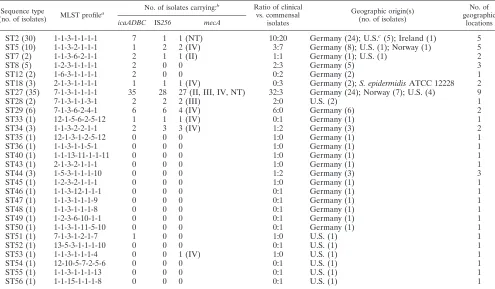
TABLE 1. MLST profiles of S. epidermidis isolates carryingicaandmecAgenes and the IS256element and distribution of the isolates among the different sequence types
Sequence type
(no. of isolates) MLST profile
a No. of isolates carrying:
b Ratio of clinical vs. commensal
isolates
Geographic origin(s) (no. of isolates)
No. of geographic
locations icaADBC IS256 mecA
ST2 (30) 1-1-3-1-1-1-1 7 1 1 (NT) 10:20 Germany (24); U.S.c(5); Ireland (1) 5
ST5 (10) 1-1-3-2-1-1-1 1 2 2 (IV) 3:7 Germany (8); U.S. (1); Norway (1) 5
ST7 (2) 1-1-3-6-2-1-1 2 1 1 (II) 1:1 Germany (1); U.S. (1) 2
ST8 (5) 1-2-3-1-1-1-1 2 0 0 2:3 Germany (5) 3
ST12 (2) 1-6-3-1-1-1-1 2 0 0 0:2 Germany (2) 1
ST18 (3) 2-1-3-1-1-1-1 1 1 1 (IV) 0:3 Germany (2);S. epidermidisATCC 12228 2 ST27 (35) 7-1-3-1-1-1-1 35 28 27 (II, III, IV, NT) 32:3 Germany (24); Norway (7); U.S. (4) 9
ST28 (2) 7-1-3-1-1-3-1 2 2 2 (III) 2:0 U.S. (2) 1
ST29 (6) 7-1-3-6-2-4-1 6 6 4 (IV) 6:0 Germany (6) 2
ST33 (1) 12-1-5-6-2-5-12 1 1 1 (IV) 0:1 Germany (1) 1
ST34 (3) 1-1-3-2-2-1-1 2 3 3 (IV) 1:2 Germany (3) 2
ST35 (1) 12-1-3-1-2-5-12 0 0 0 1:0 Germany (1) 1
ST36 (1) 1-1-3-1-1-5-1 0 0 0 1:0 Germany (1) 1
ST40 (1) 1-1-13-11-1-1-11 0 0 0 1:0 Germany (1) 1
ST43 (1) 2-1-3-2-1-1-1 0 0 0 1:0 Germany (1) 1
ST44 (3) 1-5-3-1-1-1-10 0 0 0 1:2 Germany (3) 3
ST45 (1) 1-2-3-2-1-1-1 0 0 0 1:0 Germany (1) 1
ST46 (1) 1-1-3-12-1-1-1 0 0 0 0:1 Germany (1) 1
ST47 (1) 1-1-3-1-1-1-9 0 0 0 0:1 Germany (1) 1
ST48 (1) 1-1-3-1-1-1-8 0 0 0 0:1 Germany (1) 1
ST49 (1) 1-2-3-6-10-1-1 0 0 0 0:1 Germany (1) 1
ST50 (1) 1-1-3-1-11-5-10 0 0 0 0:1 Germany (1) 1
ST51 (1) 7-1-3-1-2-1-7 1 0 0 1:0 U.S. (1) 1
ST52 (1) 13-5-3-1-1-1-10 0 0 0 0:1 U.S. (1) 1
ST53 (1) 1-1-3-1-1-1-4 0 0 1 (IV) 1:0 U.S. (1) 1
ST54 (1) 12-10-5-7-2-5-6 0 0 0 0:1 U.S. (1) 1
ST55 (1) 1-1-3-1-1-1-13 0 0 0 0:1 U.S. (1) 1
ST56 (1) 1-1-15-1-1-1-8 0 0 0 0:1 U.S. (1) 1
a
Allelic profiles of the sequence types consisting of the allele numbers of the genes analyzed (from left to right: carbamate kinase [arcC], shikimate 5-dehydrogenase [aroE], glycerol kinase [glpK], guanylate kinase [gmk], phosphate acetyltransferase [pta], triosephosphate isomerase [tpiA], and acetyl coenzyme A acetyltransferase [yqi]).
b
SCCmectype(s) is in parentheses. c
U.S., United States.
on May 15, 2020 by guest
http://jcm.asm.org/
strands by direct, automated sequencing of the PCR products by use of an ABI Prism 310 DNA sequencer (Applied Biosystems, Darmstadt, Germany) or a LiCor model 4000L DNA sequencer (LiCor Biosciences, Lincoln, NE). Nucle-otide sequences were compared to known alleles for each locus via the MLST website (http://www.mlst.net). For each isolate a seven-digit allelic profile was established which defined a sequence type (ST). Clonal analysis of the STs was performed using BURST, a Web-implemented clustering algorithm (http://www .mlst.net), which divides MLST data sets into groups of related isolates and predicts the founding genotype of each clonal complex (28). STs with at least five of seven identical alleles were defined as a clonal group.
Recombination analysis. Recombination rates were estimated by pairwise comparison of the nucleotide sequence of each single-locus variant (SLV; i.e., an ST which differs from another ST in only one locus and remains unchanged in the other six) with that of its predicted clonal ancestor as described previously (6). Briefly, BURST was used to identify SLVs and their associated ancestor STs. The nucleotide sequences of the various alleles were compared to each other using Sequence Output, a Macintosh program available from the MLST website, which allows for displaying polymorphic sites. An allele was determined to have resulted from point mutation when two criteria were fulfilled: first, when the allele differed only in one nucleotide site from its putative ancestor allele, and second, when the allele was unique within the data set of this study and within the data deposited in the mlst.net database. Recombination was determined to have occurred when an allele differed in more than one nucleotide site or when the allele varied in a single site but corresponded to a known allele found elsewhere in the database.
RESULTS
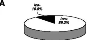
MLST profiles andicapresence in clinical and commensal isolates.MLST revealed a total of 28 different sequence types (STs) among the 118 S. epidermidis isolates (Table 1). Se-quence types ST2 to ST29 corresponded to known allelic pro-files in the mlst.netS. epidermidisdatabase, whereas ST33 to ST56 represented novel sequence types. Entire nucleotide se-quences of all alleles are available via the mlst.net database. The majority of the clinical strains (58 of 65; 89.2%) wereica positive (Fig. 1A) and displayed nine different STs (Fig. 1B). In this respect, ST27 was a remarkable clone. It harbored almost half of all clinical isolates tested (32 of 65; 49.2%) and the bulk of theica-positive isolates (35 of 62; 56.4%) (Fig. 1B). Among commensal strains of healthy volunteers, theica operon was found in only 4 of 41 isolates (9.7%); three of these belonged
to ST27 and one to ST12. Commensal strains from hospital staff, however, carried the ica operon in 8 of 11 isolates (72.7%). These isolates were grouped into ST2 (n⫽ 3), ST7 (n⫽1), ST12 (n⫽1), ST18 (n⫽1), ST33 (n⫽1), and ST34 (n⫽1).
Clonal relationship of the STs.BURST was used to divide the 118 isolates into clonal complexes by comparison of their allelic profiles (Fig. 2). This analysis indicated that the majority of isolates are closely related to each other and form a single major group or clonal complex (Fig. 2). ST2, which was the sequence type that generated the most abundant single-locus variants (SLVs), was considered the common ancestor clone. A total of 30 isolates, bothicapositives and negatives (7 versus 23), belonged to ST2. ST27, the clone that represented the majority of theica-positive isolates (56.4%) and 49.2% of the clinical isolates (Fig. 1), is a single-locus variant of ST2 differ-ing only in thearcCallele. Interestingly, this clone is a source of isolates which carry, concomitantly with the ica operon, genes that mediate methicillin resistance (mecA) and the in-sertion sequence IS256(see below).
Detection of the ica genes in an unrelated genetic back-ground.BURST analysis identified four additional STs which were unrelated to the major clonal group (Fig. 2). Each of these STs was represented by one isolate. ST33 and ST35 shared five identical alleles and were therefore regarded as a minor group (Table 1 and Fig. 2). ST40 and ST54 are single-tons and are unrelated to each other and to any other ST identified in this study. Interestingly, ST33 represented anica -positive strain, indicating that the ica operon can also be present in S. epidermidis lineages that differ from the ST2-derived clonal complex.
SCCmectyping and IS256detection.In previous studies we have shown a correlation between the presence of the ica operon, methicillin resistance, and detection of the insertion sequence IS256in clinicalS. epidermidisisolates (17). Methi-cillin resistance in staphylococci is mediated by themecAgene, which is located on a large, mobile DNA element termed SCCmec(16). Four major SCCmectypes have been identified for which mobility and repeated transfer into different genetic backgrounds ofS. aureusand coagulase-negative staphylococci has been demonstrated (5, 8, 32). IS256 is an insertion se-quence element which is part of the aminoglycoside resistance-mediating transposon Tn4001but also occurs independently in multiple copies in the genomes of staphylococci and entero-cocci. IS256has the capacity to influence expression of theica operon and subsequent biofilm formation by reversible inser-tion into theicaoperon and its regulatory genes as well as by chromosomal rearrangements (2, 14, 35, 37). To investigate whether SCCmeccassettes and IS256occur in specific STs, all isolates were tested for the presence of these genes. Of the 118
S. epidermidisstrains tested, 43 (37 clinical isolates and 6
[image:3.585.54.198.71.137.2]com-mensal isolates) weremecApositive. Four of the commensal isolates originated from hospital staff members. SCCmecII was detected in 13 strains, SCCmecIII in 14 strains, and SCCmecIV in 13 strains. ThreemecA-positive isolates were nontypeable, and none of the strains carried an SCCmecI cassette (Table 1). ST27, the most prevalent ica-positive clone, contained 27 (62.8%) of the 43 methicillin-resistant isolates. Four different SCCmeccassettes were found in these 27 mecA-positive iso-lates: SCCmecII (n⫽ 12), SCCmecIII (n ⫽ 12), SCCmecIV
FIG. 1. Proportion ofica-positive and -negativeS. epidermidis iso-lates (A) and distribution of different sequence types (B) among clin-icalS. epidermidisstrains.
on May 15, 2020 by guest
http://jcm.asm.org/
(n⫽1), and SCCmecNT (n⫽2). IS256was detected in 45 of
118S. epidermidisstrains: 40 clinical strains and 5 commensals.
Four of the five IS256-positive commensals were obtained from health care workers. IS256-carrying strains were detected in nine STs which contained bothica-positive and SCCmec -positive isolates, and ST27 again represented the clone con-taining the most IS256-positive isolates (28 of 45; 62.2%). Taken together, these data suggest that SCCmec and IS256 occur jointly in specificS. epidermidisclones which harbor the icaoperon and occur preferentially in the hospital environment.
Geographic distribution ofica-positive clones.Table 1 sum-marizes the properties of each ST with respect toicaand IS256 presence, including SCCmectyping as well as its geographic distribution.ica-positive STs were preferentially found in med-ical facilities. Interestingly, these clones were not restricted to a specific geographic area.ica-positive ST27 isolates were iden-tified in hospitals from four regions in Germany, in one hos-pital in Norway, and at four locations in the United States. Except for ST12, ST28, ST33, and ST51, which represent only one or two isolates, respectively, allica-positive STs were de-tected in more than one hospital (Table 1).
Estimates of recombination rates.To address the question of whetherS. epidermidisstrains diverge by point mutation or by recombination, a pairwise comparison of single-locus vari-ants (SLV) was done by analysis of their polymorphic sites (data not shown). An allele was considered as likely to have arisen by point mutation when (i) it differed only in one nu-cleotide from the corresponding ancestor allele and (ii) the allele was unique in the data set and the database on theS.
epidermidis mlst.net site. In contrast, recombination was
as-sumed when the allele differed in more than one nucleotide and/or was already present in the MLST database (6). Table 2 shows that 12 of 15 SLVs likely evolved by recombination and only 3 of 15 by point mutation, suggesting that recombination is the major mechanism for divergence ofS. epidermidisstrains.
DISCUSSION
Prevalence of ST27 and otherica-positive clones in hospital settings.Employing a recently established MLST scheme, this study demonstrated that the majority of the S. epidermidis infections analyzed in this report are caused by a single clone
FIG. 2. BURST analysis was used to identify clonal complexes. The predicted clonal ancestor is indicated in the central ring; SLVs are shown in the middle and double-locus variants in the outer ring. Red labels mark the number ofica-positive isolates, blue numbers indicate IS256-positive strains, and green labels designate the number and the type of the SCCmeccassettes identified. I, SCCmecI; II, SCCmecII; III, SCCmecIII; IV, SCCmecIV; NT, none typeable; MS, methicillin susceptible. The blue dotted line marks the dominantica-positive sequence type ST27.
on May 15, 2020 by guest
http://jcm.asm.org/
(i.e., ST27) whose presence is widespread within hospital set-tings in Germany. Interestingly, this sequence type was also detected in a Norwegian hospital and in medical facilities in the United States. In addition to ST27, 11 otherica-positive clones with similar properties were identified. The presence of these clones in the hospital environment is in agreement with a recent MLST study on the distribution of SCCmeccassettes in clinical S. epidermidis isolates (32). Wisplinghoff and co-workers analyzed a North American collection of 67 methicil-lin-resistant and -susceptibleS. epidermidisisolates from blood cultures and prosthetic valve endocarditis. Nine of the 32 MLST profiles identified in the SCCmecstudy coincide with ica-positive STs identified in our report, i.e., ST2, ST5, ST7, ST8, ST12, ST18, ST27, ST28, and ST29. Moreover, there was good agreement with respect to the SCCmectypes identified in the STs of both reports. At present we do not know whether the isolates analyzed by Wisplinghoff et al. are alsoica and IS256 positive, but their origination from device-associated infections makes it very likely that they are also biofilm formers. ST27 and otherica-positive clones were rarely found outside of medical facilities, suggesting that they are highly adapted to the hospital environment and differ from commensalS.
epider-midisin the community. It is conceivable that patients who are
admitted to a hospital are soon colonized by these biofilm-forming, multiresistant S. epidermidis isolates and that this newly acquired endogenous microflora represents the origin for a later infection. This hypothesis is supported by a recent ecological study in bone marrow transplantation patients dem-onstrating that these individuals carryica-,mecA-, and IS256 -positiveS. epidermidisisolates as commensals (26). Coloniza-tion of hospital staff byica-positiveS. epidermidis isolates in our report highlights again the presence of such strains in the hospital environment. However, larger studies are necessary to elucidate whether or not the personnel might function as trans-mitters of biofilm-forming strains to patients. Moreover, it will be interesting to elucidate the possible impact of other viru-lence-associated factors such as the phenol-soluble modulins or other biofilm-mediating genes such asaapandbhpin these clones (4, 25, 33).
Origin and attributes of ST27 isolates.In addition toica, the majority of ST27 isolates carry different SCCmeccassettes and multiple copies of the insertion sequence IS256. It is unknown why ST27 has become the dominant ST within nosocomial environments in Germany; however, the answer may be related to the increased reliance on implanted medical devices within immunocompromised patients. These devices represent a suit-able habitat for colonization, and the extended use of antibi-otics and disinfectants generates a highly selective pressure for the generation of resistant variants. Occupation of this niche requires specific prerequisites which are obviously met by ST27. Theicaoperon enables the bacteria to stick to polymer and metal surfaces, and the ability to gain resistance determi-nants helps ST27 isolates withstand hostile external conditions. The presence of different SCCmeccassettes in ST27 suggests repeated, independent transfer of SCCmecDNA into this ge-netic background and is an indication of the capacity of ST27 to exchange genetic material. The question of whether or not theicagenes are also subject to horizontal gene transfer must remain open at this stage of experimental work. However, the detection of the ica operon in an unrelated S. epidermidis background in our study and the identification ofica homo-logues in pathogenic Escherichia coli and a broad range of other bacterial species support the hypothesis of mobility and lateral transfer of biofilm-encoding genes (31). The recent publication of the genome sequences of a methicillin-resistant ica-positive S. epidermidis clinical strain and an ica-negative commensal isolate revealed that most of the genetic variations between the two genomes are attributed to genomic islands, phages, and integrated plasmids (11, 34). In this regard, it was concluded that lateral gene transfer between staphylococci and other low-GC gram-positive bacteria is common and contrib-utes considerably to resistance and virulence development ofS.
epidermidis(11). Interestingly, we observed in studies of the
[image:5.585.43.547.81.271.2]spontaneous emergence of biofilm-negative variants that DNA encompassing theica operon is unstable and can be readily deleted from the S. epidermidis genome (P. D. Fey and W. Ziebuhr, unpublished data). Instability of genetic material is often an indication of mobility, and in this respect it is also
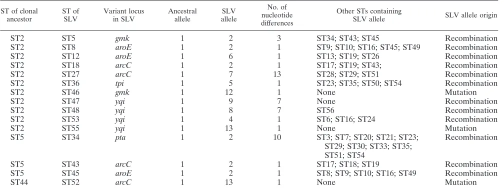
TABLE 2. Variant alleles of SLVs of the common ancestor and estimates of recombination rates
ST of clonal ancestor
ST of SLV
Variant locus in SLV
Ancestral allele
SLV allele
No. of nucleotide differences
Other STs containing
SLV allele SLV allele origin
ST2 ST5 gmk 1 2 3 ST34; ST43; ST45 Recombination
ST2 ST8 aroE 1 2 1 ST9; ST10; ST16; ST45; ST49 Recombination
ST2 ST12 aroE 1 6 1 ST13; ST19; ST26 Recombination
ST2 ST18 arcC 1 2 1 ST17; ST19; ST43; Recombination
ST2 ST27 arcC 1 7 13 ST28; ST29; ST51 Recombination
ST2 ST36 tpi 1 5 1 ST23; ST35; ST50; ST54 Recombination
ST2 ST46 gmk 1 12 1 None Mutation
ST2 ST47 yqi 1 9 7 None Recombination
ST2 ST48 yqi 1 8 7 ST56 Recombination
ST2 ST53 yqi 1 4 1 ST6; ST16; ST24 Recombination
ST2 ST55 yqi 1 13 1 None Mutation
ST5 ST34 pta 1 2 10 ST3; ST7; ST20; ST21; ST23;
ST29; ST30; ST33; ST35; ST51; ST54
Recombination
ST5 ST43 arcC 1 2 1 ST17; ST18; ST19 Recombination
ST5 ST45 aroE 1 2 1 ST8; ST9; ST10; ST16; ST49 Recombination
ST44 ST52 arcC 1 13 1 None Mutation
on May 15, 2020 by guest
http://jcm.asm.org/
conceivable that theica operon represents mobile DNA that has been lost in the commensalS. epidermidisisolates. Another finding of our study is that the generation of SLVs withinS.
epidermidisseems to occur generally by recombination rather
than by point mutation. This is in contrast to what has been described inS. aureus studies, where clonal complexes were shown to expand primarily by point mutation (6). The molec-ular background for this difference between the two species remains to be elucidated, but it might reflect again the capacity
ofS. epidermidisto acquire and incorporate foreign DNA.
It is tempting to speculate that ST27 isolates represent an ideal genetic background for biofilm and resistance genes, re-sulting in well-adapted strains which are then selected in the hospital environment. The presence of multiple copies of IS256in the ST27 genome might support this adaptation pro-cess by an ongoing generation of novel phenotypic and geno-typic variants. Thus, involvement of IS256in genome flexibility has been shown for the modulation of biofilm, resistance, and global regulatory gene expression (2, 3, 21, 22, 35, 37). There-fore, the combination of biofilm formation, antibiotic resis-tance, and genetic flexibility may explain why ST27 has become the dominant ST within medical facilities. The data of this report provide novel insights into the population structure ofS.
epidermidisand may suggest new infection control measures
for the management of these infections in the future.
ACKNOWLEDGMENTS
We are grateful to our colleagues who provided us with Staphylo-coccus epidermidisisolates: K. Naber, R. Marre, E. Straube, D. Mack, F. Go¨tz, and W. Thomas (Germany); P. Meyer (Norway); V. D. Fowler, G. L. Archer, and R. J. Sheretz (United States); and J. P. O’Gara (Ireland). We thank M. C. Enright and S. O’Hanlon (Univer-sity of Bath, Bath, United Kingdom) for data entry into the mlst.net database.
The work of S.K. and W.Z. was supported by the Deutsche For-schungsgemeinschaft SFB479 and the Federal Ministry of Education and Research (BMBF03F0401C) and that of K.O. by the Deutsche Forschungsgemeinschaft SFB630. P.D.F. was supported by grant 5RO1 AI49311 from the National Institute of Allergy and Infectious Diseases.
REFERENCES
1.Cho, S. H., K. Naber, J. Hacker, and W. Ziebuhr.2002. Detection of the icaADBCgene cluster and biofilm formation inStaphylococcus epidermidis isolates from catheter-related urinary tract infections. Int. J. Antimicrob. Agents19:570–575.
2.Conlon, K. M., H. Humphreys, and J. P. O’Gara.2004. Inactivations ofrsbU andsarAby IS256represent novel mechanisms of biofilm phenotypic vari-ation inStaphylococcus epidermidis. J. Bacteriol.186:6208–6219. 3.Couto, I., S. W. Wu, A. Tomasz, and H. de Lencastre.2003. Development of
methicillin resistance in clinical isolates ofStaphylococcus sciuriby transcrip-tional activation of themecAhomologue. J. Bacteriol.185:645–653. 4.Cucarella, C., C. Solano, J. Valle, B. Amorena, I. Lasa, and J. R. Penades.
2001. Bap, aStaphylococcus aureussurface protein involved in biofilm for-mation. J. Bacteriol.183:2888–2896.
5.Enright, M. C., D. A. Robinson, G. Randle, E. J. Feil, H. Grundmann, and B. G. Spratt.2002. The evolutionary history of methicillin-resistant Staphy-lococcus aureus(MRSA). Proc. Natl. Acad. Sci. USA99:7687–7692. 6.Feil, E. J., J. E. Cooper, H. Grundmann, D. A. Robinson, M. C. Enright, T.
Berendt, S. J. Peacock, J. M. Smith, M. Murphy, B. G. Spratt, C. E. Moore, and N. P. Day.2003. How clonal isStaphylococcus aureus? J. Bacteriol.
185:3307–3316.
7.Fey, P. D., J. S. Ulphani, F. Go¨tz, C. Heilmann, D. Mack, and M. E. Rupp.
1999. Characterization of the relationship between polysaccharide intercel-lular adhesin and hemagglutination inStaphylococcus epidermidis. J. Infect. Dis.179:1561–1564.
8.Fitzgerald, J. R., D. E. Sturdevant, S. M. Mackie, S. R. Gill, and J. M. Musser.2001. Evolutionary genomics ofStaphylococcus aureus: insights into the origin of methicillin-resistant strains and the toxic shock syndrome epi-demic. Proc. Natl. Acad. Sci. USA98:8821–8826.
9.Frebourg, N. B., S. Lefebvre, S. Baert, and J. F. Lemeland.2000. PCR-based assay for discrimination between invasive and contaminatingStaphylococcus epidermidisstrains. J. Clin. Microbiol.38:877–880.
10.Galdbart, J. O., J. Allignet, H. S. Tung, C. Ryden, and N. El Solh.2000. Screening forStaphylococcus epidermidismarkers discriminating between skin-flora strains and those responsible for infections of joint prostheses. J. Infect. Dis.182:351–355.
11.Gill, S. R., D. E. Fouts, G. L. Archer, E. F. Mongodin, R. T. Deboy, J. Ravel, I. T. Paulsen, J. F. Kolonay, L. Brinkac, M. Beanan, R. J. Dodson, S. C. Daugherty, R. Madupu, S. V. Angiuoli, A. S. Durkin, D. H. Haft, J. Vamathevan, H. Khouri, T. Utterback, C. Lee, G. Dimitrov, L. Jiang, H. Qin, J. Weidman, K. Tran, K. Kang, I. R. Hance, K. E. Nelson, and C. M. Fraser.
2005. Insights on evolution of virulence and resistance from the complete genome analysis of an early methicillin-resistantStaphylococcus aureusstrain and a biofilm-producing methicillin-resistant Staphylococcus epidermidis strain. J. Bacteriol.187:2426–2438.
12.Go¨tz, F.2002. Staphylococcus and biofilms. Mol. Microbiol.43:1367–1378. 13.Hall-Stoodley, L., J. W. Costerton, and P. Stoodley.2004. Bacterial biofilms:
from the natural environment to infectious diseases. Nat. Rev. Microbiol.
2:95–108.
14.Handke, L. D., K. M. Conlon, S. R. Slater, S. Elbaruni, F. Fitzpatrick, H. Humphreys, W. P. Giles, M. E. Rupp, P. D. Fey, and J. P. O’Gara.2004. Genetic and phenotypic analysis of biofilm phenotypic variation in multiple Staphylococcus epidermidisisolates. J. Med. Microbiol.53:367–374. 15.Heilmann, C., O. Schweitzer, C. Gerke, N. Vanittanakom, D. Mack, and F.
Go¨tz.1996. Molecular basis of intercellular adhesion in the biofilm-forming Staphylococcus epidermidis. Mol. Microbiol.20:1083–1091.
16.Ito, T., K. Okuma, X. X. Ma, H. Yuzawa, and K. Hiramatsu.2003. Insights on antibiotic resistance ofStaphylococcus aureusfrom its whole genome: genomic island SCC. Drug Resist. Updates6:41–52.
17.Kozitskaya, S., S. H. Cho, K. Dietrich, R. Marre, K. Naber, and W. Ziebuhr.
2004. The bacterial insertion sequence element IS256occurs preferentially in nosocomialStaphylococcus epidermidisisolates: association with biofilm for-mation and resistance to aminoglycosides. Infect. Immun.72:1210–1215. 18.Lane, D. J.1991. 16S/23S rRNA sequencing, p. 115–175.InE. Stackebrandt
and M. Goodfellow (ed.), Nucleic acids techniques in bacterial systematics. John Wiley & Sons, New York, N.Y.
19.Mack, D., W. Fischer, A. Krokotsch, K. Leopold, R. Hartmann, H. Egge, and R. Laufs.1996. The intercellular adhesin involved in biofilm accumulation of Staphylococcus epidermidisis a linear beta-1,6-linked glucosaminoglycan: purification and structural analysis. J. Bacteriol.178:175–183.
20.Mack, D., J. Riedewald, H. Rohde, T. Magnus, H. H. Feucht, H. A. Elsner, R. Laufs, and M. E. Rupp.1999. Essential functional role of the polysac-charide intercellular adhesin ofStaphylococcus epidermidisin hemagglutina-tion. Infect. Immun.67:1004–1008.
21.Maki, H., N. McCallum, M. Bischoff, A. Wada, and B. Berger-Ba¨chi.2004. tcaAinactivation increases glycopeptide resistance inStaphylococcus aureus. Antimicrob. Agents Chemother.48:1953–1959.
22.Maki, H., and K. Murakami.1997. Formation of potent hybrid promoters of the mutantllmgene by IS256transposition in methicillin-resistant Staphy-lococcus aureus. J. Bacteriol.179:6944–6948.
23.O’Brien, F. G., T. T. Lim, F. N. Chong, G. W. Coombs, M. C. Enright, D. A. Robinson, A. Monk, B. Said-Salim, B. N. Kreiswirth, and W. B. Grubb.2004. Diversity among community isolates of methicillin-resistantStaphylococcus aureusin Australia. J. Clin. Microbiol.42:3185–3190.
24.Oliveira, D. C., and H. de Lencastre.2002. Multiplex PCR strategy for rapid identification of structural types and variants of themecelement in methi-cillin-resistantStaphylococcus aureus. Antimicrob. Agents Chemother.46:
2155–2161.
25.Rohde, H., C. Burdelski, K. Bartscht, M. Hussain, F. Buck, M. A. Horst-kotte, J. K. Knobloch, C. Heilmann, M. Herrmann, and D. Mack.2005. Induction ofStaphylococcus epidermidisbiofilm formation via proteolytic processing of the accumulation-associated protein by staphylococcal and host proteases. Mol. Microbiol.55:1883–1895.
26.Rohde, H., M. Kalitzky, N. Kroger, S. Scherpe, M. A. Horstkotte, J. K. Knobloch, A. R. Zander, and D. Mack.2004. Detection of virulence-associ-ated genes not useful for discriminating between invasive and commensal Staphylococcus epidermidis strains from a bone marrow transplant unit. J. Clin. Microbiol.42:5614–5619.
27.Rupp, M. E., P. D. Fey, C. Heilmann, and F. Gotz.2001. Characterization of the importance ofStaphylococcus epidermidisautolysin and polysaccharide intercellular adhesin in the pathogenesis of intravascular catheter-associated infection in a rat model. J. Infect. Dis.183:1038–1042.
28.Smith, J. M., N. H. Smith, M. O’Rourke, and B. G. Spratt.1993. How clonal are bacteria? Proc. Natl. Acad. Sci. USA90:4384–4388.
29.Urwin, R., and M. C. Maiden.2003. Multi-locus sequence typing: a tool for global epidemiology. Trends Microbiol.11:479–487.
30.Vuong, C., J. M. Voyich, E. R. Fischer, K. R. Braughton, A. R. Whitney, F. R. DeLeo, and M. Otto.2004. Polysaccharide intercellular adhesin (PIA) pro-tectsStaphylococcus epidermidisagainst major components of the human innate immune system. Cell Microbiol.6:269–275.
31.Wang, X., J. F. Preston III, and T. Romeo.2004. ThepgaABCDlocus of
on May 15, 2020 by guest
http://jcm.asm.org/
Escherichia colipromotes the synthesis of a polysaccharide adhesin required for biofilm formation. J. Bacteriol.186:2724–2734.
32.Wisplinghoff, H., A. E. Rosato, M. C. Enright, M. Noto, W. Craig, and G. L. Archer. 2003. Related clones containing SCCmec type IV predominate among clinically significantStaphylococcus epidermidisisolates. Antimicrob. Agents Chemother.47:3574–3579.
33.Yao, Y., D. E. Sturdevant, and M. Otto.2005. Genomewide analysis of gene expression inStaphylococcus epidermidisbiofilms: insights into the patho-physiology ofS. epidermidisbiofilms and the role of phenol-soluble modulins in formation of biofilms. J. Infect. Dis.191:289–298.
34.Zhang, Y. Q., S. X. Ren, H. L. Li, Y. X. Wang, G. Fu, J. Yang, Z. Q. Qin, Y. G. Miao, W. Y. Wang, R. S. Chen, Y. Shen, Z. Chen, Z. H. Yuan, G. P. Zhao, D. Qu, A. Danchin, and Y. M. Wen.2003. Genome-based analysis of virulence genes in a non-biofilm-formingStaphylococcus epidermidis strain (ATCC
12228). Mol. Microbiol.49:1577–1593.
35.Ziebuhr, W., K. Dietrich, M. Trautmann, and M. Wilhelm.2000. Chromo-somal rearrangements affecting biofilm production and antibiotic resistance in aStaphylococcus epidermidisstrain causing shunt-associated ventriculitis. Int. J. Med. Microbiol.290:115–120.
36.Ziebuhr, W., C. Heilmann, F. Go¨tz, P. Meyer, K. Wilms, E. Straube, and J. Hacker.1997. Detection of the intercellular adhesion gene cluster (ica) and phase variation inStaphylococcus epidermidisblood culture strains and mu-cosal isolates. Infect. Immun.65:890–896.
37.Ziebuhr, W., V. Krimmer, S. Rachid, I. Loessner, F. Go¨tz, and J. Hacker.
1999. A novel mechanism of phase variation of virulence inStaphylococcus epidermidis: evidence for control of the polysaccharide intercellular adhesin synthesis by alternating insertion and excision of the insertion sequence element IS256.Mol. Microbiol.32:345–356.