Int J Clin Exp Pathol 2016;9(2):969-977
www.ijcep.com /ISSN:1936-2625/IJCEP0018912
Original Article
Long non-coding RNA MALAT1 promote triple-negative
breast cancer progression by regulating miR-204
expression
Lixia Li, Zhicheng Yang, Yuzhou Wang, Ying Zhang, Yijin Zhou, Wennan Wang, Lanzhen Lin, Wenmei Su, Zhixiong Yang
Department of Oncology, Affliated Hospital of Guangdong Medical University, Zhanjiang, Guangdong, China
Received November 1, 2015; Accepted December 26, 2015; Epub February 1, 2016; Published February 15, 2016
Abstract: Recent studies demonstrated that lncRNAs have a critical role in the regulation cancer progression and metastasis. However, little is known about the mechanism through which MALAT1 exerts its oncogenic activity, and the interaction between MALAT1 and microRNA remains largely unknown. In the present study, we reported that MALAT1 was up-regulated in triple-negative breast cancer (TNBC) tissues. Knockdown of MALAT1 inhibited proliferation, motility and increased apoptosis in vitro. In vivo study indicated that knockdown of MALAT1 inhibited tumor growth. Patients with high MALAT1 expression had poorer overall survival time than those with low MALAT1
expression. In addition, our findings demonstrate a reciprocal negative control relationship between MALAT1 and
miR-204: downregulation of MALAT1 increased expression of miR-204, while overexpression of miR-204 decreased MALAT1 expression. We proposed that MALAT1 exerted its function through the miR-204. In summary, we proposed that MALAT1 may be a target for TNBC therapy.
Keywords: miR-204, MALAT1, triple-negative breast cancer
Introduction
Breast cancer is a common malignancy, with an incidence of more than 1,000,000 new cases and 370,000 deaths annually worldwide [1]. Accounting for about 20% of breast cancers, triple-negative breast cancer (TNBC) refers to breast cancers that do not express the genes for estrogen receptor (ER), progesterone recep-tor (PR) and the Her2/neu receprecep-tor [2]. Treatment of TNBC is challenging because the lack of targeted therapy, aggressive behavior and relatively poor prognosis [3]. Thus, it is
urgent to find out new strategy for the treat -ment of TNBC.
It is well documented that protein-coding genes account for only about 2% of the human genome, whereas the majority of transcripts consist of the non-coding RNAs, including microRNAs and long non-coding RNAs (lncRNAs). MicroRNAs (miRNAs) are small non-coding RNAs of about 22 nucleotides in length that negatively regulate coding gene [4] or
lncRNA expression [5]. It is well documented that miRNAs are involved in diverse biological processes, including differentiation, prolifera-tion, apoptosis, and tumorigenesis [6, 7]. MiR-204 has been reported to function as a tumor suppressor in various cancers [8, 9]. A previous report suggests that miR-204 may be a tumor suppressor in breast cancer [10]. However, the underlying mechanism of miR-204 in breast cancer development was still poorly explored. Although it is well known that miRNAs can tar-get a number of protein-coding genes, little is known whether miRNAs/lncRNAs can also tar-get lncRNAs/miRNAs. Recently, a number of lit-eratures documented the regulatory networks between miRNAs and lncRNAs [11, 12].
Unlike the miRNAs, long ncRNAs (lncRNAs) are
by definition >200 nt in length. Metastasis
[13-15]. In the current study, we investigated the
clinical significance of MALAT1 on TNBC and confirmed its biological functions by in vitro and
in vivo assays. In addition, we also documented the reciprocal regulation of miR-204 and MALAT1.
Materials and methods
Cells culture and TNBC patients tissues
The breast cancer cell lines were purchased from the Cell Bank of the Shanghai Institutes for Biological Sciences (Chinese Academy of Sciences, Shanghai, China).
A total of 129 female breast cancer patients who were diagnosed by histo-pathology in the
Affiliated Hospital of Guangdong Medical
University from October 2005 to September 2011 were obtained. Specimens were
formalin-fixed and embedded in paraffin by standard
methodology after obtained during surgery. All breast cancer patients gave written consent for their tissue samples to be used for research purposes. This study was conducted with the
approval of the Ethical and Scientific Committees of Guangdong Medical University
with the permit number of 778NLSY03. RNA isolation, reverse transcription, and quan-titative real-time PCR
Total RNA from either culture cells or tissue samples was isolated with the use of Trizol reagent (Invitrogen). The samples were reverse transcribed into cDNA with different RT primers by using Revert Ace kit (Takara, Japan). PCR
primers of MALAT1 and GAPDH were previously
described [16-18]. To detect miR-204 and U6 expression, RNA samples were reverse tran-scribed into cDNA using Revert Ace
transcrip-tase by specific stem-loop RT primers accord -ing to the manufacturer’s instruction. The primers used for miR-204 and U6 were described in previous report [19]. Transcript levels were measured against an endogenous
control by qPCR using the SYBR Green I fluoro -genic dye using the Mastercycler ep realplex
system (Eppendorf, Germany).
Lentivirus vector construction and infection
Short-hairpin RNA directed against human lncRNA MALAT1 was ligated into the
LV10-CMV-RFP-Puro vector (GenePharma, Shanghai,
China). The empty vector was used as a nega-tive control (NC). The viruses were packaged in 293T cells according to standard protocols and the virus particles were harvested 72 h later. Cells were infected with virus particles plus 8
μg/ml Polybrene (Sigma, St Louis, Missouri,
USA), followed by selected with puromycin for up to 7 days.
Oligonucleotide transfection
MiR-204 mimic and miR control were
pur-chased from GenePharma (Shanghai, China).
Cells transfection was carried out using Lipofectamine 2000.
Dual luciferase reporter gene assays
The fragment from MALAT1 containing the
pre-dicted miR-204 binding site was amplified by PCR and cloned into a pmirGlO Dual-luciferase
miRNA Target Expression Vector (Promega, Madison, WI, USA) and this vector was named
wt-MALAT1. To test the binding specificity, the
corresponding mutant was created by mutating the miR-204 seed region binding site and this vector was named mut-MALAT1. The luciferase assay was performed by using the dual Luciferase reporter assay system (Promega) 48 h after transfection.
MTT, Boyden and flow cytometry assay
The MTT assay was carried out as previously described [20].
For Boyden assay, the transwell chambers were
coated with 100 μl BD Matrigel overnight in cell
incubator. Then the cells were added to upper transwell chambers. A medium containing 10% FBS was added to the lower wells. After 48 h
incubation, cells were fixed and stained, and
the nonmigratory cells were scraped from the
upper part of the filter. Then the migrated cells
were stained with 0.2% crystal violet solution and counted.
To detect cell apoptosis, the cells were harvest-ed and stainharvest-ed with 7-AAD and Annexin-V-FITC. Flow cytometry data was analyzed by BD FACSDiva software V6.1.3 (BD Biosciences). Animal studies
MALAT1 promote breast cancer progression by miR-204
Experimentation and the Ethic Committee of
Guandong Medical University (Permit Number:
2014-08-XF818). The nude mice were kept in
[image:3.612.95.522.72.258.2]pathogen free environment with 12-hour light/ dark cycle, controlled humidity and tempera-ture. For the in vivo tumor growth studies, 1 × Figure 1. A: MALAT1 was upregulated in TNBC tissues compared with their adjacent normal breast tissues. B: MALAT1 expression in TNBC cell lines was relatively high compared to that in MCF-10A cells.
[image:3.612.95.522.317.619.2]106 LV-miR-204 or LV-miR-ctrl cells were
inject-ed subcutaneously in the upper back of BALB/ C-nu/nu athymic nude mice. The length and width of the tumors were measured every 5 days using a digital caliper and tumor volumes were calculated using the formula Volume (mm3) = L × W2/2 (length L, mm; width W, mm).
Four weeks after injection, mice were
anesthe-tized with chloral hydrate and sacrificed by cer -vical dislocation. Then the tumors were removed and weighed.
Statistical analysis
SPSS 13.0 software was used to analyze the data. Survival analysis was performed using the Kaplan-Meier method. A log rank test was used to compare different survival curves. A
two-tailed Student’s t-test was used for the comparison between two independent groups. One-way ANOVA was used to determine the dif-ferences between groups. P values of <0.05
were considered as statistically significant.
Results
MALAT1 expression was upregulated in TNBC tissues and cell lines
[image:4.612.89.520.72.489.2]First, we analyzed the relative expression levels of MALAT1 by using real-time qPCR in 38 pairs of human TNBC tissues and adjacent normal breast tissues. The results showed that MALAT1 was upregulated in TNBC tissues compared with their adjacent normal breast tissues (Figure 1A). Subsequently, the expression of
MALAT1 promote breast cancer progression by miR-204
MALAT1 was evaluated in TNBC lines (MDA-MB-231, Hs578T and MDA-MB-468) and nor-mal immortalized MCF-10A cells. It was found that MALAT1 expression in TNBC cell lines was relatively high compared to that in MCF-10A cells (Figure 1B).
Knockdown of MALAT1 inhibited the cell pro -liferation, invasion, and promoted apoptosis of TNBC cells
The significant increase of MALAT1 in TNBC tis -sues and cell lines prompted us to explore the
possible biological significance of MALAT1 in
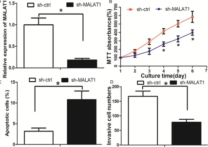
TNBC tumorigenesis. First, the lentivirus sh-MALAT1 was used to knock down sh-MALAT1 in Hs578T cells (Figure 2A). The MTT assay revealed that the cell proliferation was decreased in sh-MALAT1 group when com-pared with sh-ctrl group (Figure 2B). Flow-cytometric analysis was performed to deter-mine whether apoptosis was a contributing factor to cell proliferation inhibition. It was found that the apoptosis rate was increased in sh-MALAT1 group when compared with sh-ctrl group (Figure 2C). We then evaluated the
[image:5.612.91.524.68.481.2]abili-ties of cell invasion, which was a significant
aspect of cancer progression. The cell invasion ability was decreased in sh-MALAT1 group when compared with sh-ctrl group (Figure 2D).
Taken together, these results reflected that
knockdown of MALAT1 had tumor-suppressive effects that could inhibit cell proliferation and invasion and promote apoptosis in TNBC. Identification of microRNAs that were targeted by MALAT1
The online software program starbase v2.0 (http://starbase.sysu.edu.cn/mirLncRNA.php) [21] was used to search for miRNAs that have complementary base pairing with MALAT1. The microRNAs that formed complementary base pairing with MALAT1 were shown in Supplementary Table 1. We then examined the expression of these microRNAs in response to MALAT1 knockdown. As found in Figure 3A, there was a list of microRNAs that were upregu-lated more than 3-fold in response to MALAT1 inhibition. We focused on miR-204, which was of the greatest fold-change in response to MALAT1 knockdown. We further examined the expression of miR-204 in 38 pairs of human TNBC tissues and adjacent normal breast tis-sues used above. The result showed that miR-204 expression was downregulated in TNBC tissues when compared with adjacent normal breast tissues (Figure 3B). In addition, restora-tion of miR-204 decreased TNBC cell prolifera-tion and invasion, and increased the rate of apoptosis (Figure 3C-E).
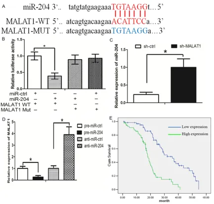
suggested that the binding sites are vital for the reciprocal repression of MALAT1 and miR-204.
We next asked whether there was a reciprocal repression between MALAT1 and miR-204. It was found that knockdown of MALAT1 increased miR-204 expression (Figure 4C). Interestingly, overexpresion of miR-204 decr- eased MALAT1 expression, while inhibition of miR-204 increased MALAT1 expression (Figure 4D). These data suggest that there was a reciprocal repression between MALAT1 and miR-204.
The relationships between expression of MALAT1 and clinical parameters in TNBC patients
To further investigate the clinicopathological
and prognostic significance of MALAT1 levels in
TNBC patients, the levels of MALAT1 in a large cohort of 129 TNBC tissues (including the tis-sues used before) were examined by real-time PCR. The median value of all 129 TNBC sam-ples was chosen as the cut-off point for sepa-rating tumors with low-level expression of MALAT1 from high-level expression MALAT1 tumors. Thus, 65 (50.3%) TNBC patients had low-level expression of MALAT1, while 64 (49.7%) TNBC patients had high-level expres-sion of MALAT1. The clinicopathologic charac-teristics of the TNBC patients and MALAT1 expression are shown in Table 1. High expres-sion of MALAT1 correlated with late TNM stage
Table 1. Clinicopathologic characteristics of the breast cancer pa-tients and MALAT1 expression
MALAT1
Variables (n = 129)All case Low expression (n = 65) High expression (n = 64) P Value*
Age (years)
≤50 54 29 25 0.523
>50 75 36 39
Tumor size (cm)
≤2 59 26 33 0.188
>2 70 39 31
TNM stage
I-II 77 32 45 0.015*
III-IV 52 33 19
Distant metastasis
Yes 54 36 18 0.002*
No 75 29 46
(*P <0.05).
Reciprocal repression of MALAT1 and miR-204
Luciferase reporter con-structs were generated to
confirm the direct binding
between MALAT1 and miR-204. The wild-type (wt) MALAT1 and mutant (mut) MALAT1 was shown in
MALAT1 promote breast cancer progression by miR-204
(P = 0.015) and higher metastasis (P = 0.002),
although there was no significant association
between age and tumor size. In addition, we found that TNBC patients with high expression of MALAT1 had poorer overall survival time (Figure 4E).
Knockdown of MALAT1 inhibited TNBC cells growth and invasion in vivo
We next investigated whether knockdown of MALAT1 against tumor growth and metastasis in vivo. As expect, knockdown of MALAT1 decreased growth and tumor weight of subcu-taneous xenograft tumors in nude mice (Figure 5A and 5B). In the experimental metastasis studies, knockdown of MALAT1 established smaller lung metastatic colonies than the con-trol group (Figure 5C). These data revealed that inhibition of MALAT1 inhibited TNBC cells growth and invasion in vivo.
Discussion
Mounting evidence demonstrated that ncRNAs
played a significant role in cancer pathogenesis
[22, 23]. During the past decade, researches
on miRNAs have dominated the field of ncRNA
regulation, including in TNBC [24, 25]. However, the role of lncRNAs in TNBC is still largely unknown. Recently, we and other researchers have provided new insights into lncRNAs in TNBC progression [26-28]. MALAT1, mapped to human chromosome 11q13, was originally
sion. On the other hand, miR-204 mimic repressed MALAT1 level while miR-204 inhibi-tor upregulated MALAT1 level. We further explored the mechanism of such a feedback loop. It was found that MALAT1 and miRNA-204 bind to the same RISC complex. Based on the fact that lncRNAs may participate in the ‘com-petitive endogenous RNAs (ceRNA)’ regulatory network and act as endogenous miRNA spong-es to bind to miRNAs and regulate their func-tion [33, 34], we proposed that MALAT1 may regulate miR-204 expression in such way. Emerging evidence documented that miRNAs mediate post-transcriptional control of gene expression by binding to the 3’-untranslated regions of protein coding genes, we supposed that the way that miR-204 promoted the down-regulation of MALAT1 is somewhat similar to the miRNA-mediated silencing of protein-cod-ing genes. MiR-204 has been found to be down-regulated in a variety of carcinomas and exhib-its tumor-suppressive activity [35, 36]. We proposed that MALAT1 exert its function by negatively regulated miR-204, and thereby pro-moted TNBC progression.
In summary, our findings first revealed that
MALAT1 functioned as an oncogene in TNBC and promoted TNBC progression through
inter-acting with miR-204. In addition, our findings
deepened the understanding of lncRNA regula-tory network and may help to develop potential therapeutic strategy for TNBC.
Acknowledgements
This work was supported by grant from Sci-
ence and Technology of Guangdong province
Foundation (Number: 2014A020212459, 20- 13B022000092).
Disclosure of conflict of interest
None.
Address correspondence to: Drs. Wenmei Su and Zhixiong Yang, Department of Oncology, Affliated
Hospital of Guangdong Medical University, Zhanjiang, Guangdong, China. E-mail: suwenmei123@hotmail. com (WMS); [email protected] (ZXY)
References
[1] Hassan MS, Ansari J, Spooner D, Hussain SA. Chemotherapy for breast cancer (Review). On-col Rep 2010; 24: 1121-31.
[2] Beg S, Siraj AK, Prabhakaran S, Jehan Z, Aja-rim D, Al-Dayel F, Tulbah A, Al-Kuraya KS. Loss of PTEN expression is associated with aggres-sive behavior and poor prognosis in Middle Eastern triple-negative breast cancer. Breast Cancer Res Treat 2015; 151: 541-53.
[3] Spanheimer PM, Lorenzen AW, De Andrade JP,
Kulak MV, Carr JC, Woodfield GW, Sugg SL,
Weigel RJ. Receptor Tyrosine Kinase Expres-sion Predicts Response to Sunitinib in Breast Cancer. Ann Surg Oncol 2015; 22: 4287-94. [4] Guo H, Ingolia NT, Weissman JS, Bartel DP.
Mammalian microRNAs predominantly act to decrease target mRNA levels. Nature 2010; 466: 835-40.
[5] Paraskevopoulou MD, Georgakilas G, Kostou -las N, Reczko M, Maragkakis M, Dalamagas
TM, Hatzigeorgiou AG. DIANA-LncBase: experi
-mentally verified and computationally predict -ed microRNA targets on long non-coding RNAs. Nucleic Acids Res 2013; 41: D239-45.
[6] Kloosterman WP, Plasterk RH. The diverse functions of microRNAs in animal develop-ment and disease. Dev Cell 2006; 11: 441-51. [7] Skommer J, Rana I, Marques FZ, Zhu W, Du Z,
Charchar FJ. Small molecules, big effects: the role of microRNAs in regulation of cardiomyo-cyte death. Cell Death Dis 2014; 5: e1325. [8] Xia Z, Liu F, Zhang J, Liu L. Decreased
Expres-sion of MiRNA-204-5p Contributes to Glioma Progression and Promotes Glioma Cell Growth,
Migration and Invasion. PLoS One 2015; 10: e132399.
[9] Sun Y, Yu X, Bai Q. miR-204 inhibits invasion and epithelial-mesenchymal transition by tar-geting FOXM1 in esophageal cancer. Int J Clin Exp Pathol 2015; 8: 12775-83.
[10] Wang X, Qiu W, Zhang G, Xu S, Gao Q, Yang Z.
MicroRNA-204 targets JAK2 in breast cancer and induces cell apoptosis through the STAT3/ BCl-2/survivin pathway. Int J Clin Exp Pathol 2015; 8: 5017-25.
[11] Juan L, Wang G, Radovich M, Schneider BP,
Clare SE, Wang Y, Liu Y. Potential roles of mi-croRNAs in regulating long intergenic
noncod-ing RNAs. BMC Med Genomics 2013; 6 Suppl
1: S7.
[12] Jalali S, Bhartiya D, Lalwani MK, Sivasubbu S, Scaria V. Systematic transcriptome wide analy-sis of lncRNA-miRNA interactions. PLoS One 2013; 8: e53823.
[13] Zheng HT, Shi DB, Wang YW, Li XX, Xu Y,
Tripa-thi P, Gu WL, Cai GX, Cai SJ. High expression of
lncRNA MALAT1 suggests a biomarker of poor prognosis in colorectal cancer. Int J Clin Exp Pathol 2014; 7: 3174-81.
[14] Gutschner T, Hammerle M, Eissmann M, Hsu J, Kim Y, Hung G, Revenko A, Arun G, Stentrup M, Gross M, Zornig M, MacLeod AR, Spector DL,
MALAT1 promote breast cancer progression by miR-204
critical regulator of the metastasis phenotype of lung cancer cells. Cancer Res 2013; 73: 1180-9.
[15] Han Y, Liu Y, Nie L, Gui Y, Cai Z. Inducing cell
proliferation inhibition, apoptosis, and motility reduction by silencing long noncoding ribonu-cleic acid metastasis-associated lung adeno-carcinoma transcript 1 in urothelial adeno-carcinoma of the bladder. Urology 2013; 81: 201, e1-7. [16] Guo F, Guo L, Li Y, Zhou Q, Li Z. MALAT1 is an
oncogenic long non-coding RNA associated with tumor invasion in non-small cell lung can-cer regulated by DNA methylation. Int J Clin Exp Pathol 2015; 8: 15903-10.
[17] Tano K, Mizuno R, Okada T, Rakwal R, Shibato J, Masuo Y, Ijiri K, Akimitsu N. MALAT-1 enhanc-es cell motility of lung adenocarcinoma cells by
influencing the expression of motility-related
genes. FEBS Lett 2010; 584: 4575-80. [18] Lang AH, Drexel H, Geller-Rhomberg S, Stark N,
Winder T, Geiger K, Muendlein A. Optimized allele-specific real-time PCR assays for the de -tection of common mutations in KRAS and BRAF. J Mol Diagn 2011; 13: 23-8.
[19] Li W, Jin X, Zhang Q, Zhang G, Deng X, Ma L.
Decreased expression of miR-204 is associat-ed with poor prognosis in patients with breast cancer. Int J Clin Exp Pathol 2014; 7: 3287-92. [20] van Meerloo J, Kaspers GJ, Cloos J. Cell sensi -tivity assays: the MTT assay. Methods Mol Biol 2011; 731: 237-45.
[21] Li JH, Liu S, Zhou H, Qu LH, Yang JH. starBase v2.0: decoding miRNA-ceRNA, miRNA-ncRNA and protein-RNA interaction networks from large-scale CLIP-Seq data. Nucleic Acids Res 2014; 42: D92-7.
[22] Peng HH, Zhang YD, Gong LS, Liu WD, Zhang Y.
Increased expression of microRNA-335 pre-dicts a favorable prognosis in primary gallblad-der carcinoma. Onco Targets Ther 2013; 6: 1625-30.
[23] Yeh YM, Chuang CM, Chao KC, Wang LH. Mi-croRNA-138 suppresses ovarian cancer cell invasion and metastasis by targeting SOX4 and HIF-1alpha. Int J Cancer 2013; 133: 867-78.
[24] Erturk E, Cecener G, Tezcan G, Egeli U, Tunca B, Gokgoz S, Tolunay S, Tasdelen I. BRCA muta -tions cause reduction in miR-200c expression
in triple negative breast cancer. Gene 2015;
556: 163-9.
[25] Liu P, Tang H, Chen B, He Z, Deng M, Wu M, Liu X, Yang L, Ye F, Xie X. miR-26a suppresses tu-mour proliferation and metastasis by targeting metadherin in triple negative breast cancer. Cancer Lett 2015; 357: 384-92.
[26] Wang YL, Overstreet AM, Chen MS, Wang J, Zhao HJ, Ho PC, Smith M, Wang SC. Combined
inhibition of EGFR and c-ABL suppresses the
growth of triple-negative breast cancer growth through inhibition of HOTAIR. Oncotarget 2015; 6: 11150-61.
[27] Eades G, Wolfson B, Zhang Y, Li Q, Yao Y, Zhou
Q. lincRNA-RoR and miR-145 regulate invasion in triple-negative breast cancer via targeting ARF6. Mol Cancer Res 2015; 13: 330-8. [28] Pickard MR, Williams GT. Regulation of apopto
-sis by long non-coding RNA GAS5 in breast
cancer cells: implications for chemotherapy. Breast Cancer Res Treat 2014; 145: 359-70. [29] Ji P, Diederichs S, Wang W, Boing S, Metzger R,
Schneider PM, Tidow N, Brandt B, Buerger H, Bulk E, Thomas M, Berdel WE, Serve H, Muller-Tidow C. MALAT-1, a novel noncoding RNA, and thymosin beta4 predict metastasis and surviv-al in early-stage non-smsurviv-all cell lung cancer. Oncogene 2003; 22: 8031-41.
[30] Wang J, Su L, Chen X, Li P, Cai Q, Yu B, Liu B, Wu W, Zhu Z. MALAT1 promotes cell prolifera-tion in gastric cancer by recruiting SF2/ASF. Biomed Pharmacother 2014; 68: 557-64. [31] West JA, Davis CP, Sunwoo H, Simon MD,
Sa-dreyev RI, Wang PI, Tolstorukov MY, Kingston RE. The long noncoding RNAs NEAT1 and MALAT1 bind active chromatin sites. Mol Cell 2014; 55: 791-802.
[32] Hirata H, Hinoda Y, Shahryari V, Deng G, Naka -jima K, Tabatabai ZL, Ishii N, Dahiya R. Long Noncoding RNA MALAT1 Promotes Aggressive Renal Cell Carcinoma through Ezh2 and Inter-acts with miR-205. Cancer Res 2015; 75: 1322-31.
[33] Tay Y, Rinn J, Pandolfi PP. The multilayered
complexity of ceRNA crosstalk and competi-tion. Nature 2014; 505: 344-52.
[34] Cesana M, Cacchiarelli D, Legnini I, Santini T, Sthandier O, Chinappi M, Tramontano A, Boz-zoni I. A long noncoding RNA controls muscle differentiation by functioning as a competing endogenous RNA. Cell 2011; 147: 358-69. [35] Li D, Liu Y, Li H, Peng JJ, Tan Y, Zou Q, Song XF,
Du M, Yang ZH, Tan Y, Zhou JJ, Xu T, Fu ZQ, Feng JQ, Cheng P, Chen T, Wei D, Su XM, Liu
HY, Qi ZC, Tang LJ, Wang T, Guo X, Hu YH,
Zhang T. MicroRNA-1 promotes apoptosis of hepatocarcinoma cells by targeting apoptosis inhibitor-5 (API-5). FEBS Lett 2015; 589: 68-76.
Supplementary Table 1. The microRNAs that formed complementary base pairing with MALAT1
Name Mir accession Gene name Target sites Bio complex Clip read num Cancer num
hsa-miR-503-5p MIMAT0002874 MALAT1 1 22 13004 6
hsa-miR-197-3p MIMAT0000227 MALAT1 2 6 0 5
hsa-miR-92b-3p MIMAT0003218 MALAT1 1 8 2984 5
hsa-miR-28-5p MIMAT0000085 MALAT1 1 8 880 5
hsa-miR-25-3p MIMAT0000081 MALAT1 1 8 2984 5
hsa-miR-370-3p MIMAT0000722 MALAT1 1 6 0 4
hsa-miR-149-5p MIMAT0000450 MALAT1 1 6 0 4
hsa-miR-155-5p MIMAT0000646 MALAT1 1 8 18 4
hsa-miR-378a-3p MIMAT0000732 MALAT1 1 6 0 4
hsa-miR-23b-3p MIMAT0000418 MALAT1 2 11 952 4
hsa-miR-506-3p MIMAT0002878 MALAT1 2 8 7033 4
hsa-miR-135b-5p MIMAT0000758 MALAT1 1 6 0 3
hsa-miR-129-5p MIMAT0000242 MALAT1 1 7 1 3
hsa-miR-200c-3p MIMAT0000617 MALAT1 2 8 759 3
hsa-miR-17-5p MIMAT0000070 MALAT1 1 7 11 3
hsa-miR-20a-5p MIMAT0000075 MALAT1 1 7 11 3
hsa-miR-203a MIMAT0000264 MALAT1 2 7 929 3
hsa-miR-1 MIMAT0000416 MALAT1 2 9 2097 3
hsa-miR-23a-3p MIMAT0000078 MALAT1 2 11 952 3
hsa-miR-181c-5p MIMAT0000258 MALAT1 1 6 0 3
hsa-miR-125a-3p MIMAT0004602 MALAT1 1 7 1 3
hsa-miR-216a-5p MIMAT0000273 MALAT1 1 10 3465 3
hsa-miR-26b-5p MIMAT0000083 MALAT1 2 7 1740 3
hsa-miR-185-5p MIMAT0000455 MALAT1 1 9 5421 3
hsa-miR-206 MIMAT0000462 MALAT1 2 9 2097 3
hsa-miR-455-5p MIMAT0003150 MALAT1 1 11 126 3
hsa-miR-363-3p MIMAT0000707 MALAT1 1 8 2984 3
hsa-miR-200b-3p MIMAT0000318 MALAT1 2 8 759 2
hsa-miR-200a-3p MIMAT0000682 MALAT1 2 8 2683 2
hsa-miR-429 MIMAT0001536 MALAT1 2 8 759 2
hsa-miR-30e-5p MIMAT0000692 MALAT1 1 8 2633 2
hsa-miR-30c-5p MIMAT0000244 MALAT1 1 8 2633 2
hsa-miR-181b-5p MIMAT0000257 MALAT1 1 6 0 2
hsa-miR-181a-5p MIMAT0000256 MALAT1 1 6 0 2
hsa-miR-205-5p MIMAT0000266 MALAT1 3 10 3034 2
hsa-miR-146b-5p MIMAT0002809 MALAT1 1 4 4873 2
hsa-miR-378c MIMAT0016847 MALAT1 1 6 0 2
hsa-miR-141-3p MIMAT0000432 MALAT1 2 8 2683 2
hsa-miR-26a-5p MIMAT0000082 MALAT1 2 7 1740 2
hsa-miR-135a-5p MIMAT0000428 MALAT1 1 6 0 2
hsa-miR-4306 MIMAT0016858 MALAT1 1 9 5421 2
hsa-miR-494-3p MIMAT0002816 MALAT1 1 8 4775 2
hsa-miR-328-3p MIMAT0000752 MALAT1 1 8 5413 2
hsa-miR-140-5p MIMAT0000431 MALAT1 2 7 1457 2
hsa-miR-22-3p MIMAT0000077 MALAT1 1 6 1457 2
hsa-miR-142-3p MIMAT0000434 MALAT1 1 14 161 2
MALAT1 promote breast cancer progression by miR-204
hsa-miR-217 MIMAT0000274 MALAT1 2 20 12989 2
hsa-miR-216b-5p MIMAT0004959 MALAT1 2 8 96 2
hsa-miR-425-5p MIMAT0003393 MALAT1 1 6 0 2
hsa-miR-143-3p MIMAT0000435 MALAT1 1 7 1198 2
hsa-miR-145-5p MIMAT0000437 MALAT1 1 6 0 2
hsa-miR-146a-5p MIMAT0000449 MALAT1 1 4 4873 2
hsa-miR-590-3p MIMAT0004801 MALAT1 2 7 936 2
hsa-miR-876-5p MIMAT0004924 MALAT1 1 6 0 2
hsa-miR-873-5p MIMAT0004953 MALAT1 1 7 801 2
hsa-miR-204-5p MIMAT0000265 MALAT1 1 8 845 2
hsa-miR-23c MIMAT0018000 MALAT1 2 11 952 2
hsa-miR-20b-5p MIMAT0001413 MALAT1 1 7 11 2
hsa-miR-106a-5p MIMAT0000103 MALAT1 1 7 11 2
hsa-miR-194-5p MIMAT0000460 MALAT1 1 6 0 1
hsa-miR-202-3p MIMAT0002811 MALAT1 1 9 2478 1
hsa-miR-3167 MIMAT0015042 MALAT1 1 6 0 1
hsa-miR-320d MIMAT0006764 MALAT1 2 7 1198 1
hsa-miR-1297 MIMAT0005886 MALAT1 2 7 1740 1
hsa-miR-92a-3p MIMAT0000092 MALAT1 1 8 2984 1
hsa-miR-485-5p MIMAT0002175 MALAT1 1 7 1 1
hsa-miR-154-5p MIMAT0000452 MALAT1 1 7 1510 1
hsa-miR-211-5p hsa-miR-211-5p MALAT1 1 8 845 1
hsa-miR-124-3p MIMAT0000422 MALAT1 2 8 7033 1
hsa-miR-1271-5p MIMAT0005796 MALAT1 2 9 5601 1
hsa-miR-30a-5p MIMAT0000087 MALAT1 1 8 2633 1
hsa-miR-93-5p MIMAT0000093 MALAT1 1 7 11 1
hsa-miR-106b-5p MIMAT0000680 MALAT1 1 7 11 1
hsa-miR-96-5p MIMAT0000095 MALAT1 2 9 5601 1
hsa-miR-383-5p MIMAT0000738 MALAT1 1 22 13004 1
hsa-miR-491-5p MIMAT0002807 MALAT1 1 7 937 1
hsa-miR-32-5p MIMAT0000090 MALAT1 1 8 2984 1
hsa-miR-374b-5p MIMAT0004955 MALAT1 1 13 938 1
hsa-miR-374a-5p MIMAT0000727 MALAT1 1 13 938 1
hsa-miR-224-5p MIMAT0000281 MALAT1 2 11 952 1
hsa-miR-101-3p MIMAT0000099 MALAT1 1 22 13004 0
hsa-miR-320b MIMAT0005792 MALAT1 2 7 1198 0
hsa-miR-346 MIMAT0000773 MALAT1 1 6 0 0
hsa-miR-708-5p MIMAT0004926 MALAT1 1 8 880 0
hsa-miR-613 MIMAT0003281 MALAT1 2 9 2097 0
hsa-miR-376a-3p MIMAT0000729 MALAT1 1 8 4737 0
hsa-miR-376b-3p MIMAT0002172 MALAT1 1 8 4737 0
hsa-miR-544a MIMAT0003164 MALAT1 1 7 729 0
hsa-miR-422a MIMAT0001339 MALAT1 1 6 0 0
hsa-miR-144-3p MIMAT0000436 MALAT1 1 6 0 0
hsa-miR-338-3p MIMAT0000763 MALAT1 3 11 3574 0
hsa-miR-320c MIMAT0005793 MALAT1 2 7 1198 0
hsa-miR-150-5p MIMAT0000451 MALAT1 1 7 2087 0
hsa-miR-519d-3p MIMAT0002853 MALAT1 1 7 11 0
hsa-miR-378b MIMAT0014999 MALAT1 1 6 0 0
hsa-miR-367-3p MIMAT0000719 MALAT1 1 8 2984 0
hsa-miR-3139 MIMAT0015007 MALAT1 1 8 880 0
hsa-miR-320a MIMAT0000510 MALAT1 2 7 1198 0
hsa-miR-30b-5p MIMAT0000420 MALAT1 1 8 2633 0
hsa-miR-30d-5p MIMAT0000245 MALAT1 1 8 2633 0
hsa-miR-384 MIMAT0001075 MALAT1 4 7 873 0
hsa-miR-378f MIMAT0018932 MALAT1 1 6 0 -1
hsa-miR-4429 MIMAT0018944 MALAT1 2 7 1198 -1
hsa-miR-4262 MIMAT0016894 MALAT1 1 6 0 -1
hsa-miR-378i MIMAT0019074 MALAT1 1 6 0 -1
hsa-miR-378d MIMAT0018926 MALAT1 1 6 0 -1
hsa-miR-378h MIMAT0018984 MALAT1 1 6 0 -1
hsa-miR-378e MIMAT0018927 MALAT1 1 6 0 -1
hsa-miR-4465 MIMAT0018992 MALAT1 2 7 1740 -1
hsa-miR-4644 MIMAT0019704 MALAT1 1 9 5421 -1