O R I G I N A L R E S E A R C H
Long Noncoding RNA MALAT1 Promotes the
Development of Colon Cancer by Regulating
miR-101-3p
/STC1 Axis
This article was published in the following Dove Press journal: OncoTargets and Therapy
Chunyan Luan1 Yongzhu Li1 Zhigang Liu2 Cunxin Zhao1
1Department of Gastroenterology, Yidu
Central Hospital of Weifang, Weifang,
Shandong 262500, People’s Republic of
China;2Department of Cardiology, Yidu
Central Hospital of Weifang, Weifang,
Shandong 262500, People’s Republic of
China
Purpose:Colon cancer (CC) is a leading cause of cancer-related deaths worldwide. This study aimed to clarify the effect of long noncoding RNA (lncRNA) metastasis-associated lung adenocarcinoma transcript 1 (MALAT1) on CC progression and the potential mechanism.
Methods: CC cell lines HCT116 and HT29 were selected for functional analysis. The
expression of MALAT1,microRNA (miR)-101-3p, and stanniocalcin 1 (STC1) in CC tissues
and cells were measured by quantitative reverse transcription PCR (qRT-PCR). Cell prolif-eration, apoptosis, migration and invasion were measured by Cell Counting Kit-8 (CCK-8),
flow cytometry, wound scratch and transwell assay, respectively. The target relationships
(MALAT1 and miR-101-3p, miR-101-3p and STC1) were validated by dual-luciferase
reporter and RNA pull-down assay.
Results:The expression of MALAT1 was elevated in CC tissues compared with adjacent normal tissues and was associated with lymph node metastasis, depth of invasion and tumor-node-metastasis (TNM) stage. Up-regulation of MALAT1 promoted the proliferation, migration, and invasion and inhibited the apoptosis of CC cells; while MALAT1 knockdown exhibited opposite
results. MiR-101-3pwas a target of MALAT1, which was negatively regulated by MALAT1.
Silencing of miR-101-3p reverses the anti-tumor effect of MALAT1 knockdown on CC cells.
STC1 was a target ofmiR-101-3p, which was negatively regulated bymiR-101-3p. Silencing of
STC1 reverses the tumor promoting effects of MALAT1 up-regulation and miR-101-3p down-regulation on CC cells.
Conclusion: MALAT1 may function as an oncogene in CC progression by affecting the
miR-101-3p/STC1 axis, providing a hopeful therapeutic option for CC.
Keywords:colon cancer, MALAT1,miR-101-3p, proliferation, apoptosis, migration, invasion
Introduction
Colon cancer (CC) and rectal cancer (RC) are collectively called colorectal cancer
(CRC).1CC is the fourth frequent cancer and thefifth cause of cancer-related deaths
all over the world, leading to approximately 1,096,601 new cases and 551,269 deaths in
2018.2During the past two decades, the incidence and mortality of CRC in Chinese are
increased.3The common risk factors of CRC are family history of CRC,4inflammatory
bowel disease (IBD),5consumption of red and processed meat,6sedentary lifestyles,7
smoking8 and bibulosity.9 The therapeutic strategies for CC mainly include surgical
resection, radiotherapy, chemotherapy, targeted therapy, and immunotherapy.10It is very
important to study the underlying mechanisms involving the occurrence and develop-ment of CC.
Correspondence: Zhigang Liu
Department of Cardiology, Yidu Central Hospital of Weifang, No. 4138, Linglongshan South Road, Qingzhou,
Weifang, Shandong 262500, People’s
Republic of China Tel +86 13563601871
Email [email protected]
OncoTargets and Therapy
Dove
press
open access to scientific and medical research
Open Access Full Text Article
OncoTargets and Therapy downloaded from https://www.dovepress.com/ by 118.70.13.36 on 25-Aug-2020
Long noncoding RNAs (lncRNAs) are non-coding RNA molecules of more than 200 nucleotides in length without
protein-coding capacity.11LncRNAs function as tumor
sup-pressors or oncogenic genes by modulating multiple cellular
and biological processes in human cancers.12For examples,
the up-regulation of lncRNA TUG1 promotes the
prolifera-tion of CC cells.13 LncRNA ATB is up-regulated in CC
tissues and serves as a predictor for the poor prognosis of
CC patients.14LncRNA-ZEB2-AS1 is up-regulated in CC
and exerts an oncogenic role in CC via regulating the
miR-143/bcl-2 axis.15On the contrary, Xiong et al have validated
that LINC01082 suppresses the development of CC.16
LncRNA metastasis-associated lung adenocarcinoma tran-script 1 (MALAT1) plays an oncogenic role in CRC through
regulating tumor growth and metastasis.17Previous studies
have proved that MALAT1 exerts important regulatory role on CRC progression through targeting certain microRNAs
(miRNAs), such asmiR-194-5p,18miR-145,17miR-129-5p19
andmiR-106b-5p.20The potential regulatory mechanisms of
MALAT1 in CC still need to be elucidated.
MiRNAs are a category of noncoding RNAs with about 22 nucleotides long, which exert key regulatory functions in animals and plants by modulating message RNAs
(mRNAs).21 Abnormal expression of miRNAs has been
shown to be involved in the occurrence of CC.22,23 For
examples, miR-378 inhibits the proliferation, migration
and invasion of CC cells by inhibiting SDAD1.24MiR-195
is a potential diagnostic marker of CC, which can inhibit the
proliferation and metastasis of CC cells.25In contrast,
miR-27apromotes the proliferation and invasiveness of CC cells
by targeting SFRP1.26MiR-101-3p promotes the
develop-ment of CRC by targeting SNHG6.27Because the
regula-tory mechanisms of miRNAs on CC are complex, we intended to explore more about the regulatory effects and
underlying mechanisms ofmiR-101-3pon CC progression.
In this study, the clinical significance of MALAT1 in
CC was analyzed. The regulatory effects of MALAT1 on the proliferation, apoptosis, migration, and invasion of CC cells were explored. Additionally, the potential regulatory
mechanism of MALAT1/miR-101-3p/STC1 in CC was
determined. Our findings may provide a potential
thera-peutic target for CC.
Materials and Methods
Clinical Samples
CC tissues and adjacent normal tissues were collected from 62 CC patients (33 males and 29 females, 38~72 years old,
median 52.6 years old) who had undergone surgery in our hospital from Jan 2017 to Oct 2018. All the samples were
histologically confirmed and preserved in liquid nitrogen
for further analysis. All the patients involved in this study did not receive any preoperative chemotherapy or radio-therapy. The current research obtained approval from the Ethics Committee of our hospital. Written informed consent was obtained from each patient.
Cell Culture and Transfection
CC cell lines HCT116 (ATCC® CCL-247) and HT29
(ATCC® HTB-38) were purchased from American Type
Culture Collection (ATCC; Manassas, VA, USA), and human normal colon epithelial cell line NCM 460 (CM-H203) was purchased from GAINING BIOLOGICAL (Shanghai, China). All cells were cultured in Roswell Park Memorial Institute 1640 Medium (RPMI1640; Gibco, Grand Island, NY, USA) containing 10% fetal bovine serum (Gibco,
Grand Island, NY, USA) at 37°C with 5% CO2.
The overexpression plasmid of MALAT1 (LV-MALAT1) , the small interfering RNA RNA) against MALAT1 MALAT1-1 and si-MALAT1-2), si-RNA against STC1 (si-STC1), and the negative controls (LV-NC and si-NC) were purchased from GenePharma (Shanghai, China). The target-ing sequences for si-MALAT1-1, si-MALAT1-2, si-STC1
and si-NC were 5ʹ-GGCAAUGUUUUACACUAUUTT-3ʹ,
5ʹ-CACAGGGAAAGCGAGTGGTTGGTAA-3ʹ, 5ʹ-CTGC
TTAAACAAAGCAGTATA-3ʹ and 5ʹ-UUCUCCGAACG
UGUCACGUTT-3ʹ, respectively. In addition, miR-101-3p
mimics, miR-101-3p inhibitor and the negative controls
(mimics NC and inhibitor NC) were purchased from
Ribobio (Guangzhou, China). When reaching 80% confl
u-ence, cells were transfected with the above agents using Lipofectamine 3000 (Invitrogen, Carlsbad, CA, USA) for 48 h.
Quantitative Real-Time PCR (qRT-PCR)
Total RNA was extracted from CC tissues and cells using
TRIzol reagent (Thermo Fisher Scientific, Waltham, MA,
USA) based on the instructions. Subsequently, RNA was used for the synthesis of complementary DNA (cDNA). The qRT-PCR assay was conducted using SYBR Green Master Mix (Roche Diagnostics, Basel, Switzerland) on
a 7500 PCR System (Thermo Fisher Scientific). The
rela-tive expression of MALAT1, STC1 or miR-101-3p was
normalized to Glyceraldehyde-3-phosphate dehydrogenase (GAPDH) or U6, respectively. The expression level was
OncoTargets and Therapy downloaded from https://www.dovepress.com/ by 118.70.13.36 on 25-Aug-2020
calculated using 2−ΔΔCt method. Primers used in this research were shown as follows:
MALAT1, forward primer: 5ʹ-AAAGCAAGGTCTC
CCCACAAG-3ʹ, reverse primer: 5ʹ-GGTCTGTGCTAGA
TCAAAAGGCA-3ʹ;
STC1, forward primer: 5ʹ- GCAGGAAGAGTGCTA
CAGCAAG-3ʹ, reverse primer: 5ʹ- CATTCCAGCAGGC
TTCGGACAA-3ʹ;
miR-101-3p, forward primer: 5ʹ-TCCGAAAGTCAA
TAGTGTC-3ʹ, reverse primer: 5ʹ-GTGCAGGGTCCGAG
GT-3ʹ;
GAPDH, forward primer: 5ʹ-AGAAGGCTGGGGCTC
ATTTG-3ʹ, reverse primer: 5ʹ-AGGGGCCATCCACAGT
CTTC-3ʹ;
U6, forward primer: 5ʹ-CTCGCTTCGGCAGCACA-3ʹ,
reverse primer: 5ʹ-AACGCTTCACGAATTTGCGT-3ʹ.
Cell Counting Kit-8 (CCK-8) Assay
Cell viability was measured using CCK-8 assay kit (Beyotime, Shanghai, China) according to the instructions. HCT116 and
HT29 cells (5 × 10³) were seeded into 96-well plate, and
cultured for 24, 48, 72, and 96 h, respectively. 10μL CCK-8
solution was then added into each well. After the reaction, the optical density at 450 nm (OD450) was measured using a microplate reader (Molecular Devices, Sunnyvale, CA, USA) to assess the cell viability.
Cell Apoptosis Assay
Apoptotic cells were analyzed using Annexin V-propidium iodide (PI) kit (Keygen, Jiangsu, China). In brief, the transfected HCT116 and HT29 cells were double-stained with Annexin V-EGFP and PI for 10 min in the dark. The apoptosis rate (percentage of cells in the right quadrants)
was examined by aflow cytometry (BD Biosciences, San
Jose, CA, USA).
Migration Assay
Wound scratch assay was applied to assess the migration of HCT116 and HT29 cells in vitro. The transfected CC cells
(2 × 10⁵) were seeded into 6-well plates, and cultured
over-night. A 10-microliter sterile pipette tip was used to gen-erate a scratch across the diameter of each well. After three times of washing with PBS to remove the suspended cells, the plates were continually incubated for 48 h at 37°C. The wound scratches were photographed using a digital camera system at 0 h and 48 h. The wound width was measured using Image J software (NIH, Bethesda, MD, USA). Cell migration rate was calculated by the following formula:
Cell migration rate = (0 h scratch width−48 h scratch
width)/0 h scratch width × 100%.
Transwell Invasion Assay
Transwell invasion assay was applied to assess the invasion of HCT116 and HT29 cells in vitro. The transfected CC cells in non-serum medium were placed into the upper chamber
pre-coated with 100 μL Matrigel (Corning, Corning, NY,
USA). Complete medium was placed in the lower chamber. At 24 h post-incubation, cells in the lower chamber were
fixed and stained with 0.2% crystal violet. Positive stained
cells were counted in 4 randomly selectedfields under a light
microscope (×200; Nikon, Tokyo, Japan) using ImageJ (National Institutes of Health, Bethesda, MD, USA).
Dual-Luciferase Reporter Assay
The binding sites between MALAT1 andmiR-101-3pand
between STC1 andmiR-101-3pwere predicted by StarBase
(http://starbase.sysu.edu.cn/). To further confirm the target relationship, the binging sequences of MALAT1 and STC1, and the mutant sequences were inserted into pGL3 (Promega, Madison, WI, USA) to generate MALAT1-WT (wild-type), MALAT1-MUT (mutant-type), STC1-WT, and STC1-MUT (Ribobio). HCT116 cells were co-transfected
with MALAT1-WT or MALAT1-MUT and miR-101-3p
mimics or mimics NC. HT29 cells were co-transfected
with MALAT1-WT or MALAT1-MUT and miR-101-3p
inhibitor or inhibitor NC. HCT116 and HT29 cells were further co-transmitted with STC1-WT or STC1-MUT and
miR-101-3pmimics ormiR-NC. Cell transfection was
per-formed using Lipofectamine 3000 (Invitrogen). After 48 h of transfection, luciferase activity was detected by Dual-Luciferase Reporter Assay Kit (Promega).
RNA Pull-Down Assay
HCT116 cells were lysed using RIPA Lysis buffer (Invitrogen), and then incubated with Biotinylated (Bio)-NC, Bio-MALAT1 -Wt or Bio-MALAT1-Mut (GenePharma Company) for 1 h. The cell lysate was then incubated with Dynabeads M-280 Streptavidin (Invitrogen) at 4°C for 3 h. The eluants were used
for the detection of miR-101-3p expression via qRT-PCR
assay.
Statistical Analysis
All data derived from 3 parallel repetition experiments were expressed as mean ± standard deviation (SD). Statistical analysis was performed by SPSS 22.0 (SPSS Inc., Chicago,
IL, USA). T-test (two groups) and One-way analysis of
OncoTargets and Therapy downloaded from https://www.dovepress.com/ by 118.70.13.36 on 25-Aug-2020
variance (ANOVA) followed by Tukey’s multiple compar-isons test (multi-groups) were used for comparison. Pearson χ2
test was used to evaluate the differences of clinicopatho-logical variables between high and low groups (MALAT1 expression). Spearman correlation analysis was used to
con-firm the correlation between MALAT1 and miR-101-3p
expression.P< 0.05 was considered statistically significant.
Results
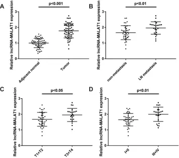
The Expression of MALAT1 Is
Up-Regulated in CC Tissues
The expression of MALAT1 in CC tissues and adjacent normal tissues was measured by qRT-PCR. The expression of MALAT1 was upregulated in CC tissues compared with
adjacent normal tissues (P< 0.001,Figure 1A). As shown in
Figure 1B, the expression of MALAT1 in CC tissues with
lymph-node (LN) metastasis was significantly higher than
that in tissues with non-metastasis (P< 0.01). The expression
of MALAT1 expression was significantly higher in CC
tis-sues with invasive depth (T3 + T4) compared with tistis-sues
with invasive depth (T1 + T2) (P < 0.05, Figure 1C). In
addition, the expression of MALAT1 was significantly
higher in CC tissues with tumor-node-metastasis (TNM) III + IV than tissues with TNM I + II (P< 0.01,Figure 1D). The clinicopathological characteristics of 62 patients with CC
were shown inTable 1. According to the median expression
level of MALAT1 in CC tissues, CC patients were divided into high (MALAT1 expression > median, N = 31) and low
groups (MALAT1 expression≤median, N = 31). The
expres-sion of MALAT1 was associated with the depth of invaexpres-sion, lymph node metastasis and TNM stage, but not associated with gender, age, tumor size, differentiation and tumor site. Taken together, these results suggested that MALAT1 might be involved in CC progression.
LncRNA MALAT1 Promotes the
Proliferation and Inhibits the Apoptosis of
CC Cells
With the application of qRT-PCR assay, the up-regulated expression of MALAT1 was detected in HCT116 and
Figure 1LncRNA MALAT1 is overexpressed in CC tissues. (A) Relative expression of MALAT1 in CC tissues and adjacent normal tissues (N = 62),P< 0.001. (B) Relative MALAT1 expression in CC tissues with and without lymph-node (LN) metastasis (N = 62),P< 0.01. (C) Relative expression of MALAT1 in CC tissues with depth of invasion T1 + T2 and T3 + T4 (N = 62),P< 0.05. (D) The relative expression of MALAT1 in CC tissues with TNM stage I + II and III + IV (N = 62),P< 0.01. Each experiment was performed in three replicates.
OncoTargets and Therapy downloaded from https://www.dovepress.com/ by 118.70.13.36 on 25-Aug-2020
HT29 cells compared with NCM 460 cells (P < 0.01,
Figure 2A). In order to investigate the role of MALAT1 in the development of CC, MALAT1 was overexpressed in HCT116 cells and silenced in HT29 cells. HCT116 cells in the LV-MALAT1 group exhibited a higher MALAT1
expression than that in the LV-NC group (P < 0.001,
Figure 2B); HT29 cells transfected with si-MALAT1-1 or si-MALAT1-2 exhibited a decreased MALAT1 expression
compared with cells transfected with si-NC (P < 0.01,
Figure 2B). Subsequently, cell proliferation and apoptosis were detected. Overexpression of MALAT1 promoted the
proliferation (P < 0.05, Figure 2C) and repressed the
apoptosis of HCT116 cells (P < 0.01, Figure 2D).
Silencing of MALAT1 exhibited contrary results in HT29
cells. Altogether, our results indicated that MALAT1 could promote cell proliferation and inhibit cell apoptosis in CC.
MALAT1 Promotes the Migration and
Invasion of CC Cells
The migration and invasion of transfected CC cells were measured by wound scratch and Transwell invasion assay,
respectively. Overexpression of MALAT1 significantly
enhanced the migration and invasion of HCT116 cells;
meanwhile, knockdown of MALAT1 significantly
inhib-ited the migration and invasion of HT29 cells (P < 0.01,
Figure 3A and B). To sum up, MALAT1 promotes the migration and invasion of CC cells.
MALAT1 Targets
miR-101-3p
To clarify the regulatory mechanism of MALAT1 in CC, the potential downstream targets of MALAT1 were predicated by StarBase (http://starbase.sysu.edu.cn/). A binding site between
MALAT1 andmiR-101-3pwas predicated (Figure 4A). The
target relationship between MALAT1 and miR-101-3p was
further validated by a dual-luciferase reporter assay.
Overexpression ofmiR-101-3psignificantly reduced the
luci-ferase activity of MALAT1-WT in HCT116 cells, whereas
knockdown ofmiR-101-3pelevated the luciferase activity of
MALAT1-WT in HT29 cells (P < 0.05,Figure 4B andC).
RNA pull-down assay showed that miR-101-3pwas pulled
down by Bio-MALAT1-Wt, which further validated that
MALAT1 could directly bind to miR-101-3p (P < 0.05,
Figure 4D). Next, we explored the effect of MALAT1 on the
expression of miR-101-3p in HCT116 and HT29 cells. As
shown inFigure 4E, overexpression of MALAT1 significantly
inhibitedmiR-101-3pexpression in HCT116 cells, while
silen-cing of MALAT1 promotedmiR-101-3pexpression in HT29
cells (P< 0.05). In addition, the expression ofmiR-101-3pwas decreased in CC tissues compared with adjacent normal
tis-sues (P < 0.001, Figure 4F). In CC tissues, there was
a negative correlation between MALAT1 and miR-101-3p
expression (r =−0.3754,P= 0.0026,Figure 4G). The above
data manifested that MALAT1 may be a sponge of
miR-101-3p, which could inversely modulatemiR-101-3pexpression.
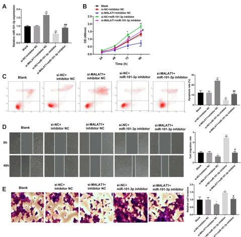
Silencing of
miR-101-3p
Reverses the
Effect of MALAT1 Knockdown on CC
Progression
To investigate whether MALAT1 is involved in the
regula-tion ofmiR-101-3pon CC progression, rescue experiments
were implemented. The qRT-PCR assay manifested that
Table 1 Clinicopathologic Characteristics and MALAT1 Expression in 62 Patients with Colon Cancer
Characteristics n = 62 MALATI Expression
p-value
High (n=31)
Low (n=31)
Gender 0.7991
Male 33 16 17
Female 29 15 14
Age, years 0.0754
≤60 31 12 19
>60 31 19 12
Tumor size, cm 0.2970
≤5 38 17 21
>5 24 14 10
Depth of invasion 0.0180*
T1+T2 39 15 24
T3+T4 23 16 7
Differentiation 0.3074
Well or moderate 34 15 19
Poor 28 16 12
Lymph node
Metastasis 38 14 24 0.0091*
Negative 24 17 7
Positive
TNM stage 0.0038*
I–II 39 14 25
III–IV 23 17 6
Tumor site 0.6098
Colon 34 16 18
Rectum 28 15 13
Note:*Presented significantly different between high and low groups at P < 0.05.
OncoTargets and Therapy downloaded from https://www.dovepress.com/ by 118.70.13.36 on 25-Aug-2020
miR-101-3pinhibitor effectively decreased the expression of
miR-101-3p in HCT116 cells. Knockdown of MALAT1
increased the expression of miR-101-3p in HCT116 cells,
and the promoting effect was reverted bymiR-101-3p
inhi-bitor (P < 0.01, Figure 5A). In addition, silencing of
MALAT1 significantly inhibited the proliferation, migration
and invasion of HCT116 cells. On the contrary, silencing
miR-101-3psignificantly promoted the proliferation,
migra-tion and invasion of HCT116 cells (P < 0.05, Figure 5B,
D and E). The results of apoptosis were contrary with
proliferation (P< 0.01,Figure 5C). Notably, silencing
miR-101-3preverses the effects of MALAT1 knockdown on the
proliferation, apoptosis, migration and invasion of HCT116
cells (P < 0.05, Figure 5B-E). These results indicated
MALAT1 knockdown might mediate CC progression via
regulatingmiR-101-3p.
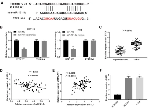
MiR-101-3p
Targets STC1
The potential downstream targets of miR-101-3p were
further analyzed. A binding site between STC1 and
miR-101-3pwas predicated by StarBase (Figure 6A). The target
relationship between STC1 andmiR-101-3pwas further
con-firmed by dual-luciferase reporter assay. Overexpression of
miR-101-3psignificantly decreased the luciferase activity of
STC1-WT in HCT116 and HT29 cells (P< 0.01,Figure 6B).
In addition, the mRNA expression of STC1 was significantly
higher in CC tissues than that in adjacent normal tissues
(P < 0.001, Figure 6C). The mRNA expression of
STC1 was negatively correlated with the expression of
miR-101-3p (r = −0.361, P = 0.0039, Figure 6D), and
positively correlated with the expression of MALAT1
(r = 0.3378,P= 0.0073,Figure 6E). The mRNA expression
of STC1 was also significantly higher in CC cell lines
(HCT116 and HT29 cells) than that in NCM 460 cells
(P < 0.01, Figure 6F). These results indicated that STC1
was a downstream target of MALAT1 andmiR-101-3p.
Silencing of STC1 Reverses the Effects of
MALAT1 Up-Regulation and miR-101-3p
Down-Regulation on CC Progression
The regulatory relationship between STC1 and
miR-101-3p was further identified by that overexpression of
miR-101-3p significantly decreased the mRNA expression of
STC1 in HCT116 and HT29 cells (P< 0.01, Figure 7A).
Rescue experiments were then performed to investigate the regulatory mechanism of STC1 involving MALAT1
andmiR-101-3pin CC cells. The viability, migration, and
Figure 2LncRNA MALAT1 promotes the proliferation and inhibits the apoptosis of CC cells. (A) Relative expression of MALAT1 in NCM 460 cells, HCT116 and HT29 cells. **P< 0.01 vs NCM 460. (B) Relative expression of MALAT1 in transfected HCT116 and HT29 cells. (C) The proliferation of transfected HCT116 and HT29 cells. (D) The apoptosis of transfected HCT116 and HT29 cells. *P< 0.05, **P< 0.01, ***P< 0.001 vs LV-NC;#
P< 0.05,##
P< 0.01 vs si-NC. Each experiment was performed in three replicates.
OncoTargets and Therapy downloaded from https://www.dovepress.com/ by 118.70.13.36 on 25-Aug-2020
invasion of HT29 cells were significantly decreased in si-STC1 group compared with those in si-NC group
(P < 0.01). In addition, silencing of STC1 significantly
reversed the promoting effects of LV-MALAT1 and
miR-101-3p inhibitor on the viability, migration, and invasion
of HT29 cells (P< 0.05, Figure 7B–D).
Discussion
CC is one of the most prevalent and life-threatening
can-cers in the world.2LncRNA MALAT1 plays an important
biological role in the development of CC.19 In this study,
we found that the expression of MALAT1 was associated
with lymph node metastasis, depth of invasion and TNM stage. Furthermore, the up-regulation of MALAT1
contrib-uted to the development of CC via targetingmiR-101-3p/
STC1 in vitro.
Previous studies have proved that MALAT1 is up-regulated in various types of cancers. The expression of MALAT1 is higher in ovarian cancer (OC) tissues and four OC cell lines compared with the normal ovary tissues and
normal ovarian epithelial cell line, respectively.28
MALAT1 is up-regulated in non-small cell lung cancer,
and MALAT1 knockdown inhibits the cell metastasis.29
MALAT1 is highly expressed in gastric cancer patients
Figure 3LncRNA MALAT1 promotes the migration and invasion of CC cells. (A) The migration of transfected HCT116 and HT29 cells. (B) The invasion of transfected HCT116 and HT29 cells. **P< 0.01 vs LV-NC,##
P< 0.01 vs si-NC. Each experiment was performed in three replicates.
OncoTargets and Therapy downloaded from https://www.dovepress.com/ by 118.70.13.36 on 25-Aug-2020
with distant metastasis, functioning as an oncogene.30The expression of MALAT1 in cancerous tissues of CRC is 2.26 times higher than that in noncancerous tissues, which serves as a negative prognostic marker for CRC patients in
stage II/III.31 Consistently, the expression of MALAT1
expression is up-regulated in CRC tissues,32 and is
con-sidered as a poor prognostic factor of CRC.18
Analogously, we observed that MALAT1 expression was up-regulated in CC tissues and HCT116 and HT29 cells. Additionally, the expression of MALAT1 was elevated in
Figure 4MALAT1 targetsmiR-101-3p. (A) The putative binding site between MALAT1 andmiR-101-3pwas predicted by the StarBase database. (BandC) Dual-luciferase reporter assay in HCT116 and HT29 cells. *P< 0.05 vs mimics NC;#
P< 0.05 vs inhibitor NC. (D) RNA pull-down assay in HCT116 cells. *P< 0.05 vs Bio-NC. (E) Relative expression ofmiR-101-3pin HCT116 cells transfected with LV-MALAT1 or LV-NC, and in HT29 cells transfected with si-MALAT1-1 or si-NC. *P< 0.05 vs LV-NC;#
P< 0.05 vs si-NC. (F) Relative expression ofmiR-101-3pin CC tissues and adjacent normal tissues,P< 0.001. (G) Correlation analysis of MALAT1 andmiR-101-3pexpression in CC tissues, r =−0.3754,P= 0.0026. Each experiment was performed in three replicates.
OncoTargets and Therapy downloaded from https://www.dovepress.com/ by 118.70.13.36 on 25-Aug-2020
CC tissues with LN metastasis, invasive depth (T3 + T4),
and/or TNM stage (III + IV). These findings indicated
a high potential of MALAT1 for predicting the poor prog-nosis of CC patients.
MALAT1 affects cell proliferation and apoptosis in
a variety of cancers, including CC.33 Wu et al have
found that MALAT1 knockdown markedly promotes the colony formation and induces the apoptosis of CC
cells.19 Zhang et al have found that MALAT1
knock-down inhibits the apoptosis and promotes the
prolifera-tion of CC cells.34 Likewise, we found that
overexpression of MALAT1 enhanced the proliferation and restrained the apoptosis of HCT116 cells, and the silencing of MALAT1 exhibited opposite results in HT29 cells. These results indicated a tumor-promoting role of MALAT1 in CC.
Figure 5Silencing of miR-101-3p reverses the anti-tumor effect of MALAT1 knockdown on CC progression. (A) Relative expression ofmiR-101-3pin transfected HCT116 cells. (B) The proliferation of transfected HCT116 cells. (C) The apoptosis of transfected HCT116 cells. (D) The migration of transfected HCT116 cells. (E) The invasion of transfected HCT116 cells. *P< 0.05, **P< 0.01 vs si-NC + inhibitor NC;#P< 0.05,##P< 0.01 vs si-MALAT1 + inhibitor NC. Each experiment was performed in three replicates.
OncoTargets and Therapy downloaded from https://www.dovepress.com/ by 118.70.13.36 on 25-Aug-2020
MALAT1 serves as a metastasis promoter in CRC, neuro-blastoma, osteosarcoma, and lung cancer, or a metastasis
sup-pressor in glioma, CRC, breast cancer, and glioma.35Ji et al
have verified that MALAT1 overexpression promotes the
proliferation and migration of CRC in vitro and in vivo via binding to SFPQ and releasing oncogene PTBP2 from SFPQ/
PTBP2 complex.36Herein, similar results were observed. The
up-regulation of MALAT1 promoted the migration and inva-sion of HCT116 cells. Conversely, the silencing of MALAT1 inhibited the migration and invasion of HT29 cells. All these results indicated a pro-migration and invasion role of MALAT1 in CC.
LncRNAs act as competing for endogenous RNAs (ceRNAs) to sponge miRNAs to participate in the occurrence
and development of gastric cancer, liver cancer, and CC.37,38
MALAT1 is involved in the development of CC by interacting with various miRNAs. Xu et al have proved that the silencing of MALAT1 suppresses the viability and metastasis of CRC
cells through spongingmiR-145.17Wu et al have shown that
MALAT1-miR203-DCP1A axis is associated with the
malig-nancy of CC.39Sun et al have demonstrated that MALAT1
enhances the proliferation and inhibits the apoptosis of OC
cells via sponging miR-503-5p.40 Herein, miR-101-3p was
predicted to be a target gene of MALAT1 by StarBase, and this target relationship was validated by dual-luciferase
repor-ter assay and RNA pull-down assay in CC cells.MiR-101-3p
plays a tumor suppressor role in diverse human cancers, such as OC,41gastric cancer,42lung cancer43and cervical cancer.44
In this study, we found that the expression ofmiR-101-3pwas
significantly down-regulated in CC tissues, and was inversely
correlated with MALAT1 expression in CC tissues. Thus, we
deduced thatmiR-101-3palso exerts a tumor suppressor role
in CC. Our following functional analysis revealed that the
silencing ofmiR-101-3pevidently enhanced the proliferation,
migration, invasion, and restrained the apoptosis of HT29 cells. These results illustrated the tumor suppressor role of
A
C
B
hsa-miR-101-3p Position 72-78 of STC1 WT
STC1 Mut
STC1 WT STC1 Mut
0.0 0.5 1.0 1.5 Relativ e lu c if e ra se ac ti v it y miR-NC miR-101-3p mimics ** HCT116
STC1 WT STC1 Mut
0.0 0.5 1.0 1.5 R e la ti v e lucif er as e a ct ivit y miR-NC miR-101-3p mimics ** HT29
Adjacent tissues Tumor 0 1 2 3 4 5 Relative expression of S T C
1 P < 0.001
E
0.0 0.2 0.4 0.6 0.8 1.0
0 1 2 3 4 5
Relative expression of miR-101-3p
Relaviv e expressio n o f ST C
1 r = -0.361
P = 0.0039
5' ...ACACGUCAAUUGAGUGUACUGUG... 3' 5' ...ACACCAGUUUUGAGUGUACUGUG...3'
3' AAGUCAAUAGUGUCAUGACAU 5'
NCM 460
HCT
116 HT29
0 1 2 3 4 5 Relative e xpression of S TC 1 ** **
D
F
0 1 2 3 4 5
0 1 2 3
Relative expression of STC1
Relative expression o f M ALAT 1
r = 0.3378
P = 0.0073
Figure 6MiR-101-3ptargets STC1. (A) The putative binding site betweenmiR-101-3pand STC1 was predicted by the StarBase database. (B) Dual-luciferase reporter assay in HCT116 and HT29 cells. **P< 0.01 vs miR-NC. (C) Relative expression of STC1 in CC tissues and adjacent normal tissues,P< 0.001. (D) Correlation analysis of STC1
andmiR-101-3pexpression in CC tissues, r =−0.361,P= 0.0039. (E) Correlation analysis of STC1 and MATAL1 expression in CC tissues, r = 0.3378,P= 0.0073. (F)
Relative expression of STC1 in HCT116 and HT29 cells. **P< 0.01 vs NCM 460. Each experiment was performed in three replicates.
OncoTargets and Therapy downloaded from https://www.dovepress.com/ by 118.70.13.36 on 25-Aug-2020
miR-101-3pin CC. Rescue experiments were then performed to clarify the regulatory relationship between MALAT1 and
miR-101-3pin CC. The results showed that silencing of
miR-101-3p reversed the anti-tumor effect of MALAT1
knock-down on CC cells. Thesefindings indicated that silencing of
MALAT1 may inhibit the development of CC through
target-ingmiR-101-3p.
MiRNAs play key regulatory roles in the occurrence
and development of CC by regulating specific genes. Zeng
et al have proved that miR-7 inhibits the proliferation and
migration of CC cells by targeting FAK.45Koo et al have
found that miR-4779 induces the apoptosis and cell cycle arrest of CC cells through direct targeting PAK2 and
CCND3.46 Chandramouli et al have shown that ectopic
expression of miR-101 markedly inhibits the proliferation and motility of CC cells through targeting PGE2 receptor
EP4.47In this study, STC1 was identified to be a target of
miR-101-3p. STC1 is a glycoprotein hormone involved in
calcium/phosphate homeostasis.48 STC1 plays a key
reg-ulatory role in the occurrence and development of various
cancers, such as breast cancer,49 cervical cancer,50
retinoblastoma,51 and prostate cancer.52 In this study, the
expression of STC1 was up-regulated in CC tissues and
cell lines, and inversely correlated with miR-101-3p
expression in CC tissues. Silencing of STC1 significantly
inhibited the proliferation, migration, invasion of HT29
Migration Invasion si-NC si-STC1
A
B
C
si-NC si-S TC1 si-S TC1 +LV -MALAT 1 si-S TC1 +miR-101 -3p inhi bito r 0.0 0.5 1.0 1.5 Cell migratio n rat e HT29 * ** # *# si-NC si-S TC1 si-S TC1 +LV -MA LAT 1 si-S TC1+ miR-101 -3p inhi bito r 0.0 0.5 1.0 1.5 R e la ti v e cell in vasio n ** # HT29 *# * si-NC 0 h 24 hD
HCT116 HT29 0.0 0.5 1.0 1.5 Relative mR NA exp re s sio n o f S TC 1 miR-NC miR-101-3p mimics ** **0 24h 48h 72h 96h
0.0 0.5 1.0 1.5 2.0 C e ll viability(OD 450 ) si-NC si-STC1 ** * HT29 si-STC1+LV-MALAT1 * # # si-STC1+miR-101-3p inhibitor si-STC1+LV-MALAT1 si-STC1 si-STC1+LV-MALAT1
si-STC1 +miR-101-3p inhibitor
si-STC1 +miR-101-3p inhibitor
Figure 7Silencing of STC1 reverses the tumor promoting effects of MALAT1 up-regulation and miR-101-3p down-regulation on CC progression. (A) Relative expression of STC1 in transfected HT29 cells. **P< 0.01 vs si-NC. (B) The proliferation of transfected HT29 cells. (C) The migration of transfected HT29 cells. (D) The invasion of transfected HT29 cells. *P< 0.05, **P< 0.01 vs si-NC;#
P< 0.05 vs si-STC1. Each experiment was performed in three replicates.
OncoTargets and Therapy downloaded from https://www.dovepress.com/ by 118.70.13.36 on 25-Aug-2020
cells. These results illustrated a tumor promoting role of STC1 in CC. Furthermore, rescue experiments showed that the tumor promoting effects of MALAT1
overexpres-sion andmiR-101-3psilencing on CC cells were reversed
by STC1 silencing. Thesefindings indicated that silencing
of MALAT1 may inhibit the development of CC through
targetingmiR-101-3p/STC1 axis.
Conclusions
MALAT1 was upregulated in CC tissues and cells, func-tioning as an oncogene in CC development. MALAT1 expression was associated with lymph node metastasis, depth of invasion and TNM stage in patients with CC. MALAT1 promoted the proliferation, migration, and inva-sion, and inhibited the apoptosis of CC cells via targeting
miR-101-3p/STC1 axis. The silencing of MALAT1 might
be a hopeful therapeutic option for CC.
Ethics Approval and Consent
Statement
This study was conducted after obtaining local ethical committee approval of Yidu Central Hospital of Weifang. Written informed consent was obtained from patients over the age of 18 years and parents of patients under the age of 18 years. This was conducted in accordance with the Declaration of Helsinki.
Author Contributions
All authors contributed to data analysis, drafting or revising
the article, gavefinal approval of the version to be published,
and agree to be accountable for all aspects of the work.
Disclosure
The authors declare that they have no conflicts of interest
to disclose.
References
1. Paschke S, Jafarov S, Staib L, et al. Are colon and rectal cancer two different tumor entities? A proposal to abandon the term colorectal cancer.Int J Mol Sci.2018;19:9. doi:10.3390/ijms19092577 2. Bray F, Ferlay J, Soerjomataram I, Siegel RL, Torre LA, Jemal A.
Global cancer statistics 2018: GLOBOCAN estimates of incidence and mortality worldwide for 36 cancers in 185 countries. CA Cancer J Clin.2018;68(6):394–424. doi:10.3322/caac.21492
3. Feng RM, Zong YN, Cao SM, Xu RH. Current cancer situation in China: good or bad news from the 2018 global cancer statistics? Cancer Commun.2019;39(1):019–0368. doi:10.1186/s40880-019-0368-6 4. Roos VH, Mangas-Sanjuan C, Rodriguez-Girondo M, et al. Effects of
family history on relative and absolute risks for colorectal cancer: a systematic review and meta-analysis.Clin Gastroenterol Hepatol. 2019;13(19):30994–30995.
5. Shawki S, Ashburn J, Signs SA, Huang E. Colon cancer: inflammation-associated cancer. Surg Oncol Clin N Am. 2018;27 (2):269–287. doi:10.1016/j.soc.2017.11.003
6. Chao A, Thun MJ, Connell CJ, et al. Meat consumption and risk of colorectal cancer. JAMA. 2005;293(2):172–182. doi:10.1001/ jama.293.2.172
7. Pahwa M, Harris MA, MacLeod J, Tjepkema M, Peters PA, Demers PA. Sedentary work and the risks of colon and rectal cancer by anatomical sub-site in the Canadian census health and environ-ment cohort (CanCHEC). Cancer Epidemiol. 2017;49:144–151. doi:10.1016/j.canep.2017.06.004
8. Sharp L, McDevitt J, Brown C, Comber H. Smoking at diagnosis significantly decreases 5-year cancer-specific survival in a population-based cohort of 18 166 colon cancer patients. Aliment Pharmacol Ther.2017;45(6):788–800.
9. Lee S, Woo H, Lee J, Oh JH, Kim J, Shin A. Cigarette smoking, alcohol consumption, and risk of colorectal cancer in South Korea: a case-control study. Alcohol. 2019;76:15–21. doi:10.1016/j.alcohol. 2018.06.004
10. Zhang Y, Chen Z, Li J. The current status of treatment for colorectal cancer in China: A systematic review. Medicine. 2017;96 (40):0000000000008242. doi:10.1097/MD.0000000000008242 11. Lagarde J, Uszczynska-Ratajczak B, Carbonell S, et al.
High-throughput annotation of full-length long noncoding RNAs with capture long-read sequencing. Nat Genet. 2017;49(12):1731–1740. doi:10.1038/ng.3988
12. Bhan A, Soleimani M, Mandal SS. Long noncoding RNA and cancer: a new paradigm.Cancer Res.2017;77(15):3965–3981. doi:10.1158/ 0008-5472.CAN-16-2634
13. Zhai HY, Sui MH, Yu X, et al. Overexpression of long non-coding RNA TUG1 promotes colon cancer progression. Med Sci Monit. 2016;22:3281–3287. doi:10.12659/MSM.897072
14. Yue B, Qiu S, Zhao S, et al. LncRNA-ATB mediated E-cadherin repres-sion promotes the progresrepres-sion of colon cancer and predicts poor prognosis. J Gastroenterol Hepatol.2016;31(3):595–603. doi:10.1111/ jgh.13206
15. Liu A, Liu L. Long non-coding RNA ZEB2-AS1 promotes prolifera-tion and inhibits apoptosis of colon cancer cells via miR-143/bcl-2 axis.Am J Transl Res.2019;11(8):5240–5248.
16. Xiong W, Qin J, Cai X, et al. Overexpression LINC01082 suppresses the proliferation, migration and invasion of colon cancer.Mol Cell Biochem.2019;20(10):019–03607.
17. Xu Y, Zhang X, Hu X, et al. The effects of lncRNA MALAT1 on proliferation, invasion and migration in colorectal cancer through regulating SOX9.Mol Med.2018;24(1):52.
18. Wu S, Sun H, Wang Y, et al. MALAT1 rs664589 polymorphism inhibits binding to miR-194-5p, contributing to colorectal cancer risk, growth, and metastasis. Cancer Res.2019;79(20):5432–5441. doi:10.1158/0008-5472.CAN-19-0773
19. Wu Q, Meng WY, Jie Y, Zhao H. LncRNA MALAT1 induces colon cancer development by regulating miR-129-5p/HMGB1 axis.J Cell Physiol.2018;233(9):6750–6757. doi:10.1002/jcp.26383
20. Zhuang M, Zhao S, Jiang Z, et al. MALAT1 sponges miR-106b-5p to promote the invasion and metastasis of colorectal cancer via SLAIN2 enhanced microtubules mobility. EBioMedicine. 2019;41:286–298. doi:10.1016/j.ebiom.2018.12.049
21. Bartel DP. MicroRNAs: genomics, biogenesis, mechanism, and function. Cell. 2004;116(2):281–297. doi:10.1016/S0092-8674(04) 00045-5
22. Strubberg AM, Madison BB. MicroRNAs in the etiology of color-ectal cancer: pathways and clinical implications. Dis Model Mech. 2017;10(3):197–214. doi:10.1242/dmm.027441
23. Amirkhah R, Schmitz U, Linnebacher M, Wolkenhauer O, Farazmand A. MicroRNA-mRNA interactions in colorectal cancer and their role in tumor progression. Genes Chromosomes Cancer. 2015;54(3):129–141. doi:10.1002/gcc.22231
OncoTargets and Therapy downloaded from https://www.dovepress.com/ by 118.70.13.36 on 25-Aug-2020
24. Zeng M, Zhu L, Li L, Kang C. miR-378 suppresses the proliferation, migration and invasion of colon cancer cells by inhibiting SDAD1.
Cell Mol Biol Lett.2017;22:12. doi:10.1186/s11658-017-0041-5 25. Li B, Wang S, Wang S. MiR-195 suppresses colon cancer
prolifera-tion and metastasis by targeting WNT3A.Mol Genetics Genomics. 2018;293(5):1245–1253. doi:10.1007/s00438-018-1457-y
26. Ba S, Xuan Y, Long ZW, Chen HY, Zheng SS. MicroRNA-27a promotes the proliferation and invasiveness of colon cancer cells by targeting SFRP1 through the Wnt/beta-catenin signaling pathway.Cell Physiol Biochem.2017;42(5):1920–1933. doi:10.1159/000479610
27. Shao Q, Xu J, Deng R, et al. SNHG 6 promotes the progression of colon and rectal adenocarcinoma via miR-101-3p and Wnt/beta-ca-tenin signaling pathway. BMC Gastroenterol. 2019;19(1):163. doi:10.1186/s12876-019-1080-3
28. Wu L, Wang X, Guo Y. Long non-coding RNA MALAT1 is upregulated and involved in cell proliferation, migration and apoptosis in ovarian cancer. Exp Ther Med. 2017;13(6):3055–3060. doi:10.3892/ etm.2017.4304
29. Guo F, Guo L, Li Y, Zhou Q, Li Z. MALAT1 is an oncogenic long non-coding RNA associated with tumor invasion in non-small cell lung cancer regulated by DNA methylation.Int J Clin Exp Pathol. 2015;8(12):15903–15910.
30. Xia H, Chen Q, Chen Y, et al. The lncRNA MALAT1 is a novel biomarker for gastric cancer metastasis. Oncotarget. 2016;7 (35):56209–56218. doi:10.18632/oncotarget.10941
31. Zheng HT, Shi DB, Wang YW, et al. High expression of lncRNA MALAT1 suggests a biomarker of poor prognosis in colorectal cancer.Int J Clin Exp Pathol.2014;7(6):3174–3181.
32. Si Y, Yang Z, Ge Q, et al. Long non-coding RNA Malat1 activated autophagy, hence promoting cell proliferation and inhibiting apopto-sis by sponging miR-101 in colorectal cancer.Cell Mol Biol Lett. 2019;24(50):019–0175. doi:10.1186/s11658-019-0175-8
33. Li ZX, Zhu QN, Zhang HB, Hu Y, Wang G, Zhu YS. MALAT1: a potential biomarker in cancer. Cancer Manag Res. 2018;10:6757–6768. doi:10.2147/CMAR.S169406
34. Zhang J, Li Q, Xue B, He R. MALAT1 inhibits the Wnt/beta-catenin signaling pathway in colon cancer cells and affects cell proliferation and apoptosis. Bosnian J Basic Med Sci. 2019. doi:10.17305/ bjbms.2019.4408
35. Sun Y, Ma L. New insights into long non-coding RNA MALAT1 in cancer and metastasis.Cancers.2019;11:2. doi:10.3390/cancers11020216 36. Ji Q, Zhang L, Liu X, et al. Long non-coding RNA MALAT1 promotes tumour growth and metastasis in colorectal cancer through binding to SFPQ and releasing oncogene PTBP2 from SFPQ/PTBP2 complex.Br J Cancer.2014;111(4):736–748. doi:10.1038/bjc.2014.383
37. Tam C, Wong JH, Tsui SKW, Zuo T, Chan TF, Ng TB. LncRNAs with miRNAs in regulation of gastric, liver, and colorectal cancers: updates in recent years. Appl Microbiol Biotechnol. 2019;103 (12):4649–4677.
38. Wu M, Li W, Huang F, et al. Comprehensive analysis of the expres-sion profiles of long non-coding RNAs with associated ceRNA net-work involved in the colon cancer staging and progression.Sci Rep. 2019;9(1):16910. doi:10.1038/s41598-019-52883-2
39. Wu C, Zhu X, Tao K, et al. MALAT1 promotes the colorectal cancer malignancy by increasing DCP1A expression and miR203 downregulation.Mol Carcinog. 2018;57(10):1421–1431. doi:10.1002/ mc.22868
40. Sun Q, Li Q, Xie F. LncRNA-MALAT1 regulates proliferation and apoptosis of ovarian cancer cells by targeting miR-503-5p.Onco Targets Ther.2019;12:6297–6307. doi:10.2147/OTT.S214689 41. Liang H, Yu T, Han Y, et al. LncRNA PTAR promotes EMT and
invasion-metastasis in serous ovarian cancer by competitively bind-ing miR-101-3p to regulate ZEB1 expression.Mol Cancer.2018;17 (1):119. doi:10.1186/s12943-018-0870-5
42. Cao C, Xu Y, Du K, et al. LINC01303 functions as a competing endogenous RNA to regulate EZH2 expression by sponging miR-101-3p in gastric cancer. J Cell Mol Med. 2019;23 (11):7342–7348. doi:10.1111/jcmm.14593
43. Zhang H, Wang X, Hu B, Zhang F, Wei H, Li L. Circular RNA ZFR accelerates non-small cell lung cancer progression by acting as a miR-101-3p sponge to enhance CUL4B expression. Artif Cells Nanomedicine Biotechnol. 2019;47(1):3410–3416. doi:10.1080/ 21691401.2019.1652623
44. Fan MJ, Zou YH, He PJ, Zhang S, Sun XM, Li CZ. Long non-coding RNA SPRY4-IT1 promotes epithelial-mesenchymal transition of cer-vical cancer by regulating the miR-101-3p/ZEB1 axis.Biosci Rep. 2019;39:6. doi:10.1042/BSR20181339
45. Zeng CY, Zhan YS, Huang J, Chen YX. MicroRNA7 suppresses human colon cancer invasion and proliferation by targeting the expression of focal adhesion kinase. Mol Med Rep. 2016;13 (2):1297–1303. doi:10.3892/mmr.2015.4643
46. Koo KH, Kwon H. MicroRNA miR-4779 suppresses tumor growth by inducing apoptosis and cell cycle arrest through direct targeting of PAK2 and CCND3. Cell Death Dis. 2018;9(2):77. doi:10.1038/ s41419-017-0100-x
47. Chandramouli A, Onyeagucha BC, Mercado-Pimentel ME, et al. MicroRNA-101 (miR-101) post-transcriptionally regulates the expression of EP4 receptor in colon cancers. Cancer Biol Ther. 2012;13(3):175–183. doi:10.4161/cbt.13.3.18874
48. Yoshiko Y, Aubin JE. Stanniocalcin 1 as a pleiotropic factor in mammals.Peptides.2004;25(10):1663–1669. doi:10.1016/j.peptides. 2004.04.015
49. Chang AC, Doherty J, Huschtscha LI, et al. STC1 expression is associated with tumor growth and metastasis in breast cancer.
Clin Exp Metastasis. 2015;32(1):15–27. doi:10.1007/s10585-014-9687-9
50. Pan X, Jiang B, Liu J, et al. STC1 promotes cell apoptosis via NF-kappaB phospho-P65 Ser536 in cervical cancer cells.Oncotarget. 2017;8(28):46249–46261. doi:10.18632/oncotarget.17641
51. Song WP, Zheng S, Yao HJ, et al. Different transcriptome profiles between human retinoblastoma Y79 cells and an etoposide-resistant subline reveal a chemoresistance mechanism. BMC Ophthalmol. 2020;20(1):92. doi:10.1186/s12886-020-01348-6
52. Bai Y, Xiao Y, Dai Y, et al. Stanniocalcin 1 promotes cell proliferation via cyclin E1/cyclindependent kinase 2 in human prostate carcinoma.
Oncol Rep.2017;37(4):2465–2471. doi:10.3892/or.2017.5501
OncoTargets and Therapy
Dove
press
Publish your work in this journal
OncoTargets and Therapy is an international, peer-reviewed, open access journal focusing on the pathological basis of all cancers, potential targets for therapy and treatment protocols employed to improve the management of cancer patients. The journal also focuses on the impact of management programs and new therapeutic
agents and protocols on patient perspectives such as quality of life, adherence and satisfaction. The manuscript management system is completely online and includes a very quick and fair peer-review system, which is all easy to use. Visit http://www.dovepress.com/ testimonials.php to read real quotes from published authors.
Submit your manuscript here:https://www.dovepress.com/oncotargets-and-therapy-journal
OncoTargets and Therapy downloaded from https://www.dovepress.com/ by 118.70.13.36 on 25-Aug-2020