Original Article
Gadd153 deficiency attenuates abdominal aortic
aneurysm formation in mice
Huiying Zhao1*, Guiying Chen2*, Haifeng Wang1
1Genetic Diagnosis Center, The First Hospital of Jilin University, Changchun 130021, China; 2Department of Cardiology, The First Affiliated Hospital of Harbin Medical University, Harbin 150001, Heilongjiang, China. *Co-first authors.
Received February 4, 2015; Accepted March 30, 2015; Epub January 1, 2018; Published January 15, 2018
Abstract: Abdominal aortic aneurysms (AAAs) are a chronic inflammatory vascular disease for which pharmacologi -cal treatments are not available. Gadd153 is closely associated with the onset of vascular smooth muscle cells (VSMCs) apoptosis. However, a role for Gadd153 in AngII-induced AAA formation is currently unknown. In our study, lentiviral-mediated silencing of Gadd153 through small RNA interference was performed in mice, which was further used for the establishment of mouse experimental AAA induced by infusion of angiotensin II (AngII). We found that
Gadd153 deficiency prevented AngII-induced AAA formation in mice 14 days post perfusion compared with wild-type control mice. Moreover, Gadd153 deficiency significantly reduced lesion macrophage and CD4+ T-cell content, T-cell proliferation, SMC apoptosis, and matrix metalloproteinase expression. In vitro studies revealed that Gadd153
deficiency regulated microvessel growth and monocyte migration. In addition, Gadd153 deficiency also affected AAA lesion Mac-3 macrophage accumulation or CD31 microvessel numbers. In conclusion, our study demonstrates that Gadd153 plays an essential role in AngII-induced AAA formation by promoting inflammatory cells proliferation and
vascular SMC apoptosis affecting MMPs expression.
Keywords: Gadd153, angiotensin II, abdominal aortic aneurysms, matrix metalloproteinases
Introduction
Abdominal aortic aneurysm (AAA) is one of leading causes of mortality worldwide, which might be associated with male sex, advanced age, hypertension, hypercholesterolemia, coro-nary artery disease, atherosclerosis, and ciga-rette smoking [1-3]. Considered that surgical treatment is associated with high postopera-tive mortality (up to 6%) [4], further epidemio-logic and pathoepidemio-logic studies are urgently need-ed to reveal the causes of AAA development, progression, and ultimate rupture.
Although the causes of AAA are not completely understood, it is widely known that pathologic features of aneurysm contribute to its
patho-genesis, including local inflammation, increased oxidative stress, significant matrix degradation
and smooth muscle cell apoptosis [4, 5]. Apo-
ptosis of VSMCs is significantly increased,
accompanied by reduced VSMC density within the medial layer of aneurysmal aortic tissue.
The increased degree of VSMC apoptosis has
already been widely found in AAA, which might
be closely triggered by local inflammation and
increased oxidative stress [6, 7]. Structural degeneration of aortic tissue at the cellular level contributes to aneurysmal formation. For example, overexpression of catalase in vascu-lar smooth muscle cells prevents the formation
of abdominal aortic aneurysms [8]. To explore
the underlying pathways involved in AAA, many studies were widely performed. For example, a recent study has demonstrated store operated calcium entry (SOCE) can modulate cell behav-ior through Orai1 in AAA VSMC apoptosis [9].
These results strongly suggest a better under -standing of the mechanisms involved in AAA may identify new targets that could be manipu-lated pharmacologically or biologically to halt disease progression.
The growth arrest and DNA damage inducible gene 153 (GADD153) encodes Gadd153 pro -tein (also called CHOP-10), which belongs to a
member of the CCAAT/enhancer-binding protein
The expression levels of Gadd153 are very low
in normal growing cells and are highly induced in response to a variety of cellular stresses, including glucose deprivation, exposure to alkylating agents, oxidative stress, and other growth-arresting situations [11-13]. Further-
more, microinjection of Gadd153 into NIH-3T3 fibroblasts induces G1 arrest [14], and the tran -sient expression of Gadd153 into different tumor cell lines also leads to growth arrest and
apoptosis [15]. Thus, a number studies have
proved that Gadd153 is closely associated with the onset of vascular smooth muscle cells
(VSMCs) apoptosis, regulated by local inflam -mation and oxidative stress [16], indicating a potential role of Gadd153 in AAA pathogenesis. But to date no study has addressed the rela-tionship between expression of Gadd153 and
AAA progression. Therefore, we hypothesized
that altering expression of Gadd153 partici-pates directly in AAA pathogenesis and
exam-ined our hypothesis by using Gadd153-deficient
mice induced by lentiviral-mediated silencing of
SOD1 through small RNA interference and
angiotensin II-induced experimental AAA mod-els, thereby potentially offer a novel molecular therapy for AAA treatment.
Materials and methods
Lentiviral vector production
The full length of human H1-RNA promoter was cloned into pBluescript SK (+) by using the primers (forward) and CATACAGAGCGACAATC-TTACTTGAGACTATGTCTT (reverse). The design
of the reverse primer incorporates BglII and HindIII sites juxtaposed on the transcriptional
start site into which DNA sequences containing
siRNA hairpins can be cloned [17]. siRNA oligo-nucleotides were designed that contained a sense strand of 22 (Gadd153) nucleotide
sequences followed by a short spacer
(TTC-AAGAGA), the reverse complement of the sense
strand, and five thymidines as an RNA poly -merase III transcriptional stop signal. Oligos were annealed and cloned into the BglII-HindIII site. For cloning into lentivectors, the complete human H1-RNA promoter plus the siRNA
cas-sette was PCR-amplified by introducing XbaI
sites both upstream and downstream of the
sequence and cloned into a unique NheI site of the 3’LTR of a lentiviral vector containing cyto
-megalovirus (CMV)-Gadd153. This
CMV-Gadd-153 cassette was deleted to generate LV-si- Gadd153.
293T cells were cotransfected with a plasmid
expressing Gadd153 together with a plasmid
expressing siRNA specific for Gadd153
(si-Gadd153). Recombinant lentiviruses were
pro-duced by transient transfection in 293T cells
using the calcium-phosphate method as de- scribed [18, 19]. Infectious lentiviruses were harvested at 48 and 72 h post transfection and
filtered through 0.22-μm-pore cellulose acetate filters. Recombinant lentiviruses were concen -trated by ultracentrifugation (2 h at 50,000 × g)
and subsequently purified on a sucrose 20%
gradient (2 h at 46,000 × g) as described [18, 19].
Animal surgery
All surgical procedures were performed as
pre-viously described [20]. Briefly, we first anesthe
-tized 40-d-old mice with 4% isoflurane and maintained them on 1.5% isoflurane anesthe -sia. Following laminectomy, lentiviral vectors were bilaterally injected at two sites separated by 2 mm in the lumbar L3-L4 region using a ste-reotaxic frame. Using a 5-µl Hamilton syringe with a 34-gauge needle, we injected 1.5 µl of concentrated viral solutions (60,000 ng of p24 antigen/ml) per site (0.75 mm below dura) with
a rate of 0.5 µl/min. The needle was then left in
place for an additional 5 min and gently with-drawn. After surgery, we injected animals sub-cutaneously with a single dose of carprofen (5
mg/kg) to limit inflammatory reaction resulting
from the surgery.
PCR detection
Viral and siRNA integration were detected by
PCR analysis. Fifty to 100 ng of DNA were used
in a 25-µl reaction. Primers spanning the H1-siGadd153 cassette were U3 forward
(5’-GGGCAGCTGTTCCAGACAACTTA-3’) and U3 reverse (5’-GCTTGTCTTTTGCGTGATGGGA-3’).
U3-H1 primers, which amplify the H1 portion of the H1-siRNA cassette, were U3 forward in combination with H1 promoter internal primer
H1 reverse 5’-CGTACGGGCCCGTGGTCTCATAC-AGAACTT-3’. The PCR conditions were 94°C
denaturation for 3 min followed by 40 cycles of
Gadd153 reverse (5’-GCGTCCTTGAAGGGTAA-GAT-3’).
Mouse AAA model and lesion characterization
Wild-type (Gadd153+/+) and lentiviral-mediat
-ed Gadd153-deficient (Gadd153-/-) male mice
at 20 weeks of age were infused with angioten-sin II (Ang II; 0.8 mg/kg per day, up to 14 days) to produce experimental AAA mouse models,
as previously described [21]. The aortic tissues
were harvested from 10 mice each group at 7
and 14 days postperfusion. The presence of an
AAA and the scoring of AAA pathology were
determined using a classification scheme described previously [21]. Two researchers
measured aortic diameters independently. On determination of the AAA incidence and
classi-fication, another investigator matched the
scored AAAs to the genotypes of the mice. Studies were performed by the approval of the
Animal Care and Use Committees in The First Affiliated Hospital of Harbin Medical University.
SMC apoptosis
SMC apoptosis was performed using primary cultured Gadd153-/- and wild type mouse
aor-tic SMCs on an 8-well chamber slide. The apop
-tosis of confluent SMCs was induced by treat -ment of 60 mol/L pyrrolidine dithiocarbamate for 24 h. Apoptotic cells were detected with In
Situ Cell Death Detection Kit according to the
manufacturer’s instructions.
Mean blood pressure measurements
Mean systolic blood pressures were measured
in conscious mice using a computerized
tail-cuff method (BP-2000 Visitech Systems). Mice
were acclimatized to the system for 1 week
prior to the initiation of studies and systolic blood pressure was measured 5 days per week until study termination.
Cell proliferation assay and transmigration as-say
CD4+T cells were purified from splenocytes by
depleting major histocompatibility complex
class II-positive cells and CD8+T cells using anti-mouse I-Ab and CD8 monoclonal antibod
-ies (BD Biosciences), followed by complement
depletion, as described previously [22]. Mono- cytes were isolated from peripheral blood by Percoll (Sigma, CA, USA) gradient
centrifuga-tion. T-cell and monocyte proliferation were
assessed with the Cell Titer 96AQ Assay kit,
according to the manufacturer’s instructions.
T-cell and monocyte transmigration assay was
performed on a type I collagen (100 ng/25 µl per well in a pH7.0 HEPES buffer)-precoated 96-well chemotaxis plate, according to the manufacturer’s instructions.
Aortic ring assay
A 96-well plate was coated with 50 µl of Matrigel. A 1-mm long mouse aortic ring from
WT mouse or Gadd153-/-mice mouse was laid on top of the solidified Matrigel and covered with 100 µl of Matrigel. After solidification at
room temperature, 150 µl of RPMI (with 10% FBS) was added to each well. After 7 to 10 days of culture, the aortas were photographed, and
the endothelial outgrowth was analyzed using
ImagePro Plus software and presented as
square millimeters. Basic fibroblast growth fac -tor (bFGF) (10 ng/ml) was used as a positive control
Histology and immunohistochemistry
Mice were euthanized at the designated time
points (7 or 14 days), and abdominal aortic sec-tions were stained with hematoxylin and eosin or Verhoeff-Van Geison (elastin) stain for histo-logical analysis or with antibodies against
mac-rophage marker protein Mac-3 or CD4+ for immunohistochemical analysis. Quantitation of
immunohistochemistry was performed by determining the ratio of the number of Mac-3 or
CD4+ positive cells to the total number of hematoxylin-positive nuclei per field (at × 400 magnification).
Quantitation of mRNA expression
Real-time quantitative polymerase chain reac
-tion analysis for COX-2, CD68, cathepsins B
and cathepsins K, MMP2, and MMP9 was
per-formed using TaqMan gene expression assays and was analyzed by the ÄÄCt method with GADPH as the endogenous control.
Statistical analyses
For comparing two groups on a continuous response variable, a two-sample Student’s t-test was used after verifying that data met
constraints of normality and equivalence of
variance to permit parametric analysis. A
two groups on a continuous response variable.
Percent incidence of AAAs was analyzed by
Fisher’s exact test. P values < 0.05 were
con-sidered to be statistically significant. All data
are represented as mean ± SEM. Results
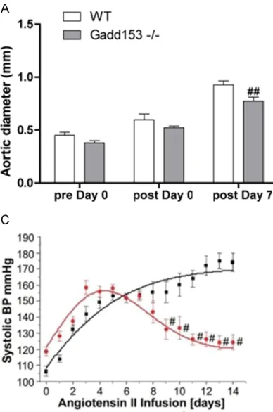
Gadd153 deficiency attenuates the incidence and severity of Ang II-induced AAAs
We first determined the effect of Gadd153 defi -ciency on AAA development in mice treated with lentiviral-mediated silencing of Gadd153 through small RNA interference
(Gadd153-/-mice). At the 7-day time point, no significant dif -ferences were observed in aortic diameters
between Gadd153-/-mice and wild type (WT)
mice before and immediately after Ang II infu-sion. At 7 days postperfusion, Gadd153-/-mice
and WT mice also showed no significant differ -ences in aortic diameters, and none of these
mice developed AAA, as defined by a 100%
increase in aortic diameter (Figure 1A). In Gadd153-/-mice used for the 14-day time point, both preperfusion and immediate post-perfusionaortic diameters were larger than
those from WT mice. At 14 days postperfusion, however, aortic diameters from WT mice were
not significantly different from those immedi
-ately postperfusionbut were significantly larger
than those from Gadd153-/-mice (P <001) (Figure 1B). In addition, at 14-day time point, all
WT mice (10 of 10) developed AAA, but not all
Gadd153-/-mice (3 of 10) formed AAA, indicat-ing that Gadd153 plays an essential role in AAA formation.
The MBP was measured by an intracarotid
telemetry method. We found that MBP of Gadd153-/-mice was slightly higher than that of
WT mice at baseline (i.e., day 0 Ang II infusion;
Figure 1C). Moreover, MBP was increased in both groups up to day 14 during Ang II infusion
(WT: 109-147 mmHg; Gadd153-/-: 118-150
mmHg; Figure 1C). Thereafter, MBP of Gadd-153-/-mice was reduced due to Gadd153 defi -ciency compared with continued increasing
MBP in WT mice (Figure 1C).
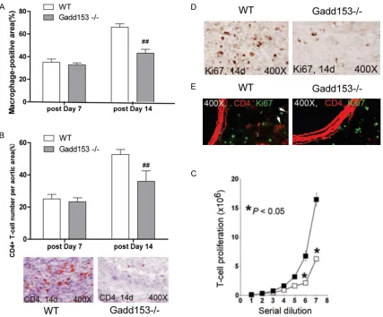
Effects of Gadd153 deficiency on inflamma -tory cells in AAA lesions
Inflammatory cell infiltration is a crucial event
contributing to AAA development. As showed in Figure 2A, a significantly higher levels of lesion macrophage contents was found in WT mice
than Gadd153-/-mice at both 7-day and 14-day Figure 1. Reduced abdominal aortic aneurysm (AAA) formation in Gadd153-/-mice. A, B. Aortic
diameters in both Gadd153-/-mice and WT mice
were measured preperfusion, immediately post-perfusion, and at 7 and 14 days post perfusion. C. Changes in mean blood pressure (MBP) in AngII–
infused Gadd153-/-mice (Red) and WT mice (WT). The MBP was monitored by an intracarotid-telem
-etry method continuously for 14 days. Data are
mean ± SEM. P < 0.05 is considered statistically
[image:4.612.92.286.72.365.2]time point (Figure 2A). Moreover, we showed
that AAA lesions from WT mice contained more higher numbers of CD4+ T cells than that of
Gadd153-/-AAA lesions at 14 days postperfu-sion (Figure 2B), suggesting that Gadd153 activity might be involved in the migration or
proliferation of macrophage and T-cell.
To test these possibilities, we performed
Boyden chamber cell transmigration assay by using stromal cell-derived factor-1 as a che-moattractant. As showed in Figure 2C, both
macrophage and CD4+ T cells from WT mice
proliferated much faster than those Gadd153-/-mice. Furthermore, the number of
proliferat-ing Ki67+ cells were significantly reduced in Gadd153-/-mice, compared with those in WT
mice at 14 days postperfusion (Figure 2D). To further determine the number of CD4+ T and Ki67+ cells directly, we performed the co-immunostaining by using CD4 and
anti-Ki67 monoclonal antibody in AAA lesions.
Among all Ki67+ proliferating cells, the percent
-ages of CD4+T cells were significantly lower in Gadd153-/-mice than WT mice (Figure 2E),
confirming a crucial of Gadd153 in macrophage and T-cell proliferation in vivo.
Effects of Gadd153 deficiency on Ang II-induced vascular cell apoptosis in AAA lesions
Apoptosis of SMC is a key factor that contrib-utes tunica media thinning in AAA lesions,
whereas infiltrating leukocytes release apop -Figure 2. Gadd153 activities on inflammatory cells proliferation. Deficiency of Gadd153 reduced abdominal aortic aneurysm (AAA) lesion Mac-3+ macrophage-positive area (A) and CD4+ T-cell numbers (B) in the adventitia at 14
days post perfusion. In vitro cell proliferation assay showed that Gadd153 deficiency impaired CD4+ T-cell pro
-liferation (C). In AAA lesion adventitia, numbers of Ki67-positive cells (D) and percentage of CD4+ T-cells among Ki67-positive cells (E; arrows indicate Ki67+; CD4+ T cells) were also reduced in Gadd153-/-mice at 14 days post perfusion. The number of mice per group is indicated in each bar. All data are mean ± SE. P < 0.05 is considered
[image:5.612.93.520.71.425.2]totic stimuli to promote vascular cell
apopto-sis[23]. The deficiency of Gadd153 inhibited Ang II-induced cell apoptosis significantly AAA
lesion. As showed in Figure 3A, the number of
terminal deoxynucleotidyl transferase UTP nick-end labeling (TUNEL)-positive cells were
reduced in whole AAA lesions and in the media (Figure 3B, mainly SMCs) from Gadd153-/-mice
compared with those in WT mice at 14 days
postperfusion. Consistent with this observa-tion, medial SMC loss in AAA lesions from
Gadd153-/-mice also was significantly impaired
at this time point (Figure 3C), although both lesion cell apoptosis and medial SMC loss were
not significantly different between both groups
at the 7-day time point (Figure 3A and 3C). To
examine the contribution of Gadd153 to SMC apoptosis, we induced SMC apoptosis with
pyr-rolidine dithiocarbamate. The SMCs from WT
mice was stimulated to apoptosis by treatment with pyrrolidine dithiocarbamate, which was on the contrast to SMC from Gadd153-/-mice, suggesting a protective role of Gadd153 in SMCs (Figure 3D).
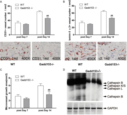
Effects of Gadd153 deficiency on Ang II-induced angiogenesis in AAA lesions
To investigate the role of Gadd153in angiogen
-esis, AAA lesions were collected from WT mice
and Gadd153-/-mice. We found that numbers
of CD31+ microvessels in Gadd153-/-mice was reduced by Gadd153 deficiency at 14 days
postperfusion and of proangiogenic laminin-5
γ2+ vessels at both 7 days and 14 days post
-perfusion, compared with WT mice (Figure 4A, 4B). In vitro aortic ring angiogenesis assay showed defects of microvessels sprouting from aortic rings from Gadd153-/-mice with or with-out angiogenic factor bFGF (Figure4C). To
assess whether the absence of Gadd153 affected the expression or activities of other
proteases, we performed RT-PCR in MHEC from Gadd153-/-mice and Gadd153+/+mice dem -onstrated that the absence of Gadd153 reduced cathepsin S, cathepsin B and cathep-sin K mRNA levels in MHEC (Figure 4D), sug-gesting that reduced angiogenesis in Gadd153-Figure 3. Gadd153 activities on lesion cell apoptosis. Abdominal aortic aneurysm (AAA) lesion adventitia (A) and
medial (B) terminal deoxynucleotidyl transferased UTP nick-end labeling (TUNEL)-positive cell numbers were signifi
-cantly lower in Gadd153-/-mice than in WT mice at 14 days post perfusion. At the same time point, lesion medial smooth muscle cell (SMC) loss was reduced in Gadd153-/-mice (C). The number of mice per group is indicated in each bar. Aortic SMCs from Gadd153-/-mice were resistant to pyrrolidine dithiocarbamate (PDTC)-induced apopto
[image:6.612.93.523.70.350.2]/-mice was caused in part by reduced EC prote-ase expression and activities.
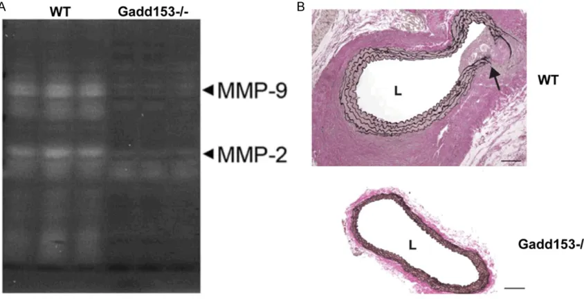
Effects of Gadd153 deficiency on Ang II-induced MMP2 and MMP9 expression in AAA pathogenesis
MMPs have been known to be mechanistically implicated in the pathogenesis of AAAs, and
MMP2 and MMP9, in particular, are identified a
concerted role in AAA initiation and progres-sion [24]. As shown in Figure 5A and 5B, Ang II
treatment significantly increased MMP2 mRNA
expression at 14-day in both group of mice. However, in Gadd153-/-mice, the increase in
MMP2 expression was significantly lower when analyzed after Ang II infusion compared with WT mice (Figure 5A). However, after 14 days of
Ang II infusion, MMP9 expression was signifi
[image:7.612.93.520.74.462.2]-cantly increased in WT mice, which was reversed by Gadd153 deficiency (Figure 5A). Increased MMP expression has been associat-ed with the proteolytic degradation and elastin breakage that occur during aneurysmal expan-sion. Verhoeff-van Gieson staining of abdomi-nal aortic segments from Gadd153-/-mice after 14 days of Ang II infusion showed regions of breakage and discontinuity of the medial elas-tin layer (Figure 5B).
Figure 4. Gadd153 function in neovascularization in abdominal aortic aneurysms (AAA) lesions. CD31+ (A) and proangiogenic laminin-5 fragment γ2+ (B) microvessel numbers were reduced in AAA lesions from Gadd153-/-mice. The number of mice per group is indicated in each bar. Both measurements are from the entire lesion including ad -ventitia and media. Aortic ring assay in vitro demonstrated impaired microvessel sprouting from Gadd153-/-mouse
Discussion
The pathological mechanism involving in the
development and progression of aneurysm is
still unclear. Thus, identification of potential
causes and medical treatment is a major chal-lenge in modern vascular medicine. Our study
identified the essential role of Gadd153 in Ang
II-induced AAA development and we provided
first evidence that Gadd153 contributed to AAA formation by regulating monocyte and T-cell
recruitment, lesion cell proliferation, angiogen-esis and MMP expression, suggesting that
Gadd153 deficiency exerts protective roles in
Ang II-induced experimental AAA mice models. A number of studies have already proved that Ang II infusion results in the formation of AAAs in mice [25, 26], due to its multiple bioactive
effects, including medial degeneration, inflam -mation, thrombus for-mation, and rupture of the abdominal aorta, therefore acting as a critical factor in Ang II-induced AAA development [26]. In addition, our studies also showed that Ang II infusion caused an abnormal rapid rise in blood
pressure in the Gadd153-/-mice and WT mice.
However, blood pressure of Gadd153-/-mice began to progressively fall after day 7 and 14 of Ang II infusion. In contrast, blood pressure of
WT mice increased continuously throughout
the course of Ang II infusion, indicating a pro-tective role of Gadd153 in Ang II infusion-induced AAA development.
The inflammatory cell population in AAA lesions
is another crucial factor that contributes to Ang II-induced AAA mice models [27]. After treated with Ang II up to 14 days, macrophage content
and T-cell number in lesions from Gadd153-/-mice, were both significantly reduced com
-pared with WT mice. Moreover, the transmigra
-tion of monocytes or T cells from Gadd153-/-mice was higher than that of WT Gadd153-/-mice, which may explain reduced T cells and macrophages
in AAA lesions from Gadd153-/-mice, although
we found that the deficiency of Gadd153 has inhibited Ang II-induced monocytes and T-cell
proliferation. Here, we demonstrated that
Gadd153 contributed to AAA lesion inflamma -tory cell accumulation.
Next, we assessed the effects of Gadd153 in
PDTC-induced SMC apoptosis. The apoptosis of
aortic SMC in vitro from Gadd153-/-mice was
markedly reduced compared with cells from WT
mice, similar results of trend was found AAA
lesion cell apoptosis measured by TUNEL, indi -cating that Gadd153 is essential to SMC apop-tosis and to AAA lesion cell apopapop-tosis, which is consistent with previous reports of Gadd153-Figure 5. Matrix metalloproteinase (MMP) expression is attenuated in the aortas of Gadd153-/-mice. Quantitative polymerase chain reaction (PCR) analysis of MMP2 mRNA expression in the abdominal aortas of Gadd153-deficient
[image:8.612.94.516.73.289.2]induced SMC apoptosis [28]. For example, a number studies have proved that Gadd153 is closely associated with the onset of vascular smooth muscle cells (VSMCs) apoptosis,
regu-lated by local inflammation in AAA pathogene -sis [16]. Next, in vitro studies of aortic ring assay and immunostaining of AAA lesion
sec-tions have revealed that Gadd153 deficiency
resulted in the reduction of microvessel or
CD31+ microvessel numbers in AAA lesions
from Gadd153-/-mice. Consistently, the
num-ber of CD31+ or laminin-5 γ2+ microvessel was significantly fewer in Gadd153-/-mice AAA lesions than in WT mouse AAA lesions, sug -gesting the effects of Gadd153 in angiogene-sis. Finally, we investigated how Gadd153 regu-lated AAA expansion. Considered that multiple proteases in AAA lesions are responsible for the extracellular elastin degradation [29], we examined the expression of cysteinyl cathep-sins and matrix metallopreteinases. As a result, cathepsins K, S and L, MMP2 and 9 were
sig-nificant by absence of Gadd153. A mechanism
research has revealed that CatL and other cathepsins (e. g., CatS and CatK.) formed
sequestered and acidic environments into in
cultured human monocyte derived
macro-phages with water-insoluble elastin fibers [30, 31]. Thus, we demonstrated that Gadd153 defi -ciency degraded extracellular elastin in the aor-tic wall and led to AAA expansion by down-regu-lation of most cathepsins and MMPs expres-sions in MHEC.
Taken together, our data from experimental
AAA models of Gadd153-/-mice reveals the essential role of Gadd153 in AAA formation
and development. Thus, pharmacological inhib -itors for selective inhibition of Gadd153 might
benefit patients with AAA, atherosclerosis, and other cardiovascular diseases. Therefore,
Gadd-153 will be of an interesting target in the future studies of cardiovascular diseases.
Disclosure of conflict of interest
None.
Address correspondence to: Dr. Haifeng Wang, Genetic Diagnosis Center, The First Hospital of Jilin University, Changchun 130021, China. Tel: +86-15843079628; Fax: +86+86-15843079628; E-mail:
References
[1] Nordon IM, Hinchliffe RJ, Loftus IM and
Thompson MM. Pathophysiology and epide -miology of abdominal aortic aneurysms. Nat Rev Cardiol 2011; 8: 92-102.
[2] Blanchard JF, Armenian HK and Friesen PP. Risk factors for abdominal aortic aneurysm: results of a case-control study. Am J Epidemiol 2000; 151: 575-583.
[3] Lederle FA, Nelson DB and Joseph AM.
Smokers‘ relative risk for aortic aneurysm compared with other smoking-related diseas-es: a systematic review. J Vasc Surg 2003; 38: 329-334.
[4] Poldermans D, Bax JJ, Kertai MD, Krenning B, Westerhout CM, Schinkel AF, Thomson IR,
Lansberg PJ, Fleisher LA, Klein J, van Urk H, Roelandt JR and Boersma E. Statins are asso-ciated with a reduced incidence of periopera-tive mortality in patients undergoing major noncardiac vascular surgery. Circulation 2003; 107: 1848-1851.
[5] Rowe VL, Stevens SL, Reddick TT, Freeman MB, Donnell R, Carroll RC and Goldman MH.
Vascular smooth muscle cell apoptosis in an-eurysmal, occlusive, and normal human aor-tas. J Vasc Surg 2000; 31: 567-576.
[6] Lopez-Candales A, Holmes DR, Liao S, Scott MJ, Wickline SA and Thompson RW. Decreased
vascular smooth muscle cell density in medial degeneration of human abdominal aortic an-eurysms. Am J Pathol 1997; 150: 993-1007. [7] Henderson EL, Geng YJ, Sukhova GK,
Whittemore AD, Knox J and Libby P. Death of
smooth muscle cells and expression of
media-tors of apoptosis by T lymphocytes in human
abdominal aortic aneurysms. Circulation 1999; 99: 96-104.
[8] Parastatidis I, Weiss D, Joseph G and Taylor
WR. Overexpression of catalase in vascular smooth muscle cells prevents the formation of abdominal aortic aneurysms. Arterioscler
Thromb Vasc Biol 2013; 33: 2389-2396.
[9] Bailey M, Young R, Rode B, Foster R, Li J and
Beech D. Significance of store operated calci -um entry in h-uman abdominal aortic aneu-rysm vascular smooth muscle cells (1057.3). FASEB J 2014; 28: 1057-1053.
[10] Cao Z, Umek RM and McKnight SL. Regulated expression of three C/EBP isoforms during
adi-pose conversion of 3T3-L1 cells. Genes Dev
1991; 5: 1538-1552.
[11] Mohan C, Sathyamurthy M and Lee GM. A role
of GADD153 in ER stress-induced apoptosis in
recombinant Chinese hamster ovary cells. Biotechnol Bioprocess Eng 2012; 17: 446-455.
[12] Weng TI, Wu HY, Chen BL, Jhuang JY, Huang
protein deficiency aggravates acute pancreati -tis and associated lung injury. World J Gas- troenterol 2013; 19: 7097-7105.
[13] Guyton KZ, Xu Q and Holbrook NJ. Induction
of the mammalian stress response gene
GADD153 by oxidative stress: role of AP-1
element. Biochem J 1996; 314: 547-554. [14] Oyadomari S, Mori M. Roles of CHOPGADD153
in endoplasmic reticulum stress. Cell Death Differ 2003; 11: 381-389.
[15] Maytin EV, Ubeda M, Lin JC and Habener JF. Stress-inducible transcription factor CHOP/ gadd153 induces apoptosis in mammalian cells via p38 kinase-dependent and -indepen-dent mechanisms. Exp Cell Res 2001; 267: 193-204.
[16] Tang JR, Nakamura M, Okura T, Takata Y,
Watanabe S, Yang ZH, Liu J, Kitami Y and Hiwada K. Mechanism of oxidative
stress-in-duced GADD153 gene expression in vascular
smooth muscle cells. Biochem Biophys Res Commun 2002; 290: 1255-1259.
[17] Raoul C, Abbas-Terki T, Bensadoun JC, Guillot S, Haase G, Szulc J, Henderson CE and
Aebischer P. Lentiviral-mediated silencing of
SOD1 through RNA interference retards dis -ease onset and progression in a mouse model of ALS. Nat Med 2005; 11: 423-428.
[18] Cockrell AS and Kafri T. Gene delivery by lenti -virus vectors. Mol Biotechnol 2007; 36: 184-204.
[19] Dykxhoorn DM, Novina CD and Sharp PA.
Killing the messenger: short RNAs that silence gene expression. Nat Rev Mol Cell Biol 2003; 4: 457-467.
[20] Ralph GS, Radcliffe PA, Day DM, Carthy JM, Leroux MA, Lee DC, Wong LF, Bilsland LG,
Greensmith L, Kingsman SM, Mitrophanous
KA, Mazarakis ND and Azzouz M. Silencing mu
-tant SOD1 using RNAi protects against neuro -degeneration and extends survival in an ALS model. Nat Med 2005; 11: 429-433.
[21] Gitlin JM, Trivedi DB, Langenbach R and Loftin CD. Genetic deficiency of cyclooxygenase-2 at -tenuates abdominal aortic aneurysm forma-tion in mice. Cardiovasc Res 2007; 73: 227-236.
[22] Shi GP, Villadangos JA, Dranoff G, Small C, Gu
L, Haley KJ, Riese R, Ploegh HL and Chapman
HA. Cathepsin S required for normal MHC
class II peptide loading and germinal center development. Immunity 1999; 10: 197-206.
[23] Zhang J, Bockler D, Ryschich E, Klemm K,
Schumacher H, Schmidt J and Allenberg JR.
Impaired Fas-induced apoptosis of T lympho -cytes in patients with abdominal aortic aneu-rysms. J Vasc Surg 2007; 45: 1039-1046. [24] Wang YX, Martin-McNulty B, da Cunha V,
Vincelette J, Lu X, Feng Q, Halks-Miller M,
Mahmoudi M, Schroeder M, Subramanyam B,
Tseng JL, Deng GD, Schirm S, Johns A, Kauser K, Dole WP and Light DR. Fasudil, a Rho-kinase
inhibitor, attenuates angiotensin II-induced ab-dominal aortic aneurysm in apolipoprotein
E-deficient mice by inhibiting apoptosis and
proteolysis. Circulation 2005; 111: 2219-2226.
[25] Daugherty A, Manning MW and Cassis LA.
Angiotensin II promotes atherosclerotic lesions
and aneurysms in apolipoprotein E-deficient
mice. J Clin Invest 2000; 105: 1605-1612. [26] Daugherty A and Cassis LA. Mouse models of
abdominal aortic aneurysms. Arterioscler.
Thromb Vasc Biol 2004; 24: 429-434.
[27] Tang EH, Shvartz E, Shimizu K, Rocha VZ, Zheng C, Fukuda D, Shi GP, Sukhova G and Libby P. Deletion of EP4 on bone marrow-de
-rived cells enhances inflammation and angio -tensin II-induced abdominal aortic aneurysm
formation. Arterioscler. Thromb Vasc Biol
2011; 31: 261-269.
[28] Cheng WP, Hung HF, Wang BW and Shyu KG.
The molecular regulation of GADD153 in apop -tosis of cultured vascular smooth muscle cells by cyclic mechanical stretch. Cardiovasc Res 2008; 77: 551-559.
[29] Sun J, Zhang J, Lindholt JS, Sukhova GK, Liu J,
He A, Abrink M, Pejler G, Stevens RL, Thompson RW, Ennis TL, Gurish MF, Libby P and Shi GP.
Critical role of mast cell chymase in mouse ab-dominal aortic aneurysm formation. Circulation 2009; 120: 973-982.
[30] Reddy VY, Zhang QY and Weiss SJ. Pericellular mobilization of the tissue-destructive cysteine
proteinases, cathepsins B, L, and S, by human monocyte-derived macrophages. Proc Natl Acad Sci U S A 1995; 92: 3849-3853.
[31] Punturieri A, Filippov S, Allen E, Caras I, Murray R, Reddy V and Weiss SJ. Regulation of elasti-nolytic cysteine proteinase activity in normal

![Bis{(E) 3 [(diethylmethylammonio)methyl] N [3 (N,N dimethylsulfamoyl) 1 methylpyridin 4 ylidene] 4 methoxyanilinium} tetraiodide pentahydrate](data:image/gif;base64,R0lGODlhAQABAIAAAP///wAAACH5BAEAAAAALAAAAAABAAEAAAICRAEAOw==)