Original Article
Global functional connectivity density increased in
treatment of refractory compulsive behaviour
Langlang Cheng1*, Qinggang Chen2*, Xiaoyan Ma2*, Jidong Xiong1, Xinyu Guo2, Lina Wang2, Baohua Zhang3, Xiang Mu3, Wei Liu4, Suling Chen1
1Department of Psychiatry, Wenzhou Seventh People’s Hospital, Wenzhou 325000, China; 2Department of
Psychi-atry, Neuroimaging Laboratory, Tianjin Anding Hospital, Tianjin Mental Health Center, Tianjin, China; 3Department
of Psychiatry, Ankang Hospital of Tianjin Public Security Bureau Tianjin, Tianjin 300240, China; 4Department of
Psychiatry, The First Affiliated Hospital of Harbin Medical University, Harbin 150001, Heilongjiang Province, China.
*Equal contributors.
Received December 30, 2017; Accepted September 10, 2018; Epub March 15, 2019; Published March 30, 2019
Abstract: Previous studies have reported widespread aberrant functional connectivity (FC) in patients with
obses-sive-compulsive disorder (OCD). Global FC density (gFCD) can reflect aberrant FC in terms of connection numbers
and can be used as a supplementary index for traditional FC methods, which focus on connectivity strength. How-ever, to the best of our knowledge, few studies have examined the relationship between gFCD and compulsive behaviour or obsessional thoughts, particularly in treatment-refractory OCD patients. The current study aimed to explore gFCD relative to refractory compulsive behaviour (compulsion) in treatment-refractory OCD patients. We ad-opted FC density mapping (FCDM) to investigate gFCD features in treatment-refractory OCD patients with obsessive compulsive behaviour (TRC) as a primary clinical symptom. Twenty-three TRC and 23 matched treatment-refractory OCD patients with obsessive thought (TRO) as a primary clinical symptom were enrolled in the study. gFCD was ad-opted to examine differences between TRC and TRO patients. The results revealed that TRC patients demonstrated
increased gFCD in the right sensorimotor cortex. Moreover, the findings suggested abnormal
hyper-information-communication in TRC patients compared with TRO patients. The right sensorimotor cortex is a key component of the sensorimotor network and plays an essential role in the modulation of somatic movements. Abnormal
hyper-information-communication in this brain region may be a specific pathological characteristic of TRC. The current results, combined with previous findings, support the hypothesis that TRC has specific clinical symptoms with corre -sponding neural bases. However, our current study was limited by the absence of a healthy control group, preventing
verification of our hypothesis. Nonetheless, our findings provide an experimental approach for clarification of this
hypothesis in further studies.
Keywords: Treatment-refractory compulsion, functional connectivity density, right sensorimotor cortex, functional connection numbers
Introduction
In the last two decades, advances in functi- onal magnetic imaging techniques (fMRI) have enabled many investigations of the neural pa- thological features of obsessive-compulsive di- sorder (OCD) patients [1-6]. Evidence from pre-vious studies has helped enhance the under-standing of the pathophysiological characteris-tics of OCD [6-10]. Currently, in terms of the brain connectome, the most widely accepted hypothesis of OCD is that dysconnectivity am- ong some brain regions, such as the orbital lobe, frontal lobe, thalamus, and hippocampus, is the pathophysiological basis of OCD [11-14].
invaluable information for the further study of the neural basis of the unique symptoms of OCD, particularly in treatment-refractory OCD. However, to the best of our knowledge, most previous studies investigated functional con-nectivity (FC) differences between OCD pati- ents and healthy controls, while few studies have explored the pathological features of the
specific symptoms of OCD, particularly in treat -ment-refractory OCD patients [1-16].
Previous studies have mainly investigated the strength of FC, which represents the strength of temporal correlations between two brain
the last 6 years, Tomasi et al. developed a method for measuring FC numbers, which
can reflect the number of voxels connected to
other voxels in the whole brain and expose aberrant FC from a connection number per-spective [19-22]. Contrary to FC strength, FC density (FCD) represents the relationship of one voxel to other voxels [22-25]. FCD was recently adopted to investigate abnormal FC in several mental disorders, including attention
deficit hyperactivity disorder (ADHD), depres -sion, alcohol addiction, and schizophrenia, re-
vealing many useful findings [20-29]. All of the
studies mentioned above support the notion that FCD can be used as a method for exploring the aberrant brain connectome from a connec-tion number perspective, and can provide use-ful information.
FCD mapping (FCDM) has been adopted to investigate aberrant FCD. FCDM uses an ultra-fast logic algorithm that is usually adopted to calculate global FCD in samples [24]. Most previous studies have focused on FC strength, while few studies have reported gFCD differ-ences between TRC and TRO patients. In the current study, we used FCDM to investigate functional connection number differences be- tween TRC and TRO patients. We hypothesized that TRC patients would exhibit a different ab- errant global FCD pattern compared with TRO patients.
Material and methods
Samples
The present study included 23 treatment-re- fractory OCD patients with compulsive behav-iour as a primary clinical symptom and 23 well-matched patients with obsessive thought as a primary clinical symptom. All patients were enrolled from outpatients and hospitalized pa-
tients at the First Affiliated Hospital of Harbin
Medical University. Two senior psychiatrists us- ed the Structured Clinical Interview for DSM-IV (SCID) [30] to identify TROCD patients with compulsive behaviour or obsessive thought as a primary clinical symptom according to addi-tional relevant criteria (such as treatment-re- fractory). The Yale-Brown Obsessive Compul- sive Scale (Y-BOCS) [31] was adopted to as- sess the severity of compulsive behaviour and obsessive thought. The Medical Research Ethi- cs Committee of our University approved our
study. All patients were fully informed of the
risks and benefits of the study, and written
informed consent was obtained from each participant.
Image acquisition
A GE Signa HDxT 3.0 T MRI scanner (GE Com- pany, USA) was used to acquire the MRI data. Foam pads were used to minimize head motion. Earplugs were used to reduce scanner noise. Sagittal 3D T1-weighted images were acquired with a brain volume sequence with the follow-ing parameters: repetition time (TR) = 8.2 ms; echo time (TE) = 3.2 ms; inversion time (TI) =
450 ms; fractional anisotropy (FA) = 12; field of
view (FOV) = 256 mm2; matrix = 256 × 256; slice thickness = 1 mm, no gap. A total of 188 sagittal slices were acquired. Resting-state fMRI data were acquired by a gradient-echo single-short echo planar imaging sequence with the following parameters: TR = 2000 ms, TE = 45 ms; FOV = 220 mm2; matrix = 64 × 64; FA = 90°; slice thickness = 4 mm; gap = 0.5 mm. A total of 32 slices and 180 volumes were acquired. All subjects were asked to keep their eyes closed, keep their heads still, relax, think of nothing, and not fall asleep during fMRI scanning.
fMRI data preprocessing
The resting-state fMRI data were preprocessed
using SPM8 (http://www.fil.ion.ucl.ac.uk/spm).
First, 10 volumes for each patient were discard-ed to allow the signal to equilibrate and the patients to adapt to the scanning noise. The remaining volumes were then corrected for the acquisition time delay between slices. Realignment was used to correct head motion between different time points. Subjects whose heads moved more than 2 mm or 2° were excluded. In addition, we compared frame-wise
displacement (FD), which reflects volume-to-volume changes in head position. No signifi -cant differences were observed in FD (t = 0.61, P = 0.57) between the two patient groups (TRC: 0.110 ± 0.007, TRO: 0.116 ± 0.002).
Some covariates, such as first-time derivations
and average blood-oxygen-level dependent (BOLD) signals of the ventricular and white mat-ter were regressed out. Signal spikes induced
by head motion significantly contaminated the final resting-state fMRI results even after
[32]. Therefore, spike volumes were regressed
out when the FD of a specific volume was great -er than 0.5. The datasets w-ere then bandpass
filtered using a frequency range of 0.01-0.08
Hz. In the normalization step, subjects’ struc-tural images were linearly co-registered with the mean functional image and the structural images were linearly co-registered to Montreal Neurological Institute (MNI) space. Ultimately,
each filtered functional volume was spatially
normalized to MNI space by co-registration parameters and resampled into 3-mm3 voxels. Grey matter volume calculation
We used the voxel-based morphometry (VBM) method to calculate the grey matter volume (GMV) of each voxel using the VBM8 toolbox (http://dbm.neuro.uni-jena.de/vbm.html). Stru- ctural images were divided into GM,
cerebro-spinal fluid (CSF) and white matter (WM) using
a standard segmentation template. After initi-
al affine registration of the GM concentration
map into MNI space, GM concentration images were non-linearly warped though diffeomorphic anatomical registration using the exponentiat-ed Lie algebra (DARTEL) method. The results were then resampled to a voxel size of 3 mm3. The relative GMV of each voxel was obtained by multiplying the GM concentration map by the non-linear determinants that were derived from the spatial normalization step. Next, the GMV images were smoothed with a Gaussian kernel of 6 × 6 × 6 mm full-width at half maximum (FWHM). Spatial preprocessing was performed and the smoothed GMV maps were used for statistical analyses.
gFCD calculation
An in-house script that was written in the Linux platform was used to calculate each voxel’s
previous studies [27]. Therefore, the gFCD cal-culation was restricted to the cerebral GM mask. The gFCD at a given voxel, x0, was cal- culated as the total number of functional con-nections, k(x0), between x0 and all other voxels in the entire brain. This calculation was repeat-ed for all x0 voxels in the entire brain. Grand mean scaling of gFCD was performed by divi- ding by the mean value of all brain voxels to increase the normality of the distribution. Fi- nally, the gFCD maps were spatially smoothed using a 6 × 6 × 6 mm FWHM Gaussian kernel. Statistical analysis
gFCD differences between the groups were calculated using a voxel-wise method with a general linear model with age and gender as nuisance variables. The permutation-based in- ference tool for non-parametric statistics in FMRIB’s diffusion toolbox (FSL 4.0, http://www. fmrib.ox.ac.uk/fsl) was used to complete this analysis. The number of permutations was set
to 5000 and the significance threshold was set
at P < 0.05. After family-wise error (FWE) cor-rection, we used the threshold-free cluster enhancement (TFCE) option in FSL. To exclu- de the possible effects of GMV on aberrant gFCD, we repeated the group comparisons with GMV as an additional covariate of no inter-est. The average gFCD of each cluster with
sig-nificant group differences was extracted for
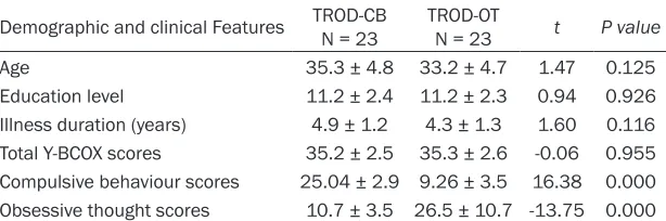
[image:3.612.93.400.85.187.2]each subject. The partial correlation method was used to calculate the correlations be- tween gFCD and clinical symptoms, illness duration and Y-BCOX scores. Age and gender effects were also controlled and multiple com-parisons were corrected using the Bonferroni method (P < 0.05). Correlation analyses be- tween gFCD and obsessive-compulsive symp-toms were completed in a voxel-wise manner in Table 1. The demographic and clinical features of the patients
Demographic and clinical Features TROD-CB N = 23 TROD-OT N = 23 t P value
Age 35.3 ± 4.8 33.2 ± 4.7 1.47 0.125
Education level 11.2 ± 2.4 11.2 ± 2.3 0.94 0.926
Illness duration (years) 4.9 ± 1.2 4.3 ± 1.3 1.60 0.116 Total Y-BCOX scores 35.2 ± 2.5 35.3 ± 2.6 -0.06 0.955 Compulsive behaviour scores 25.04 ± 2.9 9.26 ± 3.5 16.38 0.000 Obsessive thought scores 10.7 ± 3.5 26.5 ± 10.7 -13.75 0.000
TROD-CB: Treatment-refractory obsessive-compulsive disorder with obviously compulsive behaviour; TROD-OT: Treatment-refractory obsessive-compulsive disorder with obviously obsessive thought; Y-BCOX: Yale-Brown Obsessive-Compulsive Scale.
gFCD according to the method described by To- masi and Volkow [22-27]. We used Pearson’s linear correlation to as- sess the strength of FC among voxels. A
correla-tion coefficient of R >
0.6 between two voxels
indicated a significant
connection. This thre-
shold was confirmed as
the entire brain. A linear regression model was used to conduct the correlation analyses, with age and gender as covariates of no interest. Correction for multiple comparisons was con-ducted using the FWE method (P < 0.05). Two-sample t-tests were used to detect differences in age, education level, and illness duration. The chi-square test was used to compare the
gender ratio. P < 0.05 was set as the signifi -cance threshold.
Results
Demographic and clinical features
The demographic and clinical features of the patients are shown in Table 1. No significant
differences were found in gender, age, educa-tion, illness duraeduca-tion, or illness severity. Be- cause anti-obsessive-compulsive therapeutic agents are very complex, we did not list them in the table. Ten types of agents were used in these patients, and most patients were taking two or three therapeutic agents.
Comparison of global FCD between the two groups
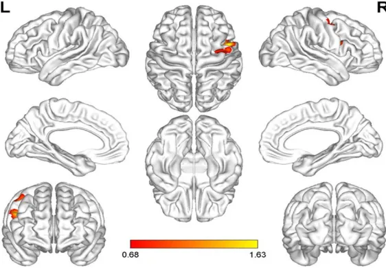
In the current study, we found that the TRC group demonstrated increased global FCD in the right sensorimotor cortex compared with the TRO group (Figure 1). We did not find a sig
-nificant correlation between increased global
FCD and the severity of TRC symptoms.
ently induces disturbed neural spontaneous activity in the brain, thereby generating abnor-mal thoughts or behaviours of patients [33, 34]. This possibility was proposed in several previous studies reporting abnormal global FCD in patients with mental disorders [22-29,
33]. The current findings also support this
proposal.
Although we did not find a relationship
be-tween increased global FCD and the severity of
TRC symptoms, our findings provide informa -tion that may be useful for facilitating under-standing of the neural pathological features
of specific symptoms in treatment-refractory
OCD. TRC patients demonstrated increased
[image:4.612.91.372.73.268.2]global FCD in the sensorimotor cortex, reflect -ing increased functional connection numbers and an increased number of brain neuron con-nections in this region. The neurons that cons- titute the sensorimotor cortex are thought to participate in the information processing circuit [34-37]. Increased global FCD in the sensorim-otor cortex plays a key role in the modulation of somatic movement [38-41]. Therefore, we pos-tulated that hyper-information-communication in the sensorimotor cortex and other brain regions in the whole brain may cause compul-sive behaviour in TRC patients. Similarly, some previous studies reported that the sensorimo-tor cortex also participates in the regulation of emotional and executive processing [42-44]. The reciprocal actions of these regions may represent the neural basis underlying the sym- Figure 1. Global FCD difference between the TRC and TRO groups.
Discussion
ptoms of patients who become treatment-re- fractory.
Previous studies reported that some brain FC alterations were correlated with some symp-toms of OCD [45-47]. However, the current re- sults revealed no correlation between incre- ased global FCD and compulsion symptom scores in TRC patients. Several factors may explain this phenomenon. First, in previous studies, researchers mainly focused on func-tional differences between OCD patients and healthy controls. Second, compared with OCD studies, fewer studies have investigated treat-ment-refractory OCD. Third, even fewer studies
have examined the specific symptoms of OCD or treatment-refractory OCD. The specific symp -toms of OCD examined in the current study
were defined according to clinical symptom
expression patterns rather than formal
diag-nostic classification.
Because the current study was limited by the absence of a healthy control group, we did not use a multiple comparison analysis method to further investigate functional alterations in the groups. This limitation should be addressed in future studies.
Three important limitations of the current stu- dy should be considered. First, as mentioned above, we did not include a healthy control group for conducting multiple comparison anal-yses, reducing the strength of our conclusions. Second, we only compared TRC and TRO patients in this study, and did not enrol OCD patients who responded well to treatment. Th- is also limits the strength of our conclusions.
Third, we did not control for the influence of
therapeutic agents on the results. This is a widespread problem because these kinds of agents are complex and methods for normal-ization are not available. These limitations sh- ould be addressed in future studies to clarify the current results.
Ideally, future research will involve long-term
follow-up studies with a large sample of
first-episode drug-naïve OCD patients and a multi-ple-mode MRI method to dynamically charac-terize the trajectory of brain features with ill-
ness progression, observe treatment efficacy
and brain alterations induced by the treat-
ment, describe the specific dynamic trajecto -ries of brain features related to clinical
symp-toms, and explore specific treatment targets
and the pathological mechanisms involved in failed responses to treatment. Although these research methods are time-consuming, such studies would be valuable for accelerating the progress of understanding the pathological basis of OCD and providing an objective index for psychiatrists to develop precise treatments in future, particularly for treatment-refractory patients.
TRC patients exhibited increased global FCD in the right sensorimotor cortex, indicating that hyper-information-communication among the sensorimotor cortex and other brain regions may be a feature of TRC. Because the current study contained several limitations, the str- ength of our conclusions was restricted. None-
theless, our preliminary findings can provide
useful background information for future stu- dies.
Acknowledgements
This project was supported by the foundation of Tianjin Public Security Bureau (2016KYSA- KY038 to Baohua Zhang).
Disclosure of conflict of interest
None.
Address correspondence to: Suling Chen, Depart- ment of Psychiatry, Wenzhou Seventh People’s Hospital, No.552, Xishandong Road, Ouhai District, Wenzhou 325000, China. Tel: +86 537 66390203; E-mail: [email protected]; Xiang Mu, Department of Psychiatry, Ankang Hospital of Tianjin Public Security Bureau, No.2 Jiangchang Road, Heibei District, Tianjin 300240, China. Tel: +86 22 243- 90203; E-mail: [email protected]; Wei
Liu, Department of Psychiatry, The First Affiliated
Hospital of Harbin Medical University, 23 Youzheng Road, Nangang District, Harbin 150001, Heilong- jiang Province, China. Tel: +86 451 53643849; E-mail: [email protected]
References
[1] Takagi Y, Sakai Y, Lisi G, Yahata N, Abe Y, Nishi-da S, Nakamae T, Morimoto J, Kawato M, Naru-moto J, Tanaka SC. A neural marker of obses-sive-compulsive disorder from whole-brain functional connectivity. Sci Rep 2017; 7: 7538. [2] Fan J, Zhong M, Gan J, Liu W, Niu C, Liao H,
mode, central executive, and salience net-works in obsessive-compulsive disorder. J Af-fect Disord 2017; 223: 106-114.
[3] Fan J, Zhong M, Zhu X, Gan J, Liu W, Niu C, Liao H, Zhang H, Yi J, Tan C. Resting-state functional connectivity between right anterior insula and right orbital frontal cortex correlate with insight level in obsessive-compulsive disorder. Neuro-image Clin 2017; 15: 1-7.
[4] Voon V, Droux F, Morris L, Chabardes S, Boug-erol T, David O, Krack P, Polosan M. Decisional impulsivity and the associative-limbic subtha-lamic nucleus in obsessive-compulsive disor-der: stimulation and connectivity. Brain 2017; 140: 442-456.
[5] Tian L, Meng C, Jiang Y, Tang Q, Wang S, Xie X, Fu X, Jin C, Zhang F, Wang J. Abnormal func-tional connectivity of brain network hubs asso-ciated with symptom severity in treatment-na-ive patients with obsesstreatment-na-ive-compulstreatment-na-ive disord- er: a resting-state functional MRI study. Prog Neuropsychopharmacol Biol Psychiatry 2016; 66: 104-111.
[6] Abe Y, Sakai Y, Nishida S, Nakamae T, Yamada
K, Fukui K, Narumoto J. Hyper-influence of the
orbitofrontal cortex over the ventral striatum in obsessive-compulsive disorder. Eur Neuropsy-chopharmacol 2015; 25: 1898-1905.
[7] Peng ZW, Xu T, He QH, Shi CZ, Wei Z, Miao GD, Jing J, Lim KO, Zuo XN, Chan RC. Default net-work connectivity as a vulnerability marker for obsessive compulsive disorder. Psychol Med 2014; 44: 1475-1484.
[8] Sakai Y, Narumoto J, Nishida S, Nakamae T, Yamada K, Nishimura T, Fukui K. Corticostria-tal functional connectivity in non-medicated patients with obsessive-compulsive disorder. Eur Psychiatry 2011; 26: 463-469.
[9] Dunlop K, Woodside B, Olmsted M, Colton P, Giacobbe P, Downar J. Reductions in cortico-striatal hyperconnectivity accompany success-ful treatment of obsessive-compulsive disor-der with dorsomedial prefrontal rTMS. Neuro- psychopharmacology 2016; 1: 1395-403. [10] Göttlich M, Krämer UM, Kordon A, Hohagen F,
Zurowski B. Resting-state connectivity of the amygdala predicts response to cognitive be-havioral therapy in obsessive compulsive dis-order. Biol Psychol 2015; 111: 100-109. [11] Vaghi MM, Vértes PE, Kitzbichler MG,
Apergis-Schoute AM, van der Flier FE, Fineberg NA, Sule A, Zaman R, Voon V, Kundu P, Bullmore
ET, Robbins TW. Specific frontostriatal circuits for impaired cognitive flexibility and goal-direct -ed planning in obsessive-compulsive disorder: evidence from resting-state functional connec-tivity. Biol Psychiatry 2017; 81: 708-717. [12] Moreira PS, Marques P, Soriano-Mas C,
Magal-hães R, Sousa N, Soares JM, Morgado P. The
neural correlates of obsessive-compulsive dis-order: a multimodal perspective. Transl Psy-chiatry 2017; 7: e1224.
[13] Bonmassar G, Makris N. The virtual patient simulator of deep brain stimulation in the ob-sessive compulsive disorder based on connec-tome and 7 Tesla MRI data. Cogn Int Conf Adv Cogn Technol Appl 2014; 2014: 235-238. [14] Shin DJ, Jung WH, He Y, Wang J, Shim G, Byun
MS, Jang JH, Kim SN, Lee TY, Park HY, Kwon JS. The effects of pharmacological treatment on functional brain connectome in obsessive-compulsive disorder. Biol Psychiatry 2014; 75: 606-614.
[15] Peng ZW, Xu T, He QH, Shi CZ, Wei Z, Miao GD, Jing J, Lim KO, Zuo XN, Chan RC. Default net-work connectivity as a vulnerability marker for obsessive compulsive disorder. Psychol Med 2014; 44: 1475-1484.
[16] Bleich-Cohen M, Hendler T, Weizman R, Fara-gian S, Weizman A, Poyurovsky M. Working memory dysfunction in schizophrenia patients with obsessive-compulsive symptoms: an fMRI study. Eur Psychiatry 2014; 29: 160-166. [17] Glomb K, Ponce-Alvarez A, Gilson M, Ritter P,
Deco G. Resting state networks in empirical and simulated dynamic functional connectivi-ty. Neuroimage 2017; 159: 388-402.
[18] Biswal BB, Van Kylen J, Hyde JS. Simultaneous
assessment of flow and BOLD signals in rest -ing-state functional connectivity maps. NMR Biomed 1997; 10: 165-170.
[19] Battiston F, Nicosia V, Chavez M, Latora V. Mul-tilayer motif analysis of brain networks. Chaos 2017; 27: 047404.
[20] Liu C, Zhang W, Chen G, Tian H, Li J, Qu H, Cheng L, Zhu J, Zhuo C. Aberrant patterns of local and long-range functional connectivity densities in schizophrenia. Oncotarget 2017; 8: 48196-48203.
[21] Zou K, Gao Q, Long Z, Xu F, Sun X, Chen H, Sun X. Abnormal functional connectivity density in
first-episode, drug-naive adult patients with
major depressive disorder. J Affect Disord 2016; 194: 153-158.
[22] Cohen AD, Tomasi D, Shokri-Kojori E, Nencka AS, Wang Y. Functional connectivity density mapping: comparing multiband and conven-tional EPI protocols. Brain Imaging Behav 2018; 12: 848-859.
[23] Tomasi D, Volkow ND. Association between functional connectivity hubs and brain net-works. Cereb Cortex 2011; 21: 2003-2013. [24] Tomasi D, Volkow ND. Functional connectivity
hubs in the human brain. Neuroimage 2011; 57: 908-917.
[26] Zhuo C, Zhu J, Wang C, Qu H, Ma X, Tian H, Liu M, Qin W. Brain structural and functional dis-sociated patterns in schizophrenia. BMC Psy-chiatry 2017; 17: 45.
[27] Zhuo C, Zhu J, Qin W, Qu H, Ma X, Tian H, Xu Q, Yu C. Functional connectivity density altera-tions in schizophrenia. Front Behav Neurosci 2014; 8: 404.
[28] Zhuo C, Zhu J, Wang C, Qu H, Ma X, Qin W. Dif-ferent spatial patterns of brain atrophy and global functional connectivity impairments in major depressive disorder. Brain Imaging Be-hav 2017; 11: 1678-1689.
[29] Qin W, Xuan Y, Liu Y, Jiang T, Yu C. Functional connectivity density in congenitally and late blind subjects. Cereb Cortex 2015; 25: 2507-2516.
[30] First M, Spitzer R, Gibbon M, Williams J. Struc-tured clinical interview for DSM-IV axis I disor-ders (SCID-I), clinician version, administration booklet. American Psychiatric Pub 2012. [31] Goodman WK, Price LH, Rasmussen SA,
Ma-zure C, Fleischmann RL, Hill CL, Heninger GR, Charney DS. “The Yale-brown obsessive-com-pulsive scale. I. Development, use, and reliabil-ity”. Arch Gen Psychiatry 1989; 46: 1006-1011.
[32] Power JD, Barnes KA, Snyder AZ, Schlaggar BL and Petersen SE. Spurious but systematic cor-relations in functional connectivity MRI net-works arise from subject motion. Neuroimage 2012; 59: 2142-2154.
[33] Vancassel S, Blondeau C, Lallemand S, Cador M, Linard A, Lavialle M, Dellu-Hagedorn F. Hy-peractivity in the rat is associated with sponta-neous low level of n-3 polyunsaturated fatty acids in the frontal cortex. Behav Brain Res 2007; 180: 119-126.
[34] Wreden CC, Meng JL, Feng W, Chi W, Marshall ZD, Heckscher ES. Temporal cohorts of lin-eage-related neurons perform analogous func-tions in distinct sensorimotor circuits. Curr Biol 2017; 27: 1521-1528, e4.
[35] Brovelli A, Badier JM, Bonini F, Bartolomei F,
Coulon O, Auzias G. Dynamic reconfiguration of
visuomotor-related functional connectivity net-works. J Neurosci 2017; 37: 839-853. [36] Shoykhet M, Middleton JW. Cardiac
arrest-in-duced global brain hypoxia-ischemia during development affects spontaneous activity or-ganization in rat sensory and motor thalamo-cortical circuits during adulthood. Front Neural Circuits 2016; 10: 68.
[37] LeBlancq MJ, McKinney TL, Dickson CT. ZIP it: neural silencing is an additional effect of the PKM-zeta inhibitor zeta-inhibitory peptide. J Neurosci 2016; 36: 6193-6198.
[38] Morita T, Saito DN, Ban M, Shimada K, Oka-moto Y, Kosaka H, Okazawa H, Asada M, Naito E. Self-face recognition shares brain regions active during proprioceptive illusion in the right inferior fronto-parietal superior longitudinal fasciculus III network. Neuroscience 2017; 348: 288-301.
[39] Fiess J, Rockstroh B, Schmidt R, Steffen A. Emotion regulation and functional neurologi-cal symptoms: does emotion processing con-vert into sensorimotor activity? J Psychosom Res 2015; 79: 477-483.
[40] Johnson MD, Miocinovic S, McIntyre CC, Vitek JL. Mechanisms and targets of deep brain stimulation in movement disorders. Neurother-apeutics 2008; 5: 294-308.
[41] Fanselow EE, Connors BW. Navigating a senso-rimotor loop. Neuron 2005; 45: 329-330. [42] Kolesar TA, Bilevicius E, Kornelsen J. Salience,
central executive, and sensorimotor network functional connectivity alterations in failed back surgery syndrome. Scand J Pain 2017; 16: 10-14.
[43] Zamorano AM, Cifre I, Montoya P, Riquelme I, Kleber B. Insula-based networks in profession-al musicians: evidence for increased function-al connectivity during resting state fMRI. Hum Brain Mapp 2017; 38: 4834-4849.
[44] García AM, Sedeño L, Herrera Murcia E, Couto B, Ibáñez A. A lesion-proof brain? Multidimen-sional sensorimotor, cognitive, and socio-af-fective preservation despite extensive damage in a stroke patient. Front Aging Neurosci 2017; 8: 335.
[45] Tian L, Meng C, Jiang Y, Tang Q, Wang S, Xie X, Fu X, Jin C, Zhang F, Wang J. Abnormal func-tional connectivity of brain network hubs asso-ciated with symptom severity in treatment-na-ive patients with obsesstreatment-na-ive-compulstreatment-na-ive disor- der: a resting-state functional MRI study. Prog Neuropsychopharmacol Biol Psychiatry 2016; 66: 104-111.
[46] Abe Y, Sakai Y, Nishida S, Nakamae T, Yamada
K, Fukui K, Narumoto J. Hyper-influence of the
orbitofrontal cortex over the ventral striatum in obsessive-compulsive disorder. Eur Neuropsy-chopharmacol 2015; 25: 1898-1905.
![Quetiapine augmentation of SRIs in treatment refractory obsessive compulsive disorder: a double blind, randomised, placebo controlled study [ISRCTN83050762]](data:image/gif;base64,R0lGODlhAQABAIAAAP///wAAACH5BAEAAAAALAAAAAABAAEAAAICRAEAOw==)