Original Article
Effect of task-oriented training combined with
vibration therapy on upper limb function in
patients with hemiplegia after stroke
Hui Dang1, Zhenghong Chen2
1Department of Rehabilitation Medicine, Danzhou People’s Hospital, Danzhou, Hainan Province, China; 2Department of Rehabilitation Medicine, The First Affiliated Hospital of Sun Yat-sen University, Guangzhou,
Guangdong Province, China
Received March 16, 2019; Accepted May 13, 2019; Epub July 15, 2019; Published July 30, 2019
Abstract: Objective: To study the effect of task-oriented training combined with vibration therapy on upper limb function in patients with hemiplegia after stroke. Methods: A total of 108 patients with hemiplegia after stroke were randomly assigned to control group (patients who received routine upper limb training, n=36), observation group 1 (patients who received routine upper limb training plus vibration therapy, n=36), and observation group 2 (patients who received routine upper limb training plus vibration therapy plus task-oriented training, n=36). Excluding the patients who withdrew from the study, there were 95 cases in total, including 30 cases in the control group, 32 cases in observation group 1 and 33 cases in observation group 2. The Fugl-Meyer Assessment Scale (FMA), Wolf
Motor Function Test (WMFT), modified Ashworth Spasticity Rating Scale (MAS) score and maximal grip strength of
all patients were evaluated and compared after treatment. Results: There was no difference in FMA score among the three groups before treatment (P>0.05). There was no difference in FMA, WMFT, MAS scores and maximal grip strength before and after treatment in the control group (all P>0.05), but there were statistical differences in FMA, MAS scores and maximal grip strength before and after treatment in observation groups 1 and 2 (all P<0.05). After treatment, the FMA score and maximal grip strength in observation group 1 were higher than those in control group while MAS score was lower than that in control group, with statistical difference (all P<0.05). FMA, WMFT scores and maximal grip strength in observation group 2 were higher than those in the control group and observation group 1, while MAS score was lower than that in the other groups, with statistical difference (all P<0.05). Conclusion:
Task-oriented training combined with vibration therapy has a significant effect on the rehabilitation of upper limb
dysfunction in patients with hemiplegia after stroke, which is worthy of clinical application.
Keywords: Task-oriented training, vibration therapy, hemiplegia after stroke, upper limb function
Introduction
Stroke has a high incidence, disability and mor-tality rate in China. Studies have found that the incidence of stroke is 1,114.8 per 100,000 people in China, of which 114.8 people die per year, and more than 70% of patients are over 60 years old [1]. Hemiplegia often occurs in stroke patients. Walking function of lower limbs can often be recovered through rehabilitation training, while the functional recovery of upper limbs is poor. A study found that about 30-60% of patients still cannot recover after 6 months of rehabilitation training [2]. The reason for this recovery rate may be related to the occurrence of symptoms such as pain and discomfort of
the muscles [6]. For stroke hemiplegic patients with upper limb dysfunction, there is less re- search on applying the two training methods together. Therefore, this study aimed to investi-gate the improvement of upper limb function of patients with hemiplegia after stroke by com-bining task-oriented training with vibration the- rapy.
Materials and methods
Clinical data
This study was approved by the ethics Com- mittee of The First Affiliated Hospital of Sun Yat-sen University. A total of 108 stroke pati- ents with hemiplegia admitted to the rehabili- tation center of The First Affiliated Hospital of Sun Yat-sen University from October 2016 to October 2018 were included in this study. The digital random control method was applied in the study. A total of 36 patients who received routine upper limb training were randomly se- lected as the control group, 36 patients who received routine upper limb training plus vibra-tion therapy as observavibra-tion group 1, and 36 patients who received routine upper limb train-ing plus vibration therapy plus task-oriented training as observation group 2. Excluding the patients who withdrew from the study, there were 95 cases in total, including 30 cases in the control group, 32 cases in observation group 1 and 33 cases in observation group 2. All the patients were aged 18-69 years, with an average age of 59.5±8.7 years, and signed con-sent forms.
Inclusion criteria
Patients diagnosed with stroke [7]; patients who suffered from primary stroke; patients sup-ported by head CT or magnetic resonance imaging (MRI); patients with stable symptoms and clear consciousness after treatment; pa- tients with stroke occurring unilaterally, with a duration of less than 18 months; patients ag- ed under 70; patients with less than 3 points in the modified Ashworth Scale (MAS) score [8]; patients with no limb pain; patients’ Brun- nstrom stage 3 or above [5].
Exclusion criteria
Patients complicated with heart and lung dys-function; patients with fracture trauma and
bone hypoplasia; patients not cooperating to complete the training; patients who did not sign the informed consent.
Methods
Routine upper limb training: Patients were given upper limb training, one hour at a time, five times a week for four weeks, including upper limb muscle strength training and rou-tine training of daily living ability.
Task-oriented training: To strengthen the com-mon movement components of the patient’s affected upper limbs, the patients lifted the affected upper limbs to reach the mouth with their hands. To induce the separation move-ment of the affected side, the patients pushed away the towel on the table with the stretch of elbow joint. The patients tried to eat steamed bread with the affected hands. The patients tried to eat long-shaped food such as cucum-bers and bananas with the affected hands. Vibration therapy: The trained therapist per-formed one-to-one treatment. The subjects sit on the Wellengang vibrator manufactured by SVG Company in Germany, with the vibration frequency range of 5-15 Hz and the amplitude range of 1-6 mm. The training intensity incre- ased but not exceeding the load. The subjects were seated in an armless chair in front of the vibrating platform board (0.18 m in height, 0.72 m in width and 0.51 m in depth), with their shoulders flexed by 90 degrees and elbows slightly bent, and with their torso bent so that their palms could contact the circuit board in the middle of the platform board.
The control group received routine upper limb training. Vibration therapy plus routine upper limb training in observation group 1: Patients were given upper limb training for half an hour at a time, then followed by vibration therapy training for half an hour. The training was per-formed five times a week for four weeks.
Efficacy evaluation
The patients were evaluated before treatment and 4 weeks after treatment.
Main outcome measures: Fugl-Meyer Motor Function Assessment Scale (FMA): Assessment of the upper limbs: The main assessment con-tents include the coordination of flexors and extensors in large joints of the upper limbs, the stability of wrist joints, the coordination of small joints and the corresponding speed [9]. There are 33 projects, each with a maximal score of 2 points, a total score of 66 points. Wolf Motor Function Test (WMFT) is an evaluation of the hand functional status in performing task-ba- sed activities, which includes 15 items [10]. The evaluation of the completion quality of each item is divided into 6 grades from 0 to 5, with each grade scoring 0 to 5 points and a total score of 75 points.
Secondary outcome measures: MAS score: Th- is score is a tool for evaluating muscle spasm [8]. It is divided into 6 grades: 0, 1, 1+, 2, 3 and
4. The higher the grade, the more sever the spasm. This study quantifies it into 6 grades with scores of 0, 1, 1.5, 2, 3 and 4, respective-ly. Test of maximal grip strength of the affected hand: Selecting a grip dynamometer from the same manufacturer, all patients were allowed
to sit in chairs at a rest state for the measure-ment. Each measurement required the grip strength of the affected hand to last for 3 sec-onds in order to measure its maximal value [11].
Statistical methods
The SPSS 22.0 statistical software was used to statistically analyze the collected data. The continuous variables were expressed as the mean ± standard deviation (_x ± sd). Data accorded with normal distribution and homoge-neity of variance in group was compared by independent sample t-test or paired t-test, oth-erwise, by rank sum test. One-way analysis of variance (ANOVA) was used for multiple-group comparison, and differences detected were fur- ther analyzed by least significant difference (LSD) method. The difference was statistically significant with P<0.05.
Results
Comparison of general data and baseline data among the three groups
[image:3.612.91.523.84.205.2]There was no significant difference among the three groups in gender, age, course of disease and affected limb position, which could be compared (all P>0.05). See Table 1.
Table 1. Comparison of general data and baseline data among the three groups
Item Control group Observation group I Observation group II χ2/F P
Gender 0.010 0.995
Male 19 20 21
Female 11 12 12
Age 60.0±9.0 57.8±8.5 60.6±8.0 0.623 0.503
Course of disease 8.1±4.3 6.6±3.6 7.2±4.8 0.789 0.369
Affected limb position 4.101 0.129
Left 18 18 12
Right 12 14 21
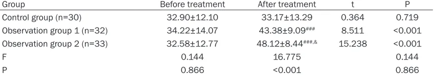
Table 2. Comparison of FMA score among the three groups
Group Before treatment After treatment t P
Control group (n=30) 32.90±12.10 33.17±13.29 0.364 0.719
Observation group 1 (n=32) 34.22±14.07 43.38±9.09### 8.511 <0.001
Observation group 2 (n=33) 32.58±12.77 48.12±8.44###,& 15.238 <0.001
F 0.144 16.775 0.144
P 0.866 <0.001 0.866
Note: FMA, Fugl-Meyer Assessment Scale. Compared with the control group after treatment, ###P<0.001; compared with
[image:3.612.91.524.241.316.2]Comparison of FMA score among the three groups
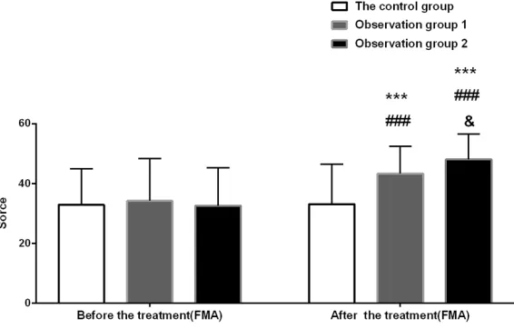
[image:4.612.93.376.74.255.2]There was no difference in FMA score among the three groups before treatment (P>0.05). There was no difference in FMA score before and after treatment in the control group (P> 0.05), but there were statistical differences before and after treatment in observation gr- oups 1 and 2 (P<0.001). After treatment, the FMA score in observation groups 1 and 2 was higher than that in the control group, with sta-tistical differences, and observation group 2 was higher than observation group 1, with sta-tistical differences (P<0.05). See Table 2 and Figure 1.
Comparison of WMFT score among the three groups
There was no difference in WMFT score among the three groups before treatment (P>0.05). There was no difference in WMFT score be- fore and after treatment in the control group (P>0.05), but there were statistical differences before and after treatment in observation gr- oups 1 and 2 (P<0.05). After treatment, there was no statistical difference in WMFT score between the control group and observation group 1 (P>0.05), while observation group 2 was higher than the control group and obser- vation group 1, with statistical differences (P< 0.05). See Table 3 and Figure 2.
servation group 2 was higher than observation group 1, with statistical difference (P<0.05). See Table 4 and Figure 3.
Comparison of MAS score among the three
groups
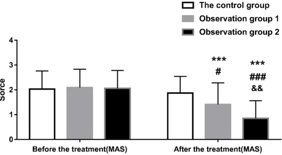
There was no difference in MAS score among the three groups before treatment (P>0.05). There was no difference in MAS score before and after treatment in the control group (P> 0.05), but there were statistical differences before and after treatment in observation gr- oups 1 and 2 (P<0.001). After treatment, the MAS score in observation groups 1 and 2 was lower than that in the control group, with sta- tistical differences, and observation group 2 were lower than observation group 1, with sta-tistical differences (P<0.05). See Table 5 and Figure 4.
Discussion
Stroke patients are injured in the cerebral func-tional area, so the training is performed main- ly for the rehabilitation of this area [12, 13]. Task-oriented training is mainly to set goals for patients to take part in corresponding mean-ingful and practical functional activities. Dif- ferent from the traditional exercise training, it is designed according to the exercise control and exercise learning theories and the pati- ent’s functional loss. With the different
func-Figure 1. Comparison of FMA score among the three groups. FMA, Fugl-Meyer Assessment Scale. Compared with the same group before treatment, ***P<0.001; compared with the control group after treatment, ###P<0.001; compared with observation group 1 after treatment, &P<0.05.
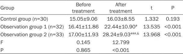
Comparison of the maximal grip strength among the three groups
tional loss of the patients, the corresponding target and exercise activities were designed, and the patients were guided accordingly to achieve the goal of improving motor function [14]. This training mode is based on the activi-ties in daily life, allowing patients to participate actively and independently, and combining with
[image:5.612.91.523.87.164.2]cles after the vibration of muscles, thus obta- ining specific response curative effects. It was initially applied in sports with the functions of relieving muscle pain, improving muscle str- ength and increasing coordination of move-ment, etc., and then in the rehabilitation field. A study found that the grip strength, spasticity, Table 3. Comparison of WMFT score among the three groups
Group Before treatment After treatment t P
Control group (n=30) 40.10±12.13 39.57±13.23 0.734 0.469
Observation group 1 (n=32) 41.16±14.11 43.47±9.49 2.367 0.024
Observation group 2 (n=33) 39.21±12.64 52.06±8.43###,&&& 12.884 <0.001
F 0.182 11.779
P 0.834 <0.001
Note: WMFT, Wolf Motor Function Test. Compared with the control group after treatment, ###P<0.001; compared with
[image:5.612.92.389.209.408.2]observa-tion group 1 after treatment, &&&P<0.001.
Figure 2. Comparison of WMFT score among the three groups. WMFT, Wolf Mo-tor Function Test. Compared with the same group before treatment, *P<0.05, ***P<0.001; compared with the control group after treatment, ###P<0.001; com-pared with observation group 1 after treatment, &&&P<0.001.
Table 4. Comparison of the maximal grip strength among the three groups
Group treatmentBefore treatmentAfter t P
Control group (n=30) 15.05±9.06 16.03±8.55 1.332 0.193 Observation group 1 (n=32) 16.41±11.86 22.44±10.90# 13.535 <0.001 Observation group 2 (n=33) 17.00±11.93 28.24±9.03###,& 13.968 <0.001
F 0.145 12.799
P 0.865 <0.001
Note: Compared with the control group after treatment, #P<0.05, ###P<0.001; compared
with observation group 1 after treatment, &P<0.05.
the life and training, whi- ch is beneficial to the recovery of patients’ fun- ctions [15]. Some stud-ies have found that the use of assisted task-ori-ented training can im- prove the blood circula-tion in hands of stroke patients, and can also relieve spasm symptoms and relieve hand stiffne- ss [16]. Two other stud-ies have analyzed the feasibility and effective-ness of task-oriented tr- aining for stroke patients, and found that it can si- gnificantly improve the hand function of the pa- tients [17, 18]. Compar- ed with passive training, research has shown that active training for pati- ents is more conducive to recruiting the cerebral motor cortex, strength-ening the interaction be- tween different function-al regions, thus improv-ing the recovery and re- modeling of hand func-tions [19].
[image:5.612.92.396.504.595.2]mus-pain and quality of life were improved after treatment in upper limb muscles with local vibration therapy [20]. Another study on the
[image:6.612.91.382.70.226.2]control group and observation group I with vibration therapy alone. It may be that task- oriented training is a targeted training, not only
[image:6.612.91.384.313.401.2]Figure 3. Comparison of the maximal grip strength among the three groups. Compared with the same group before treatment, ***P<0.001; compared with the control group after treatment, #P<0.05, ###P<0.001; compared with obser-vation group 1 after treatment, &P<0.05.
Table 5. Comparison of MAS score among the three groups
Group treatment After treatmentBefore t P
Control group (n=30) 2.03±0.73 1.87±0.67 1.863 0.077 Observation group 1 (n=32) 2.09±0.74 1.41±0.87# 6.723 <0.001 Observation group 2 (n=33) 2.06±0.72 0.85±0.71###,&& 12.477 <0.001
χ2 0.053 14.257
P 0.948 <0.001
Note: MAS, modified Ashworth Spasticity Rating Scale. Compared with the control group after treatment, #P<0.05, ###P<0.001; compared with observation group 1 after
treatment, &&P<0.01.
Figure 4. Comparison of MAS score among the three groups. MAS, modified
Ashworth Spasticity Rating Scale. Compared with the same group before treat-ment, ***P<0.001; compared with the control group after treatment, #P<0.05, ###P<0.001; compared with observation group 1 after treatment, &&P<0.01.
[image:6.612.93.384.456.616.2]a single muscle but multiple muscles of the whole body are involved together in the pro- cess of completing the set target. With the cooperation of vibration therapy, more exerci- se units can participate in and coordinate with each muscle group, and improve the patients’ control of movement [26, 27].
Limitations of this research: Due to the small number of observation cases and the short observation time, this research will further expand the sample size and extend the obser-vation time for further study.
To sum up, task-oriented training combined with vibration therapy has a significant effect on the rehabilitation of upper limb dysfunction in patients with hemiplegia after stroke, which is worthy of clinical application.
Disclosure of conflict of interest
None.
Address correspondence to: Zhenghong Chen, De-
partment of Rehabilitation Medicine, The First
Affi-liated Hospital of Sun Yat-sen University, No.58 Zhongshan Second Road, Guangzhou 510080, Guangdong Province, China. Tel: +86-1372531- 3052; E-mail: [email protected]
References
[1] Wang W, Jiang B, Sun H, Ru X, Sun D, Wang L, Wang L, Jiang Y, Li Y and Wang Y. Prevalence, incidence, and mortality of stroke in China. Circulation 2017; 135: 759.
[2] Jin J, Allison BZ, Wang X and Neuper C. A com-bined brain-computer interface based on P300 potentials and motion-onset visual evok- ed potentials. J Neurosci Methods 2012; 205: 265-276.
[3] Cirstea MC and Levin MF. Improvement of arm movement patterns and endpoint control de-pends on type of feedback during practice in stroke survivors. Neurorehabil Neural Repair 2007; 21: 398-411.
[4] Arba F, Ali M, Quinn TJ, Hankey GJ, Lees KR, Inzitari D; VISTA Collaboration. Lacunar in-farcts, depression, and anxiety symptoms one year after stroke. J Stroke Cerebrovasc Dis 2016; 25: 831-834.
[5] Yoo C and Park J. Impact of task-oriented train-ing on hand function and activities of daily liv-ing after stroke. J Phys Ther Sci 2015; 27: 2529-2531.
[6] Gusso S, Munns CF, Colle P, Derraik JG, Biggs
JB, Cutfield WS and Hofman PL. Effects of
whole-body vibration training on physical func-tion, bone and muscle mass in adolescents and young adults with cerebral palsy. Sci Rep 2016; 6: 22518.
[7] The writing group of guidelines for the diagno-sis and treatment of acute ischemic stroke in the cerebrovascular disease group of the soci-ety of neurology, Chinese medical association. guidelines 2010 for the diagnosis and treat-ment of acute ischemic stroke in China. Chinese General Practice 2011; 14: 4013-4017.
[8] Yaşar E, Adigüzel E, Kesikburun S, Yenihayat I,
Yılmaz B, Alaca R and Tan AK. Assessment of
forearm muscle spasticity with sonoelastogra-phy in patients with stroke. Br J Radiol 2016; 89: 20160603.
[9] Gladstone DJ, Danells CJ, Black SE. The Fugl-Meyer assessment of motor recovery after stroke: a critical review measurement proper-ties. Neurorehabil Neural 2002; 16: 232-240. [10] Wolf S, Catlin P, Ellis M, Archer A, Morgan B
and Piacentino A. Assessing Wolf motor func-tion test as outcome measure for research in patients after stroke. Stroke 2001; 32: 1635-1639.
[11] De Smet L, Vercammen A. Grip strength in chil-dren. J Pediatr Orthop B 2001; 10: 352-354. [12] Tang CZ, Zhao ZY, Chen C, Zheng XH, Sun FF,
Zhang XL, Tian J, Fan MX, Wu Y and Jia J. Decreased functional connectivity of homo-topic brain regions in chronic stroke patients: a resting state fMRI study. PLoS One 2016; 11: e152875.
[13] Kumar P, Kathuria P, Nair P and Prasad K. Prediction of upper limb motor recovery after subacute ischemic stroke using diffusion ten-sor imaging: a systematic review and meta-analysis. J Stroke 2016; 16: 50-59.
[14] Choi JU, Kang SH. The effects of patient-cen-tered task-oriented training on balance
activi-ties of daily living and self-efficacy following
stroke. J Phys Ther Sci 2015; 27: 2985-2988. [15] Park J. Effects of task-oriented training on
up-per extremity function and up-performance of daily activities in chronic stroke patients with impaired cognition. J Phys Ther Sci 2016; 28: 316-318.
[16] Bissolotti L, Villafañe JH, Gaffurini P, Orizio C, Valdes K and Negrini S. Changes in skeletal muscle perfusion and spasticity in patients with poststroke hemiparesis treated by robotic assistance (Gloreha) of the hand. J Phys Ther Sci 2016; 28: 769-773.
[17] Vanoglio F, Bernocchi P, Mulè C, Garofali F, Mora C, Taveggia G, Scalvini S and Luisa A.
Feasibility and efficacy of a robotic device for
[18] Etzel JA, Valchev N, Gazzola V and Keysers C. Is brain activity during action observation modu-lated by the perceived fairness of the actor? PLoS One 2016; 11: e0145350.
[19] Shin JH, Kim MY, Lee JY, Jeon YJ, Kim S, Lee S, Seo B and Choi Y. Effects of virtual reality-based rehabilitation on distal upper extremity function and health-related quality of life: a single-blinded, randomized controlled trial. J Neuroeng Rehabil 2016; 13: 17.
[20] Costantino C, Galuppo L and Romiti D. Short-term effect of local muscle vibration treatment versus sham therapy on upper limb in chronic post-stroke patients: a randomized controlled trial. Eur J Phvs Rehabil Med 2017; 53: 32-40. [21] Ko MS, Sim YJ, Kim DH and Jeon HS. Effects of
three weeks of whole-body vibration training on joint-position sense, balance, and gait in children with cerebral palsy: a randomized controlled study. Physiother Can 2016; 68: 99-105.
[22] Maarten S, Oron L, Marc VB and Swinnen SP. Corticospinal excitability changes following prolonged muscle tendon vibration. Neuro- report 2003; 14: 1901-1905.
[23] Karin R and Rothwell JC. Differential effect of muscle vibration on intracortical inhibitory cir-cuits in humans. J Physiol 2003; 551: 649-660.
[24] Christophe D, Machteld R and Sabine V. Strength increase after whole-body vibration compared with resistance training. Med Sci Sports Exerc 2003; 35: 1033-1041.
[25] Bohannon RW. Adequacy of simple measures for characterizmg impairment in upper limb strength following stroke. Percept Mot Skills 2004; 99: 813-817.
[26] Cheng HY, Yu YC, Wong AM, Tsai YS and Ju YY. Effects of an eight-week whole body vibration on lower extremity muscle tone and function in children with cerebral palsy. Res Dev Disabil 2015; 38: 256-261.