Effect of the diet amended with risk elements
contaminated soil on risk elements content in tissues
and hematological parameters of rats
J. Száková
1, Z. Novosadová
1, V. Zídek
2, A. Fučíková
1, J. Zídková
3,
D. Miholová
1, P. Tlustoš
11Faculty of Agrobiology, Food and Natural Resources, Czech University of Life Sciences Prague,
Prague, Czech Republic
2Institute of Physiology, Academy of Sciences of the Czech Republic, Prague, Czech Republic 3Faculty of Food and Biochemical Technology, Institute of Chemical Technology Prague,
Prague, Czech Republic
ABSTRACT: Penetration of rat organisms by risk elements (As, Cd, Pb) originated from differently contami-nated soils as well as interactions between the risk elements and selected essential macro- and microelements were investigated. Rat diet contained 10% of individual soils (based on dry weight) which were: (i) Fluvisol heavily polluted by As, Cd, Zn, and Pb, (ii) Luvisol contaminated by As, Cd, and Zn, and (iii) uncontami-nated Chernozem. Male Wistar rats used for the experiment were housed in cages in a room with controlled temperature for 60 days and fed ad libitum on the mentioned diet. The levels of both risk and essential elements were measured in liver, kidney, and bones of the animals and main biochemical parameters were determined to assess potential toxic effect of the soil-derived risk elements. Alternatively, in vitro Physio- logically Based Bioavailability Test (PBET) was used for evaluation of the bioaccessible pool of elements in the soil-amended diets. The element contents in the rat tissues reflected the risk element contents in the contaminated soils. Bioaccessibility and bioavailability of the risk elements decreased in the order Cd > As > Pb and were influenced by the soil physicochemical parameters. Significant changes in essential elements (Cu, Fe, Mg, P) deposition in rat tissues attended the high levels of the risk elements. Total leukocyte count in the blood of the rats fed the heavy polluted soil indicated adverse effect of soil-derived risk elements on biochemical parameters of the animals.
Keywords:risk elements; soil; soil ingestion; liver; kidney; bones; Rattus norvegicus
Former mining and smelting areas represent sig-nificant environmental risk due to the contamina-tion of soil with potentially toxic metals, most often risk elements (Šichorová et al., 2004). There are two main factors affecting the population exposure to the risk elements: ingestion of plants produced in the contaminated areas and ingestion or inhala-tion of contaminated soil dust. Pruvot et al. (2006) demonstrated that risk elements in soils or plants of kitchen gardens, lawns, and playgrounds could be potentially transferable particularly to small chil-dren. In this context, two important terms can be
montmorillonite. Karadas and Kara (2011) men-tioned potential risk of soil ingestion in the vicin-ity of zinc, lead, and manganese mines. Fischer et al. (2003) observed significant changes of selected hematological parameters in the blood of children long-term exposed to enhanced concentrations of lead and cadmium close to a smelter. Phagocytic activity and some hematological parameters are known to be highly susceptible to alterations of nutritional and environmental factors including trace elements (Kostič et al., 1993).
Oral bioavailability of metals can be assessed us-ing animal models in vivo as human surrogates. The animals tested can be swine (Rodriguez and Basta, 1999), mice (Sheppard et al., 1995), rats, rabbits, and monkeys (Ruby et al., 1996). The animals can be exposed to either single dose of contaminated soil suspension (Ellickson et al., 2001) or to long-term consumption of the diet amended with the contaminated soil (Hetiarachchi et al., 2003). At the end of the experiment, the element contents in the individual organs, tissues, and excreta of the animals are determined to assess bioavailability of the individual elements. Using this model, Ellickson et al. (2001) determined oral bioavailability of the standard reference soil 0.7% and 37.8% for Pb and As, respectively. These models provide valuable contaminant bioavailability data but have the disad-vantage of being time-consuming, requiring weeks or months for completion, expensive, and demand-ing a highly specialized personnel.
Physiologically Based Bioavailability Test (PBET) according to Ruby (1996), and Simple Bioavailability Extraction Test (SBET) according to Lee et al. (2006) are also reasonable and frequently used methods for evaluating bioaccessible pool of ele-ments in contaminated soils correlating fairly well with the results from in vivo tests from Sprague-Dawley rat (Ruby et al., 1996) and mouse (Juhasz et al., 2010) models. The former method includes
in vitro test system for predicting bioaccessibil-ity of metals from a solid matrix and incorporates gastrointestinal tract parameters representative of a human (including stomach and small intestinal pH and chemistry, soil-to-solution ratio, stomach mixing, and stomach emptying rates). Rieuwerts et al. (2006) applied this method for assessment of oral bioaccessibility of arsenic in household dust in the vicinity of a former mining site. For the stomach phase of PBET, bioaccessibility of As reached up to 10‒20% and in the intestine phases even to 30‒45% of the potential implications for human health.Our
previous results (Tremlová et al., 2010) demon-strated higher portions of elements extractable with
in vitro simulated gastric solution as compared to the soil extraction methods used for prediction of plant-available element portions. For instance, arsenic belongs to the elements characterized by limited plant-availability and its extractability with 0.01 mol/lsolution of CaCl2 did not exceed 1% of the total content whereas the soil extraction with simulated gastric solution reached up to 4% of the total arsenic content. Therefore, potential risk of direct soil ingestion in contaminated areas should be taken into account and further investigated.
Potential bioaccessibility and bioavailability of soil elements have been well described in literature in a number of in vitro and in vivo experiments. However, the inhabitants living in element-nated area have long-term been exposed to contami-nated soil and the impact of risk elements should be evaluated also from biochemical, physiological, and hematological points of view. Chronic exposure to risk elements resulting in continued bioaccumula-tion has been linked to various adverse effects like disturbance of enzyme functions, enhancement of lipid peroxidation, etc. (Amaral et al., 2007). Lack of relevant information resulted in our decision to as-sess the potential impact of soil ingestion on hema-tological and biochemical parameters in rats (Rattus norvegicus). The main objective of this investigation was to assess the possibility of risk elements (As, Cd, Pb) penetration from soil containing high lev-els of these elements to ret organisms. Interactions between risk elements and selected essential ele-ments were studied as well. Hypothesis: (i) the risk elements contained in the contaminated soils and ingested by animals are reflected in increased risk element contents of the animal tissues; (ii) exposure of rats to the soil-amended diet leads to the imbal-ance of essential elements in the animal organisms.
MATERIAL AND METHODS
Experimental design
accord-ing to the experimental design for 60 days. Feed and water were supplied to the animals ad libitum, feed consumption and body weight of the animals were monitored weekly. The control group was fed untreated semi-synthetic diet consisting of wheat coarse meal (50%), fish meal (13%), soybean meal (14%), CaHPO4 (0.28%), limestone (1.12%), alfalfa hay (4%), mineral additives (AMINOVITAN STER PLUS, Biofaktory Ltd., Prague, Czech Republic) (1%), feeding yeast (7.5%), wheat germs (4.5%), and oat meal (10%). Defined portions (10% of final diet weight) of individual soils were added to the feed of the treated groups. The soils used for diet amendment were: (i) Fluvisol from the alluvium of the Litavka River, Czech Republic, heavily pol-luted by wastes from smelter setting pits (Soil L), (ii) Luvisol from Kutná Hora, a soil contaminated by arsenic, cadmium, and zinc mainly due to tail-ings of silver mining in the Middle Ages (Soil K), and (iii) uncontaminated Chernozem (Soil S). The total element contents in the soils, diet, and soil-amended diet are summarized in Table 1 and main physicochemical parameters of the soils in Table 2. After 60 days of the experiment the animals were euthanized by exsanguination after anaesthetizing with ether, and whole blood, liver, kidney, and right femur were sampled. The sampled tissues were kept at –18°C until analyses, blood samples were treated by K2EDTA and immediately used for determina-tion of hematological parameters.
Analytical methods
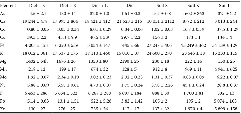
[image:3.595.64.534.516.737.2]Pressurized wet ashing was performed as follows: an aliquot (~500 mg of dry matter) of freeze-dried liver and whole kidney samples from the individual animals, or the experimental diet, were weighed into a digestion vessel. Concentrated nitric acid (8.0 ml) and 30% H2O2 (2.0 ml) (both Analytika Ltd., Prague, Czech Republic) were added. The mixture was heated in an Ethos 1 (MLS GmbH, Leutkirch, Germany) microwave assisted wet digestion system at 220°C for 30 min. After cooling, the digest was quantitatively transferred into a 20 ml glass tube and filled up to the volume with deionized water (Jankovská et al., 2010). A certified reference ma-terial BCR 185R Bovine Liver was applied for the quality assurance of analytical data. In this mate-rial, the certified values of elements represented (in mg/kg): 0.544 ± 0.017 Cd , 277 ± 5 Cu, 11.07 ± 0.29 Mn, 0.172 ± 0.009 Pb, and 138.6 ± 2.1 Zn while in our experiment we reached the values (in mg/kg): 0.537 Cd, 271 Cu, 11.3 Mn, 0.164 Pb, and 141 Zn. Total concentrations of trace elements in the soils were determined in the digests obtained by the fol-lowing decomposition procedure: aliquots (0.5 g) of air-dried soil samples were decomposed in a digestion vessel with a mixture of 8 ml of con-centrated nitric acid, 5 ml of hydrochloric acid, and 2 ml of concentrated hydrofluoric acid. The mixture was heated in an Ethos 1 (MLS GmbH,
Table 1. Total contents of elements in the individual soils, diet, and diet amended with 10% of the soil (mg/kg of dry matter)
Element Diet + S Diet + K Diet + L Diet Soil S Soil K Soil L
Leutkirch, Germany) microwave assisted wet diges-tion system at 210°C for 33 min. After cooling, the digest was quantitatively transferred into a 50 ml Teflon® vessel and evaporated to dryness at 160°C.
The digest was then dissolved in a 3 ml of nitric and hydrochloric acid mixture (1 : 3), transferred into a 25 ml glass tube, filled up with deionized water, and kept at laboratory temperature until measure-ment. A certified reference material RM 7001 Light Sandy Soil was applied for the quality assurance of analytical data. In this material, the certified values of elements represented (in mg/kg): 12.3 ± 1.1 As, 0.32 ± 0.05 Cd, 89.6 ± 4.2 Cr, 30.8 ± 0.9 Cu, 540 ± 20 Mn, 31.9 ± 1.6 Ni, 43.8 ± 3.7 Pb, and 120 ± 7 Zn. In our experiment we reached the following values (in mg/kg):13.6 As, 0.31 Cd, 87.2 Cr, 30.8 Cu, 521 Mn, 27.0 Ni, 40.1 Pb, and 120 Zn.
To determine the element contents in bones, freeze-dried femur samples were decomposed by a modified dry ashing procedure as follows: whole bone was weighed to 1 mg into a borosilicate glass test-tube and decomposed in a mixture of oxidiz-ing gases (O2 + O3 + NOx) at 400°C for 10 h in a dry mode mineralizer Apion (Tessek Ltd., Prague, Czech Republic). The ash was dissolved in 20 ml of 5% HNO3 (electronic grade purity, Analytika Ltd., Prague, Czech Republic) and kept at the labora-tory temperature until measurement (Miholová et al., 1993). Blank samples represented 15% of the total number of the digests, detection limit was calculated as mean + triple standard deviation of the blanks.
For the evaluation of bioaccessible portions of elements in soils and experimental diets the PBET test (Ruby et al., 1996; Oomen et al., 2003) was ap-plied in two phases as follows:
(i) Gastric solution – 1l volumetric flask: 1.25 g of pepsin, 0.5 g of citric acid, 0.5 g of malate, 0.84 ml of lactic acid, 1 ml of acetic acid mixed with deion-ized water and pH of the mixture was adjusted with concentrated HCl to 2.5 (± 0.05). Then 0.5 g of the
sample was mixed in 250 ml polypropylene bottle with 50 ml of the prepared gastric solution. The bottle was placed in a shaker bath at 37°C, shaked at 150 rpm for 1 h. After centrifugation a 5 ml ali-quot was taken off and measured for content of risk elements.
(ii) Pancreatic solution: 5 ml of the fresh gas-tric solution was added to the remaining reaction mixture and pH was adjusted to 7 with saturated NaHCO3 solution. Then 25 mg of pancreatin and 87.5 mg of bile salts were added. The sample was shaken once more in the bath for 2 h, centrifuged, and finally the extract was measured (Intawongse and Dean, 2008).
The total contents of the elements in the digests and extracts were determined by optical emission spectroscopy with inductively coupled plasma (ICP-OES) with axial plasma configuration Varian Vista-PRO, equipped with autosampler SPS-5 (Varian, Mulgrave, Australia). Measurement con-ditions were for all lines: power 1.2 kW, plasma flow 15.0 l/min, auxilliary flow 0.75 l/min, nebulizer flow 0.9 l/min. For determination of low concentrations of Cd, Cr, Ni, and Pb in the digests electrothermal atomic absorption spectrometry (ETAAS) using the instrument VARIAN AA280Z (Varian, Belrose, Australia) equipped with GTA120 graphite tube at-omizer was applied. The contents of Ca, Mg, and K in the digests and extracts were determined by us-ing flame atomic absorption spectrometry (Varian 280FS, Varian, Belrose, Australia).
[image:4.595.63.533.115.179.2]Of the hematological parameters, total number of erythrocytes (Er, T/l), hemoglobin (Hb, g/100 ml), he-matocrit value (Hct, %), mean cell volume (MCV, fl), and total number of leukocytes (Le, G/l) were de-termined in the whole blood stabilized by K2EDTA. All parameters were determined on the NIHON KOHDEN MEK 5208 analyzer (NIHON KOHDEN, Tokyo, Japan). For the determination of the leuko-cytes and hemoglobin values the blood hemolysis solution ISOTONAC 3 MEK 640 was used.
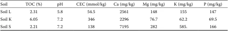
Table 2. Main physicochemical parameters of the experimental soils and available contents of the main nutrients according to Mehlich III soil extraction procedure (Mehlich, 1984)
Soil TOC (%) pH CEC (mmol/kg) Ca (mg/kg) Mg (mg/kg) K (mg/kg) P (mg/kg)
Soil L 2.31 5.8 54.5 2561 148 155 147
Soil K 6.05 7.2 346 2296 76.7 62.2 69.5
Soil S 2.21 7.2 138 7195 282 585. 166
Statistics
The data obtained were subjected to Dixon’s test for identification of outliers (significance level α = 0.05) using Microsoft Excel 2007. Subsequently, two-way analysis of variance and linear correlation analysis were applied at the significance level α = 0.05 using STATISTICA Version 9.1 program (StatSoft, 2011).
RESULTS
The element contents in the diets of the indi-vidual treatments and in the soils used for the diet amendment are summarized in Table 1. The relatively high contents of As, Cd, and Pb in soils K and L resulted in significantly (P < 0.05) increased contents of these elements in the amended diets. Moreover, higher contents of Fe, Cu, Mn, Ni, and Zn in all the soils compared to the control diet led to increased contents of these elements in the amended diets. In the opposite, low contents of Ca, Mg, and P lowered the contents of these elements in the amended diets compared to the control. Whereas the highest Cd, Mn, Mo, Pb, and Zn contents were found in soil L, the highest As content was found in soil K, as expected according to the contamination sources of the individual soils. The element con-tents extractable with simulated gastric solution
(Table 3) confirmed higher bioaccessible contents of risk elements in the contaminated soils as well as in the soil-amended diets. However, the extractable element portions (Figure 1) showed higher extract-ability of risk elements from soils K and L corre-sponding with the contamination level where the extractable portions of elements increased in the order soil S > soil K > soil L for Cd, Cu, Fe, Pb, and Zn, and opposite pattern for As, Mn, and P. The ef-fect of soil physicochemical parameters (especially CEC and Cox levels) was not significant unlike the results published by Tremlová et al. (2010). Except for K and Mg, the control diet showed better ex-tractability of elements compared to soils resulting in the decrease of extractable element portions in the case of the soil amended diets. Concerning the element contents with simulated pancreatic solution (Table 4), lower extractable portions of most of the elements were observed (Figure 2) due to sorption of elements on biomass and organic matter of the sample matrix (Turner and Ip, 2007; Turner and Price, 2008; Tremlová et al., 2010). In this case, an exception occurred for arsenic and iron in the soil samples where the extractable element amounts were markedly increased.
[image:5.595.65.535.515.735.2]During the animal experiment no apparent dif-ferences between the individual variants of the experiment were reported. Body weight, body weight gain, and feed consumption of the animals were monitored weekly. In all the three parameters,
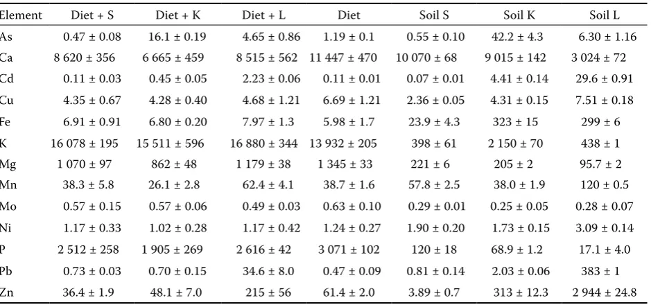
Table 3. Contents of elements in the individual soils, diet, and diet amended with 10% of the soil extractable with simulated gastric solution (mg/kg of dry matter)
Element Diet + S Diet + K Diet + L Diet Soil S Soil K Soil L
one-way ANOVA did not show significant differ-ences between the individual treatments. Total body weight gain varied between 256 ± 31 g (diet +
soil S) and 270 ± 38 g (control) whereas total feed consumption per group increased from 7.5 kg (diet + soil K) to 8 kg (control). The contents of
0 25 50 75 100
As Ca Cd Cu Fe K Mg Mn Mo Ni P Pb Zn
diet + S diet + K
diet
(A)
(%)
diet + L
0 25 50 75 100
As Ca Cd Cu Fe K Mg Mn Mo Ni P Pb Zn soil S soil K soil L (B)
[image:6.595.67.361.84.419.2](%)
Figure 1. Element portions extractable with simulated gastric solution from the individual diets (A) and from the experimental soils (B)
L = Fluvisol, K = Luvisol, S = uncontami-nated Chernozem
Table 4. Contents of elements in the individual soils, diet, and diet amended with 10% of the soil extractable with simulated pancreatic solution (mg/kg of dry matter)
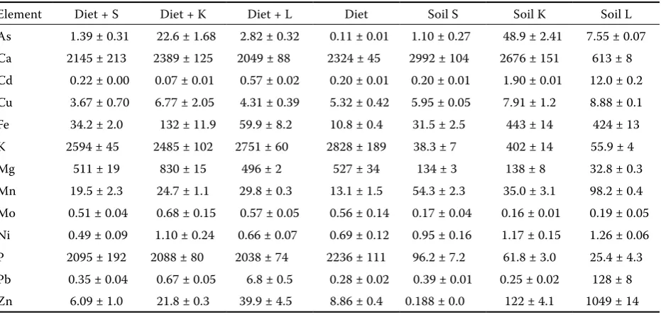
Element Diet + S Diet + K Diet + L Diet Soil S Soil K Soil L
As 1.39 ± 0.31 22.6 ± 1.68 2.82 ± 0.32 0.11 ± 0.01 1.10 ± 0.27 48.9 ± 2.41 7.55 ± 0.07 Ca 2145 ± 213 2389 ± 125 2049 ± 88 2324 ± 45 2992 ± 104 2676 ± 151 613 ± 8 Cd 0.22 ± 0.00 0.07 ± 0.01 0.57 ± 0.02 0.20 ± 0.01 0.20 ± 0.01 1.90 ± 0.01 12.0 ± 0.2 Cu 3.67 ± 0.70 6.77 ± 2.05 4.31 ± 0.39 5.32 ± 0.42 5.95 ± 0.05 7.91 ± 1.2 8.88 ± 0.1 Fe 34.2 ± 2.0 132 ± 11.9 59.9 ± 8.2 10.8 ± 0.4 31.5 ± 2.5 443 ± 14 424 ± 13 K 2594 ± 45 2485 ± 102 2751 ± 60 2828 ± 189 38.3 ± 7 402 ± 14 55.9 ± 4
Mg 511 ± 19 830 ± 15 496 ± 2 527 ± 34 134 ± 3 138 ± 8 32.8 ± 0.3
Mn 19.5 ± 2.3 24.7 ± 1.1 29.8 ± 0.3 13.1 ± 1.5 54.3 ± 2.3 35.0 ± 3.1 98.2 ± 0.4 Mo 0.51 ± 0.04 0.68 ± 0.15 0.57 ± 0.05 0.56 ± 0.14 0.17 ± 0.04 0.16 ± 0.01 0.19 ± 0.05 Ni 0.49 ± 0.09 1.10 ± 0.24 0.66 ± 0.07 0.69 ± 0.12 0.95 ± 0.16 1.17 ± 0.15 1.26 ± 0.06 P 2095 ± 192 2088 ± 80 2038 ± 74 2236 ± 111 96.2 ± 7.2 61.8 ± 3.0 25.4 ± 4.3 Pb 0.35 ± 0.04 0.67 ± 0.05 6.8 ± 0.5 0.28 ± 0.02 0.39 ± 0.01 0.25 ± 0.02 128 ± 8 Zn 6.09 ± 1.0 21.8 ± 0.3 39.9 ± 4.5 8.86 ± 0.4 0.188 ± 0.0 122 ± 4.1 1049 ± 14
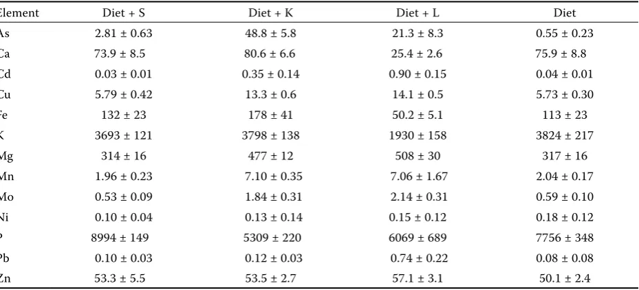
[image:6.595.66.532.519.741.2]Table 5. Total element contents in the liver of the experimental animals (mg/kg of dry matter)
Element Diet + S Diet + K Diet + L Diet
As 2.81 ± 0.63 48.8 ± 5.8 21.3 ± 8.3 0.55 ± 0.23
Ca 73.9 ± 8.5 80.6 ± 6.6 25.4 ± 2.6 75.9 ± 8.8
Cd 0.03 ± 0.01 0.35 ± 0.14 0.90 ± 0.15 0.04 ± 0.01
Cu 5.79 ± 0.42 13.3 ± 0.6 14.1 ± 0.5 5.73 ± 0.30
Fe 132 ± 23 178 ± 41 50.2 ± 5.1 113 ± 23
K 3693 ± 121 3798 ± 138 1930 ± 158 3824 ± 217
Mg 314 ± 16 477 ± 12 508 ± 30 317 ± 16
Mn 1.96 ± 0.23 7.10 ± 0.35 7.06 ± 1.67 2.04 ± 0.17
Mo 0.53 ± 0.09 1.84 ± 0.31 2.14 ± 0.31 0.59 ± 0.10
Ni 0.10 ± 0.04 0.13 ± 0.14 0.15 ± 0.12 0.18 ± 0.12
P 8994 ± 149 5309 ± 220 6069 ± 689 7756 ± 348
Pb 0.10 ± 0.03 0.12 ± 0.03 0.74 ± 0.22 0.08 ± 0.08
Zn 53.3 ± 5.5 53.5 ± 2.7 57.1 ± 3.1 50.1 ± 2.4
the investigated elements in the rat liver are sum-marized in Table 5 confirming significant impact of increased levels of As, Cd, and Pb (P < 0.05) in the diets amended with soils K and L compared to both the control diet and that amended with
soil S. The contents of these risk elements in liver tissues reflected their contents in the individual diets. Similar pattern was observed for some es-sential elements such as Mo and Mn whereas for Zn no differences in the liver were reported
regard-Figure 2. Element portions extractable with simulated pancreatic solution from the individual diets (A) and from the experimental soils (B)
L = Fluvisol, K = Luvisol, S = uncontami-nated Chernozem
0 25 50 75
As Ca Cd Cu Fe K Mg Mn Mo Ni P Pb Zn diet + S
diet (A)
diet + K diet + L (%)
0 25 50 75
As Ca Cd Cu Fe K Mg Mn Mo Ni P Pb Zn soil S soil K soil L (B)
[image:7.595.67.532.529.740.2]less of different Zn levels in the amended diets. On the contrary, the contents of nutrients (Ca, Fe, K, and P) decreased significantly with the increasing Cd and Pb levels in the liver. Significant values of correlation coefficients between Cd and nutrient contents varied from r = ‒0.61 to r = ‒0.88, and between Pb and nutrient contents the values varied from r = ‒0.36 to r = ‒0.87. In the case of As, in-creasing As levels in the liver resulted in dein-creasing
[image:8.595.67.533.100.324.2]P levels in these tissues (r = 0.81) whereas Ca and K remained unaffected and Fe increased due to high As content in As-contaminated soil K. In the opposite, Mg contents in the liver tissues increased significantly with the increasing Cd (r = 0.85) and Pb (r = 0.69) contents in the diet and subsequently in the liver tissue. Compared to the liver tissue, Cd and Pb contents in the kidney were significantly higher (P < 0.05) whereas As levels were almost
Table 6. Total element contents in the kidney of the experimental animals (mg/kg of dry matter)
Element Diet + S Diet + K Diet + L Diet
As 2.87 ± 0.99 47.2 ± 9.6 16.7 ± 6.1 1.09 ± 0.60
Ca 123 ± 49 73.1 ± 39.5 54.3 ± 28.6 114 ± 25
Cd 0.18 ± 0.37 1.65 ± 1.13 3.54 ± 1.76 0.11 ± 0.05
Cu 10.6 ± 1.6 22.4 ± 3.5 9.89 ± 0.89 12.2 ± 1.9
Fe 48.3 ± 20.7 28.1 ± 2.2 19.1 ± 4.4 46.7 ± 10.3
K 2260 ± 525 1770 ± 613 1576 ± 490 2466 ± 330
Mg 198 ± 64 132 ± 70 105 ± 15 212 ± 34
Mn 0.49 ± 0.15 0.26 ± 0.08 0.19 ± 0.04 0.44 ± 0.11
Mo 0.30 ± 0.10 0.29 ± 0.11 0.36 ± 0.11 0.12 ± 0.10
Ni 0.17 ± 0.02 0.23 ± 0.05 0.17 ± 0.05 0.34 ± 0.07
P 8130 ± 961 6976 ± 428 6539 ± 103 7916 ± 568
Pb 0.16 ± 0.03 0.40 ± 0.08 20.8 ± 2.8 0.09 ± 0.02
Zn 39.9 ± 3.2 29.1 ± 8.6 27.1 ± 8.2 36.2 ± 4.4
L = Fluvisol, K = Luvisol, S = uncontaminated Chernozem
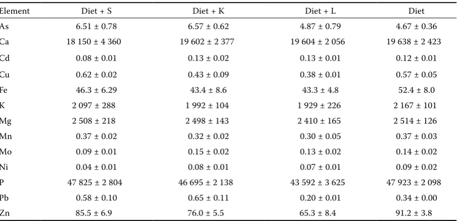
Table 7. Total element contents in the bones of the experimental animals (mg/kg of dry matter)
Element Diet + S Diet + K Diet + L Diet
As 6.51 ± 0.78 6.57 ± 0.62 4.87 ± 0.79 4.67 ± 0.36
Ca 18 150 ± 4 360 19 602 ± 2 377 19 604 ± 2 056 19 638 ± 2 423
Cd 0.08 ± 0.01 0.13 ± 0.02 0.13 ± 0.01 0.12 ± 0.01
Cu 0.62 ± 0.02 0.43 ± 0.09 0.38 ± 0.01 0.57 ± 0.05
Fe 46.3 ± 6.29 43.4 ± 8.6 43.3 ± 4.8 52.4 ± 8.0
K 2 097 ± 288 1 992 ± 104 1 929 ± 226 2 167 ± 101
Mg 2 508 ± 218 2 498 ± 143 2 410 ± 165 2 514 ± 126
Mn 0.37 ± 0.02 0.32 ± 0.02 0.30 ± 0.05 0.37 ± 0.03
Mo 0.09 ± 0.01 0.15 ± 0.02 0.13 ± 0.02 0.14 ± 0.02
Ni 0.04 ± 0.01 0.08 ± 0.01 0.07 ± 0.01 0.09 ± 0.02
P 47 825 ± 2 804 46 695 ± 2 138 43 592 ± 3 625 47 923 ± 2 098
Pb 0.58 ± 0.10 0.65 ± 0.11 0.20 ± 0.01 0.34 ± 0.00
Zn 85.5 ± 6.9 76.0 ± 5.5 65.3 ± 8.4 91.2 ± 3.8
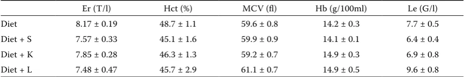
[image:8.595.65.534.512.738.2]identical (Table 6). Concerning the other micro- and macroelements, the interpretation of the kidney data is comparable to that of the liver samples regard-less of different amounts of most elements. Some exceptions were in Mn and Mg where significantly lower contents of these elements were observed in the kidney of the animals fed the diet amended with soils L and K. Significantly increased Cu content in the kidney of the animals fed soil-derived risk-element amended diet was observed only in the case of soil K. Concerning the element contents in bones (Table 7), no significant differences occurred within all the experimental groups of animals. No effect was observed even in the case of Pb, the element typi-cally deposited in calcified tissues (bones and teeth). Due to this property, lead is considered the so-called osteotrophic element (Gerhardsson, 1996; Hac and Krechinak, 1996). In our case, however, the Pb levels in the bones of the animals fed Pb-contaminated diet amended with soil L even tended to decrease as compared to the other experimental groups. Gregus and Klaasen (1986) also described higher percentage of lead in the liver of Pb-exposed rats if compared to the bones. The levels of the investigated hema-tological parameters are summarized in Table 8. Statistical evaluation of the data showed significant increase (P < 0.05) of the total leukocyte count in the blood of the animals fed the diet amended with extremely contaminated soil L, whereas the other measured parameters remained unchanged. Similar results were described by Fučíková et al. (1995) in the blood of rats fed the diet supplemented with cadmium containing yeasts and Adham et al. (2011) in the blood of wild Meriones libycus living in a Cd- and Pb-polluted area.
DISCUSSION
The results proved substantial effect of the
[image:9.595.63.532.102.180.2]con-in rat organisms. Significant side effects of the soil-derived risk elements such as substantial changes in micro- and macroelement levels in the rat or-ganisms as well as changes in some hematological parameters were reported. The contents of As, Cd, and Pb in the liver and kidney of rats reflected the contents of these elements in the soils. Similarly, Mascolo et al. (2004) investigated the effect of in-gestion of clay materials on element concentrations (including As, Cd, and Pb) in selected tissues of rats during a 6-day trial. Three groups of rats were fed three different types of clays (plus normal food). Another group was fed only conventional food and taken as a control experiment. The clays were se-lected according to their concentration of chemical elements, ranging from very low to very high values. As in our experiment, the authors did not observe any visible macro-toxic effect on the treated ani-mals. Nevertheless, the trace element contents of the analyzed organs including kidney and liver were strictly related to the clay trace element contents: upon increasing the clay trace element contents, the contents in the organs also went up and higher element contents were found in kidney compared to liver. This is fully in accordance with our findings. However, different bioavailability of the individu-al elements decreasing in the order Cd > As > Pb was determined by both in vitro and in vivo ex-periments. The effect of soil physicochemical pa-rameters (especially CEC and Cox levels) was not significant unlike the results published by Tremlová et al. (2010). Lower bioavailability of soil-derived Pb compared to As was mentioned also by Ellickson et al. (2001). In the case of Cd, Schilderman et al. (1997) observed lower (by 60%) bioavailability of soil-derived Cd compared to the ingestion of pure Cd salt. In our case, the effect of soil physicochemi-cal parameters was observed, especially in the level of cation exchange capacity (Table 2) where higher bioavailability of elements was determined in the
Table 8. Average contents of selected hematological parameters
Er (T/l) Hct (%) MCV (fl) Hb (g/100ml) Le (G/l)
Diet 8.17 ± 0.19 48.7 ± 1.1 59.6 ± 0.8 14.2 ± 0.3 7.7 ± 0.5
Diet + S 7.57 ± 0.33 45.1 ± 1.6 59.9 ± 0.9 14.1 ± 0.1 6.4 ± 0.4
Diet + K 7.85 ± 0.28 46.3 ± 1.3 59.2 ± 0.7 14.9 ± 0.3 6.9 ± 0.8
Diet + L 7.48 ± 0.47 45.7 ± 2.9 61.1 ± 0.7 14.9 ± 0.5 9.6 ± 0.8
The element contents in animal tissues were, however, affected by various element inter-actionsresulting insignificant changes in macro- and micro-nutrient contents as described above. Matovič et al. (2011) reported that the exposure to Cd reduces the levels of essential elements such as Mg and Zn, which may have adverse health ef-fects. Whittaker et al. (2011) investigated the effect of oral exposure of rats to Pb, Cd, and As mix-tures in drinking water (5 mg/l of As, 10 mg/l of Cd, 25 mg/l of Pb) on induction of the indicators of oxidative stress. They compared also the sin-gle element exposure of rats and reported signifi-cantly increased level of Cu in kidney of animals exposed to the element mixture compared to the single element dose. On the other hand, Yu and Beynen (2001) observed that the high-arsenic diet (100 mg/kg of As) significantly elevated kidney copper concentration. Higher liver As content in iron-deficient rats was observed by Paul et al. (2002). Although protective effect of Zn against Cd toxicity was described (Rogalska et al., 2011), no interaction of these elements was observed in our case. Ognjanovič et al. (2003) observed increased activity of antioxidant defense enzymes: copper and zinc containing superoxide dismutase, cata-lase, glutathione peroxidase, glutathione reductase, and glutathione-S-transferase as well as concentra-tions of non-enzymatic components of antioxidant defense system: reduced glutathione, vitamin C, and vitamin E in rats after acute exposure to Cd. In our experiment, however, no changes in Zn lev-els in rat tissues were observed compared to Cu and the potential role of copper and zinc contain-ing superoxide dismutase remains questionable. Whittaker et al. (2011) explained the increasing Cu contents in the rat organism by the induction of metallothionein in the kidney as a result of ex-posure to trace elements such as lead or cadmium. Because copper is also bound by metallothionein, accumulation of copper in the kidney may be attrib-utable to metallothionein induction. This reason is most probably responsible for the increased Cu levels in the liver and kidney of the animals in our experiment. These findings need to be proved by the direct determination of metallothionein levels in the tissues in further research.
Prigge et al. (2002) reviewed the effect of Cd on hematological parameters of animals and men-tioned significant decrease of hemoglobin values if the rat diet contained 31 mg/kg of Cd. However, lower levels of iron in the exposed rat organisms
were reported in this case. Similar effect was ob-served by Hiratsuka et al. (1996) in rats intravenously exposed to Cd. Also Turgut et al. (2007) described that high cadmium and lead decreased iron ab-sorption and negatively affected hematological pa-rameters. Except for lower Fe levels in the treated rat tissues, no significant changes in hematologi-cal parameters were registered in our experiment. Evidently, the changes in iron levels described by the above-mentioned authors were due to higher contents of risk elements in the diet compared to our experiment. However, increased total leukocyte count at the highest element level confirmed ad-verse effect of risk elements on the animal organism (Fučíková et al., 1995; Adham et al., 2011).
Noble et al. (2010) evaluated potential risk of soil ingestion by inhabitants of a mining district in Australia. Near the mine, total As concentra-tions in the soil were between 16 and 946 mg/kg and total Pb concentrations were between 12 and 430 mg/kg. The authors concluded that an average toddler (12 kg) would need to consume at least 1.5 g (and most likely 12 g) of soil per day to show some symptoms of As toxicity. They also inferred that daily exposure of small children to soil is not typical and, therefore, the health risk resulting from the soil ingestion in the vicinity of the mine is minimal. However, as confirmed by our experiment, the rate of potential risk stemming from the contaminated soil ingestion differs at each locality and depends on the risk-element content levels in the soil in the vicinity of the particular mine and also on bio-availability of the individual elements. Therefore, in the Czech Republic localities exhibiting high risk-elements soil penetration should be paid a special attention in order to assess potential bioavailability of the risk elements and to determine the potential health risk for the inhabitants.
REFERENCES
Adham K.G., Al-Eisa N.A., Farhood M.H. (2011): Impact of heavy metal pollution on the hemogram and serum biochemistry of the Libyan jird, Meriones libycus. Che-mosphere, 84, 1408–1415.
Amaral A., Cabral C., Guedes C., Rodrigues A. (2007): Apoptosis, metallothionein, and bioaccessible metals in domestic mice (Mus musculus L.) from a human-inhabited volcanic area. Ecotoxicology, 16, 475‒481. Ellickson K.M., Meeker R.J., Gallo M.A., Buckley B.T.,
from a NIST standard reference soil material. Archives of Environmental Contamination and Toxicology, 40, 128‒135.
Fischer A.B., Georgieva R., Nikolova V., Halkova J., Bainova A., Hristeva V., Penkov D., Alandjiisk D. (2003): Health risk for children from lead and cad-mium near a non-ferrous smelter in Bulgaria. Inter-national Journal of Hygiene and Environmental Health, 206, 25‒38.
Fučíková A., Slámová A., Száková J., Cibulka J., Heger J. (1995): The influence of dietary cadmium on hema-tological parameters and phagocytic activity of leuko-cytes in rats. Živočišná výroba, 40, 15‒18.
Gerhardsson L. (1996): In vivo measurement of lead in bone in longterm exposed lead smelter workers. Ar-chives of Environmental Health, 14, 22‒28.
Gibaldi M. (1984): Biopharmaceutics and clinical phar-macokinetics. 3rd Ed. Lea and Febiger, Philadelphia,
USA.
Gregus Z., Klaasen C.D. (1986): Disposition of metals in rats: A comparative study of fecal, urinary, and bil-iary excretion and tissue distribution of eighteen met-als. Toxicology and Applied Pharmacology, 85, 24‒38. Hac E., Krechinak J. (1996): Lead levels in bone and hair
of rats treated with lead acetate. Biological Trace Ele-ment Research, 52, 293‒301.
Hettiarachchi G.M., Pierzynski G.M., Oehme F.W., Son-mez O., Ryan J.A. (2003): Treatment of contaminated soil with phosphorus and manganese oxide reduces lead absorption by Sprague-Dawley rats. Journal of Environmental Quality, 32, 1335‒1345.
Hiratsuka H., Katsuta O., Toyota N., Tsuchitani M., Ume-mura T., Marumo F. (1996): Chronic cadmium expo-sure-induced renal anemia in ovariectomized rats. Toxicology and Applied Pharmacology,137, 228–236. Intawongse M., Dean J.R. (2008): Use of the physiologically-based extraction test to assess the oral bioaccessibility of metals in vegetableplants grown in contaminated soil. Environmental Pollution, 152, 60–72.
Jankovská I., Vadlejch J., Száková J., Miholová D., Kunc P., Knížková I., Čadková Z., Langrová I. (2010): Ex-perimental studies on the cadmium accumulation in the cestode Monienzia expansa (Cestoda:
Anoplo-cephalidae) and its final host (Ovis aries).
Experimen-tal Parasitology, 126, 130‒134.
Juhasz A.L., Weber J., Naidu R., Gancarz D., Rofe A., Todor D., Smith E. (2010): Determination of cadmium relative bioavailability in contaminated soils and its prediction using in vitro methodologies. Environmen-tal Science and Technology, 44, 5240‒5247.
Karadas C., Kara D. (2011): In vitro gastro-intestinal
ity in contaminated soils. Environmental Science and Pollution Research, 18, 620‒628.
Kostič M.M., Ognjanovič B., Dimitrijevič S., Žikič R.V., Štajn A., Rosič G.L., Živkovič R.V. (1993): Cadmium-induced changes of antioxidant and metabolic status in red-blood-cells of rats ‒ in-vivo effects. European Journal of Haematology, 51, 86‒92.
Lee S.W., Lee B.T., Kim J.Y., Kim K.W., Lee J.S. (2006): Human risk assessment for heavy metals and as con-tamination in the abandoned metal mine areas, Korea. Environmental Monitoring and Assessment, 119, 233‒244.
Ljung K., Oomen A., Duits M., Selinus O., Berglund M. (2007): Bioaccessibility of metals in urban playground soils. Journal of Environmental Science and Health, Part A, 42, 1241–1250.
Mascolo N., Summ V., Tateo F. (2004): In vivo experi-mental data on the mobility of hazardous chemical elements from clays. Applied Clay Science, 25, 23–28. Matović V., Buha A., Bulat Z., Dukic-Cosic D. (2011):
Cadmium toxicity revisited: focus on oxidative stress induction and interactions with zinc and magnesium. Arhiv za Higijenu Rada i Toksikologiju,62, 65‒76. Mehlich A. (1984): Mehlich 3 Soil Test Extractant: A
modification of Mehlich 2 Extractant. Communica-tions in Soil Science and Plant Analysis, 15, 1409‒1416. Miholová D., Mader P., Száková J., Slámová A., Svatoš
Z. (1993): Czechoslovakian biological certified refer-ence materials and their use in the analytical quality assurance system in a trace element laboratory. Fre-senius Journal of Analytical Chemistry, 345, 256‒260. Mrad I., Ghorbel A., Lambert J.F., Che M. (1997): Cad-mium adsorption on Al-pillared montmorillonite. Journal de Chimie Physique et de Physico-Chimie Bio- logique, 94, 1883‒1896.
Noble R.R.P., Hough R.M., Watkins R.T. (2010): Enrich- ment and exposure assessment of As, Cr and Pb of the soils in the vicinity of Stawell, Victoria, Australia. En-vironmental Geochemistry and Health, 32, 193–205. Ognjanović B.I., Pavlović S.Z., Maletić S.D., Žikić R.V.,
Štajn A.Š, Radojičić R.M., Saičić Z.S., Petrović V.M. (2003): Protective influence of vitamin E on antioxi-dant defense system in the blood of rats treated with cadmium. Physiological Research, 52, 563‒570. Oomen A.G., Rompelberg C.J.M., Bruil M.A., Dobbe
C.J.G., Pereboom D.P.K.H., Sips A.J.A.M. (2003): De-velopment of an in vitro digestion model for estimation of bioaccessibility of soil contaminants. Archives of Environmental Contamination and Toxicology, 44, 281‒287.
arsenic in tissues of iron-deficient rats. Toxicology Letters, 135, 193‒197.
Prigge E., Baumert H.P., Muhle H. (2002): Effects of di-etary and inhalative cadmium on hemoglobin and hematocrit in rats. Biological Trace Element Research, 59, 585‒590.
Pruvot C., Douay F., Hervé F., Waterlot C. (2006): Heavy metals in soil, crops and grass as a source of human exposure in the former mining areas. Journal of Soils and Sediments, 6, 215‒220.
Rieuwerts J.S., Searle P., Buck R. (2006): Bioaccessible arsenic in the home environment in southwest Eng-land. Science of the Total Environment, 371, 89‒98. Rodriguez R.R., Basta N.T. (1999): An in vitro
gastroin-testinal method to estimate bioaccessible arsenic in contaminated soils and solid media. Environmental Science and Technology, 30, 642‒475.
Rogalska J., Pilat-Marcinkiewicz B., Brzóska M.M. (2011): Protective effect of zinc against cadmium he-patotoxicity depends on this bioelement intake and level of cadmium exposure: A study in a rat model. Chemico-Biological Interactions, 193, 191–203. Ruby M.V., Davi A., Schoo R., Eberl S., Sellston C.M.
(1996): Estimation of lead and arsenic bioavailability using a physiologically based extraction test. Environ-mental Science and Technology, 30, 422–430. Ruby M.V., Schoof R., Brattin W., Goldade M., Post G.,
Harnois M., Mosby D.E., Casteel S.W., Berti W., Car-penter M., Edwards D., Cragin D., Chappell W. (1999): Advances in evaluating the oral bioavailability of in-organics in soil for use in human health risk assess-ment. Environmental Science and Technology, 33, 3697‒3705.
Schilderman P.A.E.L., Moonen E.J.C., Kempkers P., Kle-injans J.C.S. (1997): Bioavailability of soil-adsorbed cadmium in orally exposed male rats. Environmental Health Perspectives, 105, 234‒238.
Sheppard S.C., Eveden W.G., Schwartz W.J. (1995): In-gested soil ‒ bioavailability of sorbed lead, cadmium,
cesium, iodine, and mercury. Journal of Environmen-tal Quality, 24, 498‒505.
Šichorová K., Tlustoš P., Száková J., Kořínek K., Balík J. (2004): Horizontal and vertical variability of heavy metals in the soil of a polluted area. Plant, Soil and Environment, 50, 525–534.
StatSoft (2011): STATISTICA (Data analysis software system). Version 9.1. StatSoft® Inc., Tulsa, USA. Tremlová J., Száková J., Tlustoš P. (2010): An assessment
of possible effect of risk elements contained in soil on human organism. Chemické Listy, 104, 349–352. (in Czech)
Turgut S., Polat A., Inan M., Turgut G., Emmungil G., Bican M., Karakus T.Y., Genç O. (2007): Interaction between anemia and blood levels of iron, zinc, copper, cadmium and lead in children. Indian Journal of Pe-diatrics, 74, 827‒830.
Turner A., Ip K.H. (2007): Bioaccessibility of metals in dust from the indoor environment: application of a physiologically based extraction test. Environmental Science and Technology, 41, 7851‒7856.
Turner A., Price S. (2008): Bioaccessibility of platinum group elements in automotive catalytic converter par-ticulates. Environmental Science and Technology, 42, 9443‒9448.
Whittaker M.H., Wang G., Chen X.-Q., Lipsky M., Smith D., Gwiazda R., Fowler B.A. (2011): Exposure to Pb, Cd, and As mixtures potentiates the production of oxidative stress precursors: 30-day, 90-day, and 180-day drinking water studies in rats. Toxicology and Applied Pharma-cology, 254, 154–166.
Yu S., Beynen A.C. (2001): High arsenic intake raises kidney copper and lowers plasma copper concentra-tions in rats. Biological Trace Element Research, 63, 63‒70.
Received: 2012‒02‒21 Accepted after corrections: 2012‒06‒05
Corresponding Author
prof. Ing. Jiřina Száková,CSc.,Czech University of Life Sciences Prague, Faculty of Agrobiology, Food and Natural Resources, Department of Agroenvironmental Chemistry and Plant Nutrition, Kamýcká 129,