RESEARCH ARTICLE
Skin glands of an aquatic salamander vary in size and distribution
and release antimicrobial secretions effective against chytrid
fungal pathogens
Kenzie E. Pereira1,2,*, Brian I. Crother2, David M. Sever2, Clifford L. Fontenot, Jr2, John A. Pojman, Sr3, Damien B. Wilburn4and Sarah K. Woodley1
ABSTRACT
Amphibian skin is unique among vertebrate classes, containing a large number of multicellular exocrine glands that vary among species and have diverse functions. The secretions of skin glands contain a rich array of bioactive compounds including antimicrobial peptides (AMPs). Such compounds are important for amphibian innate immune responses and may protect some species from chytridiomycosis, a lethal skin disease caused by the fungal pathogensBatrachochytrium dendrobatidis (Bd) and Batrachochytrium salamandrivorans (Bsal). While the bioactivity of skin secretions against Bd has been assessed for many amphibian taxa, similar studies are lacking for Bsal, a chytrid fungus that is especially pathogenic for salamanders. We studied the skin glands and their potential functions in an aquatic salamander, the three-toed amphiuma (Amphiuma tridactylum). Skin secretions of captive adult salamanders were analyzed by RP-HPLC and tested against the growth of Bd and Bsal usingin vitroassays. We found that compounds within collected skin secretions were similar between male and female salamanders and inhibited the growth of Bd and Bsal. Thus, skin secretions that protect against Bd may also provide protection against Bsal. Histological examination of the skin glands of preserved salamanders revealed the presence of enlarged granular glands concentrated within caudal body regions. A site of potential gland specialization was identified at the tail base and may indicate specialized granular glands related to courtship and communication.
KEY WORDS: Amphibian,Amphiuma, Antimicrobial peptide, Batrachochytrium dendrobatidis,Batrachochytrium salamandrivorans, Chytridiomycosis
INTRODUCTION
Amphibian skin is unique among vertebrate classes, containing two distinct types of multicellular exocrine glands: mucous and granular (Duellman and Trueb, 1994). The repertoire of products secreted from these two gland types and distribution of glands vary greatly among different species (Sever, 2003; Woodley, 2014; Xu and Lai, 2015). Though skin glands serve diverse roles, previous studies have primarily focused on the granular glands of frogs (Anura) for their roles in poison production and potential for therapeutic use
(Gomes et al., 2007). More recently, there has been increased interest in exploring the antimicrobial properties of skin secretions and their roles in preventing diseases linked to global amphibian decline (Conlon and Sonnevend, 2010; Pukala et al., 2006; Woodhams et al., 2007).
The granular gland secretions of many amphibians contain a diverse array of antimicrobial compounds including alkaloids, proteins and antimicrobial peptides (AMPs) that exhibit broad-spectrum antimicrobial activity against bacteria, viruses and fungi (Conlon, 2011; Dahham et al., 2016; Daly, 1995; Mina et al., 2015). These antimicrobial compounds, particularly AMPs, comprise an important component of the amphibian innate immune response and may help protect some species from emerging infectious diseases such as chytridiomycosis (Woodhams et al., 2007), a lethal skin disease that has been linked to amphibian population declines and extinctions worldwide and is caused by the fungal pathogens Batrachochytrium dendrobatidis(Bd) (Longcore et al., 1999; Voyles et al., 2009) andBatrachochytrium salamandrivorans(Bsal) (Martel et al., 2013; Voyles et al., 2009).These fungi infect hosts through the action of mobile zoospores that embed and mature within the keratinized epithelium of amphibian skin (Berger et al., 2005; Martel et al., 2013). There is growing evidence that antimicrobial substances secreted onto the skin surface prevent chytridiomycosis by limiting the burden of infection (Woodhams et al., 2007). Research has primarily focused on the antimicrobial properties of anuran skin secretions partially because salamander (Urodela) populations have been comparatively less afflicted by Bd (Conlon et al., 2013; Rollins-Smith et al., 2002b). However, with the discovery of a new species of a chytrid fungus (Bsal) that is especially pathogenic for urodele amphibians (Martel et al., 2014), it is essential to test the skin secretions of salamanders for anti-chytrid properties.
In addition to their roles in antimicrobial defense, the granular glands of some species also serve specialized functions in nutrient storage (Williams and Larsen, 1986), predator defense (Brodie et al., 1991; Brodie and Smatresk, 1990), chemosensory communication (Woodley, 2015) and reproduction (Sever, 2003). Unlike‘ordinary’ granular glands that are randomly scattered throughout the skin,
‘specialized’ granular glands often exhibit unique morphologies and are concentrated within specific parts of the body (Woodley, 2014). Likewise, specialized mucous gland types have also been described (Brizzi et al., 2002; Sever, 1976; Wilburn et al., 2017). For example, while ordinary mucous glands constitutively release products rich in mucopolysaccharides and facilitate cutaneous gas exchange, specialized mucous glands synthesize protein pheromones used during courtship and mating (Woodley, 2015). Though the occurrence of specialized glands is widespread among terrestrial salamanders, the presence of similar glands in aquatic species remains largely unexplored (Rupp and Sever, 2018). Received 30 April 2018; Accepted 31 May 2018
1Department of Biological Sciences, Duquesne University, Pittsburgh, PA 15282, USA.2Department of Biology, Southeastern Louisiana University, Hammond, LA 70402, USA.3Department of Chemistry, Louisiana State University, Baton Rouge, LA 70803, USA.4Department of Genome Sciences, University of Washington, Seattle, WA 98195, USA.
*Author for correspondence ([email protected])
K.E.P., 0000-0002-1810-6730
Journal
of
Experimental
We studied the skin glands and their potential functions in adult three-toed amphiuma (Amphiuma tridactylum), one of three aquatic salamander species comprising the Amphiumidae (Petranka, 1998). Two species, A. tridactylum and Amphiuma means, are among the largest salamanders, attaining total lengths of 100–110 cm, whereasAmphiuma pholeteris relatively small, rarely exceeding 30 cm (Powell et al., 2016). Amphiumid salamanders inhabit various freshwater environments throughout the southeastern USA and do not appear to be vulnerable to chytridiomycosis (Chatfield et al., 2012; Petranka, 1998). Chatfield et al. (2012) surveyed A. tridactylum (n=37), A. means (n=11), A. pholeter (n=1) and non-specified amphiumid species (n=6) for the presence of Bd in Florida, Mississippi and Louisiana. Although 23 of the surveyed individuals were infected with the fungal pathogen, none showed any symptoms of disease, suggesting that skin secretions may serve a protective role against chytridiomycosis.
As for other aquatic salamander species, the presence of specialized gland types in the skin of A. tridactylum remains unknown. While detailed descriptions of cloacal glands involved in spermatophore production (males) and sperm storage (females) have been reported, similar studies are lacking for mucous and granular glands (Sever, 1992). Because specialized glands have been related to amphibian behaviors, including courtship and reproduction, identifying regions of gland specialization in A. tridactylummay provide valuable insight towards understanding the behaviors of this elusive salamander.
The first goal of this study was to determine whether the skin secretions of captive A. tridactylum were effective at inhibiting the growth of Bd and Bsal usingin vitrogrowth inhibition assays. Because chytridiomycosis has not been reported for wild populations, we predicted that the skin secretions of adultA. tridactylumwould exhibit efficacy against chytrid fungi (Bd and Bsal) in vitro. The second goal of this study was to examine the skin histology of preserved A. tridactylumfor evidence of gland specialization by searching for regions of increased gland size and frequency. Based on studies of other salamander taxa, we predicted that evidence of gland specialization would also be present inA. tridactylum.
MATERIALS AND METHODS Skin secretions
Animal care
One male (snout–vent length, SVL=75.5 cm, mass=2.2 kg) and one female (SVL=71 cm, mass=1.8 kg)A. tridactylum(Cuvier, 1827) were collected from East Baton Rouge Parish, Baton Rouge, LA, USA, in 2014 under Louisiana Department of Wildlife and Fisheries Permit No. 100-3510-934. Adult salamanders were housed communally for 3 years in a 208 l aquarium with filtration.
Partial water changes were conducted weekly. Animals were exposed to an 11 h:13 h light:dark cycle and sustained on tilapia (Oreochromissp.) and salmon (Salmo salar) dusted with vitamin supplement (Rep-Cal Research Labs, Los Gatos, CA, USA).
Collection and processing
Methods utilized for the collection and processing of crude skin secretions were adapted from Woodhams et al. (2006b) and were approved by the Duquesne University Institutional Animal Care and Use Committee. Animals were submerged individually in 300 ml collection buffer (50 mmol l−1sodium chloride, 25 mmol l−1sodium acetate) for 15 min. During this time, animals were gently prodded with blunt forceps to manually induce skin secretions. The collection buffer was removed and combined with 300 µl trifluoroacetic acid (TFA). In order to remove particulates that could interfere with further processing, the collection buffer was centrifuged at 1632gfor 30 min and the supernatant was passed through a syringe filter (0.2 µm PES syringe filter, GE Healthcare Life Sciences, Pittsburgh, PA, USA). To enrich for compounds containing hydrophobic regions typical of AMPs, the filtered buffer was loaded into a sterile 50 ml syringe and passed through a C-18 Sep-Pak cartridge (cat. no. WAT 051910, Waters Corp., Milford, MA, USA) that had been wetted with 100% methanol and equilibrated in 0.1% TFA. Following passage of the collection buffer, Sep-Pak cartridges were washed in 0.1% TFA and retained compounds were eluted with 70% acetonitrile and 0.1% TFA. Wash steps, passage of the collection buffer and elution were repeated two times to increase the recovery of secreted compounds. Eluted compounds were concentrated using a Speed-Vac concentrator (Savant Instruments, Marietta, OH, USA), neutralized by addition of 100 mmol l−1ammonium bicarbonate, concentrated again and reconstituted in nanopure water (final pH∼7.5). The peptide/protein concentration of each reconstituted sample was determined using a Micro BCA Protein Assay Kit (Thermo Scientific, Rockford, IL, USA) following the manufacturer’s instructions except that bradykinin (Sigma Chemical Co., St Louis, MO, USA) was used as a peptide standard for calibration (Rollins-Smith et al., 2002b). Based on BCA peptide estimates, processed skin secretions from both animals were pooled, filter sterilized and diluted with sterile nanopure water to generate skin secretion concentrations of 1000, 500, 250, 125, 62.5 and 31.25 µg ml−1.
Reversed-phase high-performance liquid chromatography (RP-HPLC)
Compounds within processed skin secretions (25 µg) were analyzed by RP-HPLC using a C-18 reverse-phase column (Vydac, Hesperia, CA, USA; 5 mm; 4.6×150 mm) on a 2695 Alliance HPLC column equipped with a 2487 dual wavelength absorbance detector and Empower 2 software (Waters Division, Milford, MA, USA). The column was equilibrated with 0.1% TFA and analytes were separated with a linear gradient from 0 to 70% acetonitrile at 1% acetonitrile per minute, with detection at 220 and 280 nm.
Chytrid growth inhibition assays
Batrachochytrium dendrobatidis (Bd isolate JEL-197) and B. salamandrivorans (Bsal isolate AMFP) were maintained in liquid broth culture as previously described by Rollins-Smith et al. (2002a) and Martel et al. (2013). The two cultures (Bd–pass no. 42, Bsal–pass no. 68) were plated individually on TGhL agar plates containing antibiotics [ampicillin sodium salt (Sigma-Aldrich) and streptomycin sulfate (MP Biomedicals, LLC, Santa Ana, CA, USA)] and cultured for 5 days at 23°C (Bd) or 15°C (Bsal). Zoospores were List of symbols and abbreviations
AB Alcian Blue AMP antimicrobial peptide BB Bromophenol Blue
Bd Batrachochytrium dendrobatidis
Bsal Batrachochytrium salamandrivorans
H&E Hematoxylin and Eosin
MIC minimal inhibitory concentration (µg ml−1) OD optical density
PAS periodic acid–Schiff
RP-HPLC reversed-phase high-performance liquid chromatography SVL snout–vent length
TFA trifluoroacetic acid
Journal
of
Experimental
harvested by flooding each agar plate with 2–3 ml of culture broth followed by passing the collected broth through sterile nylon mesh (N301, BioDesign Inc., Carmel, NY, USA) to collect mature zoosporangia. Zoospores were enumerated using a hemocytometer and adjusted to a final concentration of 1×10⁶zoospores per ml. In a sterile 96-well microtiter plate, 50 µl zoospores were combined with 50 µl of each concentration of skin secretions in triplicate. Control wells (replicates of six) consisted of 50 µl sterile nanopure water (no skin secretions) combined with either 50 µl zoospores ( positive control) or 50 µl heat-killed zoospores (negative control). To monitor plates for microbial contamination, negative control wells (replicates of two) consisting of 50 µl culture broth combined with either 50 µl sterile nanopure water or 50 µl of each skin secretion concentration were also included. Each 96-well plate, one per chytrid species, was incubated at either 23°C (Bd) or 15°C (Bsal) for 15 days. Optical density (OD) at 490 nm was read daily with an accuSkan GO plate reader (Fisher Scientific, Hampton, NH, USA). To measure growth in each well, the OD at day 0 was subtracted from the OD at day 15. A positive change in OD was interpreted as growth, whereas a zero or negative change in OD was interpreted as no growth.
To quantify the antimicrobial properties of processed skin secretions, we determined the minimal inhibitory concentration (MIC, the lowest skin secretion concentration at which no growth was detected, as per Sheafor et al., 2008). The change in OD was fitted with a four-parameter logistic or sigmoidal regression curve using SigmaPlot software version 11.0 (Systat Software Inc., Richmond, CA, USA). The MIC for each assay was calculated as the point where the upper 95% confidence limit of the negative control (i.e. heat-killed chytrid+water) crossed the regression line.
To facilitate comparison of the relative effectiveness of skin secretions collected fromA. tridactylumwith that of other species, we also determined mean MIC equivalents per cm2 surface area. An MIC equivalent incorporates both the MIC as well as the amount (mass) of skin secretions present on the skin surface. An MIC equivalent is the total amount (mass) of skin secretions collected (µg) divided by the experimentally estimated MIC value (µg ml−1) (Woodhams et al., 2006a). The surface area of each animal was calculated using the equation: surface area (cm2)=8.42×mass (g)0.694(Whitford and Hutchison, 1967).
Skin histology
Specimens
AdultA. tridactylum(n=4) previously fixed in 10% neutral buffered formalin and stored in 70% ethanol were used for histological examination. Specimens were collected in Orleans and East Baton Rouge Parishes, LA, USA, on the following dates, with sex, SVL and museum voucher numbers in parentheses: 16 May 1946
(female, 55.8 cm, SLU 1089), 21 March 2012 (female, 49.8 cm, SLU 02903) and 24 June 2012 (male, 36 cm, SLU 02904). Locality data were unavailable for one specimen collected on 23 January 2002 (male, 43.5 cm, SLU 774). Because voucher specimen SLU 774 was an adult collected within the months of peak sperm production (i.e. December to March; Rose, 1967; Wilson, 1940) and had enlarged testes and vas deferens, it was deemed reproductively active. All other specimens were deemed reproductively non-active because collection was outside of the months of peak sperm production and small testes and vas deferens were observed (male specimen) or mature oocytes were not observed within the body cavity (female specimens).
Tissue collection and light microscopy
Pieces of skin (approximately 2×2 mm2) were removed from 18 locations of each museum specimen (Fig. 1), dehydrated in ascending ethanol dilutions (70%, 95% and 100%), cleared in toluene and paraffin embedded under vacuum for 24 h. Using a MR3 rotary microtome (RMC Instruments, Tucson, AZ, USA), 192 sequential transverse 10 µm sections were collected onto 12 albumenized slides per piece of skin. Slides were stained with Hematoxylin–Eosin (H&E) for general histology or Bromophenol Blue (BB) for proteins, or treated with the periodic acid–Schiff procedure (PAS) for neutral carbohydrates. Slides treated with PAS were counterstained with Alcian Blue (AB) at pH 2.5 for acidic mucopolysaccharides. Slides were affixed with coverslips using Permount (Kiernan, 1990; Presnell and Schreibman, 1997).
For each of the 18 sets of 12 slides, sections were randomly selected for data collection using a random number generator. Micrographs for selected sections were generated using light microscopy [Leica DM2000 compound microscope equipped with a Leica DF420 digital camera (Leica Microsystems, Wetzlar, Germany), or National Scientific DC5-163 compound microscope equipped with a digital camera (National Optical & Scientific Instruments Inc., Schertz, TX, USA)].
Gland count and gland area
To determine the mean gland count for a skin site, the number of mucous and granular glands was recorded from each of 12 randomly selected micrographs. To correct for slight differences in the size of the skin piece being analyzed, the number of glands was divided by the width (range: 1.86–2.33 mm) of the skin piece. This corrected gland count was averaged across the 12 randomly selected micrographs for each skin site within each specimen and used for statistical analyses. To determine mean gland area, we measured the acini of a total of 20 glands of each type from randomly selected sections using Imaging software [Leica Application Suite Version
2 3 4 5 6
7 8 9 10 11
13 14 15 16
12
17 18
A
B
C
[image:3.612.110.501.600.702.2]1
Fig. 1. Sites of skin excision from adultAmphiuma tridactylum(n=4).Skin pieces were collected from sites 1–18 and grouped by body region or body plane. Body region: H1(head)=1, 7, 13; B1(anterior torso)=2, 8, 14; B2(mid-torso)=3, 9, 15; B3( posterior torso)=4, 10, 16; T1(tail base)=5, 11, 17; and T2(lower tail)=6, 12, 18. Body planes: dorsal (A)=1–6; lateral (B)=7–12; and ventral (C)=13–18.
Journal
of
Experimental
3.4.0 or Motic Images Plus 2.0 (Motic China Group Co., Ltd, Hong Kong, China)]. In 21 of the 72 skin pieces examined, fewer than 20 granular glands were present (range: 0–15). Gland areas for each of the 18 skin sites were averaged within each specimen.
Statistics
Data satisfied the assumptions of parametric statistics as verified by Shapiro–Wilk tests for normality and Levene’s tests for equal variances. To determine whether mucous and granular glands differed, mean gland count and area were compared with paired t-tests. To test for differences in gland count or area among different body regions, data were analyzed with a two-way repeated measures ANOVA with body region (H1, B1, B2, B3, T1and T2) and gland type (mucous or granular) as repeated measures. To investigate differences in gland count or area among body planes, data were analyzed with a two-way repeated measures ANOVA with body plane (dorsal, lateral and ventral) and gland type (mucous or granular) as repeated measures. Significant ANOVA were followed by least significant differencespost hoctests with a Bonferroni correction.
RESULTS Skin secretions
RP-HPLC
RP-HPLC revealed multiple peaks from the skin secretions of male and female animals, with a large peak eluting at approximately 18 min (Fig. 2).
Chytrid growth inhibition
Bd (no skin secretions, positive control) grew throughout the 15 day incubation period as indicated by an increased OD on day 15 compared with that on day 0 (relatively large positive change in OD). In contrast, the OD on day 15 was similar to that on day 0 in wells containing heat-killed Bd (no skin secretions, negative control), indicating a lack of fungal growth (no change in OD). The change in OD in wells containing Bd and skin secretions decreased with increasing concentration of skin secretions. The estimated MIC of the skin secretions was 75 µg ml−1 (Fig. 3A). Likewise, Bsal (no skin secretions, positive control) grew over the 15 day incubation period as demonstrated by a positive change in OD. Similar to the effect on Bd, skin secretions inhibited the growth
of Bsal in a dose-dependent fashion, with an estimated MIC of 187 µg ml−1 (Fig. 3B). For both Bd and Bsal growth inhibition assays, there was no change in OD in wells containing only culture broth combined with either skin secretion concentrations or water, indicating a lack of microbial contamination.
Quantification of recovered skin secretions and MIC equivalents
Using manual induction and Sep-Pak processing to collect and concentrate the skin secretions of captive A. tridactylum, approximately 1.2 mg (0.54 µg g−1body mass or 0.68 µg cm−2 surface area) and 0.03 mg (0.18 µg g−1body mass or 0.21 µg cm−2 surface area) of concentrated skin secretions were recovered from one male and one female salamander, respectively. These values were then divided by experimentally estimated MIC values to calculate the mean MIC equivalent per cm2surface area against Bd to be 0.006 and against Bsal to be 0.002.
Skin histology
Identifying gland types
Morphology and staining reactions of glands were used to distinguish between mucous and granular glands. Consistent with previous studies, mucous glands were primarily basophilic in H&E, while granular glands were eosinophilic (Fig. 4A). Occasionally, a small mucous-like gland was observed containing eosinophilic demilunes and may have been indicative of a mixed gland, a third gland type unique to salamanders. Because observations of putative mixed glands were infrequent, and they shared a similar morphology to mucous glands and could not be ascertained as a third gland type, they were categorized as mucous glands.
Unlike mucous glands, granular glands were encased by myoepithelial sheaths that may be important for regulating the discharge of gland products (Fig. 4A). When treated with PAS for neutral carbohydrates, granular glands gave a strong positive reaction while mucous glands reacted weakly. Mucous glands were rich in acidic mucopolysaccharides as indicated by strong positive reactions to AB. Granular gland reactions to AB were variable. Whereas some granular glands gave a strong negative reaction to AB indicating the absence of acidic mucopolysaccharides, others gave a slight positive reaction (Fig. 4B). It was not clear whether the variable reactions to AB were an artifact of the staining
0.85
A
B
0.80 0.75 0.70 0.65 0.60 0.55 0.50 0.45 0.40 0.35 0.30 0.25 0.20 0.15 0.10 0.05 0
0 5.00 10.00 15.00 20.00 25.00 30.00 35.00 40.00 45.00 50.00 55.00 60.00 65.00 0 5.00 10.00 15.00 20.00 25.00 30.00 35.00 40.00 45.00 50.00 55.00 60.00 65.00
0.140
0.130
0.120
0.110
0.100
0.090
0.080
0.070
0.060
0.050
0.040
0.030
0.020
0.010
0
–0.010
Absorbance (220 nm)
[image:4.612.55.559.517.720.2]Time (min)
Fig. 2. Separation of compounds collected from the skin secretions ofA. tridactylumby RP-HPLC.Data are for one male (A) and one female (B).
Journal
of
Experimental
procedure or represented differences in the secretory components of separate granular gland types. Granular glands were the only gland type to show a strong positive reaction for proteins as indicated by BB (Fig. 4C).
Statistics
Mucous glands were significantly greater in number (mean±s.d., 1.7±0.29 glands per mm) than granular glands (0.59±0.11 glands per mm) (t6=7.272, P<0.001). However, granular glands were significantly larger in area (mean±s.d., 77,908±14,137 µm2) than mucous glands (41,052±4252 µm2) (t
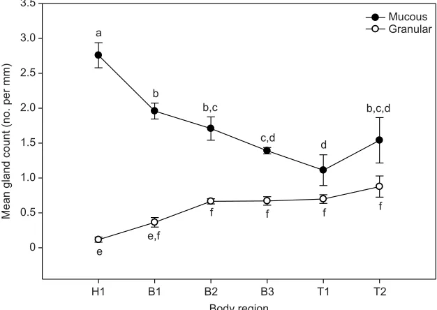
6=−4.993,P=0.002). The frequency (mean gland count) of mucous and granular glands varied across body regions. Mucous glands were significantly greater
in abundance in the head than in other body regions, while the number of granular glands was significantly greater in posterior body regions (body region×gland type interaction:F5,15=25.35,P<0.001) (Fig. 5). The two gland types were similarly distributed across all body planes (effect of body plane: F2,6=2.42, P=0.170; body plane×gland type interaction:F2,6=0.464,P=0.649).
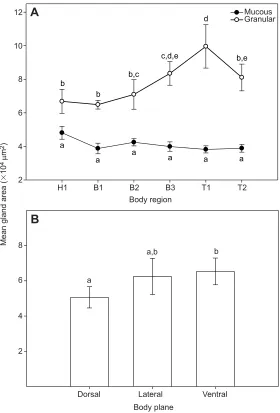
The size (mean gland area) of granular glands varied significantly across body regions (Fig. 6). Granular glands localized to posterior body regions were significantly larger than granular glands located within anterior regions. Conversely, the size of mucous glands did not significantly vary and was similar across all body regions (body region×gland type interaction: F5,15=11.607, P<0.001) (Fig. 6A). The size of granular and 0 50 100 150 200 250 300 350 400 450 500
Change in OD at 490 nm
(day 15 to day 0)
0 0.01 0.02 0.03 0.04 0.05
Bd + skin secretions
HK Bd + water (negative control) Calculated logistic curve
Adjusted R2=0.9950
MIC=75
A
Skin secretion concentration (µg ml–1)
0 50 100 150 200 250 300 350 400 450 500
0 0.02 0.04 0.06 0.08 0.10 0.12
Bsal + skin secretions
HK Bsal + water (negative control) Calculated sigmoidal curve
MIC=187
Adjusted R2=0.9783
B
5.2554
26.1956 1 +
0.0418
x
y=0.0008 +
y=–0.0016 +
–41.1002 1+e–
0.08861
[image:5.612.49.403.58.572.2]x+82.4299
Fig. 3. Growth inhibition of
Batrachochytrium dendrobatidisand Batrachochytrium salamandrivorans zoospores by skin secretions collected fromA. tridactylum.Data points for (A)B. dendrobatidis(Bd)+skin secretions and (B)B. salamandrivorans(Bsal)+skin secretions represent means±s.e. of three within-plate replicates. Data points for negative [heat-killed (HK) Bd and Bsal] and positive controls (skin secretion concentration=0 µg ml−1) represent means±s.e.m. of six within-plate replicates. Values for the minimal inhibitory concentration (MIC) were calculated as the lowest point where the upper 95% confidence limit of the negative control (horizontal dashed line) crossed the regression line. OD, optical density.
Journal
of
Experimental
mucous glands varied significantly across body planes in a similar manner (effect of body plane: F2,6=6.625, P=0.03; body plane×gland type interaction: F2,6=1.886, P=0.231) and was significantly smaller within the dorsal body plane than within the ventral plane (Fig. 6B).
Individual variation in granular gland size
Because of limitations due to sample size, statistical tests for differences in gland frequency or size based on sex or reproductive condition were not possible. However, the size of granular glands of the reproductively active male (voucher specimen SLU 774) exceeded that of non-active animals (voucher specimens SLU 1089, 02903 and 02904) in 13 out of the 18 skin pieces examined and peaked in skin pieces comprising the T1(skin pieces 5, 11 and 17) and H1 (skin piece 7) body regions (Fig. 7). Because granular gland size has been positively correlated with body size (i.e. SVL) (Holder and Glade, 1984; Saporito et al., 2010), it is noteworthy that the reproductively active male was one of the smallest specimens
included in this study. Therefore, our observations were unlikely to be a result of differences in specimen body size.
DISCUSSION
We demonstrated that skin secretions from a fully aquatic salamander, the three-toed amphiuma (A. tridactylum), inhibited the growth of Bd and Bsal, which are chytrid fungal pathogens linked to worldwide amphibian declines (Martel et al., 2013; Voyles et al., 2009). Furthermore, this is the first study to show that amphibian-derived skin secretions inhibit the growth of Bsal. We also demonstrated that granular glands were larger and more frequent in caudal body regions, suggesting that caudal granular glands may serve specialized functions. Below, we discuss each main result in turn.
Antimicrobial skin secretions
Skin secretions collected from captiveA. tridactylumexhibited antimicrobial activity against the chytrid fungal pathogens
MG
A
B
C
GG
ep
ss
ms
ep
ep
ss
GG GG MG MG
GG
MG ss
[image:6.612.59.552.58.239.2]500 µm 500 µm 500 µm
Fig. 4. Representative micrographs of histological sections ofA. tridactylumskin showing morphological and differential staining reactions of mucous and granular gland types.(A) Hematoxylin and Eosin; (B) periodic acid–Schiff, counterstained with Alcian Blue at pH 2.5; (C) Bromophenol Blue. MG, mucous glands; GG, granular glands; ep, epidermis; ss, stratum spongiosum (dermis); ms, myoepithelial sheath.
Body region
H1 B1 B2 B3 T1 T2 0
0.5 1.0
Mean gland count (no. per mm)
1.5 2.0 2.5 3.0 3.5
Mucous Granular a
b
b,c
c,d
d
b,c,d
e
e,f
f f f f
Fig. 5. Mean (±s.e.m.) gland counts of mucous and granular glands across body regions ofA. tridactylum.
n=4 salamanders for each body region. H1=head, B1–3=body, T1–2=tail. Points with the same letters (a–f ) are not statistically different.
Journal
of
Experimental
[image:6.612.48.361.511.733.2]Bd and Bsal. While research has primarily focused on anti-chytrid properties of secreted AMPs, a number of other antimicrobial compounds have also been described from amphibian skin secretions (Dahham et al., 2016; Mina et al., 2015; Zhao et al., 2006). The methods we used to process the crude skin secretions of A. tridactylum enriched for AMPs but may have included other compounds containing hydrophobic moieties, including larger proteins. While we hypothesize that AMPs present in the skin secretions ofA. tridactylumare likely responsible for thein vitro growth inhibition of Bd and Bsal, further analyses (e.g. mass spectrometry, Edmund degradation) are required to identify the specific molecular nature of the antimicrobial compounds within the skin secretions ofA. tridactylum(Rollins-Smith et al., 2002b).
Analysis by RP-HPLC, which separates compounds according to hydrophobicity, revealed that the skin secretions ofA. tridactylum are composed of a mixture of several compounds that were similar between the male and female animals and were dominated by a single compound type. Because absorbance was measured at wavelengths appropriate for the detection of peptide bonds and histological examination revealed the proteinaceous nature of granular gland secretions, these compounds likely represent peptides or proteins. Our results are consistent with those of Woodhams et al. (2006b), who reported that skin secretions of
Panamanian amphibians consisted of 5–25 peptides with little intraspecific variation. Our results from captive animals are likely representative of those in the wild because captivity has been shown to have little effect on the types of peptides synthesized and secreted by amphibian granular glands (Tennessen et al., 2009).
Skin secretions ofA. tridactyluminhibited the growth of Bd at a MIC estimated at 75 µg ml−1. MIC values against Bd have been estimated for a number of amphibian taxa and have been related to species’susceptibility to chytridiomycosis (Sheafor et al., 2008; Woodhams et al., 2007). For example, frogs that survived natural or experimental infections with Bd (i.e. have low susceptibility) generally had MIC values below 280 µg ml−1 (Woodhams et al., 2006a). In salamanders, MIC values ranging from 386 to 740 µg ml−1 have been estimated for species that generally survive infections by Bd (Sheafor et al., 2008). The low estimated MIC suggests that the skin secretions ofA. tridactylumare highly potent against Bd and may partially explain why chytridiomycosis has not been observed among amphiumid salamanders in the field despite the high prevalence of Bd infection among wild populations (Chatfield et al., 2012). Because it remains unknown whether amphiumid salamanders survive experimental infections with Bd, additional studies are needed to better understand the roles of skin secretions in protecting individuals from chytridiomycosis.
Body region
H1 B1 B2 B3 T1 T2
Mean gland area (
⫻
10
4 µ
m
2)
10
12 Mucous
Granular
a
a
a
a a a
b
b
b,c
c,d,e d
b,e
A
Body plane
Dorsal Lateral Ventral a
a,b b
B
8
6
4
2
8
6
4
[image:7.612.51.331.55.468.2]2
Fig. 6. Mean (±s.e.m.) gland area of mucous and granular glands ofA. tridactylum.(A) Area across body regions and (B) combined area of mucous and granular glands per body plane. n=4 salamanders for each body region and body plane. Points with the same letters (a–e) are not statistically different. H1=head, B1–3=body, T1–2=tail.
Journal
of
Experimental
1 2 3 4 5 6 10
12 M*
F M F
7 8 9 10 11 12
10 12 14 16
Skin piece
13 14 15 16 17 18
Head Tail
Dorsal
Lateral
Ventral 10
12 14 16
Mean
granular
gland area (
⫻
10
4 µ
m
2)
8
6
4
2
8
6
4
8
6
[image:8.612.51.354.75.716.2]4
Fig. 7. Comparison of mean granular gland area across skin pieces of reproductively active (*) and non-active male and femaleA. tridactylum.Skin pieces were collected from the dorsal (1–6), lateral (7–12) and ventral (13–18) body ofA. tridactylumand are arranged in increasing numerical order from head to tail for each set.
Journal
of
Experimental
Skin secretions ofA. tridactylumwere also effective against the growth of Bsal at a MIC estimated at 187 µg ml−1. Similar to Bd, susceptibility to Bsal is species specific (Martel et al., 2014). Though the specific mechanisms by which skin secretions inhibit the growth of chytrid fungi are poorly understood, available data indicate that AMP bioactivity is most effective against zoospores (infective stage) (Rollins-Smith et al., 2002b). Because the zoospore ultrastructure of Bsal and that of Bd are highly similar (Martel et al., 2013), we hypothesize that AMPs effective against the growth of Bd may also provide protection against Bsal by limiting infection burden. However, further research is needed to test this hypothesis. Skin secretions ofA. tridactylumwere antimicrobial at both 26°C (Bd) and 15°C (Bsal), indicating that antimicrobial function was maintained over a broad thermal range. This is consistent with previous studies in which isolated AMPs, esculentin-2P and ranatuerin-2P collected from the skin secretions of anurans [Lithobates(=Rana) pipiens], maintained anti-viral activity over a thermal range of 0–26°C (Chinchar et al., 2001). Because esulentin-2P and ranatuerin-2P also exhibited fungicidal activity against Bd (Rollins-Smith et al., 2002a), it is possible that the skin secretions of amphiumid salamanders may similarly exhibit broad-spectrum antimicrobial activity against other viral, fungal and bacterial pathogens.
MIC equivalents are an important measure for comparing the effectiveness of skin secretions across different amphibian species and consider the total amount (mass) of skin secretions (e.g. peptides) released (Woodhams et al., 2006a). Because the peptide/ protein content (µg), as quantified by BCA protein assays, of the skin secretions collected from A. tridactylum (maximum 0.54 µg g−1 body mass) was considerably lower than amounts collected from other salamanders (183.4–973 µg g−1body mass) using similar methods (Woodhams et al., 2006b), and the captive animals used in the current study did not exude copious skin secretions commonly observed when handling wild-caught
amphiumid salamanders (K. E. Pereira, unpublished
observations), our estimates of MIC equivalents are likely underestimates. In anurans, noradrenaline (norepinephrine) triggered bulk peptide release in both captive and wild animals through stimulation of the sympathetic nervous system (Hoffman and Dent, 1978; Pask et al., 2013; Woodhams et al., 2006b). In newts (Notophthalmus viridescens), acetylcholine may have a similar effect (Hoffman and Dent, 1977). However, we were unable to induce skin peptide release by immersing A. tridactylum in 200 µmol l−1acetylcholine (K. E. Pereira, unpublished data). Future studies should determine a method for inducing peptide release from salamander skin glands to better estimate MIC equivalents.
Skin histology
Similar to the skin of other adult amphibians, mucous glands were the dominant gland type and lacked apparent specialization in adult A. tridactylum(Fujikura et al., 1988). In males of some plethodontid salamanders, clusters of enlarged submandibular mucous glands (‘mental glands’) form conspicuous patches in reproductively active individuals (Sever, 2003; Woodley, 1994). Mental glands are the only specialized mucous gland type known among salamanders and have rarely been described outside of the Plethodontidae (Sever, 2003; Wilburn et al., 2017). Likewise, mental glands have never been reported for members of the Amphiumidae and nor were they observed in the reproductively active male included in this study. While mucous glands ofA. tridactylumwere most abundant in the head, the consistent size of mucous glands across the entire body suggests that these glands lack specialization and serve more general roles inAmphiuma.Because amphiumid salamanders are fossorial,
the lubricative secretions of cephalically located mucous glands may facilitate head-first burrowing activities by providing protection from abrasive skin damage (Breckenridge and Murugapillai, 1974; Jared et al., 2018).
Granular glands were scattered throughout the body surface but were most abundant in posterior areas. In addition, enlarged granular glands were identified at the tail base. Together, these observations suggest the presence of both ordinary and specialized granular gland types inA. tridactylum.While ordinary or randomly scattered granular glands likely function in the synthesis of AMPs and other bioactive compounds, the specialized glands in the tail may have additional functions in predator defense (Heiss et al., 2009), nutrient storage (Williams and Larsen, 1986), or pheromone production important for mate attraction or territorial defense (Woodley, 2015, 2014). The largest granular glands were observed at the tail base of the reproductively active male specimen, highlighting a specific site of potential gland specialization in A. tridactylum. In other salamander species, concentrations of specialized granular gland types at the tail base function in social communication (Sever, 2003; Woodley, 2015). For example, in some species of plethodontid salamander, males possess specialized granular glands located at the dorsal tail base termed ‘caudal courtship glands’. These glands hypertrophy during the breeding season and are hypothesized to produce courtship pheromones (Rupp and Sever, 2018; Sever, 1989; Trauth et al., 1993). At the tail base of other salamanders, ventral granular glands (termed
‘post-cloacal glands’) synthesize chemosignals relevant to intraspecific interactions (Chouinard, 2012; Largen and Woodley, 2008). Although we did not see differences in the skin secretions of male and female A. tridactylum using RP-HPLC analysis, no animals used for the collection of skin secretions exhibited signs of reproductive activity (e.g. swollen cloaca). To better understand the reproductive biology and social behaviors of this elusive species, future studies are needed to resolve whether granular glands located at the tail base are sexually dimorphic or undergo seasonal secretory cycles similar to courtship glands described for other species (Rupp and Sever, 2018; Sever and Siegel, 2015).
Conclusions
Collectively, our findings provide novel insight on the functions of skin glands in aquatic salamanders of the Amphiumidae. The efficacy of skin secretions against the chytrid pathogens Bd and Bsal represents an important contribution towards understanding amphibian innate immune defenses against pathogens linked to worldwide amphibian declines. In addition to their roles in antimicrobial defense, we also present the first evidence of skin gland specialization in an amphiumid salamander. The next step is to further investigate whether the in vitrogrowth inhibition of Bsal by amphibian skin secretions is related toin vivohost susceptibility. Additional studies are needed to resolve the functions of caudal granular glands in A. tridactylumto better understand their reproductive patterns.
Acknowledgements
We thank Drs Rick and Pam Feldhoff (Department of Biochemistry and Molecular Biology, School of Medicine, University of Louisville) for help with the RP-HPLC analysis and also the Southeastern Louisiana University Vertebrate Museum for allowing access to the natural history collection.
Competing interests
The authors declare no competing or financial interests.
Author contributions
Conceptualization: K.E.P., B.I.C., D.M.S., C.L.F., J.A.P., S.K.W.; Methodology: K.E.P., B.I.C., D.M.S., C.L.F., J.A.P., D.B.W., S.K.W.; Formal analysis: K.E.P., D.B.W.;
Journal
of
Experimental
Investigation: K.E.P.; Writing - original draft: K.E.P., S.K.W.; Writing - review & editing: K.E.P., B.I.C., D.M.S., C.L.F., J.A.P., D.B.W., S.K.W.; Supervision: B.I.C., S.K.W.
Funding
This research was supported by the American Museum of Natural History (Theodore Roosevelt Memorial Fund), Southeastern Louisiana University, and Duquesne University’s Bayer School of Natural and Environmental Science.
References
Berger, L., Hyatt, A. D., Speare, R. and Longcore, J. E.(2005). Life cycle stages of the amphibian chytrid Batrachochytrium dendrobatidis.Dis. Aquat. Organ. 68, 51-63.
Breckenridge, W. and Murugapillai, R.(1974). Mucous glands in the skin of
Ichthyophis glutinosus (Amphibia: Gymnophiona). Ceylon J. Sci. Biol. Sci.
11, 43-52.
Brizzi, R., Delfino, G. and Pellegrini, R.(2002). Specialized mucous glands and their possible adaptive role in the males of some species ofRana(Amphibia, Anura).J. Morphol.254, 328-341.
Brodie, E. D., Jr and Smatresk, N. J.(1990). The antipredator arsenal of fire salamanders: spraying of secretions from highly pressurized dorsal skin glands.
Herpetologica46, 1-7.
Brodie, E. D., Jr, Ducey, P. K. and Baness, E. A.(1991). Antipredator skin secretions of some tropical salamanders (Bolitoglossa) are toxic to snake predators.Biotropica23, 58-62.
Chatfield, M. W. H., Moler, P. and Richards-Zawacki, C. L.(2012). The amphibian chytrid fungus,Batrachochytrium dendrobatidis, in fully aquatic salamanders from Southeastern North America.PLoS ONE7, e44821.
Chinchar, V. G., Wang, J., Murti, G., Carey, C. and Rollins-Smith, L.(2001). Inactivation of frog virus 3 and channel catfish virus by esculentin-2P and ranatuerin-2P, two antimicrobial peptides isolated from frog skin. Virology 288, 351-357.
Chouinard, A. J.(2012). Rapid onset of mate quality assessment via chemical signals in a woodland salamander (Plethodon cinereus).Behav. Ecol. Sociobiol. 66, 765-775.
Conlon, J. M.(2011). Structural diversity and species distribution of host-defense peptides in frog skin secretions.Cell. Mol. Life Sci.68, 2303-2315.
Conlon, J. M. and Sonnevend, A.(2010). Antimicrobial peptides in frog skin secretions.Antimicrob. Peptides Methods Protoc.618, 3-14.
Conlon, J. M., Reinert, L. K., Mechkarska, M., Prajeep, M., Meetani, M. A., Coquet, L., Jouenne, T., Hayes, M. P., Padgett-Flohr, G. and Rollins-Smith, L. A. (2013). Evaluation of the skin peptide defenses of the Oregon spotted frog Rana pretiosaagainst infection by the chytrid fungus
Batrachochytrium dendrobatidis.J. Chem. Ecol.39, 797-805.
Cuvier, G. J. L. F.(1827). Sur le genre de reptile batraciens, nomméAmphiuma, et sur une nouvelle espece de ce genre (Amphiuma tridactylum).Mem. Mus. Hist.,
Nat. Paris14, 1-14.
Dahham, S. S., Hew, C., Jaafar, I. and Gam, L.(2016). The protein profiling of asian giant toad skin secretions and their antimicrobial activity. Int. J. Pharmacy
Pharmaceutical Sci.8, 88-95.
Daly, J. W.(1995). The chemistry of poisons in amphibian skin.Proc. Natl Acad. Sci. USA92, 9-13.
Duellman, W. and Trueb, L.(1994).Biology of Amphibians. Baltimore; London: John Hopkins University Press.
Fujikura, K., Kurabuchi, S., Tabuchi, M. and Inoue, S.(1988). Morphology and distribution of the skin glands in Xenopus laevis and their response to experimental stimulations: morphology.Zoolog. Sci.5, 415-430.
Gomes, A., Giri, B., Saha, A., Mishra, R., Dasgupta, S. C., Debnath, A. and Gomes, A. (2007). Bioactive molecules from amphibian skin: their biological activities with reference to therapeutic potentials for possible drug development. Heiss, E., Natchev, N., Rabanser, A., Weisgram, J. and Hilgers, H.(2009). Three types of cutaneous glands in the skin of the salamandridPleurodeles waltl. A histological and ultrastructural study.J. Morphol.270, 892-902.
Hoffman, C. W. and Dent, J. N.(1977). Effects of neurotransmitters upon the discharge of secretory product from the cutaneous glands of the red-spotted newt.
J. Exp. Zool.202, 155-161.
Hoffman, C. W. and Dent, J. N.(1978). The morphology of the mucous gland and its responses to prolactin in the skin of the red-spotted newt.J. Morphol.157, 79-97. Holder, N. and Glade, R.(1984). Skin glands in the axolotl: the creation and
maintenance of a spacing pattern.Development79, 97-112.
Jared, C., Mailho-Fontana, P. L., Marques-Porto, R., Sciani, J. M., Pimenta, D. C., Brodie, E. D. and Antoniazzi, M. M. (2018). Skin gland concentrations adapted to different evolutionary pressures in the head and posterior regions of the caecilianSiphonops annulatus.Sci. Rep.8, 3576. Kiernan, J.(1990). Carbohydrate histochemistry.Histological and Histochemical
Methods - Theory and Practice, pp. 274-306. Banbury: Scion Publishing Ltd. Largen, W. and Woodley, S. K.(2008). Cutaneous tail glands, noxious skin
secretions, and scent marking in a terrestrial salamander (Plethodon shermani).
Herpetologica64, 270-280.
Longcore, J. E., Pessier, A. P. and Nichols, D. K.(1999).Batrachochytrium
dendrobatidisgen. et sp. nov., a chytrid pathogenic to amphibians.Mycologia
91, 219-227.
Martel, A., Spitzen-van der Sluijs, A., Blooi, M., Bert, W., Ducatelle, R., Fisher, M. C., Woeltjes, A., Bosman, W., Chiers, K., Bossuyt, F. et al.(2013).
Batrachochytrium salamandrivoranssp. nov. causes lethal chytridiomycosis in
amphibians.Proc. Natl. Acad. Sci. USA110, 15325-15329.
Martel, A., Blooi, M., Adriaensen, C., Van Rooij, P., Beukema, W., Fisher, M. C., Farrer, R. A., Schmidt, B. R., Tobler, U., Goka, K. et al.(2014). Recent introduction of a chytrid fungus endangers Western Palearctic salamanders.
Science346, 630-631.
Mina, A. E., Ponti, A. K., Woodcraft, N. L., Johnson, E. E. and Saporito, R. A. (2015). Variation in alkaloid-based microbial defenses of the dendrobatid poison
frogOophaga pumilio.Chemoecology25, 169-178.
Pask, J. D., Cary, T. L. and Rollins-Smith, L. A.(2013). Skin peptides protect juvenile leopard frogs (Rana pipiens) against chytridiomycosis.J. Exp. Biol. 216, 2908-2916.
Petranka, J. W. (1998). Salamanders of the United States and Canada. Washington, DC: Smithsonian Institution Press.
Powell, R., Conant, R. and Collins, J. T.(2016).Peterson Field Guide to Reptiles and Amphibians of Eastern and Central. North America: Houghton Mifflin Harcourt.
Presnell, J. K. and Schreibman, M. P. (1997). Humason’s Animal Tissue Techniques. Baltimore: Johns Hopkins University Press.
Pukala, T. L., Bowie, J. H., Maselli, V. M., Musgrave, I. F. and Tyler, M. J.(2006). Host-defence peptides from the glandular secretions of amphibians: structure and activity.Nat. Prod. Rep.23, 368-393.
Rollins-Smith, L. A., Carey, C., Longcore, J., Doersam, J. K., Boutte, A., Bruzgal, J. E. and Conlon, J. M.(2002a). Activity of antimicrobial skin peptides from ranid frogs againstBatrachochytrium dendrobatidis, the chytrid fungus associated with global amphibian declines.Dev. Comp. Immunol.26, 471-479. Rollins-Smith, L. A., Reinert, L. K., Miera, V. and Conlon, J. M.(2002b).
Antimicrobial peptide defenses of the Tarahumara frog,Rana tarahumarae.
Biochem. Biophys. Res. Commun.297, 361-367.
Rose, F. L.(1967). Seasonal changes in lipid levels of the salamanderAmphiuma
means.Copeia1967, 662-666.
Rupp, A. E. and Sever, D. M.(2018). Histology of mental and caudal courtship glands in three genera of plethodontid salamanders (Amphibia: Plethodontidae).
Acta Zool.99, 20-31.
Saporito, R. A., Isola, M., Maccachero, V. C., Condon, K. and Donnelly, M. A. (2010). Ontogenetic scaling of poison glands in a dendrobatid poison frog.J. Zool. 282, 238-245.
Sever, D. M. (1976). Induction of secondary sexual characters in Eurycea
quadridigitata.Copeia1976, 830-833.
Sever, D. M. (1989). Caudal hedonic glands in salamanders of the Eurycea
bislineatacomplex (Amphibia: Plethodontidae).Herpetologica45, 322-329.
Sever, D. M. (1992). Comparative anatomy and phylogeny of the cloacae of salamanders (Amphibia: Caudata). III. Amphiumidae.J. Morphol.211, 63-72. Sever, D. M.(ed.) (2003). Courtship and mating glands. InReproductive Biology and
Phylogeny of Urodela, pp. 323-332. Boca Raton, FL: CRC Press.
Sever, D. M. and Siegel, D. S.(2015). Histology and ultrastructure of the caudal courtship glands of the red-backed salamander, Plethodon cinereus (Amphibia: Plethodontidae).J. Morphol.276, 319-330.
Sheafor, B., Davidson, E. W., Parr, L. and Rollins-Smith, L.(2008). Antimicrobial peptide defenses in the salamander,Ambystoma tigrinum, against emerging amphibian pathogens.J. Wildl. Dis.44, 226-236.
Tennessen, J. A., Woodhams, D. C., Chaurand, P., Reinert, L. K., Billheimer, D., Shyr, Y., Caprioli, R. M., Blouin, M. S. and Rollins-Smith, L. A.(2009). Variations in the expressed antimicrobial peptide repertoire of northern leopard frog (Rana pipiens) populations suggest intraspecies differences in resistance to pathogens.Dev. Comp. Immunol.33, 1247-1257.
Trauth, S. E., Smith, R. D., Cheng, A. and Daniel, P.(1993). Caudal hedonic glands in the dark-sided salamander, Eurycea longicauda melanopleura (Urodela: Plethodontidae).J. Arkansas Acad. Sci.47, 151-153.
Voyles, J., Young, S., Berger, L., Campbell, C., Voyles, W. F., Dinudom, A., Cook, D., Webb, R., Alford, R. A., Skerratt, L. F. et al.(2009). Pathogenesis of chytridiomycosis, a cause of catastrophic amphibian declines. Science 326, 582-585.
Whitford, W. G. and Hutchison, V. H.(1967). Body size and metabolic rate in salamanders.Physiol. Zool.40, 127-133.
Wilburn, D. B., Doty, K. A., Chouinard, A. J., Eddy, S. L., Woodley, S. K., Houck, L. D. and Feldhoff, R. C.(2017). Olfactory effects of a hypervariable multicomponent pheromone in the red-legged salamander, Plethodon shermani.
PLoS ONE12, e0174370.
Williams, T. A. and Larsen, J. H.(1986). New function for the granular skin glands of the eastern long-toed salamander,Ambystoma macrodactylum columbianum.
J. Exp. Zool. A Ecol. Genet. Physiol.239, 329-333.
Wilson, F.(1940). The life cycle of Amphiuma in the vicinity of New Orleans based on a study of the gonads and gonaducts.Anat Rec Suppl78, 104.
Journal
of
Experimental
Woodhams, D. C., Rollins-Smith, L. A., Carey, C., Reinert, L., Tyler, M. J. and Alford, R. A.(2006a). Population trends associated with skin peptide defenses against chytridiomycosis in Australian frogs.Oecologia146, 531-540. Woodhams, D. C., Voyles, J., Lips, K. R., Carey, C. and Rollins-Smith, L. A.
(2006b). Predicted disease susceptibility in a Panamanian amphibian assemblage based on skin peptide defenses.J. Wildl. Dis.42, 207-218. Woodhams, D. C., Ardipradja, K., Alford, R. A., Marantelli, G., Reinert, L. K. and
Rollins-Smith, L. A. (2007). Resistance to chytridiomycosis varies among amphibian species and is correlated with skin peptide defenses.Anim. Conserv. 10, 409-417.
Woodley, S. K. (1994). Plasma androgen levels, spermatogenesis, and secondary sexual characteristics in two species of plethodontid
salamanders with dissociated reproductive patterns. Gen. Comp.
Endocrinol.96, 206-214.
Woodley, S. K.(2014). Chemical signaling in amphibians. InNeurobiology of
Chemical Communication(ed. C. Mucignat-Caretta), pp. 255-284. Boca Raton,
FL: CRC Press.
Woodley, S.(2015). Chemosignals, hormones, and amphibian reproduction.Horm.
Behav.68, 3-13.
Xu, X. and Lai, R.(2015). The chemistry and biological activities of peptides from amphibian skin secretions.Chem. Rev.115, 1760-1846.
Zhao, Y., Jin, Y., Lee, W.-H. and Zhang, Y.(2006). Purification of a lysozyme from skin secretions ofBufo andrewsi.Comp. Biochem. Physiol. C Toxicol. Pharmacol. 142, 46-52.