Original Article
Expression of Nemo-like kinase was increased and
negatively correlated with the expression of
TCF4 in lung cancers
Xiu-Wei Zhang1, Song-Yan Chen2, Dong-Wei Xue3, Hui-Hui Xu1, Lian-He Yang2, Hong-Tao Xu2, En-Hua Wang2
1Department of Pathology, The Fourth Affiliated Hospital of China Medical University, Shenyang, China; 2Department of Pathology, The First Affiliated Hospital and College of Basic Medical Sciences of China Medical
University, Shenyang 110001, China; 3Department of Urinary Surgery, The Fourth Affiliated Hospital of China
Medical University, Shenyang, China
Received September 20, 2015; Accepted October 24, 2015; Epub November 1, 2015; Published November 15, 2015
Abstract: Nemo-like kinase (NLK), as a mitogen activated protein kinase (MAPK)-like kinase, is involved in the devel-opment of several human cancers. In this study, we explored the expression of NLK in lung squamous cell carcinoma (SCC) and adenocarcinoma tissues, and investigated the associations among NLK, β-catenin, T-cell factor 4 (TCF4), and the clinicopathological factors of lung cancers. The expressions of NLK, β-catenin, TCF4 were examined in 109 cases of lung cancers using immunohistochemistry method. The expression of NLK was observed in the nuclei of lung cancer tissues, and was significantly higher in lung cancer tissues than that in corresponding normal lung tis -sues (t = 21.636, n = 109, P < 0.001). The high expression of NLK was found in 45 cases of lung SCCs (45/49, 91.84%), which was much more than that in adenocarcinomas (38/60, 63.33%) (P = 0.001). Furthermore, the high expression of NLK was negatively correlated with TCF4 expression and positively correlated with the membranous expression of β-catenin. In conclusion, the present study demonstrated that the expression of NLK was localized in nucleus and significantly increased in lung cancers. The expression of NLK was negatively correlated with TCF4 expression and positively correlated with β-catenin membranous expression in lung cancers.
Keywords: Nemo-like kinase, T-cell factor 4, β-catenin, wnt signaling pathway, lung neoplasm
Introduction
Nemo-like kinase (NLK) is discovered as an evolutionarily conserved protein kinase from worms to humans. Similar to mitogen activated protein kinases/extracellular-signal regulated kinases (MAPKs/ERKs), NLK belongs to a pro-line-directed serine/threonine protein kinase superfamily [1, 2]. The substrates of NLK include several transcription factors, Such as T-cell factor/lymphoid enhancer factor (TCF/ LEF), c-Myb and STAT3 [3-6]. NLK can phos-phorylate TCF/LEF transcription factors and inhibit TCF/LEF-mediated transcription. So, NLK is considered a negative regulator of Wnt signaling pathway [2-4]. But, recent study found that NLK also functions as a positive regulator of Wnt signaling pathway by phosphorylating LEF1 in neural progenitor cells [7]. NLK is also considered a negative regulator of Notch
sig-naling pathway, because NLK inhibits Notch1 intracellular domain (Notch1-ICD) mediated gene expression by phosphorylating Notch1-ICD [8]. NLK interacts with p53 and enhances p53 activity and stability by blocking MDM2-mediated p53 ubiquitination and degradation [9]. NLK phosphorylates the C-terminal domain of CREB binding protein (CBP)/p300, and sup-presses the transcription activity of NF-κB, AP-1, Smad, and p53, all of which utilize CBP as a co-activator [10].
sion of NLK can decrease the expression of androgen receptor as well as inhibited andro-gen receptor-mediated transcription [12]. The low expression of NLK was correlated with glio-ma grade and poor patient outcome [13]. Recent study showed that NLK was significantly downregulated in the breast cancer tissues and was negatively correlated with c-Myb expression. NLK suppressed proliferation, induced apoptosis in breast cancer MCF-7 cells [15]. But, on the contrary, some studies revealed that NLK was associated with the pro-gression of some cancers. NLK was overex-pression in nasopharyngeal carcinomas [16], hepatocellular carcinomas [17, 18] and gall-bladder cancers [19, 20]. The positive expres-sion of NLK was associated with cancer pro-gression and shorter survival in nasopharyn-geal carcinomas [16] and gallbladder cancers [19, 20]. Knockdown of NLK can suppress cell growth and arrested cell cycle transition of liver cancer cells [17, 18], and enhance the sensitiv-ity to Taxol treatment in laryngeal cancer cells [21]. It is unclear why NLK plays different role in diverse cancers. The mechanism of NLK regu-lating cancer progression needs to be add- ressed.
Lung cancer is one of the most common can-cers in the world. The expression and function of NLK in lung cancers is unclear. In this study, we explored the expression of NLK in lung squamous cell carcinoma (SCC) and adenocar-cinoma tissues, and investigated the associa-tions among NLK, β-catenin, TCF4, and the clinicopathological factors of lung cancers, in order to gain an understanding of the role of NLK in lung cancer progression.
Materials and methods
Patients and tissue specimens
In this study, 109 lung SCC and adenocarcino-ma samples and corresponding noradenocarcino-mal lung tis-sues were obtained from patients selected ran-domly from a population that underwent sur-gery in the First Affiliated Hospital of China Medical University between 2009 and 2013. The patients included in the study were 63 men and 46 women, and aged 34-81 years with a mean age of 61 years. The histological diagno-sis and grade of differentiation were assessed by examination of hematoxylin-eosin (H/E)-stained sections according to the classification
system of the World Health Organization. The tumors were diagnosed as SCCs (n = 49) or adenocarcinomas (n = 60). These tumors were classified as well-differentiated (n = 27), mod-erately differentiated (n = 58), or poorly differ-entiated (n = 24) tumors. Forty-seven cases showed lymphatic metastasis.The tumor stage was classified as stages I-III (n = 52, 45 and 12, respectively) according to the TNM classifica-tion system of the Internaclassifica-tional Union Against Cancer. The study was conducted according to the regulations of the institutional review boards at China Medical University.
Immunohistochemistry
All resected specimens were embedded in par-affin blocks after fixed with 10% neutral-buff-ered formalin for 24 hours. Tissue blocks were cut into 4-μm sections. These sections were deparaffinized, rehydrated. The antigen retriev-al was performed by pressure cooking in a PH 6 citrate buffer for 1.5 min. Then, the sections were incubated with polyclonal rabbit anti-NLK antibody (sc-48361; 1:200; Santa Cruz Biotechnology Inc., CA), monoclonal mouse anti-β-catenin antibody (610154; 1:200; BD Transduction Laboratories, KY) or polyclonal rabbit anti-TCF4 antibody (sc-13027; 1:200; Santa Cruz Biotechnology Inc., CA) at 4°C over-night. The detection of antibodies was per-formed using the streptavidin-peroxidase me- thod. Some slides were stained in the absence of primary antibodies and served as negative controls.
Evaluation of immunostaining
nega-tive, weak, moderate, or marked, respectively. Scores from each tumor sample were multi-plied to give a final score of 0 to 12, and the tumors were finally determined, based on scores ≤ 6, and ≥ 8 as having low or high cyto-plasmic expression, respectively. When ≥ 10% of the tumor cells per specimen were stained in the nuclear or membranous regions, the sam-ple was scored as having positive nuclear or membranous expression of β-catenin.
Statistical analysis
The paired-samples t-test was used to analyze the differences in expression of NLK between the lung cancer tissues and the corresponding normal lung tissues. The Pearson Chi-Square test or the Likelihood-Ratio test was used to assay whether the expression levels of NLK, β-catenin or TCF4 were related to the
clinico-pathological factors of lung cancers. The Spearman’s correlation test was used to exam-ine the correlation among NLK, β-catenin and TCF4 levels. P values less than 0.05 were con-sidered statistically significant.
Results
The nuclear expression of NLK was increased in lung cancers than in corresponding normal lung tissues
[image:3.629.100.531.81.410.2]In this study, the expression of NLK was observed in the nuclei of lung cancer cells or normal lung epithelial cells (Figure 1). The aver-age expression rate of NLK was 63.53% ± 24.50% in lung cancer tissues, which was sig-nificantly higher than that in corresponding nor-mal lung tissues (10.90% ± 11.75%) (t = 21.636, n = 109, P < 0.001). Eight-three cases
lung cancers (P = 0.001), but was not associ-ated with the histological type (P = 0.052), TNM stage (P = 0.270), lymphatic metastasis (P = 0.055), or patients’ sex (P = 0.211) and age (P = 0.635). The expression of TCF4 was not asso-ciated with any of the clinicopathological fac-tors (data not shown). Furthermore, Spearman’s correlation test revealed that the membranous expression of β-catenin was positively correlat-ed with the cytoplasmic expression of β-catenin (Correlation coefficient = 0.268, P = 0.005), and negatively correlated with the nuclear expression of β-catenin (Correlation coefficient = 0.237, P = -0.013).
Discussion
As a MAPK-like proline-directed serine/threo-nine protein kinase, NLK can phosphorylate many transcription factors, including TCF/LEF, c-Myb, STAT3 and Notch1-ICD [3-6, 8]. So, NLK can regulate the activities of some signaling pathways, such as Wnt signaling pathway and Notch signaling pathway, and take part in the carcinogenesis and development of cancers [4, 7, 8, 22]. NLK is primarily considered as an anti-cancer factor by inhibiting the transcription activity of TCF/LEF-β-catenin complex [2, 23]. The expression of NLK was reduced in breast cancers and gliomas [13, 15]. Upregulating the expression of NLK induced apoptosis in colon cancers, prostate cancers and gliomas [11-13]. But, surprisingly and interestingly, the overex-pression of NLK was also found in nasopharyn-of lung cancers were scored as high expression
of NLK (83/109, 76.15%), meanwhile, only 14 cases of corresponding normal lung tissues were scored as high expression of NLK (14/109, 12.84%).
The high expression of NLK was negatively correlated with TCF4 expression and positively correlated with the membranous expression of β-catenin
We examined the expression of NLK, TCF4 and β-catenin in lung cancers and investigated the correlations among them. The Spearman’s cor-relation test revealed that the expression of NLK was negatively correlated with the expres-sion of TCF4 (Correlation coefficient = -0.222, P = 0.020). Furthermore, the expression of NLK was also correlated with the membranous expression of β-catenin (Correlation coefficient = 0.297, P = 0.002), but was not correlated with the abnormal cytoplasmic expression of β-catenin (Correlation coefficient = 0.065, P = 0.501) or nuclear expression of β-catenin (Correlation coefficient = -0.107, P = 0.269) (Table 1).
The high expression of NLK was more com-mon in lung SCCs than in adenocarcinomas
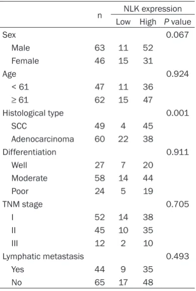
After immunostaining, we examined the asso-ciations between the expression of NLK and clinicopathological factors in lung cancers. The high nuclear expression of NLK was observed in 45 cases of lung SCCs (45/49, 91.84%),
which was much more than that in adenocarcinomas (38/- 60, 63.33%) (P = 0.001). But,the expression of NLK was not associated with the differ-entiation (P = 0.911), TNM stage (P = 0.705), lymphatic metastasis (P = 0.493), or patients’ sex (P = 0.069) and age (P = 0.924) (Table 2).
The abnormal expression of β-catenin was associated with the differentiation of lung cancers, but TCF4 expression was associated with none of the clinicopathological factors
[image:4.629.99.383.105.300.2]The abnormal expression of β-catenin was associated with the poorly differentiation of
Table 1. The correlations between the expressions of NLK, TCF4 and β-catenin in lung cancers
n
NLK expression Low High Correlation coefficient P value
TCF4 expression -0.222 0.020
Low 42 5 37
High 67 21 46
β-catenin membranous expression 0.297 0.002
+ 30 1 29
- 79 25 54
β-catenin cytoplasmic expression 0.065 0.501
Low 28 8 20
High 81 18 63
β-catenin nuclear expression -0.107 0.269
+ 14 5 9
geal carcinomas [16] hepatocellular carcino-mas [17, 18] and gallbladder cancers [19, 20]. NLK was associated with the development of nasopharyngeal carcinomas and gallbladder cancers [16, 19]. The role of NLK in diverse cancers is contradictory. Resent study revealed that besides negatively regulating the Wnt pathway, NLK also could promote the transcrip-tion activatranscrip-tion of TCF/LEF [7]. In neural progeni-tor cells, NLK phosphorylates LEF1 at Thr-155 and Ser-166. The phosphorylation of LEF1 induces itself dissociating from histone deacet-ylase HDAC1, and activates the transcription of Wnt target genes. So, one possible reason for opposite function of NLK is that the phosphory-lation level and phosphoryphosphory-lation sites of TCF/ LEF might affect the function of NLK in differ-ent cells [2].
In this study, we examined the expression and correlations of NLK in lung cancer tissues. The expression of NLK was localized in nucleus and significantly increased in lung cancers than in corresponding normal lung tissues. The high expression of NLK was more common in SCCs than in adenocarcinomas. But, we failed to find
any correlations between the expression of NLK and clinicpathological factors of lung can-cers. Furthermore, we found that the expres-sion of NLK was negatively correlated with the expression of TCF4. This result confirmed that NLK could phosphorylates TCF4 and downregu-lates the level of TCF4, further inhibits the acti-vation of Wnt signaling pathway. Other study also demonstrated that NLK was mainly local-ized in the nuclei in some cancers [19, 24] and the expression of NLK was increased in can-cers than normal tissues [16-20]. But, our results were different to the previously study in lung cancers, which indicated that the expres-sion of NLK was low and localized in cytoplasm [23]. We considered this might because of the using of different antibody of NLK, which might detect different localization of NLK. Shaw-Hallgren et al. found that the cytoplasmic local-ization of NLK induces cell apoptosis in breast cancers. The phosphorylation of NLK at Lys 155 and Thr 286 leads nuclear localization of NLK. The interaction of nuclear NLK and Heat shock protein 27 further protects cancer cells from apoptosis [24]. So, the localization of NLK might be another reason for its opposite func-tion in diverse cancers. Interestingly, we found that the expression of NLK was correlated with the membranous expression of β-catenin in lung cancers. But, its mechanism was unclear. NLK might control Wnt signaling pathway by regulating the expression or localization of β-catenin. This hypothesis needs further study.
In conclusion, the present study demonstrated that the expression of NLK was localized in nucleus and significantly increased in lung can-cers. The expression of NLK was negatively related with TCF4 expression and positively cor-related with β-catenin membranous expression in lung cancers.
Acknowledgements
This study was supported by National Natural Science Foundation of China (Grant No. 813- 72497 to H.-T. Xu and Grant No. 81301930 to L.-H. Yang) and Program for Liaoning Excellent Talents in University (Grant No. LR2015067 to H.-T. Xu).
Disclosure of conflict of interest
[image:5.629.99.297.116.410.2]None.
Table 2. The correlations between NLK ex-pression and the clinicopathological param-eters of lung cancers
n Low HighNLK expressionP value
Sex 0.067
Male 63 11 52
Female 46 15 31
Age 0.924
< 61 47 11 36
≥ 61 62 15 47
Histological type 0.001
SCC 49 4 45
Adenocarcinoma 60 22 38
Differentiation 0.911
Well 27 7 20
Moderate 58 14 44
Poor 24 5 19
TNM stage 0.705
I 52 14 38
II 45 10 35
III 12 2 10
Lymphatic metastasis 0.493
Yes 44 9 35
Address correspondence to: Dr. Hong-Tao Xu, Department of Pathology, The First Affiliated Hospital and College of Basic Medical Sciences of China Medical University, Shenyang 110001, China. Tel: 86-24-83282248; Fax: 86-24-83282248; E-mail: [email protected]
References
[1] Brott BK, Pinsky BA and Erikson RL. Nlk is a murine protein kinase related to Erk/MAP ki-nases and localized in the nucleus. Proc Natl Acad Sci U S A 1998; 95: 963-968.
[2] Ishitani T and Ishitani S. Nemo-like kinase, a multifaceted cell signaling regulator. Cell Signal 2013; 25: 190-197.
[3] Ishitani T, Ninomiya-Tsuji J, Nagai S, Nishita M, Meneghini M, Barker N, Waterman M, Bowerman B, Clevers H, Shibuya H and Matsumoto K. The TAK1-NLK-MAPK-related pathway antagonizes signalling between beta-catenin and transcription factor TCF. Nature 1999; 399: 798-802.
[4] Ishitani T, Ninomiya-Tsuji J and Matsumoto K. Regulation of lymphoid enhancer factor 1/T-cell factor by mitogen-activated protein ki-nase-related Nemo-like kinase-dependent phosphorylation in Wnt/beta-catenin signal-ing. Mol Cell Biol 2003; 23: 1379-1389. [5] Kanei-Ishii C, Ninomiya-Tsuji J, Tanikawa J,
Nomura T, Ishitani T, Kishida S, Kokura K, Kurahashi T, Ichikawa-Iwata E, Kim Y, Matsumoto K and Ishii S. Wnt-1 signal induces phosphorylation and degradation of c-Myb pro-tein via TAK1, HIPK2, and NLK. Genes Dev 2004; 18: 816-829.
[6] Kojima H, Sasaki T, Ishitani T, Iemura S, Zhao H, Kaneko S, Kunimoto H, Natsume T, Matsumoto K and Nakajima K. STAT3 regu-lates Nemo-like kinase by mediating its inter-action with IL-6-stimulated TGFbeta-activated kinase 1 for STAT3 Ser-727 phosphorylation. Proc Natl Acad Sci U S A 2005; 102: 4524-4529.
[7] Ota S, Ishitani S, Shimizu N, Matsumoto K, Itoh M and Ishitani T. NLK positively regulates Wnt/ beta-catenin signalling by phosphorylating LEF1 in neural progenitor cells. EMBO J 2012; 31: 1904-1915.
[8] Ishitani T, Hirao T, Suzuki M, Isoda M, Ishitani S, Harigaya K, Kitagawa M, Matsumoto K and Itoh M. Nemo-like kinase suppresses Notch signalling by interfering with formation of the Notch active transcriptional complex. Nat Cell Biol 2010; 12: 278-285.
[9] Zhang HH, Li SZ, Zhang ZY, Hu XM, Hou PN, Gao L, Du RL and Zhang XD. Nemo-like kinase is critical for p53 stabilization and function in
response to DNA damage. Cell Death Differ 2014; 21: 1656-1663.
[10] Yasuda J, Yokoo H, Yamada T, Kitabayashi I, Sekiya T and Ichikawa H. Nemo-like kinase suppresses a wide range of transcription fac-tors, including nuclear factor-kappaB. Cancer Sci 2004; 95: 52-57.
[11] Yasuda J, Tsuchiya A, Yamada T, Sakamoto M, Sekiya T and Hirohashi S. Nemo-like kinase in-duces apoptosis in DLD-1 human colon cancer cells. Biochem Biophys Res Commun 2003; 308: 227-233.
[12] Emami KH, Brown LG, Pitts TE, Sun X, Vessella RL and Corey E. Nemo-like kinase induces apoptosis and inhibits androgen receptor sig-naling in prostate cancer cells. Prostate 2009; 69: 1481-1492.
[13] Cui G, Li Z, Shao B, Zhao L, Zhou Y, Lu T, Wang J, Shi X, Zuo G, Zhu W and Shen A. Clinical and biological significance of nemo-like kinase ex -pression in glioma. J Clin Neurosci 2010; 18: 271-275.
[14] Wang K, Wang X, Zou J, Zhang A, Wan Y, Pu P, Song Z, Qian C, Chen Y, Yang S and Wang Y. miR-92b controls glioma proliferation and inva-sion through regulating Wnt/beta-catenin sig-naling via Nemo-like kinase. Neuro Oncol 2013; 15: 578-588.
[15] Huang Y, Jiang Y, Lu W and Zhang Y. Nemo-like kinase associated with proliferation and apop-tosis by c-Myb degradation in breast cancer. PLoS One 2013; 8: e69148.
[16] Chen S, Ma Z, Chen X and Zhang J. Prognostic significance of nemo-like kinase in nasopha -ryngeal carcinoma. Mol Med Rep 2014; 10: 131-136.
[17] Shen Q, Bae HJ, Eun JW, Kim HS, Park SJ, Shin WC, Lee EK, Park S, Park WS, Lee JY and Nam SW. MiR-101 functions as a tumor suppressor by directly targeting nemo-like kinase in liver cancer. Cancer Lett 2013; 344: 204-211. [18] Jung KH, Kim JK, Noh JH, Eun JW, Bae HJ, Xie
HJ, Ahn YM, Park WS, Lee JY and Nam SW. Targeted disruption of Nemo-like kinase inhib-its tumor cell growth by simultaneous suppres-sion of cyclin D1 and CDK2 in human hepato-cellular carcinoma. J Cell Biochem 2010; 110: 687-696.
[19] Li M, Zhang S, Wang Z, Zhang B, Wu X, Weng H, Ding Q, Tan Z, Zhang N, Mu J, Yang J, Shu Y, Bao R, Wu W, Cao Y and Liu Y. Prognostic sig-nificance of nemo-like kinase (NLK) expression in patients with gallbladder cancer. Tumour Biol 2013; 34: 3995-4000.
[20] Tan Z, Li M, Wu W, Zhang L, Ding Q, Wu X, Mu J and Liu Y. NLK is a key regulator of proliferation and migration in gallbladder carcinoma cells. Mol Cell Biochem 2012; 369: 27-33.
taxol sensitivity in laryngeal cancer. Asian Pac J Cancer Prev 2014; 14: 7137-7141.
[22] Fernandes VM, Panchapakesan SS, Braid LR and Verheyen EM. Nemo promotes Notch-mediated lateral inhibition downstream of pro-neural factors. Dev Biol 2014; 392: 334-343. [23] Lv L, Wan C, Chen B, Li M, Liu Y, Ni T, Yang Y,
Cong X, Mao G and Xue Q. Nemo-like kinase (NLK) inhibits the progression of NSCLC via negatively modulating WNT signaling pathway. J Cell Biochem 2014; 115: 81-92.


![4 (3 Methoxyphenyl) 3 [2 (4 methoxyphenyl)ethyl] 1H 1,2,4 triazol 5(4H) one](data:image/gif;base64,R0lGODlhAQABAIAAAP///wAAACH5BAEAAAAALAAAAAABAAEAAAICRAEAOw==)