Original Article
Tumor cell nuclear diameter and CD30 expression as
potential prognostic parameter in patients with
extranodal NK/T-cell lymphoma, nasal type
Junshik Hong1, Sanghui Park2*, Hae Lim Baek1, Joo Hyun Jung3, Il Gyu Kang3, Sun Jin Sym1, Jinny Park1,
Jeong Yeal Ahn4, Eun Kyung Cho1, Seon Tae Kim3, Dong Bok Shin1, Jae Hoon Lee1*
1Department of Internal Medicine, Gachon University School of Medicine, Incheon, Korea; 2Department of Pathol-ogy, Ewha Womans University School of Medicine, Seoul, Korea; 3Department of Otorhinolaryngology-Head and Neck Surgery and 4Laboratory Medicine, Gachon University School of Medicine, Incheon, Korea. *Equal contribu-tors.
Received August 6, 2012; Accepted October 1, 2012; Epub October 20, 2012; Published October 30, 2012
Abstract: Extranodal natural killer/T-cell lymphoma, nasal type (nasal ENKTL) is a distinct clinicopathologic entity of lymphoid tumors with variable size and differentiation of tumor cells. Nasal ENKTL is related to infection of the tu-mor cells with Epstein-Barr virus (EBV) and virtually all cases contain monoclonal episomal EBV DNA and detectable EBV encoded small nuclear RNAs (EBERs). Several clinical factors are known for their relation to the prognosis, but histopathologic prognostic factors of nasal ENKTL have not yet been well established. We evaluated the prognostic value of the longest nuclear diameter of EBER+ tumor cells (NDTC) along with the result of CD30 expression. Twenty two patients with newly diagnosed nasal ENKTL were evaluated regarding clinicopathologic characteristics. NDTC
was measured using a computerized image analysis system. The results were expressed as the mean diameter of ≥ 50 cells in a patient. Median of the mean NDTC of the patients was 7.32 μm (5.15-11.27). Patients with larger mean NDTC (≥ 7.35 μm) had a poorer event-free survival (EFS) than those with smaller mean NDTC (<7.35 μm; p = 0.024) and had a tendency of inferior overall survival (OS) (p = 0.08). Patients with CD30 expression had a inferior EFS (p = 0.018) and OS (p = 0.011) compared those without CD30 expression. The NDTC of EBV infected tumor cell and CD30 expression had relation to survival in the current exploratory analysis.
Keywords: Extranodal NK/T-cell lymphoma, nasal type, epstein-barr virus, CD30, prognosis, nuclear diameter
Introduction
Extranodal natural killer/T-cell lymphoma, nasal type (nasal ENKTL) is usually derived from natural killer (NK) cells or, rarely, from cytotoxic T-cells. Characteristically, Epstein-Barr virus (EBV) latently infects tumor cells in nearly all nasal ENKTLs. Nasal ENKTL is com-mon in Asia and South America but is very rare in North America and Europe [1-3]. Nasal ENKTL most frequently presents in the upper aerodigestive tract (UAT); however, it can also involve variable extranodal sites [4]. The overall prognosis of this disease is poor because of frequent relapse or resistance to treatment [5, 6]. However, most patients initially have low international prognostic index (IPI) scores, as they usually present as a localized disease
involving the head and neck with good perfor-mance status [7, 8]. Therefore, evaluating risk
prognostic factors specific to nasal ENKTL is of
paramount importance for the appropriate management of the disease.
The cytologic spectrum of nasal ENKTL is very broad. Cells may be small, medium-sized, large, or anaplastic [4]. In most cases, the lymphoma is composed of medium-sized cells or a mixture of small and large cells [4]. The correlation between tumor size and genetic status or clini-cal outcome is not clear to date. In one study,
associated with p53 mutation [10], even
though these findings were not confirmed by
other study [11]. Here, we measured the nucle-ar diameter of tumor cells and CD30 expres-sion to evaluate their prognostic value.
Materials and methods
Study sample
Clinical data and biopsied tissue of the patients with nasal ENKTL treated at a single institution, Gachon University Gil Hospital (GUGH), between
February 2000 and June 2011 were retrospec -tively analyzed. The patients were included if
they had histologic confirmation of nasal ENKTL
according to the World Health Organization cri-teria [4]; the NK/T-cell type as proven by
immu-nohistochemical, flow cytometry, or EBV in situ
hybridization analysis. The enrolled patients were without any previous treatment for lym-phoma and received at least one of the follow-ing therapies: concurrent chemo-radiotherapy, chemotherapy alone, or radiotherapy alone. Patients with extranodal NK/T-cell lymphoma without lymphomatous lesion on the upper aerodigestive tract (UAT) were excluded from the analysis. The patients underwent pre-treat-ment staging evaluation, including computed tomography (CT) or magnetic resonance imag-ing (MRI) of the head and neck, chest radiogra-phy, abdominopelvic CT, upper endoscopy for the evaluation of upper aerodigestive tract, and bilateral bone marrow trephine biopsy. The Institutional Review Board of GUGH approved the acquisition, analysis, and reporting of patient data (grant number: GIRBA 2583). Measurement of EBER-positive tumor cells
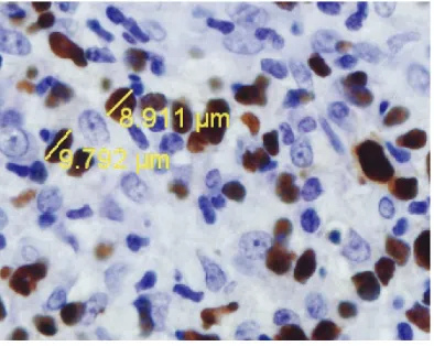
The nuclear diameter of EBER-positive tumor cells were measured using a computerized image analysis system (IMT i-Solution, Vancouver, British Columbia, Canada) that included a DP70 Digital camera (Olympus, Tokyo, Japan) installed on an Olympus BX51 light microscope and attached to a personal computer. More than 50 tumor cells were selected randomly for the measurement of the longest nuclear diameter of tumor cells (NDTC) in each case. The results of each patient were expressed as the mean diameter of the evalu-ated tumor cells. The mean diameter was used for statistical analysis.
Histopathologic examination and evaluation of EBV infection
Representative formalin-fixed paraffin-embed -ded tissues obtained from surgical resections or biopsies were submitted to immunohisto-chemistry and EBV study. Sections of the
paraf-fin-embedded tissues were cut at 4μm, placed on slides, deparaffinized in xylene, and hydrat -ed in a grad-ed series of alcohol. Sections were stained with antibody to CD30 (Dakopatts, Copenhagen, Denmark). Immunohistochemical
study was performed using a modified avidin-biotin peroxidase complex amplification and
detection system. CD30 expression was con-sidered positive when more than 50% of tumor cells showed strong membranous staining. EBV RNA was detected by an ISH (in situ
hybridiza-tion) technique. Paraffin sections were pre -treated with xylene followed by treatment with
proteinase K and hybridized with fluorescein
isothiocyanate-conjugated EBV oligonucle-otides (Novocastra, Newcastle, UK) comple-mentary to the mRNA portion of the EBER-1 and EBER-2 genes.
Statistical analyses
The relationships of mean NDTCs with clinical
variables were evaluated using Fisher’s exact test. Event-free survival (EFS) was defined as
survival free of progression, relapse, or death from lymphoma or treatment toxicity. Overall
survival (OS) was defined as survival free of
death from any cause. Survival was calculated using Kaplan-Meier method and compared by log rank test or Cox proportional hazard model.
Probability values < 0.05 were considered sig
-nificant. All values were two-sided and statisti
-cal significance was accepted at the P < 0.05
level. Results
Patient characteristics and treatment out-comes
patients (27.3%) had local tumor invasion (LTI),
which is defined as bony invasion or perforation
or invasion of the skin in a previous study [7]. Thirteen patients (59.1%) were Ann Arbor stage I or II. Most patients (16; 72.7%) received con-current chemo-radiotherapy whereas 5 and 1 received only chemotherapy and radiotherapy, respectively. Of the 20 evaluable patients, 14 patients responded to initial therapy (complete
remission in 9 patients, partial remission in 5, stable disease in 3, and progressive disease in 3). During the median follow-up duration of
19.9 months, 13 patients (59.1%) had signifi -cant events and 11 patients (50.0%) died (Ten patients were died of lymphoma progression and one for treatment-related mortality). The
median EFS was 9.7 months and EFS rate of
1-year and 3-year were 45.4% and 37.8%, respectively. The median OS was 28.2 months and OS rate of 1-year and 3-year were 75.9% and 43.6%, respectively.
Measurement of the nuclear diameter of tumor cells
Representative captured images are provided in Figure 1. The mean nuclear diameter of
EBER-positive tumor cells ranged from 5.15 μm to 11.27 μm among the 22 patients. The medi -an -and me-an value of the me-an NDTCs of the
patients were 7.32 μm, and 7.27 μm (± 1.30 as
standard deviation), respectively. Patients were separated into two groups according to the
[image:3.612.88.521.83.370.2]mean NDTC, larger NDTC (≥ 7.35 μm) and smaller NDTC group (<7.35 μm).
Table 1. Patient characteristics
Characteristics N %
Gender Male
Female 202 90.99.1
Age (year) Median
Range 15-8148
Primary site Nasal cavity (NC)
Nasopharynx or Oropharynx Both NC and naso- or oropharynx
15 5 2
68.2 22.7 9.1
Local tumor invasion No
Yes 166 72.727.3
ECOG performance status 0
1 2 3
5 12
4 1
22.7 54.5 18.2 4.5
Lactose dehydrogenase Not elevated
Elevated 139 59.140.9
B symptom No
Yes 184 81.818.2
Cervical lymph node(s) Not involved
Involved 139 59.140.9
Bone marrow involvement No
Yes 211 95.54.5
Splenomegaly at diagnosis No
Yes 1510 6040
Ann Arbor staging I
II III IV
7 6 3 6
31.8 27.3 13.6 27.3
Type of therapy Concurrent chemo-radiotherapy
Chemotherapy alone Radiotherapy alone
16 5 1
72.7 22.7 4.5
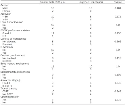
[image:3.612.92.289.409.566.2]Clinical feature and survival according to the nuclear diameter of tumor cells
The relationships of NDTC group with clinical characteristics are summarized in Table 2. No
statistically significant association was report -ed between NDTC group and clinical characteristics.
By Kaplan-Meier analysis, patients of larger
NDTC group had a inferior EFS (median 2.87 months, 95% confidence interval [CI]
2.67-3.06) compared to smaller group (median 43.37 months, 95% CI 14.10-72.6; p = 0.024 by log-rank test). Patients with larger NDTC group had a tendency of inferior OS compared to those with smaller NDTC group (p = 0.08 by log-rank test; Figure 2), although failed to reach
statistical significance.
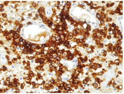
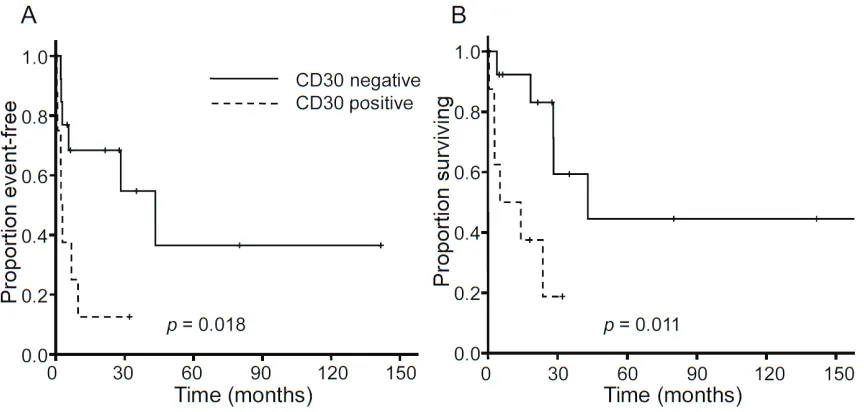
CD30 expression and patient survival
The membranous staining pattern of CD30 expression was noted. Of 22 cases, 8 (36.4%) cases were positive for CD30 (Figure 3). Patients with CD30 expression showed inferior
EFS (median 2.2 vs. 43.36 months, p = 0.018 by log-rank test) and OS (median 5.2 vs. 43.0 months, p = 0.011 by log-rank test; Figure 4). Univariate and multivariate analyses for event-free survival according to the clinicopathologic factors
In the univariate analysis, CD30 expression
and NDTC were associated with EFS along with
[image:4.612.88.523.83.449.2]age, LTI, type of therapy, performance status, and splenomegaly at the time of diagnosis (Table 3). Only LTI (Hazard Ratio [HR] 30.59, Table 2. Comparison of clinical characteristics according to nuclear diameter of tumor cells
Smaller cell (<7.35 μm) Larger cell (≥7.35 μm) P-value Gender
Male
Female 102 100 0.481
Age (year)
< 60
≥ 60 102 55 0.172
Local tumor invasion No
Yes 102 82 1.0
ECOG* performance status
0 and 1
≥ 2 111 64 0.135
Lactose dehydrogenase Not elevated
Elevated 84 55 0.666
B symptom No
Yes 102 82 1.0
Cervical lymph node(s) Not involved
Involved 66 73 0.415
Bone marrow involvement No
Yes 111 100 1.0
Splenomegaly at diagnosis No
Yes 93 46 0.192
Ann Arbor staging I and II
III and IV 93 55 0.378
Type of therapy CCRT†
Not CCRT 102 64 0.348
CD30 expression Yes
No 39 55 0.378
95% CI 3.69-253.5, p = 0.002), splenomegaly (HR 4.61, 95% CI 1.16-18.27, p = 0.03), and performance status (2.45, 95% CI 2.10-73.7, p
= 0.005) were associated with EFS in the multi -variate analysis. NDTC group and CD30
expres-sion did not show statistical significance in the
multivariate analysis, along with patient age
(>60 vs. ≤ 60 years) and type of therapy (con -current chemo-radiotherapy or not).
Discussion
Many clinical and pathological efforts have been made to identify prognostic markers in nasal ENKTL. Clinically, two major clinical prog-nostic models are applied in NK/T-cell lympho-ma: IPI and prognostic index for peripheral
T-cell lymphoma-unspecified (PIT). The IPI has
been widely used for both predicting prognosis and selecting therapeutic options in patients
with aggressive non-Hodgkin’s lymphoma.
However, its value has been challenged by nasal ENKTL because it has failed to predict prognosis in retrospective analyses [12, 13]. Recently, PIT has been carried out in other sub-types T-cell lymphoma and its improved prog-nostic value were recognized [14-16]. However, these prognostic models are based primarily on pre-treatment clinical characteristics; the path-ological or molecular factors that may predict the prognosis of nasal ENKTL have not yet been
well defined. Ki-67 proliferation rate was report -ed to be correlat-ed with a shorter disease-free
survival and OS (P < 0.05), while Ann Arbor
stage and IPI failed to predict prognosis of the patients with nasal ENKTL [17]. Recently, loss of the granzyme B protease inhibitor 9, cyclo-oxygenase-2 expression and decreased
quan-tity of tumor-infiltrating FOXP3-positive regula -tory T-cells have been associated with poor prognosis of nasal or UAT-ENKTL [18-20]. Nasal ENKTL is associated with various histo-logic changes. According to the predominant
lymphoma cells in the infiltrates, they could be classified into four histologic subtypes; small
cell type, medium-sized cell type, large cell type, and pleomorphic cell type [9]. The
majori-ty consists of polymorphous infiltrations of
small, medium, and large atypical lymphocytes
with accompanying inflammatory cells, plasma
cells, macrophages, and neutrophils. The
[image:5.612.95.515.77.273.2]asso-Figure 2. Kaplan-Meier curve of event-free survival and overall survival according to nuclear diameter of tumor cells (NDTC) group.
[image:5.612.91.290.318.470.2]ciation of tumor cell type with prognosis has not been clear until now. In one study, survival
analysis failed to demonstrate significant differ -ence among the histologic subtypes [9]. The neoplastic cells often consist only of small lym-phocytes lacking atypia and necrosis, and are easily overlooked, resulting in a misdiagnosis
as chronic inflammation [21]. We experienced
several cases of nasal ENKTL with favorable
prognosis that consisted of small infiltrating
lymphocytes lacking cytological atypia and necrosis. Therefore, we hypothesized that the small cell type of nasal ENKTL might be associ-ated with favorable prognosis and the current study showed that nasal ENKTL cases with
smaller size had favorable EFS. However, most
cases of nasal ENKTL consist of polymorphous
infiltrations of various tumor cells. Therefore, it
is nearly impossible to segregate histologic subtypes exactly. The authors measured the nuclear diameter of the EBER positive tumor cells as a surrogate for tumor cell size to avoid
subjective histologic classifications. EBV in situ
hybridization is a commonly used technique
that can be easily applied to formalin-fixed, par
-affin-embedded tissue sections and image
analyzer is also a commonly used computer-ized system. Therefore, these techniques allow for the differentiation of larger tumor cells from smaller tumor cells with convenience.
[image:6.612.93.525.70.276.2]CD30 (Ki-1) molecule is a member of the tumor necrosis factor receptor super-family and pref-erentially expressed in activated CD8+T-cell and NK-cell [22, 23]. In nasal ENKTL, the posi-tivity for CD30 has been reported in some spo-radic reports (range 20-64%) [24-28]. In one study (n = 30), patients with CD30 expression tend to have more favorable outcome [29], but other studies conducted by Kuo et al (n = 22) [9] and Gaal et al (n = 15) [24] revealed that there was no difference of survival according to CD30 expression, although it seemed to be that CD30 expression correlated with vascular Table 3. Prognostic factors affecting event-free survival in univariate and multivariate analysis
Univariate analysis
Parameters HR 95% CI P
Age (> 60 years) 4.97 1.33-18.58 0.017
Local tumor invasion (present) 10.4 2.24-48.19 0.003
Cervical nodes (involved) 1.62 0.54-4.90 0.389
ECOG* Performance status (2, 3, and 4) 11.86 2.73-51.58 0.001
CCRT† (not done) 3.99 1.30-12.22 0.015
Lactose dehydrogenase (elevated) 2.05 0.68-6.13 0.201
B symptom (present) 2.13 0.56-8.10 0.270
Splenomegaly (present) 4.63 1.37-15.66 0.014
CD30 (expressed) 3.68 1.16-11.74 0.027
NDTC‡ group (≥7.35 μm) 3.75 1.10-12.77 0.035
[image:6.612.91.524.318.430.2]*ECOG, Eastern Cooperative Oncology Group; †CCRT, concurrent chemo-radiotherapy; ‡NDTC, Nuclear diameter of tumor cells.
destruction, thrombosis, or pleomorphic cell type [9, 24]. In our study, patients with CD30
expression (≥ 50%) had inferior EFS and OS. Further study with larger patient number would define the actual relationship of CD30 expres -sion to the prognosis of nasal ENKTL.
Relatively small patient number can be a limita-tion of the current study. However, since the rar-ity of nasal ENKTL, not a few of the studies on pathologic or molecular prognostic markers on nasal ENKTL were performed with small num-bers (15 to 30) of patients [9, 10, 24, 29]. Several biomarker studies on nasal ENKTL had larger patient numbers but in those studies
patients’ tissues were collected over fifteen to even more than twenty-five years [18-20, 30];
in those studies, the development of optimal chemo-radiotherapy strategy, the advance of diagnostic technology including positron emis-sion tomography, and the improved supportive care could not be considered enough, opposite our study, which only dealt with patients in a decade recently.
Because of limited sample size, the result of multivariate analysis in the current study has only an auxiliary value and does not exclude the necessity of further investigation on NDTC and CD30 expression. Even multivariate analyses
shows conflict results according to the studies. For Example, elevated serum lactose dehydro -genase was an independent prognostic factor in several studies [6, 8, 30], but other studies do not support it [7, 12].
We measured the longest nuclear diameter of EBER-positive tumor cells for economy of time and it was the simplest method for evaluating
tumor cells as many as possible (≥ 50 cells in a
patient). Other parameters such as nuclear area or circumference may improve the prog-nostic value of tumor cell size in later studies.
Furthermore, the nuclear diameter of tumor cells in paraffin sections may vary from case to
case and there are many factors that could
affect the cell size in formalin-fixed and paraf
-fin-embedded tissue. When measuring the
mean NDTCs again within the case and among different cases, the values were well reproduc-ible. To use a size standard such as the size of red blood cells or nuclei of small lymphocytes might be better method to ensure the repro-ducibility in future studies.
In conclusion, the measurement of NDTC of EBV infected tumor cell and assessment of CD30 expression had relation to survival in the
current exploratory analysis. Further larger scale study to define the prognostic value of
them is warranted.
Address correspondence to: Dr. Jae Hoon Lee, Department of Internal Medicine, Gachon University School of Medicine, 21 Namdongdae-ro 774-gil, Namdong-gu, Incheon, 405-760, Republic of Korea.
Tel: +82-32-460-2186; Fax: +82-32-460-3233;
E-mail: [email protected]; Dr. Sanghui Park, Department of Pathology, Ewha Womans University School of Medicine, 911-1 Mok-dong, Yangcheon-gu, Seoul, Korea, 158-710, Republic of Korea; Tel:
+82-2-2650-5731; Fax: +82-2-2650-2879; E-mail:
References
[1] Aozasa K, Ohsawa M, Tajima K, Sasaki R,
Mae-da H, Matsunaga T and Friedmann I.
Nation-wide study of lethal mid-line granuloma in
Ja-pan: frequencies of wegener’s granulomatosis,
polymorphic reticulosis, malignant lymphoma and other related conditions. Int J Cancer 1989; 44: 63-66.
[2] Harris NL, Jaffe ES, Diebold J, Flandrin G,
Muller-Hermelink HK, Vardiman J, Lister TA
and Bloomfield CD. World Health Organization classification of neoplastic diseases of the he -matopoietic and lymphoid tissues: report of the Clinical Advisory Committee meeting-Airlie House, Virginia, November 1997. J Clin Oncol 1999; 17: 3835-3849.
[3] Jaffe ES, Chan JK, Su IJ, Frizzera G, Mori S, Feller AC and Ho FC. Report of the Workshop
on Nasal and Related Extranodal Angiocentric
T/Natural Killer Cell Lymphomas. Definitions,
differential diagnosis, and epidemiology. Am J Surg Pathol 1996; 20: 103-111.
[4] Swerdlow SH, Campo E, Harris NL, Jaffe ES, Pileri SA, Stein H, Thiele J, Vardiman JW and
editors. WHO Classification of Tumours of Hae -matopoietic and Lymphoid Tissue (IARC WHO
Classification of Tumours). Lyon: IARC Press;
2008.
[5] Kim GE, Cho JH, Yang WI, Chung EJ, Suh CO, Park KR, Hong WP, Park IY, Hahn JS, Roh JK and Kim BS. Angiocentric lymphoma of the head and neck: patterns of systemic failure af-ter radiation treatment. J Clin Oncol 2000; 18: 54-63.
Ko YH, Ahn YC and Park K. Extranodal nasal type NK/T-cell lymphoma: elucidating clinical
prognostic factors for risk-based stratification
of therapy. Eur J Cancer 2005; 41: 1402-1408. [7] Kim TM, Park YH, Lee SY, Kim JH, Kim DW, Im
SA, Kim TY, Kim CW, Heo DS, Bang YJ, Chang KH and Kim NK. Local tumor invasiveness is more predictive of survival than International Prognostic Index in stage I(E)/II(E) extranodal NK/T-cell lymphoma, nasal type. Blood 2005; 106: 3785-3790.
[8] Lee J, Suh C, Park YH, Ko YH, Bang SM, Lee JH, Lee DH, Huh J, Oh SY, Kwon HC, Kim HJ, Lee SI, Kim JH, Park J, Oh SJ, Kim K, Jung C, Park K and Kim WS. Extranodal natural killer T-cell lymphoma, nasal-type: a prognostic model from a retrospective multicenter study. J Clin Oncol 2006; 24: 612-618.
[9] Kuo TT, Shih LY and Tsang NM. Nasal NK/T cell lymphoma in Taiwan: a clinicopathologic study of 22 cases, with analysis of histologic sub-types, Epstein-Barr virus LMP-1 gene associa-tion, and treatment modalities. Int J Surg Pathol 2004; 12: 375-387.
[10] Quintanilla-Martinez L, Kremer M, Keller G, Na-thrath M, Gamboa-Dominguez A, Meneses A,
Luna-Contreras L, Cabras A, Hoefler H, Mohar A and Fend F. p53 Mutations in nasal natural
killer/T-cell lymphoma from Mexico: associa-tion with large cell morphology and advanced disease. Am J Pathol 2001; 159: 2095-2105. [11] Ng SB, Lai KW, Murugaya S, Lee KM, Loong SL,
Fook-Chong S, Tao M and Sng I. Nasal-type ex -tranodal natural killer/T-cell lymphomas: a clinicopathologic and genotypic study of 42 cases in Singapore. Mod Pathol 2004; 17: 1097-1107.
[12] Aviles A, Diaz NR, Neri N, Cleto S and Talavera A. Angiocentric nasal T/natural killer cell lym-phoma: a single centre study of prognostic fac-tors in 108 patients. Clin Lab Haematol 2000; 22: 215-220.
[13] Cheung MM, Chan JK, Lau WH, Ngan RK and
Foo WW. Early stage nasal NK/T-cell lympho -ma: clinical outcome, prognostic factors, and the effect of treatment modality. Int J Radiat Oncol Biol Phys 2002; 54: 182-190.
[14] Beltran B, Quinones P, Morales D, Cotrina E and Castillo JJ. Different prognostic factors for survival in acute and lymphomatous adult T-cell leukemia/lymphoma. Leuk Res 2011; 35: 334-339.
[15] Park BB, Ryoo BY, Lee JH, Kwon HC, Yang SH, Kang HJ, Kim HJ, Oh SY, Ko YH, Huh JR, Lee SS, Nam EM, Park KW, Kim JH, Kang JH, Bang SM, Park S, Kim K, Park K, Suh C and Kim WS. Clin-ical features and treatment outcomes of an-gioimmunoblastic T-cell lymphoma. Leuk Lym-phoma 2007; 48: 716-722.
[16] Rodriguez J, Conde E, Gutierrez A, Arranz R, Leon A, Marin J, Bendandi M, Albo C and Ca-ballero MD. The results of consolidation with autologous stem-cell transplantation in pa-tients with peripheral T-cell lymphoma (PTCL)
in first complete remission: the Spanish Lym -phoma and Autologous Transplantation Group experience. Ann Oncol 2007; 18: 652-657. [17] Kim SJ, Kim BS, Choi CW, Choi J, Kim I, Lee YH
and Kim JS. Ki-67 expression is predictive of prognosis in patients with stage I/II extranodal NK/T-cell lymphoma, nasal type. Ann Oncol 2007; 18: 1382-1387.
[18] Bossard C, Belhadj K, Reyes F, Martin-Garcia N, Berger F, Kummer JA, Briere J, Baglin AC,
Cheze S, Bosq J, Ribrag V, Gisselbrecht C, Mounier N and Gaulard P. Expression of the granzyme B inhibitor PI9 predicts outcome in nasal NK/T-cell lymphoma: results of a
West-ern series of 48 patients treated with first-line polychemotherapy within the Groupe d’Etude des Lymphomes de l’Adulte (GELA) trials.
Blood 2007; 109: 2183-2189.
[19] Kim WY, Jeon YK, Kim TM, Kim JE, Kim YA, Lee SH, Kim DW, Heo DS and Kim CW. Increased
quantity of tumor-infiltrating FOXP3-positive
regulatory T cells is an independent predictor for improved clinical outcome in extranodal NK/T-cell lymphoma. Ann Oncol 2009; 20: 1688-1696.
[20] Shim SJ, Yang WI, Shin E, Koom WS, Kim YB, Cho JH, Suh CO, Kim JH and Kim GE. Clinical
significance of cyclooxygenase-2 expression in
extranodal natural killer (NK)/T-cell lymphoma, nasal type. Int J Radiat Oncol Biol Phys 2007; 67: 31-38.
[21] Ham MF and Ko YH. Natural killer cell neo -plasm: biology and pathology. Int J Hematol 2010; 92: 681-689.
[22] Chiarle R, Podda A, Prolla G, Gong J, Thorbecke GJ and Inghirami G. CD30 in normal and neo-plastic cells. Clin Immunol 1999; 90: 157-164. [23] Horie R and Watanabe T. CD30: expression
and function in health and disease. Semin Im-munol 1998; 10: 457-470.
[24] Gaal K, Sun NC, Hernandez AM and Arber DA. Sinonasal NK/T-cell lymphomas in the United States. Am J Surg Pathol 2000; 24: 1511-1517.
[25] Ko YH, Ree HJ, Kim WS, Choi WH, Moon WS and Kim SW. Clinicopathologic and genotypic study of extranodal nasal-type natural killer/T-cell lymphoma and natural killer precursor lym-phoma among Koreans. Cancer 2000; 89: 2106-2116.
[27] Lu D, Lin CN, Chuang SS, Hwang WS and Huang WT. T-cell and NK/T-cell lymphomas in southern Taiwan: a study of 72 cases in a sin-gle institute. Leuk Lymphoma 2004; 45: 923-928.
[28] Schwartz EJ, Molina-Kirsch H, Zhao S, Mari-nelli RJ, Warnke RA and Natkunam Y. Immuno-histochemical characterization of nasal-type extranodal NK/T-cell lymphoma using a tissue microarray: an analysis of 84 cases. Am J Clin Pathol 2008; 130: 343-351.
[29] Mraz-Gernhard S, Natkunam Y, Hoppe RT, LeB-oit P, Kohler S and Kim YH. Natural
killer/natu-ral killer-like T-cell lymphoma, CD56+, present-ing in the skin: an increaspresent-ingly recognized entity with an aggressive course. J Clin Oncol 2001; 19: 2179-2188.