A Fast Saliency Based Iris Segmentation
Method for Colour Eye Images
Fazil Latheef 1, Robin Abraham 2
P.G. Student, Department of Electronics and Communication, ICET, Muvattupuzha, Kerala, India1
Associate Professor, Department of Electronics and Communication, ICET, Muvattupuzha, Kerala, India2
ABSTRACT: Iris recognition is regarded as the most reliable and accurate biometric identification system available. In Iris recognition system, iris segmentation is the most time consuming and critical step, in concern to the accuracy of the process. This is a new method of iris segmentation based on the saliency map of an image. In this paper, first the saliency map which provides the visually important region, has been designed to specifically locate the iris region. Then a colour based masking is done to get the iris boundary. Since this method is computationally simple, a fast and reliable method which segment eye has been proposed. The reliability of the method has been checked with different eye images. The result also show that the proposed model is faster compared to the existing methods and gives a moderate accuracy.
KEYWORDS:Iris recognition, iris segmentation, saliency map, visually important, masking. I. INTRODUCTION
Based on a unique characteristic of an individual a biometric system provides identification of an individual. Different features we consider for identification of a person are human speech, iris, finger print, face, retina etc. Among these methods the quality which makes iris recognition more important are its high degree of randomness, unchanging characteristics over time, small database size and low template matching time. This makes iris based identifications the most reliable and apt method to recognize an individual.
Iris recognition system captures an image of an individual’s eye; the iris in the image is then segmented and normalized for feature extraction process. The performance of iris recognition systems highly depends on segmentation. Segmentation is used to locate the correct iris region in an eye. This should be done accurately and correctly to remove the eyelids, reflection and eyelashes present in iris region. Also the accuracy and speed of the iris segmentation process is a critical factor in the iris recognition system.
The human visual system (HVS), receives a considerably large amount of information well beyond its capability to process all of it. To cope with large amounts of information, visual attention is one of the most important mechanisms deployed in the HVS to reduce the complexity of scene analysis. Visual attention would selectively process the important part by filtering out others to reduce the complexity of scene analysis. A saliency map of an image finds out the salient or important region of an image. So here in this paper the saliency map of the input eye image is computed to remove the eyelids and the eyelashes of the image. Then this saliency map is subjected to a colour masking for obtaining the iris boundary.
II. RELATEDWORK
The most popular iris recognition method was developed by Daugman[1-3]. He proposed an Integro-differential operator for locating the outer boundaries of iris, as well as the upper and lower eyelids. Wildes [5] used edge detection and Hough transform to localize the iris. Edge detector is applied to a grey scale iris image to generate the edge map. Gaussian filter is applied to smooth the image to select the proper scale of edge analysis. Many other researchers relied on these two techniques for locating the iris and extracting it from the others eye parts. In order to increase the pupil accuracy segmentation Kheirolahy [6] have used the optimized colour mapping to make the pupil region clear and easy to segment and they have achieved 98% accuracy by applying optimized colour mapping. Tisse et a [7]proposed a segmentation method based on integro-differential operators with a Hough Transform. This reduced the computation time and excluded potential centres outside of the eye image. Black hole search method[8] was proposed by Teo and Ewe to compute the centre and area of a pupil. Since the pupil is the darkest region in the image, this approach applies threshold segmentation method to find the dark areas in the iris image. Richard et al., developed an iris segmentation approach which was able compensate all four types of noises in order to achieve higher accuracy rate [9].
III.PRESENTATIONOFTHEMAINCONTRIBUTIONOFTHEPAPER
Most of the previous papers do not concentrated on minimising segmentation time but mainly focused on improving the accuracy. The previous algorithms are computationally complex and require high processing time. Hence these algorithms are not suitable for real-time processing of iris images. This paper considers reducing the segmentation time with almost no loss in accuracy. This paper presents a fast iris segmentation method for colour eye images. The proposed method does not require the implementation of complex relations for edge detection. So this method is computationally faster than the existing segmentation methods.
Previous studies had shown that every iris is not circular. So it is not necessary to approximate the iris boundary as a perfect circle. This paper also does not approximate the iris boundary as circular region as previous works did. Here a colour threshold masking is used to get the boundary of the iris. The segmentation also gives a good accuracy. Another main advantage of the method is that the texture and colour features extracted from the input image can be used for encoding or template creation, which is a further process in the iris recognition system. So a reliable and fast iris segmentation framework for fast segmentation is proposed.
IV.THE PROPOSED FRAMEWORK
Fig1. Basic block diagram of the model
An enhanced saliency map for the detection of the iris region is designed by the following step:
A. Feature Extraction
Here features like colour, texture and luminance are considered for extraction. These features are extracted from the input eye image. For this, the eye image is divided into small image cells. Then DCT coefficients are used to represent the energy of each cell. From the previous experiments it was seen that the cell size within visual angle can give good result. In the proposed model a cell size of 8 x 8 is used. The input image is resized such that it can be divided into 8 x 8 cells. Here also a DCT block size of 8 x 8 is used. The input RGB image is converted to YCbCr image. The YCbCr image is used due to its perceptual property.
B. Feature Map Calculation
It is a fact that the salient region of an image is noticed because, the feature contrast of this region differ from their surrounding region. So by calculating the feature contrast between the image cells, the salient region can be identified. Here a saliency value for each image cell is calculated based on the feature difference of that cell with all other cells. For this, a Gaussian model of spatial distance between the image cell is weighed with the feature difference between the image cells. The saliency value Fkiof image cell i from feature k can be computed as:
=∑
σ√ πe
σ U (1)
In this equation k represents the feature we have considered; lij denotes the spatial distance between image
cells i and j; Uki jrepresents the feature difference between image cells i and j from feature k; σ is the variance of the Gaussian model and is set as 20. Based on the centre surround difference between this cell and all other cells a saliency value is assigned. The weighting for the centre-surround differences is determined by the spatial distance between image cells. Here the difference from near image cell is given more saliency value than those from farther image cells. For the features, the feature difference Uki jbetween image cell i and j is computed as follows.
= −
+ (2)
Here Bmrepresents the feature and the denominator normalizes the feature contrast.
C. Saliency Map from Feature Map Fusion
After computing the feature maps as the Gaussian relation these feature maps are then fused linearly to obtain the saliency map of the image. This saliency map is to be enhanced to specifically obtain the iris region .this is done in the next step
D. Saliency Enhancement
The saliency map formed can be enhanced by two ways. First is to scale the saliency map with its centre bias map. This gives more importance to specifically select the salient region in the centre of the image. Second is by human visual acuity, in which the non salient regions are given less importance or neglected. This is described briefly as follows.
Eye tracking experiments have shown that there exist a bias towards the centre of the screen, when human view an image. This is known as centre bias. Also studies have shown that there will be an initial response to the centre when scene appear. It is also clear that iris region is biased to the centre of the eye image. So in this model the centre bias factor is used to enhance the saliency map formed by fusing. The centre bias factor is modelled with a Gaussian kernel, with kernel width one degree. A Central Bias Map can be obtained by using this Gaussian function on the saliency map formed. It is a fact that the centre bias is independent on the distribution of the image features, which means the centre bias is independent on the saliency map Sfcalculated from image features. So the new saliency map is calculated by considering the centre bias factor as follows.
Here the two maps are weighed by the two parameters a and b. In the experiment, we consider the saliency map Sf from image features more important than the CBM Sc from centre-bias factor, and the parameters are set as a = 0.6 and b = 0.4 empirically.
Now the second enhancement is by human visual acuity. Due to the different densities of cone photoreceptor cells in the retina the HVS is highly space-variant. The foveas have the highest density of cone photo receptor cells in the retina. So to get a clear image the focused region has to be projected on to the fovea. The retinal eccentricity becomes larger as the density of the cone photoreceptor cells decreases. The visual acuity decreases with the increased eccentricity from the fixation point [7]. This property is used to enhance the saliency map. Here the saliency map is weighed by a model of human visual sensitivity, the contrast sensitivity Cs ( f, e) [7] given by the equation 4.
( , ) = 1 C exp αf(e + e )e
(4)
Here f indicates the spatial frequency in cycles/degree; e indicates the retinal eccentricity in degree; Co
indicates the minimum contrast threshold; α indicates the spatial frequency decay constant; e2 indicates the half resolution eccentricity. From the experimental results in [7], the appropriate parameter values are chosen to be: α = 0.106, e2 = 2.3, Co = 1/64.
The retina eccentricity e between the salient pixel and non salient pixel can be calculated based on the relationship with spatial distance between image pixels. For any pixel position (i, j), the eccentricity e can be calculated by the spatial distance between this pixel and the nearest salient pixel (i0, j0) as:
= tan d′ v (5)
In this equation 5, Vindicates the viewing distance; d’ is the spatial distance between image pixels (i0, j0) and (i, j ).
By the normalizing the new saliency map with the visual sensitivity Cs ( f, e), the final saliency map S’ enhanced can be obtained as:
′ = ∗ ( , ) (6)
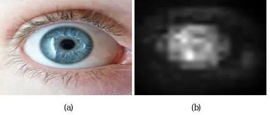
So it can be summarised that , the saliency values of centre regions in images would increase ,with the enhancement operation by the centre bias factor, while the saliency values of non-salient regions in natural scenes would decrease, with the enhancement operation by human visual acuity . As a result the saliency map would get visually better. The figure.2 show the input eye image and the saliency map obtained after enhancing.
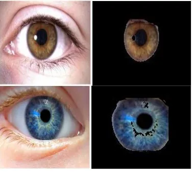
Now the salient region of the image is subjected to a colour masking process. By the saliency detection the eye lids and the eye brow noises can be removed from the input image. Then to get the iris boundary we used a colour mask which masks the white portion in the salient region of the input image. So the result obtained after masking the left and right eye image is shown in the figure.3.
Fig 3.The left and right input eye image and the segmented iris
V. EXPERIMENTAL RESULTS
In order to analyze the proposed iris segmentation algorithm, we have considered several left and right color eye images in the data sets. A reliable accuracy is obtained for the input image considered. The accuracy can be improved further by a more accurate color based thresh holding and by using a more efficient saliency map. The main advantage of this proposed method is that it does not approximate the iris boundary as circular region and so it is applicable for images which do not have exact circular iris boundaries. The disadvantage of this method is that the reflections formed on the iris region are not removed by this method. The segmentation time is calculated for various segmentation methods. The result is shown in Table-1. These results were obtained by running the different segmentation algorithms in MATLAB on a 2GHz Core2Duo processor based computing system.
VI. CONCLUSION
A new saliency based approach has been proposed for the iris segmentation of an iris recognition system. Since the model is not computationally complex this method can segment iris faster than the existing models. It also has the advantage that it does not approximate the iris boundary as circular region. So it is applicable for images which do not have circular boundaries. A reliable accuracy is obtained for the images. . The disadvantage of this method is that the reflections formed on the iris region are not removed by this method. This work can be extended by a method for removing the reflections and also improving the accuracy.
REFERENCES
[1] J. Daugman “Probing the uniqueness and randomness of Iris codes”. Proceedings of the IEEE. Vol 94 No.11, November 2006. Pp. 1927-1935 [2] J. Daugman, “How Iris Recognition Works”, IEEE Transactions on Circuits and Systems for Video Technology (CSVT), vol. 14, no. 1, 2004, pp. 21 -30
[3] J. Daugman, “High confidence visual recognition of persons by a test of statistical independence”, IEEE Trans. on Pattern Analysis and Machine Intelligence, vol. 15, no. 11, 1993, pp. 1148-1161.
[4] L. Itti, C. Koch, and E. Niebur, “A model of saliency-based visual attention for rapid scene analysis,” IEEE Trans. Pattern Anal. Mach. Intell., vol. 20, no. 11, pp. 1254–1259, Nov. 1998.
[5] R. P. Wildes. Iris Recognition: An Emerging Biometric Technology. Proc. of the IEEE, 85(9):1348–1363, Sep 1997
[6] Kheirolahy, R.; Ebrahimnezhad, H.; Sedaaghi, M.H., "Robust pupil boundary detection by optimized color mapping for iris recognition," Computer Conference, 2009. CSICC 2009. 14th International CSI, vol., no., pp.170-175, 20-21 Oct. 2009.
[7] Tisse C., Martin L., Torres L. and Robert M., (2002). Person identification technique using human iris recognition.In proceedings of ICVI’ 02, pp. 294-299.
[8] Teo, C.C. and Ewe, H.T., (2005). An efficient one-dimesional fractal analysis for iris recognition. Proceedings of the 13th WSCG International Conference in Central Europe on Computer Graphics, Visualization and Computer Vision, pp. 157-160.
[9] Richard, Yew Fatt Ng, Yong, Haur Tay and Kai Ming Mok, (2007). An effective segmentation method for iris recognition system.