Abstract: As a critical member of the p53 family of transcription factors, p63 has been implicated a role in devel-opment than in tumor formation, because p63 is seldom mutated in human cancers, while p63 null mice exhibit severe developmental abnormalities without increasing cancer susceptibility. Notably, besides the major epithelial and cardiac defect, p63 deficient mice show severe limb and craniofacial abnormalities. In addition, humans with
p63 mutations also show severe limb and digit defects, suggesting a putative role of p63 in skeletal development. There are eight p63 variants which encode for the TAp63 and ΔNp63 isoforms by alternative promoters. How these
isoforms function during skeletal development is currently largely unknown. Our recent transgenic studies suggest a role of TAP63α, but not ΔNP63α, during embryonic long bone development. However, the moderate skeletal phe -notypes in the TAP63α transgenic mice suggest requirement of additional p63 isoform(s) for the limb defects in
p63 null mice. Here, we report analysis of mouse p63 variants in MCT and ATDC5 cells, two cell models undergo hy-pertrophic differentiation and mimic the process of endochondral bone formation upon growth arrest or induction. We detected increased level of p63 variants in hypertrophic MCT cells by regular RT-PCR analysis. Further analysis
by qRT-PCR, we detected significantly upregulated level of γ variant (p<0.05), but not α or β variant (p>0.05), in
hypertrophic MCT cells than in proliferative MCT cells. Moreover, we detected upregulated TAP63γ in ATDC5 cells
undergoing hypertrophic differentiation. Our results suggest that TAp63γ plays a positive role during endochondral
bone formation.
Keywords: Mouse chondrocytes, p63 variants, TAp63γ, qRT-PCR
Introduction
p63, also called transformation related protein 63 (Trp63), is an important member of the p53 family of transcription factors [1, 2]. Given their structural similarity, p63 was originally thought to have overlapping functions with p53 by regu-lating common down-stream target genes. However, unlike p53, p63 has been majorly implicated a role in development than in tumor formation, as not many p63 mutations have been found in human cancers, while severe developmental abnormalities were observed in
p63 deficient mice that do not increase cancer susceptibility [3]. The predominant function of p63 is in epithelial development, as p63 null mice lack epidermis and other epithelia [4-6]. p63 may also be important for heart develop-ment given its cardiac defects in p63 null mouse embryos [7]. Notably, mice deficient for
p63 also show severe skeletal defects
includ-ing absent or truncated limbs and craniofacial skeletal abnormalities [1, 8]. The limb pheno-type is majorly attributed to defect in apical ectodermal ridge (AER), which is a specialized epithelium at the limb bud directing its out-growth along the axis [1, 9]. However, the short-ened limb and craniofacial skeletal changes suggest that both endochondral and intramem-branous ossifications are impaired in p63 null mice. In humans, P63 mutations are associat-ed with EEC (ectrodactyly, ectodermal dyspla-sia, and cleft lip/palate) or SHFM (split hand-split foot malformation) syndrome, which also shows similar limb defects as seen in p63 null mice [10]. These observations suggest a role of p63 in long bone development, possibly by affecting endochondral bone formation that involves critical steps of chondrocyte differen-tiation and hypertrophy (or maturation).
transcriptional variants encoding six different isoforms: TAP63α, -β, -γ and ΔNP63α, -β, -γ [11]. These p63 isoforms have been shown to play multiple functions during development and cancer formation [5]. However, the specific p63 isoforms that may play a role in bone and carti-lage development is currently largely unknown. We have recently performed p63 gain-of-func-tion studies using p63 variants TAP63α and
ΔNP63α and the (hypertrophic) chondrocyte-specific Col2a1 or Col10a1 control elements. The results suggest an insignificant role of ΔNP63α in embryonic skeletal development, while TAP63α may play distinct functions dur-ing different skeletal developmental stages [12, 13]. However, the moderate skeletal phe-notypes seen in TAP63α transgenic mice strongly suggest that additional p63 isoform(s) is required to be responsible for the severe skeletal defects seen in p63 null mice. In this manuscript, we report systematic analysis of
p63 variants in two chondrogenic cell models: MCT and ATDC5 cells [14, 15]. We detected var-ied levels of p63 transcripts in these cells with
the γ variants being more abundant. Moreover,
TAP63γ is significantly upregulated both in hypertrophic MCT cells and in ATDC5 cells undergoing hypertrophic differentiation. Our results suggest that TAp63γ promotes chondro-genesis, and thereby, plays a positive role dur-ing endochondral bone formation.
Materials and methods
Analysis of p63 variants
Based on literature review and the gene records in NCBI (National Center for Biotechnology Information) database, we performed detailed sequence analysis of the multiple mouse p63
variants with the most recently updated infor-mation. The gene structure of p63 variants was drawn based on previous studies and modified with updated information [16].
Cell culture, total RNA extraction and cDNA synthesis
BRL) and 8% CO2 as per published protocol [14, 15]. After grown until sub-confluence, these MCT cells were further cultured at either 32 °C (proliferative) or 37 °C for additional 3 days (become hypertrophic) before harvest. Total RNAs from both proliferative and hypertrophic MCT cells were isolated and reversely tran-scribed using Trizol reagent and Superscript III reverse transcriptase (Invitrogen, Carlsbad, CA) to synthesize the first strand cDNA. ATDC5 cells were maintained in a mixed DMEM/F-12 (1:1) medium (Invitrogen) with 5% FBS and 1% human insulin, transferrin, and sodium selenite (ITS, Sigma) at 37 °C and 5% CO2 [17]. Cells were then harvested at days 0, 4, 7, 10, 14, and 21 and subjected to RNA extraction and cDNA synthesis respectively as described above.
Expression analysis of genes using real-time/ qRT-PCR
The RT product was subjected to real-time or quantitative polymerase chain reaction (qRT-PCR) to show the relative mRNA levels of genes of interest. These genes include hypertrophic chondrocyte-specific Col10a1, Runx2, as well
as p63 variants and the endogenous control gene Gapdh for normalization of the RNA quali-ty and quantiquali-ty. For qRT-PCR, the cDNA tem-plates were amplified with relevant gene- or
p63 variants-specific primers (listed in Table 1) using the Bio-Rad iQ™ SYBR Green supermix and the MyiQ Real-Time PCR Detection System (Bio-Rad Hercules, CA). Relative mRNA chang-es were analyzed by manufacturer provided MyiQ Optical System Software. The mean threshold cycle number (CT values) of target genes was normalized to endogenous Gapdh
and calculated using 2ˉΔΔCt and student t-test [18, 19]. Data is collected from multiple runs of real-time PCR with duplicate templates and the relative mRNA level was compared between proliferative and hypertrophic MCT cells and between day 0 and days 4, 7, 10, 14, and 21 respectively. p<0.05 was considered statisti-cally significant fold change of mRNA level between samples.
Sequence analysis of the RT-PCR product
[image:3.612.95.524.70.280.2]Regular RT-PCR was performed using standard protocol to amplify the cDNA templates from
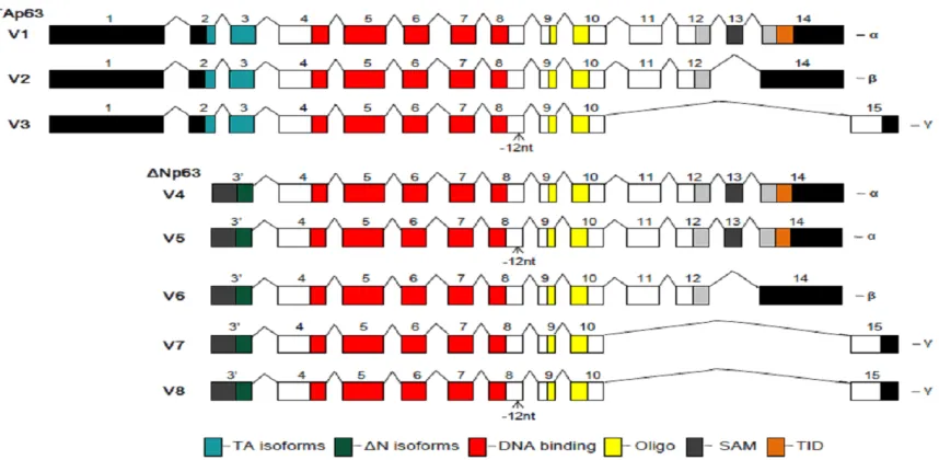
Figure 1. Structure of mouse p63 variants. There are 8 mouse p63 variants (V) encoding two subtypes of isoforms
TAp63 and ΔNP63. V1 is the longest p63 mRNA transcript encoding the longest isoform a--TAP63α, which includes
transactivation (TA), DNA-binding, Oligomerization (Oligo), Sterile alpha motif (SAM), and Transcription inhibition
(TID) domains as illustrated. Compared to V1 (or isoform a), V2 lacks a 3’ exon and encodes isoform b--TAp63β, with
a shorter and different C-terminus; V3 lacks an internal 12 nt and several 3’ exons (an alternate 3’ exon) and
en-codes isoform c--TAp63γ, with a shorter and different C-terminus; V4 (and V5, lacks an internal 12 nt) lacks several 5’ exons (an alternate 5’ exon) and encodes isoform d (and e)--ΔNp63α with a shorter and different N-terminus; V6 lacks several 5’ exons (an alternate 5’ exon) and an internal 3’ exon and encodes isoform f--ΔNp63β with shorter
and different N- and C-termini; V7 (and V8, lacks an internal 12 nt) lacks several 5’ and 3’ exons, (with alternate 5’
ATDC5 cells with designated primes as listed in
Table 2. The RT-PCR products were subjected to electrophoresis in agarose gel and ethidium bromide (EB) staining. Selected PCR product was confirmed by sequencing at the University of Illinois at Chicago’s Center for Genomic Research–DNA Services Facility.
Results
Mouse P63 variants in NCBI database
Based on the most recently updated NCBI database (October 2013), mouse p63 gene is consisted of eight transcriptional variants (vari-ants 1-8) that encode eight p63 isoforms a-h. Among these, isoform a, also called TAp63α, is the longest isoform encoded by variant1. Compared to variant1, some variants lack sev-eral 5’-exons due to the alternative use of their 5’-promoters and result in different N-termini, while some other variants lack some 3’-exons due to alternative splicing and thereby, lead to different C-termini. There are also variants that lack 12 nucleotides within internal exons and thus, lead to an isoform lacking an internal 4 aa. The structure of each of the eight variants is as illustrated (Figure 1).
Hypertrophic differentiation of MCT and ATDC5 cells
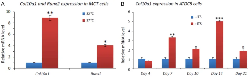
Total RNAs from MCT cells growing at 32 °C and 37 °C (for 3 days) were reversely tran-scribed and the cDNA templates were used for real-time PCR to examine the relative mRNA levels of Col10a1 and Runx2, two marker genes of chondrocyte hypertrophy or maturation.
After normalization to Gapdh, both Col10a1
(8.8 fold, p=0.009) and Runx2 (4.0 fold, p=0.04) show significant upregulation in hyper-trophic MCT cells compared to that in prolifera-tive MCT cells (Figure 2A). This result demon-strated the terminal hypertrophic differentiation of MCT cells grown for 3 days at 37 °C. Similarly, total RNAs from ATDC5 cells cultured with or without ITS for 0, 4, 7, 10, 14, and 21 days were also reversely transcribed and subjected to real-time PCR analysis of Col10a1. The results show that Col10a1 is significantly upregulated in cells maintained in ITS medium and grown for 7, 10, 14, and 21 days (but not 4 days) respectively compared to cells in day 0 without ITS induction (Figure 2B). Among these, day 14 shows the highest level of Col10a1 upregula-tion (5 fold, p<0.001). These results demon-strated the successful induction of hypertro-phic differentiation of the corresponding ATDC5 cells. The primer sequence and amplicon size of genes Col10a1, Runx2, and Gapdh for qRT-PCR are listed in Table 1.
p63 variants expression in MCT and ATDC5 cells
[image:4.612.90.521.71.206.2]To examine the expression of p63 variants in MCT cells, semi-quantitative regular RT-PCR was performed using cDNA templates from both proliferative and hypertrophic MCT cells with ΔNP63, TAp63 and variant-specific prim-ers (listed in Table 1). The results suggest more abundant of mRNA transcripts that encode iso-forms α and γ than isoform β (data not shown). qRT-PCR was then performed using the same
Figure 2. Marker gene expression in MCT and ATDC5 cells. A: Col10a1 shows approximately 9-fold (p=0.009), while
Runx2 shows approximately 4-fold (p=0.04) upregulation in hypertrophic (grown in 37 °C) MCT cells compared to that in proliferative (grown in 32 °C) MCT cells. B: ATDC5 cells were maintained in medium with or without 1% ITS. After 4 days culturing, Col10a1 mRNA transcript did not show significant change (p>0.05), but was significantly upregulated at days 7 (3.3-fold, p=0.002), 10 (2.0-fold, p=0.01), 14 (5.0-fold, p<0.001), and 21 (1.8-fold, p=0.02)
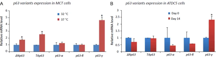
primers and cDNA templates generated from MCT cells. The result showed that both ΔNP63
(1.8 fold, p=0.04)and TAp63 (2.6 fold, p=0.002) are significantly upregulated in hypertrophic MCT cells compared to proliferative MCT cells. We also performed qRT-PCR using variant-spe-cific primers, the result showed that variants encoding isoform γ (4.6 fold, p=0.002), but not α or β, are significantly upregulated in hypertro-phic MCT cells (Figure 3A). We also examined
p63 variants in Day 14 ATDC5 cells, which show hypertrophic differentiation as indicated by highest level of Col10a1, and compared to Day 0 ATDC5 cells. We only detected upregulated
P63-γ variants in Day 14 ATDC5 cells (2.3 fold, p=0.04) compared to that in Day 0 ATDC5 cells (Figure 3B).
TAP63γ is upregulated in hypertrophic ATDC5 cells
To further determine the specific p63 variants that are expressed in ATDC5 cells, we per-formed regular RT-PCR using TAp63, ΔNP63, and γ variants-specific primers as listed in
Table 2. Similar to the findings in MCT cells, we detected mRNA transcripts of TAp63 and γ
variants (lanes 1, 1’, 3, and 3’), but not ΔNP63
variants (lanes 4, 4’, 5, and 5’), both in Day 0 ATDC5 cells and in Day 14 ATDC5 cells with ITS induction. In addition, we detected TAP63γ in Day 14 ATDC5 cells (lane 2’) but not in Day 0 ATDC5 cells (Lane 2, Figure 4A). Sequence analysis confirmed that the PCR product con-tains sequence from exon 9 of TAP63γ (Figure
in MCT cells. As illustrated, both ΔNP63 (1.8 fold, p=0.04)and TAp63 (2.6 fold, p=0.002) are significantly upregu
-lated in hypertrophic MCT cells compared to proliferative MCT cells. Using variant-specific primers, we detected significantly upregulated γ variant (4.6 fold, p=0.002), but not α or β variant (p>0.05), in hypertrophic MCT cells. B:
p63 variants were examined in Day 14 ATDC5 cells after ITS induction and compared to that in Day 0 ATDC5 cells. While the mRNA level of ΔNP63, TAp63,and variants α and β did not show significant change (p>0.05), P63-γ
vari-ants show significant upregulation after treatment with ITS for 14 days (2.3 fold, p=0.04) compared to that in Day
[image:5.612.92.520.71.186.2]0 ATDC5 cells without ITS induction.
Figure 4. TAP63γ in hypertrophic ATDC5 cells. Regular RT-PCR using TAp63, ΔNP63, and γ variants-specific primers
detected mRNA transcripts of TAp63 and γ variants, but not ΔNP63 variants, both in Day 0 ATDC5 cells and in Day 14 ATDC5 cells with ITS induction (lanes 1, 1’, 3, and 3’). TAP63γ is expressed in Day 14 ATDC5 cells (lane 2’) but not
in Day 0 ATDC5 cells (Lane 2). Sequence analysis of the PCR product (lane 2’) confirmed that it contains sequence
[image:5.612.92.521.298.433.2]variant(s) is significantly higher in hypertrophic MCT cells. In addition, we also detected signifi-cantly higher level of γ variant(s) in Day 14 hypertrophic ATDC5 cells compared to Day 0 ATDC5 cells, while the mRNA levels of ΔNP63,
TAP63, p63-α, and p63-β variants did not show significant change (Figure 3). Moreover, our regular RT-PCR detected TAp63γ variant, which was confirmed by sequencing, in ATDC5 cells undergoing hypertrophic differentiation, but not in Day 0 ATDC5 cells (Figure 4), suggesting a potential role of γ variants, especially TAp63γ
during chondrocyte maturation.
It has been shown that p63 isoforms have over-lapping and distinct functions during develop-ment and cancer formation [2]. Generally, the TAP63 isoforms act as transcription factors, while the ΔNP63 isoforms lack the main trans-activation (TA) domain and act as dominant-negative inhibitors of TA isoforms. Interestingly, p63 has been implicated a role in bone devel-opment for over a decade, however, not many studies have been reported thereafter that aim to delineate p63’s function during skeletal development. This is at least partially due to the multiple isoforms of p63 and their complex functions during development. We have recent-ly performed p63 gain-of-function studies by generating a series of transgenic mice using chondrocyte or hypertrophic chondrocyte-spe-cific Col2a1 or Col10a1 control elements to drive ΔNP63α or TAP63α variant. Phenotypic analyses of these Col2a1-ΔNP63α/TAP63α
and Col10a1-ΔNP63α/TAP63α transgenic mice suggest an insignificant role of ΔNP63α
during embryonic skeletal development, while
TAP63α may play a distinct function during dif-ferent skeletal developmental stages [12, 13]. However, as mentioned before, the skeletal phenotypes in TAP63α transgenic mice are moderate, suggesting a potential importance of other p63 isoforms in controlling bone for-mation. The fact that TAp63γ is expressed in proliferative chondrocytes and upregulated chondrocyte-specific genes Col2a1 and
Col10a1 [20]. Multiple transcription factors and signaling pathways have been shown to control the process of endochondral bone for-mation [21, 22]. The severe limb and digit defects in p63 null mice and in humans with
P63 mutations suggest a role of p63 during skeletal development, possibly by affecting the endochondral bone formation pathway [1, 8, 10, 23, 24]. p63 is consisted of multiple tran-scriptional variants encoding two types of p63 isoforms TAp63 and ΔNP63 that each has three subtypes α, β, and γ. These six p63 iso-forms have been extensively studied and are known to play distinct functions during develop-ment and cancer formation [5]. However, how
p63 and its variants function during chondro-cyte differentiation and maturation, steps criti-cal for endochondral bone formation during long bone development, is currently largely unknown.
In this manuscript, we report analysis of mouse
p63 variants by literature review and by refer-ring to the most updated NCBI database. p63 is generally known to consist of 6 isoforms, however, more human P63 transcript variants that may encode additional P63 isoforms have recently been reported [25, 26]. In mouse, based on the latest NCBI database, there are two more p63 transcript variants which make functional studies of p63 more complicated (Figure 1). To study the potential function of p63 during endochondral bone formation, we analyzed its expression in MCT and ATDC5 cells, two cell models that undergo chondro-cyte hypertrophic differentiation either by growth arrest or by induction [14, 15]. The hypertrophic MCT or ATDC5 cells show signifi-cant upregulation of Col10a1 and Runx2
er N, Bronson RT, Tabin C, Sharpe A, Caput D, Crum C, McKeon F. p63 is essential for regen-erative proliferation in limb, craniofacial and epithelial development. Nature 1999; 398: 714-8.
[9] Casanova JC, Uribe V,Badia-Careaga C, Giovin-azzo G,Torres M,Sanz-Ezquerro JJ. Apical ecto-dermal ridge morphogenesis in limb develop-ment is controlled by Arid3b-mediated regulation of cell movements. Development 2011; 138: 1195-205.
[10] van Bokhoven H, Hamel BC, Bamshad M, San-giorgi E, Gurrieri F, Duijf PH, Vanmolkot KR. van Beusekom E, van Beersum SE, Celli J, Merkx GF, Tenconi R, Fryns JP, Verloes A, Newbury-Ecob RA, Raas-Rotschild A, Majewski F, Beem-er FA, Janecke A, Chitayat D, Crisponi G, Kay-serili H, Yates JR, Neri G, Brunner HG. p63 Gene mutations in eec syndrome, limb-mam-mary syndrome, and isolated split hand-split foot malformation suggest a genotype-pheno-type correlation. Am J Hum Genet 2001; 69: 481-92.
[11] Petitjean A, Ruptier C, Tribollet V, Hautefeuille A, Chardon F, Cavard C, Puisieux A, Hainaut P, Caron de Fromentel C. Properties of the six iso-forms of p63: p53-like regulation in response to genotoxic stress and cross talk with Delt-aNp73. Carcinogenesis 2008; 29: 273-81. [12] Li F, Lu Y, Ding M, Wu G, Sinha S, Wang S,
Zheng Q. Putative function of TAP63α during
endochondral bone formation. Gene 2012; 495: 95-103.
[13] Lu Y, Abbassi S, Li F, Ding M, Wu G, Gu J, Zheng Q. Distinct function of P63 isoforms during em-bryonic skeletal development. Gene 2013; 519: 251-9.
[14] Lefebvre V, Garofalo S, de Crombrugghe B. Type X collagen gene expression in mouse chondrocytes immortalized by a temperature-sensitive simian virus 40 large tumor antigen. J Cell Biol 1995; 128: 239-45.
[15] Shukunami C, Ishizeki K, Atsumi T, Ohta Y,
Su-zuki F, Hiraki Y. Cellular hypertrophy and calcifi -cation of embryonal carcinoma-derived chon-drogenic cell line ATDC5 in vitro.J Bone Miner Res1997; 12: 1174-88.
We thank Dr. Benoit de Crombrugghe for the MCT cells. The ATDC5 cells were purchased from Experimental Center of Basic Medicine (ECBM), Fudan University, Shanghai, China. This work was partly supported by the NSFC grant (31271399 awarded to QZ, JG, and YL.) and the 2013 innovative training project for col-lege students in Jiangsu Province (awarded to HC, YG, YL, and QZ).
Disclosure of conflict of interest
All authors have no conflict of Interest.
Address correspondence to: Dr. Qiping Zheng, Department of Anatomy and Cell Biology, Rush
University Medical Center, Chicago, IL 60612, USA.
E-mail: [email protected]; Dr. Yaojuan Lu, Department of Hematology and Hematological Laboratory Science, School of Medical Science and
Laboratory Medicine, Jiangsu University, Zhenjiang
212013, China. E-mail: [email protected]
References
[1] Mills AA, Zheng B, Wang XJ, Vogel H, Roop DR, Bradley A. p63 is a p53 homologue required for limb and epidermal morphogenesis. Nature 1999; 398: 708-13.
[2] Levrero M, De Laurenzi V, Costanzo A, Gong J, Wang JY, Melino G. The p53/p63/p73 family of transcription factors: overlapping and dis-tinct functions. J Cell Sci 2000; 113: 1661-70. [3] Moll UM, Slade N. p63 and p73: roles in devel -opment and tumor formation. Mol Cancer Res 2004; 2: 371-86.
[4] Koster MI, Kim S, Mills AA, DeMayo FJ, Roop DR. p63 is the molecular switch for initiation of
an epithelial stratification program. Genes Dev
2004; 18: 126-31.
[5] Candi E, Dinsdale D, Rufini A, Salomoni P,
transcriptional variants of the human p63 gene. Nucleic Acids Res 2009; 37: 6092-104. [26] Zambelli F, Pavesi G, Gissi C, Horner DS,
Pe-sole G. Assessment of orthologous splicing iso-forms in human and mouse orthologous genes. BMC Genomics 2010; 11: 534.
[27] Koster MI,Roop DR. Transgenic mouse models provide new insights into the role of p63 in epi-dermaldevelopment. Cell Cycle2004; 3: 411-3.
[19] Pfaffl MW. A new mathematical model for
rela-tive quantification in real-time RT-PCR. Nucleic Acids Res2001; 29: e45.
[20] Mackie EJ,Ahmed YA,Tatarczuch L,Chen KS, Mirams M. Endochondral ossification: how car -tilage is converted into bone in the developing skeleton. Int J Biochem Cell Biol 2008; 40: 46-62.
[21] de Crombrugghe B, Lefebvre V, Nakashima K. Regulatory mechanisms in the pathways of cartilage and bone formation. Curr Opin Cell Biol 2001; 13: 721-7.
[22] Wu Q, Chen D, Zuscik MJ, O’Keefe RJ, Rosier RN. Overexpression of Smurf2 stimulates
en-dochondral ossification through upregulation