DIAGNOSTIC MODALITIES FOR TUBERCULOSIS
Azger Dusthackeer, Silambuchelvi Kannayan
Department of Bacteriology, National Institute for Research in Tuberculosis, ICMR, Chennai,
ARTICLE INFO ABSTRACT
Diagnostic methods with high sensitivity and specificity are highly essential for effective management. Endemic nature of the pathogen, cross
growth rate makes it’s diagnosis more complicated and urges the need for improved diagnostic methods to limit
conventional knowledge on TB and molecular genetics approaches have
considerable progress in TB diagnostics. This review highlights different methods of TB diagnosis ranging from phenotypic methods to genetic approa
pipeline for commercialization.
Copyright©2019, Azger Dusthackeer et al. This is an open use, distribution, and reproduction in any medium, provided
INTRODUCTION
Tuberculosis (TB), an all-time disease of concern is caused by bacterial pathogen Mycobacterium tuberculosis
inflicting the Lung. However, it also causes extra
TB by afflicting other organs too. TB is still one among top ten diseases with high mortality especially from South Asian and African countries. Worldwide in 2015, there was an est 10.4 million TB incidence inclusive of 1 million childhood cases (WHO, 2017). Population density, malnutrition, and ethnicity are the risk factors of TB disease progression (Narasimhan, 2013). For the initiation of treatment and curtailing the spread of infection to the close contacts of the patients, early diagnosis is very crucial since TB is highly contagious through airborne transmission.
includes classical approaches like microscopy and culture and the modern genotyping methods (López Ávalos
a slow grower, M. tuberculosis requires prolonged incubation time of 8 weeks in solid culture. Similarly, each method has its own advantages and limitations like less specificity and cross reactivity as in the case of Interferon Gamma Release Assay (IGRA). Recent developments in rapid molecular techniques have shrunken the reporting time considerably. This review is aimed to summarize antiquity to the present scenario of the disease, the problems, limitations of available diagnostic the recent advancements for rapid TB diagnosis revising the diagnostic algorithm.
ISSN: 0975-833X
Article History:
Received 25th March, 2019
Received in revised form 27th April, 2019
Accepted 20th May, 2019
Published online 30th June, 2019
Citation: Azger Dusthackeer, Silambuchelvi Kannayan, Sam Ebenezer Rajadas and Rajesh Mondal
Journal of Current Research, 11, (06), 4881-4888.
Availableonlineathttp://www.journal
Key Words:
Tuberculosis; Diagnosis; Drug Pharmacodynamics.
*Corresponding author: Rajesh Mondal
REVIEW ARTICLE
DIAGNOSTIC MODALITIES FOR TUBERCULOSIS
Azger Dusthackeer, Silambuchelvi Kannayan, Sam Ebenezer Rajadas and
Department of Bacteriology, National Institute for Research in Tuberculosis, ICMR, Chennai,
Tamilnadu, India
ABSTRACT
Diagnostic methods with high sensitivity and specificity are highly essential for effective
management. Endemic nature of the pathogen, cross-reaction in vaccinated individuals and poor growth rate makes it’s diagnosis more complicated and urges the need for improved diagnostic methods to limit progression and the spread. Advancements in technolo
conventional knowledge on TB and molecular genetics approaches have
considerable progress in TB diagnostics. This review highlights different methods of TB diagnosis ranging from phenotypic methods to genetic approaches, which are approved by WHO and are in the pipeline for commercialization.
open access article distributed under the Creative Commons Attribution provided the original work is properly cited.
time disease of concern is caused by Mycobacterium tuberculosis predominantly inflicting the Lung. However, it also causes extra-pulmonary TB by afflicting other organs too. TB is still one among top ten diseases with high mortality especially from South Asian and African countries. Worldwide in 2015, there was an estimated 10.4 million TB incidence inclusive of 1 million childhood . Population density, malnutrition, and ethnicity are the risk factors of TB disease progression
For the initiation of treatment and d of infection to the close contacts of the patients, early diagnosis is very crucial since TB is highly contagious through airborne transmission. TB diagnosis includes classical approaches like microscopy and culture and ez Ávalos, 2012). Being prolonged incubation time of 8 weeks in solid culture. Similarly, each method has its own advantages and limitations like less specificity and
cross-ma Release Assay (IGRA). Recent developments in rapid molecular techniques shrunken the reporting time considerably. This review is aimed to summarize antiquity to the present scenario of the of available diagnostic tools, rapid TB diagnosis and need for
History of Tuberculosis: In ancient times and d
19th and early 20th century TB
as consumption, phthisis, pulmonaris and the white plague (Frith, 2014). Tubercule bacilli and its host
believed to share East Africa as a common ancestral geographic location, however, there is no strong archeological evidence supporting the credence. It is quit
that, all six major lineages or clades of c
tuberculosis strains are existing in East Africa even though its global distribution is not consistent
Archeological evidence was also found in America as it was in Egyptian mummies. There are written texts stating that as early as 3300 and 2300 years ago TB
and also reached Europe (Daniel eye-opener to reveal the causative ag 1882. In 1890, as an attempt towards prepared old tuberculin from culture proteins of
which was later used as a clinical indicator of TB by Clemens von Pirquet(Robert, 2003). In spite of the addition
drugs and diagnostics tools, TB still continues to be an alarming disease with high mortality and morbidity. The emergence of multidrug and extensively drug
tuberculosis further added fuel to the fire occupy 5% of the total TB population, new cases (World Health Organization 2015) of trends focusing on the years 2008
International Journal of Current Research Vol. 11, Issue, 06, pp.4881-4888, June, 2019
DOI: https://doi.org/10.24941/ijcr.35677.06.2019
Azger Dusthackeer, Silambuchelvi Kannayan, Sam Ebenezer Rajadas and Rajesh Mondal. 2019. “Diagnostic modalities for tuberculosis
Availableonlineathttp://www.journalcra.com
z
Ebenezer Rajadas and *Rajesh Mondal
Department of Bacteriology, National Institute for Research in Tuberculosis, ICMR, Chennai,
Diagnostic methods with high sensitivity and specificity are highly essential for effective tuberculosis reaction in vaccinated individuals and poor growth rate makes it’s diagnosis more complicated and urges the need for improved diagnostic progression and the spread. Advancements in technology in combination with conventional knowledge on TB and molecular genetics approaches have helped to achieve considerable progress in TB diagnostics. This review highlights different methods of TB diagnosis ches, which are approved by WHO and are in the
Attribution License, which permits unrestricted
In ancient times and during the 18th,
has been called by names such
pulmonaris and the white plague
Tubercule bacilli and its host - Homo sapiens are believed to share East Africa as a common ancestral geographic location, however, there is no strong archeological evidence supporting the credence. It is quite interesting to note that, all six major lineages or clades of currently circulating M. strains are existing in East Africa even though its global distribution is not consistent (Albanna, 2011). also found in America as it was in There are written texts stating that as early as 3300 and 2300 years ago TB cases entered India and China Daniel, 2006). Robert Koch was the opener to reveal the causative agent for TB in the year 1882. In 1890, as an attempt towards TB therapy, Koch old tuberculin from culture proteins of M.tuberculosis
used as a clinical indicator of TB by Clemens In spite of the addition of newer drugs and diagnostics tools, TB still continues to be an alarming disease with high mortality and morbidity. The emergence of multidrug and extensively drug-resistant tuberculosis further added fuel to the fire. MDR-TB cases TB population, among which 3.5% are (World Health Organization 2015). A new analysis of trends focusing on the years 2008−2013 shows that the
INTERNATIONAL JOURNAL OFCURRENTRESEARCH
proportion of new cases to the MDR-TB remains unchanged at around 3.5% globally. As per the Drug resistance surveillance data, 44% of MDR-TB patients failed to thrive among 4,80,000 cases. Many of such cases are either not detected or not reported and even among the previously treated patients, 40% and 50% were not tested for drug resistance and HIV respectively (World Health Organization 2016a). This scenario also insists on prompt and rapid TB diagnosis.
Impediment in the Current Methods: World Health Organization (WHO) has formulated the following goals: a case detection rate of 70% and a cure rate of 85% and considers that this will have a positive effect by curtailing the TB transmission (Borgdorff 2004). According to WHO's report (Tuberculosis: Directly observed treatment short course (DOTS) case detection rate), an estimated 53% of new smear-positive cases were treated under DOTS during 2004, which was increased to 60% but still falls short of 70% target (Uplekar 2006). Sputum is the preferred specimen for the diagnosis of pulmonary tuberculosis, however sputum collection from infants and neonates is cumbersome, Hence, gastric lavage is recommended in spite of requirement in technical expertise. Presence of commensals in the pulmonary specimen poses the need for specimen processing and incorporation of antibiotics in the media in a cautious way which otherwise inhibits tubercle bacilli also. This set-up is same with the extra-pulmonary specimens but in a milder way due to significantly less normal flora. TB diagnostic methods have to be interpreted with caution since each method has its own advantages and limitations. Growth based assays and genotypic assays are the two broad disciplines of TB diagnosis, apart from the unapproved methods including the serological assays and skin tests. For the growth-based assays, liquid and solid-based media are currently used. The turnaround time for the liquid-based system is much more rapid. Fluorescent-based detection system available for reading the results is coupled with barcoded commercial reagents which makes it more expensive excluding the instrument cost and the infrastructure. However, the results should be confirmed manually by specific antigen detection using the simple immune-chromatographic method and acid-fast staining. 2 to 4 weeks’ for the culture and drug susceptibility testing (DST) is required for reporting the results. Whereas the solid based assay systems like Lowenstein Jensen and Ogawa media are mostly in-house prepared and are very economic methods involving skilled labor, but in contrast requires additional 4 weeks compared to a liquid culture which makes the method unattractive and time-consuming. Growth based assay can detect a load of 102 bacilli/ml of specimen. Smear microscopy which will be dealt with in the following sections is still a rapid method but has a poor sensitivity of 104 bacilli/ml. The promising genotypic assays such as Gene Xpert and line probe assay (LPA) targeting the TB genes are now widely used due to its rapid turnaround time. These WHO approved methods are capable of detecting Multidrug Resistant (MDR) and Extermely drug Resistant (XDR) more rapidly, but with some limitations. The list of genesre presenting the resistant markers may not be complete. Dynamic and diverse nature of tubercle bacilli begets to evolve newer strategies like efflux pumps and newer mutations to escape killing. Failure in discriminating the dead bacilli is another limitation. Early diagnosis can be made with the above-mentioned methods, but the question is how early. Growth based assays are the gold standard due to the conclusiveness in identifying the nature of the live tubercle bacilli, but it is not as rapid as mentioned earlier. Though these methods are considered gold standard
and very accurate, delay in the diagnosis can enhance the transmission of infection, worsen the disease, increase the mortality, and maybe a reason for sustained TB incidence despite the global scale-up of the DOTS strategy (Dye and Williams 2010). Unfortunately, owing to the long incubation period against the low limit of detection by the phenotypic methods we don't have the option of diagnosis except for genomic methods. This may be incredibly useful in non-endemic countries but requires cautious interpretation in the endemic countries. Initiation of appropriate TB therapyis another important factor which requiring an early diagnosis for addressing drug resistance since prescription of drugs without DST still exists among 50% of the cases in the developing world. However, additionally, it requires a minimum of two weeks post primary isolation using MGIT960 or otherwise much more sophisticated devices like Gene Xpert. However, the establishment of such a facility remains to be a burden to the government economy also. Factors responsible for the development of drug resistance are several and can be grouped broadly into the patient (defaulters) and pathogen centric (development of mutation and the dissemination). Dosage inadequacy is an important indicator which might lead to the development of mutation in the pathogen in spite of regular intake of the drugs. The sub-optimal dosage of a prescribed drug may cause an enhanced growth of the pathogen which is referred to as Hormesis (Calabrese 2014). If the isolate is a mono resistant, the sub-optimal dose of that particular drug for which the isolate is resistant may cause hormesis. In yet another scenario, in spite of the isolate being drug susceptible, suboptimal serum levels leading to poor pharmacodynamics (PD) further chains on to poor treatment outcome. Such low levels of first-line anti-TB drugs were also reported in HIV-positive patients (Gurumurthy, 2004). Variation in the prevalence of drug-resistant strains occurs and hence the (PD) also varies. The treatment guidelines have been optimized based on the in-vitro Minimum inhibitory concentration (MICs) of the effective drugs against the microbe and the drug levels along with toxicity profiles of the same in animals and patients from different geographical locations. There is a lack of evidence for such a study being conducted to determine the PD levels in the patients in every subcontinent. Being a global pandemic disease afflicting people from a wide genetic and anthropometric base, wide pharmacokinetic variability leading to variation in the MIC of anti-TB drugs may occur(Gumbo, 2010). All these factors suggest the importance of individualized therapy based on the pharmaco dynamic level to opt for the correct regimen and dosage. A revised diagnostic algorithm with focus on therapeutic drug monitoring will eliminate an element of doubt regarding the optimal therapy, which otherwise has the possibility of leading to sub-therapeutic exposure to certain drugs. The ability of M. tuberculosis to enter into phenotypically drug-resistant non-replicating, dormant state during latency is a major impediment in TB diagnosis since currently there are no growth-based tests to enumerate these dormant bacilli. This phenomenon has been demonstrated in certain smear-positive cases also (16) indicating the need for methods to detect latent TB since existing culture-based techniques detects an active population of tubercle bacilli while failing to grow non-cultivable forms.
Moore, 2004). It has high accessibility and settings with good microscopy, can detect 83% of TB transmitters (Lung, 2000). However, its overall sensitivity even in good settings is only 60% (Bruchfeld, 2000; Tessema, 2001 and Siddiqi, 2003). It cannot distinguish live from dead bacilli. Fluorescence microscopy offers 10% higher sensitivity and improved time efficiency (Steingart, 2006) has the same specificity as Z-N staining, but its value in HIV infected patients is yetunknown (Steingart, 2006). Two or three sputum samples including a morning sample were recommended to be examined and the number of bacilli has to be reported based on the International Union against Tuberculosis and Lung Disease (IUATLD) criteria. Useful as it is, sputum smear microscopy requires a minimum of 5x10³ bacilli per ml of sputum to appear positive (Wormser, 2003).
Culture methods are considered as gold standards for TB diagnosis but it requires expertise, expensive equipment and careful handling of the specimen. It is also slow and has a high risk of contamination by fast-growing bacteria. Culture has a sensitivity and specificity of 80-85% and 98% respectively (Ichiyama, 1993). To detect as few as 101 to 102 viable organisms per ml of the specimen, culture methods are required (Wood, 2007 and Murray, 2003). Enhanced sensitivity could be achieved by processing the specimen by centrifugation and overnight sedimentation preceded with any of several chemical methods. However, this may cause a slight reduction in the specificity (Steingart, 2006). Liquid and solid media are available for M. tuberculosis isolation. Apart from the advantage of using more inoculum, the use of liquid media increases isolation in less time (Wormser, 2005). Irrespective of high contamination rate it takes a shorter time of 1-3 weeks for theM. tuberculosis to propagate contrast to 3-8 weeks for a traditional egg-based solid medium like Lowenstein Jensen (L-J) media. Colonies can be detected on agar-based media after 10-12 days compared to18-24 days for egg-based solid media thereby offers an alternative to liquid media for faster isolation of M. tuberculosis. For DST, solid media require further 4-6 weeks while it is 7 days for liquid cultures. Growth detection methods based on detection of radioactivity (Bactec 460-TB), fluorescence (Bactec Growth Indicator Tube (MGIT 960), phage-based tests e.g. FASTPlaque TB-RIF and Microscopic Observation drug susceptibility Assay (MODS) are faster diagnostic methods. A meta-analysis comprising 10 different studies, the Mycobacterial Growth Indicator Tube (MGIT960), and BACTEC 460 systems showed a sensitivity and specificity of 81.5 and 99.6% and 85.8 and 99.9%, respectively among 1,381 strains from 14,745 clinical specimens. Combination of the above with solid media was found to increase, the sensitivity to 87.7 and 89.7% respectively(Moore, 2004).
Collection of 3 sputum specimens in 2 days for the routine TB diagnosis was practiced in India during the 1950s, for increased diagnostic probability. However, this was not possible practically since it demands a repeated visit by the patients. A study conducted by the Tuberculosis Research Center, Chennai, revealed that, with a good quality of sputum, the majority of TB patients can be diagnosed with the 1st sputum specimen itself. The increase in the average sensitivity improved by examining additional sputum specimen appears to be low. Examining 2 specimens on the same day instead of 3 could benefit patients and TB control programmes (Evidence-based Tuberculosis Diagnosis).
Microscopic observation drug susceptibility culture (MODS): MODS is one of the non-commercial cultures and
DST method endorsed by WHO in the year 2010 (World Health Organization, 2011). This assay is based on the observance of typical cord formation - a special characteristic feature of mycolic acid rich cell wall containing M. tuberculosis observed under an inverted phase contrast microscope. It offers a rapid turnaround time of 7 days and it is less expensive when compared to solid based assay and liquid-based commercial systems respectively. MODS has sensitivity and specificity of 95% and 100% with a high concordance for Isoniazid (97%), Rifampicin (100%), and fluoroquinolones (100%) when compared to the reference standard techniques (Moore, 2006 and Ejigu, 2008). This method requires technical expertise for identifying the cord formation and a typical BSL3 level of containment facility.
Genotypic Assays
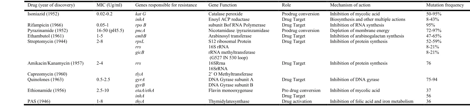
The knowledge on the complete genome of M. tuberculosis has led to the development of newer molecular TB diagnostic tools rapid turnaround time. Genotypic methods can be established by developing probes or by sequencing the specific gene of interest of the M.tbgenome. These methods have considerable advantages for scaling-up programmatic management of drug-resistant and HIV associated TB, in particular with regard to speed, standardized testing, a potential for high throughput, direct handling of the specimen without processing and reduced biosafety needs. The molecular-based kits are usually developed based on the gene responsible for the resistance or the gene that targets the cell wall synthesis, DNA Coiling, transcription and translation mechanism which is depicted in Table 1. These methods include line probe assay. (LAP) and crene expert which have been approved by WHO. INNO-LiPA Rif TB (LiPA), Genotype MTBDRplus assay (Hains Life Sciences) can be used for both diagnoses of M.tb as well as for DST. Resistance is detected by targeting the mutation in the gene which could be indicated either by the presence of the wild-type probe and the mutant probe. RMP resistance is due to the most significant mutation in the 81-bp (base pair) region of the rpoB gene encoding the β-subunit of RNA polymerase accounting for 95% of resistance. INH resistance is caused by mutations in the katGgene and promoter region of the inhA gene. LiPA is a highly sensitive and specific test for the detection of rifampicin resistance in culture isolates but not on direct clinical specimens. Moreover, it can detect only rifampicin resistance whereas newly developed, Line Probe Assay (LPA) can detect resistance towards both the first line drugs rifampicin and isoniazid. The GenoType MTBDR assays have excellent sensitivity and specificity for rifampicin resistance even when directly used on clinical specimens. In a meta-analysis, the pooled sensitivity of Hain MTBDRplus was 98% for detection of rifampicin resistance but not consistent with isoniazid resistance (77% to 90%)(Lawn, 2011).
Though these methods are rapid and have less turnaround time, LPA is recommended only for smear-positive pulmonary cases. The laboratory will also need to maintain strict quality control and avoid cross DNA contamination in addition to the need for skilled-labor and well-established infrastructure. Gene Xpert (Xpert MTB/RIF) is a cartridge-based hemi-nested real-time PCR assay which promises rapid detection within 90 minutes directly from a clinical specimen including Rifampicin (RMP) resistance. Five molecular probes are designed to bind with the wild-type (sensitive) gene of M. tuberculosis. Binding is detected as fluorescent signals from each of these probes. The signal from at least two of these probes indicates the
presence of M.tuberculosis, while a delay in binding or failure to bind, and of at least one probe indicates rifampicin resistance (Lawn et al., 2011). The limit of detection of the Xpert MTB/RIF assay in spiked sputum samples has been measured to be 131 bacilli per ml of sputum (Helb et al., 2010). Sensitivity and specificity of this assay for tuberculosis detection are 89% and 99%, respectively for pulmonary tuberculosis (Steingart et al., 2014). However, sensitivity is lower in HIV (79%) and pediatric patients (66%) (Detjen et al., 2015). Sensitivity varied from specimen to specimen in extrapulmonary cases with the highest for lymph node biopsies and cerebrospinal fluid and low in pleural fluid. Therefore this assay can only be used as a diagnostic tool but not for monitoring the treatment response, however, it can be potentially used at the point of treatment at a primary health center also. High cost, need for continuous power supply, sensitivity to high temperatures and assay throughput are major constraints for its widespread rollout. Additionally, the emerging silent mutations in the rifampicin resistance determining region (RRDR) of the rpoB are not targeted by Xpert MTB/Rif (Mathys et al., 2014). Xpert Ultra has been developed to overcome this limitation and to improve the sensitivity and specificity in detection of TB and RIF-R, respectively. Itcovers two additional amplification targets (IS6110 and IS1081), 25 different RRDR mutations spanning almost the entire rpo B RRD Rexcept IIe491Phe with increased sensitivity capable of detecting as low as 15.6 CFU/mL of sputum (Chakravorty et al., 2017; Dorman et al., 2018). This has been endorsed by WHO at the end of March 2017 and recommended it as a replacement of Xpert due to its increased sensitivity (). Cepheid has brought up a battery supported single-cartridge testing point of care unit namely GeneXpert Omni for TB and rifampicin resistance detection. This GeneXpert Omni awaits validation and approval by WHO (Garcia-Basteiro, 2018). Hain Life science has developed a second-line LPA (MTBDRsl) test and the process of the validation was also completed.
[image:4.842.7.796.73.252.2]Once the patients are diagnosed with resistant TB using rapid diagnostic methods like Xpert MTB/RIF or LPA, there existed a considerable delay in getting the phenotypic results in the cases of amplified resistance for the diagnosis of second-line drug resistance. This delay has been circumvented through the aforementioned assay, which was recognized by WHO in May 2016 targeting second-line anti-TB drug resistance (WHO, 2016). The current version of this assay targetsgyr A and gyr B for the detection of resistance to fluoroquinolones and rrs and eis for the detection of resistance to injectable anti-TB drugs. According to a Cochrane review, thisassay for the smear-positive specimens has the sensitivity and specificity of about 97%and 98% respectively and for smear-negative specimens, the same were 80%and 100%respectively. In the direct testing for fluoroquinolone resistance and the Second-Line Injectable Drug (SLID) resistance, the sensitivity and specificity were 89%and 90%for the positive specimen and smear-negative specimen the same were80%and 100%respectively. In the XDR-TB, sensitivity and specificity were 79%and 97%for smear-positive specimen while 50%and 100%for smear-negative specimen respectively (Guo, 2013). These data varied for specific drugs since there is incomplete cross-resistance among injectable drugs (Madhukar, 2016). Therefore, this test is useful as a rule-in test for XDR or pre-XDR tuberculosis, but, because of suboptimal sensitivity, it cannot be used to completely rule out resistance. Detailed understanding of the local distribution of drug-resistance mutations is required to interpret the results of this assay in the local context. Rifampicin resistance can also be detected by a highly specific and sensitive method called Pyrosequencing which requires technical expertise. It has an overall sensitivity and specificity of about 94%and 98%respectively and for the clinical specimens, the same were 89%and 99%respectively (Guo, 2003).
Table 1. Genes involved in antibiotic resistance and its function
Drug (year of discovery) MIC (Ug/ml) Genes responsible for resistance Gene Function Role Mechanism of action Mutation frequency
Isoniazid (1952) 0.02-0.2 kat G Catalase peroxide Prodrug conversion Inhibition of mycolic acid 50-95%
inhA Enoyl ACP reductase Drug Target Biosynthesis and other multiple actions 8-43%
Rifampicin (1966) 0.05-1 rpo B subunit Bof RNA Polymerase Drug Target Inhibition of RNA synthesis 95%
Pyrazinamide (1952) 16-50 (pH5.5) pncA Nicotamidase /pyrazinzamidase Prodrug conversion Depletion of membrane energy 72-97%
Ethambutol (1961) 1-5 embB Arabinosyl transferase Drug Target Inhibition of arabinogalactan synthesis 47-65%
Streptomycin (1944) 2-8 rpsL S12 ribosomal Protein Drug Target Inhibition of protein synthesis 52-59%
rrs 16S rRNA 8-21%
gicB rRNA methyltransferase (G527 IN 530 loop)
8-21%
Amikacin/Kanamycin (1957) 2-4 rrs 16SRrna
16SrRNA
Drug Target Inhibition of protein synthesis 76
Capreomycin (1960) tlyA 2’ O Methyltransferase
Quinolones (1963) 0.5-2.5 gyrA DNA Gyrase subunit A Drug Target Inhibition of DNA gyrase 75-94
gyrB DNA Gyrase subunit B
Ethionamide (1956) 2.5-10 etaA/ethA
inhA
Flavin monooxygenase Pro drug conversion Drug Target
Inhibition of mycolic acid 37
56
Immunodiagnostics: A variety of immunodiagnostic test recognizing specific host response towards TB infection is available. Tuberculin Skin Test (TST) is the oldest, however, it has limited value in the diagnosis of TB in areas with high TB and HIV prevalence and where BCG vaccination is given, because of possible false positivity (Siddiqi, 2003). This test yields false negative results in immune suppressed individuals andlacks the ability to differentiate active from past sensitization (Hans, 1999). Newer immunodiagnostic tests based on antibody detection are available however, the specificity of this test is less than 80% (Wood, 2007).
LAM-ELISA targeting Lipoarabinomannan (LAM) and
QuantiFERON-TB GOLD and T-SPOT TB assays targeting IFN-γ are specific for TB bacilli with no cross-reactivity. The sensitivity and specificity of the LAM ELISA test are unknown. While the same for QuantiFERON-TB GOLD and T-SPOT TB are 75%-95% and 90-100% respectively with possibly reduced sensitivity in HIV positive cases (Pai, 2007). However, these assays are not still recommended for TB diagnosis by WHO.
Diagnostics in the Pipeline: Although sputum has traditionally been thought to contain actively growing tubercle bacilli, transcriptomic analysis refuses this hypothesis (Garton, 2008). Importantly, a subpopulation of dormant, persisting M. tuberculosis can become active in presence of appropriate factors like resuscitation-promoting factors (RPF), a protein of pathogen origin. This underlines the great potential of RPF in improving the detection of M. tuberculosis in clinical specimens both qualitatively and quantitatively. The RPF-dependency of clinical isolateswas found to be lost after primary isolation from the specimen. Interestingly, during chemotherapy, the proportion of RPF-dependent cells increased relative to the surviving colony-forming active population (Mukamolova, 2010). This finding promises reduced detection limit by culture techniques in the presence of RPF for the pulmonary samples and also help in accessing the efficiency of chemotherapy. Previously, we used diagnostic luciferase reporter phage assay which can detect non-replicating persistor TB bacilli in patient sputum, and identified 30 additional positives, which were culture negative (Dusthackeer, 2012). Presence of viable bacilli in these samples was confirmed by reverse transcriptase-PCR for Mtb 16S rRNA gene. The use of the specific mycobacteriophages to detect the tubercle bacilli forms the basis of these luciferase reporter phage assay (LRP) and hence seems to be promising in Rifampicin susceptibility detection. Even though it has high specificity of 83% to 100% and modest, variable sensitivity of 21% to 88% at this juncture it cannot replace the culture-based assays (Pai, 2007). Newer cheaper NAAT based tests with a combination of PCR and visual readouts by lateral flow method in kit format is ideal to bring testing as near to the patient as possible (Nikam et al., 2013). The True Nat MTBTM a diagnostic kit from Molbio Diagnostics, India is chip-based nucleic acid amplification test for detection of M. tuberculosis and resistance to rifampicin from sputum samples.
The DNA extraction is doneusing Truprep-MAGTM (a nanoparticle-based protocol run on a battery operated device) and for real-time PCRTruelab-UNOTM analyzer is used (Hand-held battery operated thermal cycler).It has been evaluated in India and found to have sensitivity and specificity of 91.1% and 100% respectively when compared to the in-house nested PCR and similar to Xpert MTB/RIF. The time required for completion of testing is one hour (Nikam et al., 2013 & 2014).
Data on its feasibility in ruralcenters is essential to claim this test as decentralized, "point-of-care"use but there are no published data. The EasyNAT Diagnostic Kit for M.tb Complex has been manufactured by Ustar Biotechnologies, China. The kit performs an isothermal amplification targeting the IS6110 region with the sensitivity and specificity of 84.1% and 97.8% respectively. The sensitivity was 59.8%in smear-negative cases (Ou et al., 2014). The VereMTB assayfrom Veredus Laboratories, Singaporeis based on a combination of both PCR and microarray technology targeting resistance to rifampicin and isoniazid within 3 hrs excluding the sample extraction. Epistem Genedrive system is apaper-based digestion cum PCRkit which is very close to being released in the market for MDR and XDRTB diagnosis (Shenai et al., 2016). United States Food and Drug Administration (US FDA) has given approval for the AMPLICOR M. tuberculosis test from Roche Diagnostic Systems, the USA, and the Amplified Mycobacterium tuberculosis Direct test (MTD) from Gen-Probe, Inc., USA tests for the diagnosis of TB. However, these methods need high technical support and are costlier which stumbles the widespread adoption as diagnostic kits in TB endemic countriesir respective of it research applications (Tebruegge et al., 2014; Kambashi et al., 2001; Kivihya-Ndugga et al., 2004). The previously established PCR based COBAS Amplicor assay has been replaced by the US Food and Drug Administration (FDA) approved qualitative COBAS Taq Man MTB from Roche Diagnostics, Tokyo, Japan for use in smear-positive and/or smear-negative pulmonary disease. It targets 16S rRNA gene using TaqMan probe to detect TB with a turnaround time of 2-5 hours for analyzing 48 samples in one shot. However it could be used only for pulmonary TB detection and not for extra pulmonary cases (Jonsson, 2015).
To enable detection in low bacillary load, Loop-mediated isothermal amplification (LAMP) (Eiken Chemical Co. Ltd., Tokyo, Japan) assay developed by Notomi et al. is most commonly employed. It is an isothermal nucleic acid amplification technique, which amplifies very few copies of target DNA with high specificity, efficiency, and rapidity under isothermal conditions using a set of 4 specially designed primers and a DNA polymerase with strand displacement activity without the need for a thermal cycler (Notomi, 2000). It is recommended by WHO in August 2016 for diagnosing pulmonary TB in adults (Parida, 2008). From then on several LAMP-based assays have been developed for TB detection targeting gyrB, rrs, rimM, IS6110, hspX, mpb64and sda A gene of TB bacilli and are competent to detect Non-Tuberculus Mycobacterium also (Iwamoto, 2003; Pandey, 2008; Zhu, 2009; Aryan, 2010; Bi, 2012; Balne, 2013 and Nimesh, 2014). Varied levels of sensitivity has been reported for TB-LAMP ranging from as high as 100% for smear-positive samples to as low as 52% for smear-negative samples (Gray, 2016; Ou, 2014; Bojang, 2016; Kaku, 2016). Major limitation in LAMP is the occurrence of false positivity when the reaction tubes are exposed to aerosol contamination, hence it requires sophisticated facilities preventing such DNA contaminations (Zhu, 2009). Heretofore, LAMP has not been fully evaluated in HIV patients and children. Furthermore, it requires endorsement by WHO for testing extrapulmoary TB samples.
Conclusion
Rapid and early TB detection is crucial to improve the global TB control. The Phenotypic methods serve as the gold standard but take a prolonged time to obtain endpoint wherein the
genotypic methods are quite promising for both the detection of TB as well as itsresistance. Though there is a huge development in the molecular gene-based test, there are limitations like high cost and increased dependency on manufacturer’s support which has to be foreseen to bring the tests as a point of care test to be used in the periphery. This will have a better impact on settings where presumptive treatment is practiced especially in the extrapulmonary TB cases and in the pediatric population. More of feasibility studies need to be conducted to ensure that these new diagnostic kits to reach the market which in turn assist in the early detection and thereby help the TB control programme to reach the goal of END TB by 2025.
Conflicts of Interest: Authors declare no Conflict of Interest. Authors AZ and KN contributed equally.
REFERENCES
Albanna AS, Reed MB, Kotar K V, Fallow A, McIntosh FA, Behr MA, et al. Reduced Transmissibility of East African Indian Strains of Mycobacterium tuberculosis. PLoS One [Internet]. 2011 Sep 19;6(9):e25075. Available from: https://doi.org/10.1371/journal.pone.0025075
Aryan E, Makvandi M, Farajzadeh A, Huygen K, Bifani P, Mousavi S-L, et al. A novel and more sensitive loop-mediated isothermal amplification assay targeting IS6110 for detection of Mycobacterium tuberculosis complex. Microbiol Res. 2010 Mar;165(3):211–20.
Balne PK, Barik MR, Sharma S, Basu S. Development of a loop-mediated isothermal amplification assay targeting the mpb64 gene for diagnosis of intraocular tuberculosis. J Clin Microbiol. 2013 Nov;51(11):3839–40.
Bi A, Nakajima C, Fukushima Y, Tamaru A, Sugawara I, Kimura A, et al. A rapid loop-mediated isothermal amplification assay targeting hspX for the detection of Mycobacterium tuberculosis complex. Jpn J Infect Dis. 2012;65(3):247–51.
Bojang AL, Mendy FS, Tientcheu LD, Otu J, Antonio M, Kampmann B, et al. Comparison of TB-LAMP, GeneXpert MTB/RIF and culture for diagnosis of pulmonary tuberculosis in The Gambia. J Infect. 2016 Mar;72(3):332–7.
Borgdorff MW. New measurable indicator for tuberculosis case detection. Emerg Infect Dis [Internet]. 2004
Sep;10(9):1523–8. Available from:
https://www.ncbi.nlm.nih.gov/pubmed/15498151
Bruchfeld J, Aderaye G, Palme IB, Bjorvatn B, Kallenius G, Lindquist L. 2000. Sputum concentration improves diagnosis of tuberculosis in a setting with a high prevalence of HIV. Trans R Soc Trop Med Hyg., 94(6):677–80.
Calabrese EJ. Hormesis: from mainstream to therapy. J Cell Commun Signal [Internet]. 2014/11/01. 2014 Dec; 8(4):289–91. Available from: https://www.ncbi.nlm.nih. gov/pubmed/25366126
Chakravorty S, Simmons AM, Rowneki M, Parmar H, Cao Y, Ryan J, et al. The New Xpert MTB/RIF Ultra: Improving Detection of Mycobacterium tuberculosis and Resistance to Rifampin in an Assay Suitable for Point-of-Care Testing. MBio. 2017 Aug;8(4).
Cruciani M, Scarparo C, Malena M, Bosco O, Serpelloni G, Mengoli C. Meta-analysis of BACTEC MGIT 960 and BACTEC 460 TB, with or without solid media, for detection of mycobacteria. J Clin Microbiol [Internet].
2004 May; 42(5):2321–5. Available from: https://www. ncbi.nlm.nih.gov/pubmed/15131224
Daniel TM. The history of tuberculosis. Respir Med. 2006 Nov;100(11):1862–70.
Detjen AK, DiNardo AR, Leyden J, Steingart KR, Menzies D, Schiller I, et al. Xpert MTB/RIF assay for the diagnosis of pulmonary tuberculosis in children: a systematic review and meta-analysis. Lancet Respir Med [Internet]. 2015 Jun 1;3(6):451–61. Available from: https://doi.org/10.1016/ S2213-2600(15)00095-8
Dorman SE, Schumacher SG, Alland D, Nabeta P, Armstrong DT, King B, et al. Xpert MTB/RIF Ultra for detection of Mycobacterium tuberculosis and rifampicin resistance: a prospective multicentre diagnostic accuracy study. Lancet Infect Dis. 2018 Jan;18(1):76–84.
Dusthackeer VNA, Balaji S, Gomathi NS, Selvakumar N, Kumar V. Diagnostic luciferase reporter phage assay for active and non-replicating persistors to detect tubercle bacilli from sputum samples. Clin Microbiol Infect. 2012 May;18(5):492–6.
Dye C, Williams BG. The population dynamics and control of tuberculosis. Science. 2010 May;328(5980):856–61. Ejigu GS, Woldeamanuel Y, Shah NS, Gebyehu M, Selassie
A, Lemma E. Microscopic-observation drug susceptibility assay provides rapid and reliable identification of MDR-TB. Int J Tuberc Lung Dis. 2008 Mar;12(3):332–7. Frith J. History of tuberculosis. Part 1 - phthisis, consumption
and the white plague. J Mil Veterans Heal. 2014;22(2):29– 35.
Garcia-Basteiro AL, DiNardo A, Saavedra B, Silva DR, Palmero D, Gegia M, et al. Point of care diagnostics for tuberculosis. Pulmonology. 2018 Mar;24(2):73–85. Garton NJ, Waddell SJ, Sherratt AL, Lee S-M, Smith RJ,
Senner C, et al. Cytological and Transcript Analyses Reveal Fat and Lazy Persister-Like Bacilli in Tuberculous Sputum. PLOS Med [Internet]. 2008 Apr 1;5(4):e75.
Available from: https://doi.org/10.1371/journal
.pmed.0050075
Gray CM, Katamba A, Narang P, Giraldo J, Zamudio C, Joloba M, et al. Feasibility and Operational Performance of Tuberculosis Detection by Loop-Mediated Isothermal Amplification Platform in Decentralized Settings: Results from a Multicenter Study. Carroll KC, editor. J Clin Microbiol [Internet]. 2016 Aug 1;54(8):1984 LP – 1991.
Available from: http://jcm.asm.org/content/54/
8/1984.abstract
Gumbo T. New Susceptibility Breakpoints for First-Line
Antituberculosis Drugs Based on Antimicrobial
Pharmacokinetic/Pharmacodynamic Science and
Population Pharmacokinetic Variability. Antimicrob Agents Chemother [Internet]. 2010 Apr 1;54(4):1484 LP – 1491. Available from: http://aac.asm.org/content/ 54/4/1484.abstract
Guo Q, Zheng R-J, Zhu C-T, Zou L-L, Xiu J-F, Li J, et al. Pyrosequencing for the rapid detection of rifampicin resistance in Mycobacterium tuberculosis: a meta-analysis. Int J Tuberc Lung Dis. 2013 Aug;17(8):1008–13. Gurumurthy P, Ramachandran G, Hemanth Kumar AK,
Hans L R. Epidemiologic Basis of Tuberculosis Control. International Union Against Tuberculosis and Lung Disease. 1999.
Helb D, Jones M, Story E, Boehme C, Wallace E, Ho K, et al. Rapid Detection of <em>Mycobacterium tuberculosis</em> and Rifampin Resistance by Use of On-Demand, Near-Patient Technology. J Clin Microbiol [Internet]. 2010 Jan 1;48(1):229 LP – 237.
Available from: http://jcm.asm.org/content/48/
1/229.abstract
Ichiyama S, Shimokata K, Takeuchi J. Comparative study of a biphasic culture system (Roche MB Check system) with a conventional egg medium for recovery of mycobacteria. Aichi Mycobacteriosis Research Group. Tuber Lung Dis. 1993 Oct;74(5):338–41.
Iwamoto T, Sonobe T, Hayashi K. Loop-mediated isothermal amplification for direct detection of Mycobacterium tuberculosis complex, M. avium, and M. intracellulare in sputum samples. J Clin Microbiol. 2003 Jun;41(6):2616– 22.
Jonsson B, Lonnermark E, Ridell M. Evaluation of the Cobas TaqMan MTB test for detection of Mycobacterium tuberculosis complex. Infect Dis (London, England). 2015 Apr;47(4):231–6.
Kaku T, Minamoto F, D’Meza R, Morose W, Boncy J, Bijou J, et al. Accuracy of LAMP-TB Method for Diagnosing Tuberculosis in Haiti. Jpn J Infect Dis. 2016 Nov;69(6):488–95.
Kambashi B, Mbulo G, McNerney R, Tembwe R, Kambashi A, Tihon V, et al. Utility of nucleic acid amplification techniques for the diagnosis of pulmonary tuberculosis in sub-Saharan Africa. Int J Tuberc Lung Dis. 2001 Apr;5(4):364–9.
Kivihya-Ndugga L, van Cleeff M, Juma E, Kimwomi J, Githui W, Oskam L, et al. Comparison of PCR with the routine procedure for diagnosis of tuberculosis in a population with high prevalences of tuberculosis and human immunodeficiency virus. J Clin Microbiol. 2004 Mar;42(3):1012–5.
Lawn SD, Nicol MP. Xpert(R) MTB/RIF assay: development, evaluation and implementation of a new rapid molecular diagnostic for tuberculosis and rifampicin resistance. Future Microbiol. 2011 Sep;6(9):1067–82.
López Ávalos GG, Prado Montes de Oca E. Classic and New Diagnostic Approaches to Childhood Tuberculosis. J Trop Med [Internet]. 2012 Mar 12;2012:818219. Available
from: http://www.ncbi.nlm.nih.gov/pmc/articles/
PMC3317187/
Lung IUAT and, Disease. Sputum Examination for Tuberculosis by Direct Microscopy in Low Income
Countries [Internet]. 2000. Available from:
https://tbrieder.org/publications/books_english/microscop y.pdf
Madhukar P, Mark P N, Catharina C B. Tuberculosis Diagnostics: State of the Art and Future Directions. Microbiol Spectr. 2016;4(5):1–15.
Mathys V, van de Vyvere M, de Droogh E, Soetaert K, Groenen G. False-positive rifampicin resistance on Xpert(R) MTB/RIF caused by a silent mutation in the rpoB gene. Int J Tuberc Lung Dis. 2014 Oct;18(10):1255– 7.
Moore DAJ, Evans CAW, Gilman RH, Caviedes L, Coronel J,
Vivar A, et al. Microscopic-observation
drug-susceptibility assay for the diagnosis of TB. N Engl J Med. 2006 Oct;355(15):1539–50.
Moore DAJ, Mendoza D, Gilman RH, Evans CAW, Hollm Delgado M-G, Guerra J, et al. 2004. Microscopic observation drug susceptibility assay, a rapid, reliable diagnostic test for multidrug-resistant tuberculosis suitable for use in resource-poor settings. J Clin Microbiol., Oct;42(10):4432–7.
Mukamolova G V, Turapov O, Malkin J, Woltmann G, Barer MR. Resuscitation-promoting factors reveal an occult population of tubercle Bacilli in Sputum. Am J Respir Crit Care Med. 2010 Jan;181(2):174–80.
Murray SJ, Barrett A, Magee JG, Freeman R. Optimisation of acid fast smears for the direct detection of mycobacteria in clinical samples. J Clin Pathol [Internet]. 2003 Aug; 56(8):613–5. Available from: https://www.ncbi.nlm.nih. gov/pubmed/12890813
Narasimhan P, Wood J, MacIntyre CR, Mathai D. Risk Factors for Tuberculosis. Pulm Med [Internet]. 2013 Feb
12;2013:828939. Available from:
http://www.ncbi.nlm.nih.gov/pmc/articles/PMC3583136/ Nikam C, Jagannath M, Narayanan MM, Ramanabhiraman V,
Kazi M, Shetty A, et al. Rapid Diagnosis of Mycobacterium tuberculosis with Truenat MTB: A Near-Care Approach. PLoS One [Internet]. 2013 Jan
21;8(1):e51121. Available from:
https://doi.org/10.1371/journal.pone.0051121
Nikam C, Kazi M, Nair C, Jaggannath M, M M, R V, et al. Evaluation of the Indian TrueNAT micro RT-PCR device with GeneXpert for case detection of pulmonary tuberculosis. Int J mycobacteriology. 2014 Sep;3(3):205– 10.
Nimesh M, Joon D, Varma-Basil M, Saluja D. Development and clinical evaluation of sdaA loop-mediated isothermal amplification assay for detection of Mycobacterium tuberculosis with an approach to prevent carryover contamination. J Clin Microbiol [Internet]. 2014
Jul;52(7):2662–4. Available from:
https://www.ncbi.nlm.nih.gov/pubmed/24789191
Notomi T, Okayama H, Masubuchi H, Yonekawa T, Watanabe
K, Amino N, et al. Loop-mediated isothermal
amplification of DNA. Nucleic Acids Res. 2000 Jun; 28(12):E63.
Ou X, Li Q, Xia H, Pang Y, Wang S, Zhao B, et al. Diagnostic accuracy of the PURE-LAMP test for pulmonary tuberculosis at the county-level laboratory in China. PLoS One. 2014;9(5):e94544.
Ou X, Song Y, Zhao B, Li Q, Xia H, Zhou Y, et al. A multicenter study of cross-priming amplification for tuberculosis diagnosis at peripheral level in China. Tuberculosis (Edinb). 2014 Jul;94(4):428–33.
Pai M, Joshi R, Bandyopadhyay M, Narang P, Dogra S, Taksande B, et al. Sensitivity of a whole-blood interferon-gamma assay among patients with pulmonary tuberculosis and variations in T-cell responses during anti-tuberculosis treatment. Infection. 2007 Apr;35(2):98–103.
Pandey BD, Poudel A, Yoda T, Tamaru A, Oda N, Fukushima Y, et al. Development of an in-house loop-mediated isothermal amplification (LAMP) assay for detection of Mycobacterium tuberculosis and evaluation in sputum samples of Nepalese patients. J Med Microbiol. 2008 Apr;57(Pt 4):439–43.
Parida M, Sannarangaiah S, Dash PK, Rao PVL, Morita K. Loop mediated isothermal amplification (LAMP): a new generation of innovative gene amplification technique; perspectives in clinical diagnosis of infectious diseases. Rev Med Virol. 2008;18(6):407–21.
Robert JP. HIV & AIDS, 5Ed: a foundation for nursing and healthcare practice. 2003. 137 p.
Shenai S, Armstrong DT, Valli E, Dolinger DL, Nakiyingi L, Dietze R, et al. Analytical and Clinical Evaluation of the Epistem Genedrive Assay for Detection of <span class="named-content genus-species" id="named-content-1">Mycobacterium tuberculosis</span> G. A. L, editor. J Clin Microbiol [Internet]. 2016 Apr 1;54(4):1051 LP – 1057.
Available from: http://jcm.asm.org/content/
54/4/1051.abstract
Siddiqi K, Lambert M-L, Walley J. Clinical diagnosis of smear-negative pulmonary tuberculosis in low-income countries: the current evidence. Lancet Infect Dis. 2003 May;3(5):288–96.
Steingart KR, Henry M, Ng V, Hopewell PC, Ramsay A, Cunningham J, et al. Fluorescence versus conventional sputum smear microscopy for tuberculosis: a systematic review. Lancet Infect Dis. 2006 Sep;6(9):570–81.
Steingart KR, Ng V, Henry M, Hopewell PC, Ramsay A, Cunningham J, et al. Sputum processing methods to improve the sensitivity of smear microscopy for tuberculosis: a systematic review. Lancet Infect Dis. 2006 Oct;6(10):664–74.
Steingart KR, Schiller I, Horne DJ, Pai M, Boehme CC, Dendukuri N. Xpert(R) MTB/RIF assay for pulmonary tuberculosis and rifampicin resistance in adults. Cochrane database Syst Rev. 2014 Jan;(1):CD009593.
Tebruegge M, Ritz N, Koetz K, Noguera-Julian A, Seddon JA, Welch SB, et al. Availability and Use of Molecular Microbiological and Immunological Tests for the Diagnosis of Tuberculosis in Europe. PLoS One [Internet].
2014 Jun 12;9(6):e99129. Available from:
https://doi.org/10.1371/journal.pone.0099129
Tessema TA, Bjune G, Assefa G, Bjorvat B. 2001. An evaluation of the diagnostic value of clinical and radiological manifestations in patients attending the addis ababa tuberculosis centre. Scand J Infect Dis., 33(5):355– 61.
Theron G, Peter J, Richardson M, Warren R, Dheda K, Steingart KR. GenoType((R)) MTBDRsl assay for resistance to second-line anti-tuberculosis drugs. Cochrane database Syst Rev. 2016 Sep;9:CD010705.
Uplekar M. Stop TB Partnership & World Health Organization. The Stop TB strategy : building on and enhancing DOTS to meet the TB-related Millennium Development Goals [Internet]. Geneva; 2006. Available from: http://www.who.int/iris/handle/10665/69241 Wood R, Middelkoop K, Myer L, Grant AD, Whitelaw A,
Lawn SD, et al. Undiagnosed tuberculosis in a community with high HIV prevalence: implications for tuberculosis control. Am J Respir Crit Care Med. 2007 Jan;175(1):87– 93.
World Health Organization. Global Tuberculosis Report 2017. 2017.
World Health Organization. Global Tuberculosis Report. 2015. World Health Organization. Global Tuberculosis Report. 2016. World Health Organization. Noncommercial culture and
drug-susceptibility testing methods for screening patients at risk for multidrugresistant tuberculosis [Internet]. 2011. Available from: http://apps.who.int/iris/bitstream/ handle/10665/44601/9789241501620_eng.pdf?sequence= 1
World Health Organization. WHO meeting report of a technical expert consultation: non-inferiority analysis of Xpert MTB/RIF Ultra compared to Xpert MTB/RIF
[Internet]. 2017. Available from:
https://apps.who.int/iris/bitstream/handle/10665/254792/ WHO-HTM-TB-2017.04-eng.pdf
World Health Organization. WHO RECOMMENDATIONS ON THE USE OF THE SL-LPA [Internet]. 2016.
Available from:
http://www.who.int/tb/areas-of-work/laboratory/policy_statements
Wormser GP, Fisher MA. Manson’s Tropical Diseases, 21st Edition Edited by Gordon C. Cook and Alimuddin I. Zumla Philadelphia: W. B. Saunders, 2003. 1847 pp., illustrated. $179.00 (cloth). Clin Infect Dis [Internet]. 2003 Aug 15;37(4):609. Available from: http://dx.doi.org/ 10.1086/376995
Zhu R-Y, Zhang K-X, Zhao M-Q, Liu Y-H, Xu Y-Y, Ju C-M, et al. Use of visual loop-mediated isotheral amplification of rimM sequence for rapid detection of Mycobacterium tuberculosis and Mycobacterium bovis. J Microbiol Methods. 2009 Sep;78(3):339–43.