b
-
D-Altrose
Yuji Watanabe,aHiromi Yoshida,aKosei Takeda,a Tomohiko Ishiband Shigehiro Kamitoria*
a
Division of Structural Biology, Life Science Research Center and Faculty of Medicine, Kagawa University, 1750-1 Ikenobe, Miki-cho, Kita-gun, Kagawa 761-0793, Japan, andbFaculty of Engineering, Kagawa University, 2217-20 Hayashi-machi, Takamatsu, Kagawa 761-0396, Japan
Correspondence e-mail: [email protected]
Received 11 December 2008; accepted 6 January 2009
Key indicators: single-crystal X-ray study;T= 293 K; mean(C–C) = 0.007 A˚;
Rfactor = 0.046;wRfactor = 0.125; data-to-parameter ratio = 6.8.
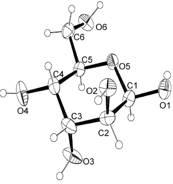
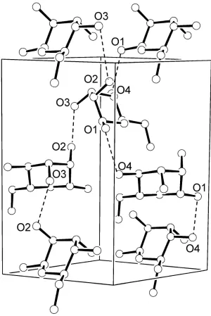
The molecule of the title compound, C6H12O6, [systematic name: (2R,3S,4R,5R,6R )-6-(hydroxymethyl)oxane-2,3,4,5-tetrol] adopts a 4C1 chair conformation with the anomeric hydroxyl group in the equatorial position. All hydroxyl groups act as donors and acceptors in hydrogen bonding and the molecule is involved in ten intermolecular O—H O inter-actions [O O = 2.672 (5)–2.776 (4) A˚ ] with eight neigh-bouring molecules. Two independent O—H O—H
helices extending along thezaxis are found in this structure.
Related literature
For the crystal structure of methyl-d-altrose, see: Gatehouse
& Poppleton (1971).
Experimental
Crystal data
C6H12O6 Mr= 180.16
Trigonal,P32 a= 7.1749 (13) A˚
V= 568.04 (16) A˚ Z= 3
CuKradiation
T= 293 (2) K 0.300.300.30 mm
Data collection
Rigaku RAPID2 diffractometer Absorption correction: none 6207 measured reflections
736 independent reflections 719 reflections withI> 2(I) Rint= 0.113
Refinement
R[F2> 2(F2)] = 0.046 wR(F2) = 0.125 S= 1.15 736 reflections 109 parameters
1 restraint
H-atom parameters constrained max= 0.24 e A˚
3 min=0.24 e A˚3
Table 1
Hydrogen-bond geometry (A˚ ,).
D—H A D—H H A D A D—H A
O1—HO1 O4i
0.82 1.97 2.743 (5) 156
O2—HO2 O3ii
0.82 1.96 2.768 (5) 169
O3—HO3 O6iii
0.82 1.88 2.672 (5) 162
O4—HO4 O1iv
0.82 1.94 2.748 (5) 167
O6—HO6 O2v
0.82 1.96 2.776 (4) 174
Symmetry codes: (i)x1;y;z; (ii)xþy;xþ2;zþ1
3; (iii)xþyþ1;xþ2;zþ 1 3;
(iv)xþy;xþ1;zþ1
3; (v)yþ1;xyþ1;z 1 3.
Data collection: PROCESS-AUTO (Rigaku, 1998); cell refine-ment: PROCESS-AUTO; data reduction: PROCESS-AUTO; program(s) used to solve structure: SHELXS97 (Sheldrick, 2008); program(s) used to refine structure:SHELXL97(Sheldrick, 2008); molecular graphics: ORTEPIII (Burnett & Johnson, 1996) and
ORTEP-3(Farrugia, 1997); software used to prepare material for publication:SHELXL97.
This study was supported in part by a Grant-in-Aid for Young Scientists (B) (19770085) from the Ministry of Education, Culture, Sports, Science and Technology of Japan, and by the Fund for Kagawa University Young Scientists 2007–8.
Supplementary data and figures for this paper are available from the IUCr electronic archives (Reference: GK2181).
References
Burnett, M. N. & Johnson, C. K. (1996).ORTEPIII. Report ORNL-6895. Oak Ridge National Laboratory, Tennessee, USA.
Farrugia, L. J. (1997).J. Appl. Cryst.30, 565.
Gatehouse, B. M. & Poppleton, B. J. (1971).Acta Cryst.B27, 871–876. Rigaku (1998).PROCESS-AUTO. Rigaku Corporation, Tokyo, Japan. Sheldrick, G. M. (2008).Acta Cryst.A64, 112–122.
Structure Reports Online
supporting information
Acta Cryst. (2009). E65, o280 [doi:10.1107/S1600536809000397]
β
-
D-Altrose
Yuji Watanabe, Hiromi Yoshida, Kosei Takeda, Tomohiko Ishi and Shigehiro Kamitori
S1. Comment
The molecular structure of β-D-altrose is shown in Fig. 1. The aldopyranose ring adopts a 4C
1 chair conformation and the
anomer hydroxyl group is in equatorial position pointing to a β-anomer structure. All bond distances and angles between non-hydrogen atoms of β-D-altrose are in the normal range, and torsion angles along C—C and C—O bonds show staggered conformations.
The crystal of β-D-altrose belongs to a trigonal crystal system, space group <it>P</it>32, which is for the first time
found in the crystal structure of aldohexoses.
S2. Experimental
D-Altrose was purchased from Sigma-Aldrich Ltd., Japan. Crystals were prepared by dissolving 20 mg of D-altrose in distilled water (4 ml). Suitable crystals for X-ray data collection were obtained by slow evaporation of this solution at 293 K.
S3. Refinement
Figure 1
Figure 2
Crystal packing of β-D-altrose, with two helices along the z axis shown as dashed lines.
(2R,3S,4R,5R,6R)-6-(hydroxymethyl)oxane-2,3,4,5-tetrol
Crystal data
C6H12O6 Mr = 180.16
Trigonal, P32
Hall symbol: P 32
a = 7.1749 (13) Å
c = 12.7415 (15) Å
V = 568.04 (16) Å3 Z = 3
F(000) = 288
Dx = 1.580 Mg m−3
Cu Kα radiation, λ = 1.54178 Å Cell parameters from 2323 reflections
θ = 7.2–68.0°
Rigaku RAPID2 diffractometer
Radiation source: fine-focus sealed tube Graphite monochromator
ω scans
6207 measured reflections 736 independent reflections
719 reflections with I > 2σ(I)
Rint = 0.113
θmax = 71.8°, θmin = 7.1° h = −8→8
k = −8→8
l = −15→14
Refinement
Refinement on F2
Least-squares matrix: full
R[F2 > 2σ(F2)] = 0.046 wR(F2) = 0.125 S = 1.15 736 reflections 109 parameters 1 restraint
Primary atom site location: structure-invariant direct methods
Secondary atom site location: difference Fourier map
Hydrogen site location: inferred from neighbouring sites
H-atom parameters constrained
w = 1/[σ2(F
o2) + (0.0525P)2 + 0.4167P]
where P = (Fo2 + 2Fc2)/3
(Δ/σ)max < 0.001
Δρmax = 0.24 e Å−3
Δρmin = −0.24 e Å−3
Special details
Geometry. All esds (except the esd in the dihedral angle between two l.s. planes) are estimated using the full covariance
matrix. The cell esds are taken into account individually in the estimation of esds in distances, angles and torsion angles; correlations between esds in cell parameters are only used when they are defined by crystal symmetry. An approximate (isotropic) treatment of cell esds is used for estimating esds involving l.s. planes.
Refinement. Refinement of F2 against ALL reflections. The weighted R-factor wR and goodness of fit S are based on F2,
conventional R-factors R are based on F, with F set to zero for negative F2. The threshold expression of F2 > σ(F2) is used
only for calculating R-factors(gt) etc. and is not relevant to the choice of reflections for refinement. R-factors based on F2
are statistically about twice as large as those based on F, and R- factors based on ALL data will be even larger.
Fractional atomic coordinates and isotropic or equivalent isotropic displacement parameters (Å2)
x y z Uiso*/Ueq
C1 0.3898 (7) 1.0634 (7) 0.3892 (3) 0.0257 (9)
H1 0.4984 1.1575 0.3381 0.031*
C2 0.4763 (7) 1.1433 (7) 0.4977 (4) 0.0299 (10)
H2 0.5069 1.2918 0.5054 0.036*
C3 0.6856 (7) 1.1365 (7) 0.5119 (4) 0.0309 (10)
H3 0.7366 1.1760 0.5843 0.037*
C4 0.6474 (7) 0.9113 (8) 0.4889 (4) 0.0288 (9)
H4 0.5474 0.8118 0.5415 0.035*
C5 0.5476 (6) 0.8366 (7) 0.3805 (3) 0.0253 (8)
H5 0.6491 0.9295 0.3267 0.030*
C6 0.4840 (8) 0.6073 (7) 0.3594 (4) 0.0306 (9)
H6A 0.6000 0.5832 0.3807 0.037*
H6B 0.3583 0.5139 0.4011 0.037*
O1 0.1989 (5) 1.0605 (6) 0.3667 (2) 0.0349 (8)
HO1 0.1136 1.0032 0.4151 0.042*
O2 0.3161 (5) 1.0090 (5) 0.5728 (3) 0.0309 (7)
O3 0.8420 (6) 1.2911 (6) 0.4415 (3) 0.0435 (9)
HO3 0.9453 1.3817 0.4750 0.052*
O4 0.8438 (6) 0.9049 (7) 0.4941 (3) 0.0441 (9)
HO4 0.8688 0.8905 0.5555 0.053*
O5 0.3537 (5) 0.8494 (5) 0.3754 (3) 0.0267 (7) O6 0.4365 (5) 0.5522 (5) 0.2508 (3) 0.0362 (8)
HO6 0.3057 0.4819 0.2428 0.043*
Atomic displacement parameters (Å2)
U11 U22 U33 U12 U13 U23
C1 0.034 (2) 0.028 (2) 0.021 (2) 0.0197 (17) −0.0044 (17) −0.0027 (16) C2 0.037 (2) 0.027 (2) 0.020 (2) 0.0118 (19) 0.0030 (18) 0.0018 (16) C3 0.027 (2) 0.035 (2) 0.018 (2) 0.0056 (18) 0.0015 (17) 0.0001 (16) C4 0.025 (2) 0.044 (2) 0.018 (2) 0.0172 (19) 0.0026 (16) 0.0059 (18) C5 0.026 (2) 0.030 (2) 0.024 (2) 0.0169 (17) 0.0009 (15) 0.0031 (16) C6 0.038 (2) 0.036 (2) 0.022 (2) 0.021 (2) 0.0038 (18) 0.0017 (18) O1 0.0384 (17) 0.0461 (18) 0.0308 (18) 0.0291 (15) 0.0000 (13) −0.0011 (14) O2 0.0343 (17) 0.0324 (16) 0.0248 (15) 0.0157 (14) 0.0036 (13) −0.0009 (13) O3 0.0348 (18) 0.0380 (19) 0.0288 (18) −0.0034 (14) 0.0043 (15) 0.0018 (15) O4 0.0302 (17) 0.077 (3) 0.0334 (19) 0.0334 (19) −0.0011 (14) 0.0063 (18) O5 0.0262 (15) 0.0278 (15) 0.0283 (15) 0.0152 (13) −0.0072 (12) −0.0043 (12) O6 0.0282 (15) 0.0430 (18) 0.0367 (19) 0.0172 (15) 0.0019 (13) −0.0121 (15)
Geometric parameters (Å, º)
C1—O1 1.389 (5) C4—H4 0.9800
C1—O5 1.435 (5) C5—O5 1.441 (5)
C1—C2 1.506 (6) C5—C6 1.495 (6)
C1—H1 0.9800 C5—H5 0.9800
C2—O2 1.435 (5) C6—O6 1.432 (6)
C2—C3 1.537 (6) C6—H6A 0.9700
C2—H2 0.9800 C6—H6B 0.9700
C3—O3 1.431 (5) O1—HO1 0.8199
C3—C4 1.526 (6) O2—HO2 0.8188
C3—H3 0.9800 O3—HO3 0.8199
C4—O4 1.434 (5) O4—HO4 0.8206
C4—C5 1.524 (6) O6—HO6 0.8199
O1—C1—O5 108.1 (3) O4—C4—H4 108.6
O1—C1—C2 114.3 (4) C5—C4—H4 108.6
O5—C1—C2 109.8 (3) C3—C4—H4 108.6
O1—C1—H1 108.2 O5—C5—C6 106.8 (3)
O5—C1—H1 108.2 O5—C5—C4 108.5 (3)
C2—C1—H1 108.2 C6—C5—C4 112.5 (3)
O2—C2—C1 108.5 (4) O5—C5—H5 109.7
O2—C2—C3 111.5 (4) C6—C5—H5 109.7
C1—C2—H2 109.4 O6—C6—H6A 109.2
C3—C2—H2 109.4 C5—C6—H6A 109.2
O3—C3—C4 110.9 (4) O6—C6—H6B 109.2
O3—C3—C2 107.5 (4) C5—C6—H6B 109.2
C4—C3—C2 110.5 (3) H6A—C6—H6B 107.9
O3—C3—H3 109.3 C1—O1—HO1 109.6
C4—C3—H3 109.3 C2—O2—HO2 109.4
C2—C3—H3 109.3 C3—O3—HO3 109.6
O4—C4—C5 109.0 (4) C4—O4—HO4 109.1
O4—C4—C3 111.5 (4) C1—O5—C5 113.6 (3)
C5—C4—C3 110.5 (4) C6—O6—HO6 109.3
O1—C1—C2—O2 58.2 (5) C2—C3—C4—C5 54.3 (5) O5—C1—C2—O2 −63.4 (4) O4—C4—C5—O5 −178.4 (3) O1—C1—C2—C3 179.6 (3) C3—C4—C5—O5 −55.6 (4) O5—C1—C2—C3 58.1 (4) O4—C4—C5—C6 63.7 (5) O2—C2—C3—O3 −174.0 (3) C3—C4—C5—C6 −173.4 (4) C1—C2—C3—O3 66.4 (4) O5—C5—C6—O6 74.4 (4) O2—C2—C3—C4 64.8 (5) C4—C5—C6—O6 −166.8 (3) C1—C2—C3—C4 −54.8 (5) O1—C1—O5—C5 170.9 (3) O3—C3—C4—O4 56.6 (5) C2—C1—O5—C5 −63.9 (4) C2—C3—C4—O4 175.7 (4) C6—C5—O5—C1 −177.1 (3) O3—C3—C4—C5 −64.8 (5) C4—C5—O5—C1 61.5 (4)
Hydrogen-bond geometry (Å, º)
D—H···A D—H H···A D···A D—H···A
O1—HO1···O4i 0.82 1.97 2.743 (5) 156
O2—HO2···O3ii 0.82 1.96 2.768 (5) 169
O3—HO3···O6iii 0.82 1.88 2.672 (5) 162
O4—HO4···O1iv 0.82 1.94 2.748 (5) 167
O6—HO6···O2v 0.82 1.96 2.776 (4) 174